Acquired immunity to amyloodiniosis is associated with an ...
9
DISEASES OF AQUATIC ORGANISMS Dis Aquat Org I Published October 8 Acquired immunity to amyloodiniosis is associated with an antibodv resnonse Charles S. Cobb, Michael G. ~evy',*, Edward J. ~ o g a ~ Departments of '~icrobiolo~y, Pathology, and Parasitology, and 'companion Animal and Special Species Medicine, North Carolina State University College of Veterinary Medicine, Raleigh, North Carolina 27606, USA ABSTRACT The dinoflagellate Amyloodlnium ocellatum, which causes amyloodiniosis or 'marine vel- vet disease', 1s one of the most senous ectoparasitic diseases plaguing warmwater manne fish culture worldwide We report that tomato clownfish Amphlpnon frenatus develop strong immunity to Amyloo- dlnlum ocellatum infection following repeated nonlethal challenges and that specific ant~bodies are associated with this response React~on of immune fish antisera against dinospore and trophont-denved antigens in Western blots indicated both shared and stage-speclfic antibody-antigen reactions A man- nan-binding-protein affinity column was used to isolate IgM-like antibody from A frenatus serum The reduced Ig consisted of one 70 kD heavy chain and one 32 kD light chain with an estimated molecular weight of 816 kD for the native molecule Immunoglobulin (Ig) isolated from immune but not non- unmune fish serum significantly inhlblted parasite infectivity In vitro An enzyme-linked immunosor- bent assay (ELISA)was developed using polyclonal rabbit antibody produced aga~nst aff~nity-punfied A frenatus Ig Anti-Amyloodlnium serum antibody was not always detectable in immune fish although serum antibody titers in immune fish increased after repeated exposure to the parasite These results suggest that there may be a localized antibody response in skin/gill epithelia1 tissue although antibody was rarely detected in skin mucus KEY WORDS: Amylood~nium ocellatum . Immunity . Arnphlprjon frenatus . Ig INTRODUCTION Amyloodinium ocellatum (Brown, 1931) is a parasite of serious consequence in food fish mariculture and the ornamental fish trade (Noga & Levy 1995). It is an ectoparasitic dinoflagellate with a triphasic life cycle consisting of infectious dinospore, feeding trophont and reproducing tomont stages. When fish populations are stressed as in mariculture or the ornamental fish trade, the parasite can rapidly increase and cause heavy mortality. Smith et al. (1992) showed that follow- ing injection of intact or sonically disrupted dino- spores, blue tilapia Oreochromis aureus exhibited an antibody response against A. ocellatum. Although sev- eral species of fish exhibit increased resistance to A. ocellatum infection (Lawler 1977, 1980, Paperna 1980), the mechanism and dynamics of resistance have not been characterized. 'Addressee for correspondence E-mail: [email protected] Cobb et al. (1998) have shown that the tomato clown- fish Amphiprion frenatus can develop immunity to Amyloodinium ocellatum infection through repeated sublethal challenges. This study was designed to address 2 questions: (1) Are specific antibodies associ- ated with protective immune response? (2) Can spe- cific antigens be identified which are involved in the response? Identification of specific protective antigens may facilitate development of a vaccine to prevent amyloodiniosis. MATERIALS AND METHODS Maintenance of fish. Aquarium-reared tomato clown- fish (4 to 13 g; 47 to 71 mm total length) were initially obtained from Aqualife Resources (Ft. Lauderdale, Florida, USA) and later from C-Quest (Salinas, Puerto Rico) (2 to 4 g; 30 to 50 mm). Fish stocks were main- tained in 150 1 round tanks containing 25 ppt artificial seawater (ASW). All fish were acclimated for at least O Inter-Research 1998 Resale of full artjcle not permitted
Transcript of Acquired immunity to amyloodiniosis is associated with an ...

DISEASES OF AQUATIC ORGANISMS Dis Aquat Org
I Published October 8
Acquired immunity to amyloodiniosis is associated with an antibodv resnonse
Charles S. Cobb, Michael G. ~ e v y ' , * , Edward J. ~ o g a ~
Departments of ' ~ i c r o b i o l o ~ y , Pathology, and Parasitology, and 'companion Animal and Special Species Medicine, North Carolina State University College of Veterinary Medicine, Raleigh, North Carolina 27606, USA
ABSTRACT The dinoflagellate Amyloodlnium ocellatum, which causes amyloodiniosis or 'marine vel- vet disease', 1s one of the most senous ectoparasitic diseases plaguing warmwater manne fish culture worldwide We report that tomato clownfish Amphlpnon frenatus develop strong immunity to Amyloo- dlnlum ocellatum infection following repeated nonlethal challenges and that specific ant~bodies are associated with this response React~on of immune fish antisera against dinospore and trophont-denved antigens in Western blots indicated both shared and stage-speclfic antibody-antigen reactions A man- nan-binding-protein affinity column was used to isolate IgM-like antibody from A frenatus serum The reduced Ig consisted of one 70 kD heavy chain and one 32 kD light chain with an estimated molecular weight of 816 kD for the native molecule Immunoglobulin (Ig) isolated from immune but not non- unmune fish serum significantly inhlblted parasite infectivity In vitro An enzyme-linked immunosor- bent assay (ELISA) was developed using polyclonal rabbit antibody produced aga~ns t aff~nity-punfied A frenatus Ig Anti-Amyloodlnium serum antibody was not always detectable in immune fish although serum antibody titers in immune fish increased after repeated exposure to the parasite These results suggest that there may be a localized antibody response in skin/gill epithelia1 tissue although antibody was rarely detected in skin mucus
KEY WORDS: Amylood~nium ocellatum . Immunity . Arnphlprjon frenatus . Ig
INTRODUCTION
Amyloodinium ocellatum (Brown, 1931) is a parasite of serious consequence in food fish mariculture and the ornamental fish trade (Noga & Levy 1995). It is an ectoparasitic dinoflagellate with a triphasic life cycle consisting of infectious dinospore, feeding trophont and reproducing tomont stages. When fish populations are stressed as in mariculture or the ornamental fish trade, the parasite can rapidly increase and cause heavy mortality. Smith et al. (1992) showed that follow- ing injection of intact or sonically disrupted dino- spores, blue tilapia Oreochromis aureus exhibited an antibody response against A. ocellatum. Although sev- eral species of fish exhibit increased resistance to A. ocellatum infection (Lawler 1977, 1980, Paperna 1980), the mechanism and dynamics of resistance have not been characterized.
'Addressee for correspondence E-mail: [email protected]
Cobb et al. (1998) have shown that the tomato clown- fish Amphiprion frenatus can develop immunity to Amyloodinium ocellatum infection through repeated sublethal challenges. This study was designed to address 2 questions: (1) Are specific antibodies associ- ated with protective immune response? (2) Can spe- cific antigens be identified which are involved in the response? Identification of specific protective antigens may facilitate development of a vaccine to prevent amyloodiniosis.
MATERIALS AND METHODS
Maintenance of fish. Aquarium-reared tomato clown- fish (4 to 13 g ; 47 to 71 mm total length) were initially obtained from Aqualife Resources (Ft. Lauderdale, Florida, USA) and later from C-Quest (Salinas, Puerto Rico) (2 to 4 g ; 30 to 50 mm). Fish stocks were main- tained in 150 1 round tanks containing 25 ppt artificial seawater (ASW). All fish were acclimated for at least
O Inter-Research 1998 Resale of full artjcle not permitted
126 Dis Aquat Org 34: 125-133, 1998
2 wk prior to experimentation and were considered unexposed to Amyloodinium ocellatum, although their precise history was not available. Since A, ocellatum dinospores are susceptible to low levels of copper, a solution of copper sulfate was used to maintain copper levels of 0.15 to 0.20 mg 1-' to control ectoparasites in maintenance tanks. Parasite returns from fish main- tained copper free were similar to those from fish removed from copper several days prior to infection.
Propagation of Amyloodinium ocellatum. A. ocella- turn (isolate 85-1 of DC-1) was propagated in cell culture using the methods developed by Noga (1987, 1989).
Production of Amyloodinium ocellatum antigens. Trophont antigen: Parasites were cultured on Amphi- prion frenatus and collected as described by Bower et al. (1987) with modifications described by Cobb et al. (1998), with the following exceptions. As soon as fish were removed fro-. the specimen cups, cold (4°C) ASW was added and the cups were kept on ice to arrest trophont development into tomonts. The troph- onts were then counted. Whole trophonts were irnme- diately frozen at -70°C. Sonicated trophonts were pre- pared by sonicating on ice at a setting of #6 with a mini-probe (Branson Sonifier, Cell Disrupter 200, Dan- bury, CT, USA) with a cycle of 15 S on and 15 S off for a total of 3 min. The sonicated material was centrifuged at 60 X g for 5 min and the protein content of the super- natant was determined using the dotMetricB 1 p1 Pro- tein Assay (Geno Technology, Inc., St. Louis, MO, USA). The sonicated material was then aliquoted and stored at -70°C.
Dinospore antigen. Amyloodinium ocellatum was cultured on Amphiprion frenatus and collected as described by Bower et al. (1987). Dinospores were used either live or were sonicated as described above.
Production of anti Amyloodinium ocellatum anti- serum. Antiserum to A. ocellatum was prepared as de- scribed by Smith et al. (1992) with the following modl- fications. Two groups of 10 fish each (3 to 4 g) were injected at Weeks 0, 2 and 4 with either live dinospores (103 g-' body wt) or whole frozen trophonts (102 g-' body wt). Serum was collected 4 d following the final injection. Antlserurn was also produced by exposing 10 fish (6 to 12 g) to a sublethal challenge of 40000 dinospores fish-' at a concentration of 200 dinospores ml-l. After 30 min, all fish were gently removed from each container and placed in separate 38 1 holding aquaria with 25 ppt ASW to allow the A. ocellatum trophonts to mature. After 3 d, each fish was dipped in distilled water (dH20) for 3 min to remove the trophonts for counting and then placed in a recovery aquarium containing 0.15 to 0.20 mg 1-' copper. This procedure was repeated 4 times at which time parasiLe recovery rate (PRR) was less than 1 %. Serum was col- lected 4, 10, and 30 d following the final challenge.
Serum and mucus collection. Blood samples (50 to 300 p1) were taken from fish via the caudal vessels with a sterile, 23-gage needle attached to a 1.0 cc syringe. Pooled blood was allowed to clot in a glass tube at room temperature for 1 h and then stored overnight at 4°C. Serum was then collected by centnfugation at 1000 X g for 10 min. and then stored at -70°C. For ELISA analysis of serum from individual fish, serum was collected and stored as above in separate 0.5 m1 microcentrifuge tubes
Mucus was collected from individual fish with a 2 X
4 mm piece of Gel Bond Film (FMC Corp., Rockland, ME, USA). The film was held at a 45" angle to the skin and scraped from the operculum to the caudal fin. The fish was then turned over and the other side of the film was used to repeat the scrape. The film was then placed in a 0.5 m1 microcentrifuge tube, vortexed with 0.1 ml phosphate buffered saline (PBS) for !O S anc! the suspension stored at -70°C.
Dinospore immobilization assay. This assay was carried out as described by Smith et al. (1993b). Quad- ruplicate 50 p1, serial, doubling dilutions of each serum sample were prepared in a 96-well flat-bottom tissue culture plate using ASW/Hank1s balanced salt solution (I02/HBSS) to give final dilutions of 10%, 5 %, 2.5%, 1.25% and 0.61% serum concentrations. Then 200 dinospores in 50 p1 of I02/HBSS were placed into each well. Motility was observed with an inverted phase contrast microscope at 5, 15,30, and 60 min and scored as 0 (majority of the dinospores immobilized), 1 (mini- mal effect, some slowing of movement and some im- mobilization), or 2 (no effect), as compared to quadru- plicate wells of dinospores in I02/HBSS without serum. Mean scores for each serum dilution were cal- culated for the different time intervals (Smith et al. 1993b).
Infectivity assay. To determine the toxicity of immune and non-immune tomato clownfish serum to Amyloodinium ocellatum, serum concentrations of 10%, 5%, 3%, 1.5% or 0.75% serum m I02/HBSS were prepared in a 96-well cell culture plate having fish gill (GIB) cells as a feeder layer (Noga 1987, 1989). Wells containing I02/HBSS without serum served as a control. Then 100 dinospores were added and troph- onts were counted following incubation at 26°C for 3 d.
To differentiate non-specific serum toxicity from a specific antibody-mediated response, 250 dinospores were exposed to affinity-purified serum Ig (500 pg ml-' I02/HBSS) isolated from immune or unexposed fish in a 96-well cell-culture plate. The plate was incubated at 26°C and trophonts were counted on Day 3.
Passive transfer of immunity. Fifty fish (8 to 14 g) were rendered immune by repeated challenge of the same fish every 2 wk for 6 wk at which time parasite recovery rate was less than 1 % (naturally immune)

Cobb et al.: Antibody I response to amyloodiniosis 127
(Cobb et al. 1998). To maintain a high level of immu- nity, fish were challenged weekly with 10000 Amyloo- djnium ocellatum dinospores fish-'. In other experi- ments (data not shown), this regimen was shown to induce high levels of anti- dinospore and anti-trophont antibodies in an ELISA assay .
Blood (50 to 100 p1) was collected from each of 50 immune fish via the caudal vessels using a 1 cc syringe with a 23-gage needle. The blood was pooled, allowed to clot in a glass tube at room temperature for 1 h and then stored overnight at 4OC. The next day serum was collected by centrifugation in pediatric serum separa- tor tubes (Microtainer, Becton Dickinson and Com- pany, Rutherford, NJ, USA) at 14000 X g for 2 min, then stored in 0.5 m1 aliquots at -70°C.
In Expt 1, 1 d prior to challenge, 0.1 rnl immune serum was injected (IP) into 10 fish (2 to 3 g) which had never been experimentally challenged (nai've). On the day after injection, the fish were sublethally chal- lenged; 3 d later, PRRs were determined.
In Expt 2, 3 d prior to challenge, serum from immune fish was thawed and injected (IP) into nai've fish (2 to 3 g) at a rate of 0.1 m1 fish-'. Injections were repeated daily for 2 additional days. On the day after the last injection, the fish were sublethally challenged (40 000 dinospores fish-'); 3 d later, PRRs were determined. Sufficient quantities of na'ive serum were unavailable therefore sterile PBS was utilized as a negative control in these experiments.
Affinity purification of Ig. Tomato clownfish im- munoglobulin (Ig) was isolated using a mannan-bind- ing protein (MBP) affinity column following the manu- facturer's instructions (ImmunoPurem IgM Purification I t , Pierce Chemical Co., Rockford, IL, USA). Briefly, the 5 m1 column was washed with 5 n ~ l of Immuno- Purem MBP column preparation buffer and then equili- brated with 20 m1 of ImmunoPurem IgM binding buffer at 4°C. Gravity provided a flow rate of 1 m1 min-l. Fish serum was diluted 1:2 with ImmunoPure@ IgM binding buffer and dialyzed overnight at 4°C in a tris-NaC1 buffer (10 mM Tris, 1.25 M NaCl, 0.02% NaN3) (Ohta et al. 1990, Nevens et al. 1992). One m1 of dialyzed serum was applied to the column. After 30 min, the column was washed with 42 m1 of binding buffer to remove unbound protein. The column was then placed at room temperature to decrease the column's binding affinity for Ig. Three m1 of ImmunoPurem IgM elution buffer was added and allowed to incubate for 1 h. In all procedures, 3 m1 fractions were collected and protein content measured using the Micro BCA Protein Assay (Pierce Chemical Co.). Eluates with significant amounts of protein ( > l 0 pg ml-l) were pooled, and then concentrated and desalted by centrifugation (Centricon-l0 micro-concentrator, Amicon, Danvers, MA, USA). The determination of protein concentration
in small samples was performed by the dotMETRICm 1 1-11 Protein Assay. Purified Ig was stored at -70°C in PBS or I02/HBSS.
SDS-PAGE. Purified Ig was analyzed by analytical sodium dodecyl sulfate polyacrylamide gel electro- phoresis (SDS-PAGE) as described by Laemmli (1970). The sample was diluted 1:2 with electrophoresis sam- ple buffer (2.3 m1 of 10% SDS, 1.0 m1 of 0.625 M Tris- HCl, pH 6.8, 1.0 m1 of glycerol and 5.2 m1 of distilled water with 2.5 % bromophenol blue) and then added to a single well of a vertical 4 % stacking gel over a 7 % resolving gel (BioRad Laboratories, Richmond, CA, USA). The above procedure was modified to analyze denatured Ig by having 2-mercaptoethanol (2ME) at 2.5% in the sample buffer and boiling of the sample prior to loading. The gel was electrophoresed for 15 rnin at 100 V and then at 200 V until the marker dye reached the bottom of the gel. The gels were then silver-stained (Daiichi 11, ISS-Emprotech, Hyde Park, MA) according to manufacturer's instructions.
Western blot analysis of inmlune and unexposed Amphiprjon frenatus serum was performed as follows. Parasite antigen (dinospore or trophont) was run on a 12 % SDS-PAGE gel as previously described and trans- ferred to a 0.2 pm nitrocellulose membrane using the BioRad Modular Mini Electrophoresis System (BioRad Laboratories Richmond, CA). Following transfer, the nitrocellulose membrane was blocked with Tris- buffered saline with 0.1 % Tween 20 and 0.1 % gelatin (TBST/gelatin) overnight at 4°C. The membrane was incubated for 3 h with A. frenatus serum diluted 1:5000 in TBST/gelatin. After 3 washes of 10 min in TBST/ gelatin, rabbit anti-tomato clownfish Ig antiserum diluted 1:5000 in TBST/gelatin was added and incu- bated for 2 h followed by another 3 washes of 10 min in TBST/gelatin. Horseradish peroxidase (HRP)-labeled goat anti rabbit antibody (Kirkegaard and Perry Labo- ratories, Inc., Gaithersburg, MD, USA) diluted 1:5000 in TBST/gelatin was added and incubated. After 2 h, the membrane was washed 3 times with TBST/gelatin and once with Tris buffered saline (TBS).
For chemiluminescent development, the Pierce Ultra Supersignal CL-HRP Substrate System (Pierce Chemi- cal Co.) was used according to manufacturer's instruc- tions. Chemiluminescent solutions A and B were mixed and added to a clean, dry incubation tray. The nitrocellulose membrane was placed into the chemi- luminescent solution and rocked gently for 60 S. The development solution was poured off and the strip was quickly wrapped with Saran Wrap@ Dow Brands L.P. Indianapolis, IN, USA). A piece of X-OMAT AR scien- tific imaging film (Eastman Kodak Co., Rochester, NY, USA) was placed on the nitrocellulose for 1 S. The film was developed using a Kodak M35A X-OMAT pro- cessor.

128 Dis Aquat Org 34: 125-133, 1998
Production of anti Amphiprion frenatus Ig antibod- ies. Polyclonal antibodies against A. frenatus Ig were produced in 2 New Zealand White rabbits by use of the RIB1 Adjuvant System (RAS) (RIB1 ImmunoChem Research, Inc., Hamilton, MO, USA) following the manufacturer's instructions. Briefly, the RAS formula- tion was reconstituted in 2 m1 of 0.15 M PBS containing 200 pg of affinity-purified Ig and warmed to 37°C prior to use. On Day 0, each rabbit was anesthetized by intramuscular injection with acepromazine and butor- phanol (0.5 mg kg-' of each drug) and 4 m1 of whole blood was collected prior to injecting 1 m1 of RAS divided as follows: 6 intradermal sites in the neck (0.05 ml); 1 intramuscular site in each hind leg (0.3 ml); 1 subcutaneous site in the neck (0.1 ml). Each rabbit was boosted on Day 21 and test bled on Day 28. Exsan- guination was performed under anesthesia on Day 31 using ketamine hydrochloride (Ketaset, Fort Dodge Laboratories, Inc., Fort Dodge, IA, USA) All sera were stored at -70°C.
Ouchterlony double diffusion. Tris-tricine buffered, ouchterlony double-diffusion gels (Monthony et al. 1978) were prepared by pipetting 8 m1 of 1 % agarose onto a 75 mm by 53 mm agarose support medium (Gel Bond Film, FMC Corp., Rockland, ME). Five mm holes were punched in the agar and 20 p1 samples were used for the assay.
Enzyme-linked immunosorbent assay (ELISA). Stock protein concentrations were 500 pg ml-' for dinospore antigen (Ag) and 250 pg ml-l for trophont Ag. The optimal concentrations of antigen (Ag), fish serum (primary antibody [Ab]), rabbit serum (sec- ondary Ab) and goat anti-rabbit HRP (tertiary Ab) were determined in accordance with the procedures in Smith et al. (1992). The optimal Ag and Ab dilutions were 1:1000, except for trophont Ag, which was diluted 1:500. Wells in which all reagents except fish serum were added were used to evaluate non-specific binding. ELISA values (EVs) as described by Hornitzky & Searson (1986) were used to compare results be- tween several ELISA plates to compensate for normal variation in plate preparation, processing and develop- ment. Positive control fish serum was generated by injecting 10 fish (6 to 12 g) with lo3 live Amyloodinium ocellatum dinospores per g of fish. Immunizations were performed on Weeks 0, 2, and 4. Serum was col- lected 4 d following the final injection.
ELISA analysis of mucus scrapings from fish were made as described above except that 100 p1 of mucus extract in PBS was substituted for fish serum.
Fluorescent antibody assay. Three 25 m1 cell-culture flasks were seeded with GIB cells (Noga 1989). Cells were conditioned for 24 h and then inoculated with 50000 dinospores flask-'. After 72 h the medium was replaced with either: (1) serum from fish receiving re-
peated nonlethal challenges diluted 1:250 in I02/ HBSS with 0.5% bovine serum albumin (BSA); (2) un- exposed fish serum diluted 1:250 in I02/HBSS with BSA; or (3) I02/HBSS only with BSA. After incubation for 1 h at room temperature with constant gentle mix- ing, the detached trophonts were transferred to 15 rnl centrifuge tubes and washed 4 times with 12 m1 of I02/HBSS using gentle centrifugation (500 X g for 5 min) between each wash. Two m1 of a 1:1000 dilution of rabbit anti-fish Ab in I02/HBSS with BSA was added to each tube and incubated for 1 h at room tem- perature with constant mixing. The trophonts were washed as before. Two m1 of a 1:1000 dilution of a 10 pg ml-l concentration of FITC-conjugated goat anti rabbit antibody (Cappel Organon Teknika, West Chester, PA, USA) in I02/HBSS with BSA was then added to each well and incubated for 1 h at room tem- perature with constant mixing. The trophonts were again washed 4 times and resuspended in 0.25 m1 of I02/HBSS. Samples were mounted on slides, dried and photographed using Technical Pan 2415 film with an Olympus Vanox AH-3 photomicroscope.
Statistical analysis. Means, standard deviations from the mean and Student's t-test, p-values were calcu- lated using Microsoft Excel for Windows.
RESULTS
Purification of Amphiprion frenatus Ig
A mannan-binding protein affinity column (Pierce Chemical Co.) successfully isolated serum Ig from both Amyloodinium ocellatum-resistant and unexposed fish. Serially eluted 3 m1 fractions were analyzed for protein. content by reading optical density at 280 nm. Fractions 2, 3, 4 and 5 showed significant protein content (OD 2 0.02 at 280 nm) and were pooled, concentrated, and desalted.
SDS-PAGE of reduced MBP column eluate revealed 2 bands representing the heavy and light chains of the fish immunoglobulin with little observable contamina- tion (Fig. 1). Comparison of the bands with molecular weight standards gave estimates of 32 kD for the light chain and 70 kD for the heavy chain. Assuming a tetramenc structure for the intact Ig molecule, its size would be 816 kD. Tomato clownfish Ig was isolated with fewer contaminants as compared to rainbow trout
lg. Ouchterlony double diffusion analysis of pre-
immune rabbit serum revealed no reactivity with fish Ig. After immunization with affinity-punfied fish Ig, 2 distinct precipitation bands were observed between tomato clownfish serum and post-immunization rabbit serum.

Cobb et al.: Antibody response to amyloodiniosis 129
Stds Rainbow Tomato trout clownfish
Molecular weights
t Immune serum
+ Non-immune serum
Serum Concentration
Fig. 2. In vjtro infectivity of Amyloodinium ocellatum dinospores cultured with serum from immune or unexposed tomato clownfish. Serum dilutions were mixed with dino- spores in a 96-well cell-culture plate having GIB cells; trophonts were counted on Day 3. Data is reported as mean number of trophonts well-' (* SD) for each treatment group.
Fig. 1. SDS-PAGE of eluate from rainbow trout Oncorhynchus Values with an asterisk (*) were significantly (P < 0.05) differ- mykiss and tomato clownfish Amphiprion frenatus serum ent at that serum concentration from an immobilized mannan-binding protein affinity column
Effect of immune serum on in vitro parasite infectivity Effects of immune serum and purified Ab on
dinospore motility Incubation of dinospores with non-immune fish serum resulted in a dose-dependent reduction in infec-
To evaluate anti-dinospore activity in serum from tivity, with 5 % serum completely inhibiting trophont clownfish that were resistant to Arnyloodinium ocella- development. Immune serum caused a significantly turn, we compared the activity of pooled sera from greater inhibition (4.96 -+ 3.1 trophonts well-') than resistant fish with pooled sera from unexposed fish, unexposed fish serum (11.00 + 2.9 trophonts well-') (p Although both sera immobilized dinospores at high 0.0005) (Fig. 2 ) . Serum concentrations of 1 or 2 % did (>5%) concentrations, no differences were observed not significantly reduce dinospore infectivity. When between sera from immune and unexposed fish the antibody fraction of immune serum was enriched (Table 1). by affinity chromatography, anti-parasite activity
remained strong (85 % inhibition), whereas activity
Table 1. Mohlity index of Amyloodinium ocellatum dino- was lost when this process was performed with serum
spores after exposure for various times to various concentra- from unexposed fish (Fig. 3). tions of immune and unexposed serum in I02/HBSS medium (quadruplicate treatments). 0: great majority of the parasites immobilized; 1: minimal effect, some slowing of movement
and some immobilization); 2: no visible effect Passive transfer of immunity
Serum Serum from Serum from concentration immune fish unexposed fish
Time(min): 5 15 30 60 5 15 30 60
10.00 % 2 1 1 1 2 1 1 0 5.00% 2 1 1 1 2 1 1 0 2.50% 2 2 2 2 2 2 2 2 1.25% 2 2 2 2 2 2 2 2 0.61 O/o 2 2 2 2 2 2 2 2 0.00% 2 2 2 2 2 2 2 2
In order to evaluate the effectiveness of serum from immune fish in transfer of immunity to naive fish, serum from immune fish which had been challenged weekly was injected into nai've fish. In Expt 1, fish given a single injection of immune serum (0.1 rnl) showed no transfer of immunity with a mean PRR of 7.2 % (k1.5) compared to PBS injected fish which had a mean PRR of 7.3% (*2.5) (p = 0.46). In Expt 2, PBS injected fish had a mean PRR of 13.3 % (* 6.0) while 3 daily injections of immune serum reduced the PRR to

130 Dis Aquat Org 34: 125-133, 1998
NOSERUM
UNEXPOSED
IMMUNE
Fig. 3. Effect of MBP-purified Ig on infectivity of Amylood- inium ocellatum &nospores in vitro. Data is reported as mean umber of trophonts we!!-' (* SD) for each t r e a t ~ e n t group. N = 24 wells treatment-' Asterisk (+) indicates that the value is significantly (p < 0.0005) different from the other treatments
7.98% (k1.5). When t-test analysis was applied to the data a calculated p-value of 0.087 gave a confidence level of 85% that passive transfer of immunity had occurred. When one outlier in the control group and one outlier in the experimental group were removed, a p-value of 0.046 was observed (confidence level 90%).
Effect of immunization on Ab titers
In order to measure anti Amyloodinium ocellatum serum Ab, an ELISA was developed using the methods described by Smith et al. (1992). Preliminary studies showed that dilutions of 1:1000 of Ag, primary Ab (fish), secondary Ab (rabbit), and terminal Ab (goat) gave optimal results. Pre-immunization rabbit serum had an average corrected optical density or COD (actual OD - OD of blank wells) of 0.003 (+0.002) while the mean post-immunization COD was 0.698 (k0.050) (p < 0.0005).
ELISA values (EVs) as described by Hornitzky & Searson (1986) were used to evaluate the reactivity of sonicated dinospore or sonicated trophont antigen with antiserum from fish injected with either whole dinospores or whole trophonts. Serum from fish which had 4 weekly trophont injections (102 g-' of fish) showed no reactivity against either dinospore or trophont antigen (EV = 0), while serum from fish which had 4 weekly injections of live dinospores (103 g-' of fish) had EV's of 96 (*28) against dinospore Ag and 37 (*16) against trophont Ag.
ELISAs were performed on serum from immune fish to evaluate the hnetics of the immune response. Ten fish previously shown to be immune to Amyloodinium
ocellatum challenge but which had not been chal- lenged for 30 d had no detectable Ab to either dino- spore or trophont Ag, although the fish were solidly immune based on parasite recovery rate in a subse- quent standard challenge. When these fish were given 2 standard challenges 1 wk apart, they did not produce EVs above background levels. However, 4 d after a third standard challenge, EVs averaged 21 (*15) against dinospore Ag and 30 (k25) against trophont Ag. By Day 10 after the third challenge, EVs had fallen to an average of 11 (kg) against lnospore Ag and to background levels against trophont Ag. ELISAs using mucus from immune fish, with only one exception, were negative (data not shown).
Antibody recognition of specific parasite antigens
Indirect fluorescent antibody showed that both trophonts and dinospores strongly fluoresced in the presence of serum from fish rendered resistant to rein- fection by repeated non-lethal parasite challenges. Most trophonts uniformly fluoresced, while some fluo- resced more intensely around specific areas (Fig. 4). Most dinospores uniformly fluoresced (not shown).
Serum from unexposed fish showed no reactivity to either dinospore or trophont Ag in Western blots, while serum from immune fish showed several bands of reac- tivity against both dinospore and trophont Ag (Fig. 5). There was a stronger reaction with dinospore Ag (more bands evident). Some bands of reactivity against trophont Ag were not evident in the dinospore Ag lane.
Fig. 4 . Indirect fluorescent antibody analysis of immune tomato clownfish serum reacted wi.th cell cultured trophonts.
Arrow indicates area of strong fluorescence
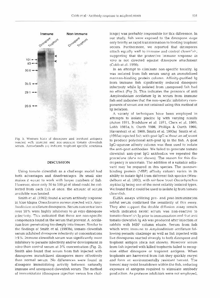
Cobb et al.: Antibody response to amyloodiniosis 131
Immune Non-Immune
Fig. 5. Western blots of dinospore and trophont antigens reacted with immune and non-immune tomato clownfish serum. Arrowheads (<) indicate trophont-specific reactions
DISCUSSION
Using tomato clownfish as a challenge model had both advantages and disadvantages. Its small size makes it easier to work with larger numbers of fish. However, since only 50 to 100 p1 of blood could be col- lected from each fish at once, the amount of serum available was limited.
Smith et al. (1992) found a serum antibody response in blue tilapia Oreochromis aureus injected with Amy- loodlnlum ocellatum dinospores. Serum concentrations over 10% were highly inhibitory to in vitro dinospore infectivity. This indicated that there are non-specific components found in the serum that prevent A. ocella- tum from penetrating too deeply into tissues. Similar to the findings of Smith et al. (1993b), tomato clownfish serum inhibited dinospore infectivity at concentrations 25 %. Immune clownfish serum was significantly more inhibitory to parasite infectivity and/or development in vitro than control serum at 3 % concentration (Fig. 2). Smith also found that serum from fish injected with dinospores immobilized dinospores more effectively than normal serum. No differences were found in &nospore immobilizing activity between naturally immune and unexposed clownfish serum. The method of immunization (dinospore injection versus live chal-
lenge) was probably responsible for this difference. In our study, fish were exposed to the dinospore stage only briefly as rapid transformation to feeding trophont occurs. Furthermore, we reported that dinospores attach equally well to immune and control clownfish, suggesting that the protective immune response in vivo is not directed against dinospore attachment (Cobb et al. 1998).
In an attempt to eliminate non-specific toxicity, Ig was isolated from fish serum using an immobilized mannan-binding protein column. Affinity-purified Ig from immune fish significantly reduced dinospore infectivity while Ig isolated from unexposed fish had no effect (Fig 3). This indicates the presence of anti Amyloodinium ocellatum Ig in serum from immune fish and indicates that the non-specific inhibitory com- ponents of serum are not retained using this method of Ig isolation.
A variety of techniques have been employed in attempts to isolate piscine Ig with varying results (Acton 1971, Bradshaw et al. 1971, Clerx et al. 1980, Lobb 1981a, b, Ourth 1986, Phillips & Ourth 1986, Havarstein et al. 1988, Smith et al. 1993a). Smith et al. (1993a) injected fish with goat IgG without an adjuvant to produce polyclonal anti-goat Ig in the fish. A goat IgG-agarose affinity column was then used to isolate the anti-goat antibodies. We failed to generate tomato clownfish anti-goat IgG antibodies we repeated the procedure (data not shown). The reason for this dis- crepancy is uncertain. The addition of a suitable adju- vant may be required in this species. The mannan- binding protein (MBP) affinity column varies in its ability to isolate IgM from different fish species (Wen- delborn et al. 1992), with rainbow trout Oncorhynchus mykiss Ig being one of the most reliably isolated types. We found that it could be used to isolate Ig from tomato clownfish.
ELISA assays utilizing pre- and post-immunization rabbit serum confirmed the sensitivity of this assay. They also support the double diffusion assay results which indicated rabbit serum was non-reactive to tomato clownfish Ig prior to immunization and that anti tomato clownfish lg Ab was produced after injection of rabbits with MBP column eluate. Serum from fish which were immune to Amyloochnium ocellatum fol- lowing parasite challenge as well as fish injected with live dinospores reacted strongly to both dinospore and trophont antigen (data not shown). However serum from fish injected with killed trophonts failed to recog- nize either dinospore or trophont antigens. When trophonts are harvested from fish they quickly encyst and form an environmentally resistant tomont. The tomont may resist breakdown within the fish, reducing exposure of antigens required to stimulate antibody production. As protease inhibitors were not employed,
132 Dis Aquat Org 34: 125-133, 1998
the antigenicity of tomonts may have been reduced by post-preparation autolytic processes. It was previously determined that injection of live dinospores prod.uce a greater response than dead, sonicated parasites (Smith et al. 1993b); therefore, dead trophonts also may be less antigenic. Alternatively, trophont antigens may be inherently less immunogenic.
Sera from immune fish which had not been chal- lenged for 30 d did not react in the ELISA to dinospore or trophont antigen, even though the fish were solidly immune to subsequent sublethal challenge. Three weekly standard challenges were required to increase serum antibody to detectable levels. Ten days after the third challenge, average EVs to dinospore antigen had dropped to half the 4 d post challenge values and aver- age EVs to trophont Ag had dropped to background levels. A wide variation in serum Ab titers between individual immune fish made the difference between Days 4 and 10 insignificant (p >0.05), but all fish were immune as measured by parasite recovery rates fol- lowing sublethal challenge (data not shown).
The existence of a localized antibody response in fish has been widely proposed and studies of skin, intestine and bile (Ourth 1980, Lobb & Clem 1981a, b, St. Louis- Cormier et al. 1984, Georgopoulou & Vernier 1986, Rombout et al. 1986, Lobb 1987, Lumsden et al. 1993) have supported such a possibility. ELISA results using skin mucus from tomato clownfish were almost com- pletely negative with only one fish showing positive results. The highly variable serum antibody levels in immune fish may indicate that protective Ab is pro- duced on the body surface and Ab may only appear in serum when sufficient quantities are produced to move from these tissues into the capillaries of the blood stream as reported for mammals (Brandtzaeg 1989). The development of mucosal antibody with strong memory may require multiple exposures at the proper site (Ganguly & Waldman 1983). In a more analogous situation, Lumsden et al. (1993) found that there was little correlation between levels of gill surface and cutaneous mucus antibody in brook trout Salvelinus fontinalis following infection with Flavobacterium branchiophilum, the etiologic agent of bacterial gill disease. Failure to detect antibody in mucus is consis- tent with Smith (1990), who found anti-dinospore activity in skin mucus of only 25% of tilapia immu- nized with dinospores. The difficulty of gill mucus col- lection without serum contamination from these small fish precluded direct measurement. The possibility that mucus antibody is of a different Ig subclass than serum Ig and therefore might not be reactive in the ELISA cannot be excluded. Examination of the gill for localized surface antibody production in the immune response of fish to Amyloodinium ocellatum infection must await use of a larger fish species.
Serum from immune fish but not unexposed fish strongly reacted with cell-cultured trophonts and dinospores (Fig. 4). A single injection of 0.1 m1 of immune serum l d prior to infection failed to affect par- asite recovery rates 3 d post infection. However, when similar injections were given on a daily basis for 3 d prior to infection, parasite recovery rates were reduced 40% when compared to fish receiving injections of sterile PBS. The unavailability of sufficient nalve serum and the necessity to substitute sterile PBS in these experiments requires that this experiment be confirmed using the proper control (nai've serum). This finding, along with the inhibitory effects of immune Ig on parasite growth/development in vitro suggest that Ab may exert its strong antiparasite effects via inter- action with parasite surface antigen.
Western blot analysis (Fig. 5) showed that serum from unexposed fish failed to react with dinospore or trophont antigens. Serum from immune fish reacted to several antigens from both dinospores and trophonts. The reaction to dinospores was stronger and included more antigens than the reaction to trophonts.
In summary, our data suggest that tomato clownfish can mount a protective antibody-mediated response to Amyloodinium ocellatum infection. An immobilization assay as well as an in vivo infection assay (Cobb et al. 1998) suggested that dinospores are probably not the target of the protective response. Further studies involving characterization of the immune response including the role of mucosal immunity, to A. ocella- turn, and further identification and characterization of parasite antigens wdl be necessary before production of a protective vaccine will be feasible.
Ackno~vledgernents. This work was supported by a grant from the College of Veterinary Medicine at North Carolina State University and Grant #NA46R6008? from the National Sea Grant College Program, National Oceanic and Atmos- phenc Administration to the North Carol~na Sea Grant Pro- gram. We thank Rob~n Gager, Zhiqin Fan and David Robi- nette for their assistance with laboratory procedures.
LITERATURE CITED
Acton RT, Weinheimer PF, Hall SJ, N~edermier W, Shelton E, Bennett JC (1971) Tetrameric immune macroglobulins in three orders of bony fishes. Proc Natl Acad Sci 68:107-111
Bower CE, Turner DT, Biever RC (1987) A standardized method of propagating the marine fish parasite, Amyloo- drnium ocellatum. J Parasitol73:85-88
Bradshaw CM, hchard AS, Sigel MM (1971) Immunologic and immunochemical studies on the gar. Lepisosteus platyrhrhincus. J lmmunol 106:1480-1487
Brandtzaeg P (1989) Overview of the mucosal immune sys- tem. Curr Top Microbial Immunol 146:13-25
Brown EM (1931) Note on a new specles of dinoflagellate from the gills and ep~dermis of marine fishes. Proc Zoo1 Soc Lond 1:345-346

Cobb et al.: Antibody response to amyloodiniosis 133
Clerx JPM, Caste1 A, B01 JF, Gerwig GJ (1980) Isolation and characterization of the immunoglobulin of pike (Esox lucius L). Vet Immunol Immunopathol 1:125-144
Cobb CS, Levy MG, Noga EJ (1998) Development of immu- nity by the tomato clownfish (An~ph~pnon frenatus) to the dinoflagellate parasite Amyloodinlum ocellatum. J Aquat Anim Health 10:259-263
Ganguly R. Waldman RH (1983) T-cell and B-cell memory on mucosal surfaces. Ann NY Acad Sci 409603-611
Georgopoulou U, Vernier J (1986) Local immunological response in the posterior segment of the rainbow trout after oral administration of macromolecules. Dev Comp lmmunol 10:529-537
Havarstein LS. Assjord PM. Ness S, Endresen C (1988) Purifi- cation and partial characterization of an IgM-like serum immunoglobulin from Atlantic salmon (Salmo salar). Dev Comp Immunol 12:??3-785
Hornitzky M, Searson J (1986) The relationship between the isolation of Brucella abortus and serological status if infected, non-vaccinated cattle. Aust Vet J 63:172-174
Laemmli UK (1970) Cleavage of structural proteins during the assembly of the head of bacteriophage T4. Nature 227: 680-685
Lawler AR (1977) Dinoflagellate (Amjrloo&nium) infestation of pompano. In: Sinderman CJ (ed) Disease diagnosis and control in North American marine aquaculture. Elsevier, Amsterdam, p 257-264
Lawler AR (1980) Studies on Amyloodinium ocellatum (Dino- flagellata) in Mississippi Sound: natural and experimental hosts. Gulf Res Rep 6 403-413
Lobb CJ (1987) Secretory imnlunity induced in catfish, Ictalu- rus punctatus, following bath immunization. Dev Comp Immunol 11: 727-738
Lobb CJ, Clem LW (1981a) The metabolic relationships of the immunoglobulins in fish serum, cutaneous mucus and bile. J Imnlunol 127(4)-1525-1529
Lobb CJ, Clem LW (1981b) Phylogeny of immunoglobulin structure and function X. Humoral immunoglobulins of the sheepshead, Archosargus probatocephalus. Dev Comp Imrnunol5:271-282
Lumsden JS, Ostland VE, Byrne PJ, Ferguson HW (1993) Detection of a distinct gill-surface antibody response fol- lowing horizontal infection and bath challenge of brook trout Salvelinus fontinalis with Flavobacterium branchio- philum, the causative agent of bacterial g111 disease. Dis Aquat Org 1 6 2 - 2 7
Monthony JF, Wallace EG. Allen DM (1978) A non-barbltal buffer for immunoelectrophoresis and zone electrophore- sis in agarose gels. Chn Chem 24:1825-1827
Nevens JR, Mallia AK, Wendt MW, Smith PK (1992) Affinity chromatographic purification of immunoglobulin M anti- bodies utilizing immobilized mannan-binding protein. J Chromatogr 597:247-256
Noga EJ (1987) Propagation in cell culture of the dinoflagel-
Editorial responsibility: Wolfgang Korting, Hannover, Germany
late Amyloodinium, an ectoparasite of marine fishes. Sci- ence 236: 1302-1304
Noga EJ (1989) Culture conditions affecting the in vitro prop- agation of Amyloodinium ocellatum. Dis Aquat Org 6: 137-143
Noga EJ, Levy MG (1995) Dinoflagellida (Phylum Sarco- mastigophora). In: Woo PTK (ed) Fish diseases and dis- orders. Vol 1 Protozoan and metazoan infections. CAB International, Wallingford, p 1-25
Ohta M, Okada M, Ymashina I , Kawasaki T (1990) The mech- anism of carbohydrate-mediated complement activation by the serum mannan-binding protein. J Biol Chem 265: 1980-1984
Ourth DD (1980) Secretory IgM, lysozyme and lymphocityes in the s h n mucus of the channel catfish, Ictaluruspuncta- tus. Dev Comp Immunol4:65-74
Ourth DD (1986) Purification and quantification of channel catfish (Ictalurus punctatus) imrnunoglobulin M. J Appl Ichthyol 3:140-143
Paperna I (1980) Amyloodinium oceUatum (Brown, 1931) (Dinoflagellida) infestations in cultured marine fish at Eilat, Red Sea: epizootiology and pathology. J Fish Dis 3: 363-372
Philhps JO, Ourth DD (1986) Isolahon and molecular weight determination of two immunoylobulin heavy chains In the channel catfish. Ictalurus puncatus. Comp Biochem Phys- iol 85B:49-54
Rombout JH, Blok U, Lamers CH, Egberts E (1986) Immu- nization of carp Cyprinus carplo, w ~ t h a Vibno anguil- larum bacterin: indications for a common mucosal lrnmune system. Dev Comp Immunol 10.341-351
Smith SA (1990) The immune response of blue tilapia. Ore- ochromis aureus to the parasitic &noflagellate, Amylood- inium ocellatum. PhD dissertation. Dept of Companion Animal and Special Species Medicine, College of Veteri- nary Medicine, North Carolina State University
Smith SA, Levy MG, Noga EJ (1992) Development of an enzyme-linked immunoadsorbant assay (ELISA) for the detection of antibody to the parasitic dinoflagellate Amy- loodinium ocellatum in Oreochromis aureus. Vet Parasitol 42:145-155
Smith SA, Gebhard DH, Housman JM, Levy MG, Noga EJ (1993a) Isolation, purification, and molecular-weight determination of serum immunoglobulin from Oreo- chromis aureus. J Aquat Anim Health 5:23-35
Smith SA, Noga EJ, Levy MG, Gerig TM (199313) Effect of immune serum from tilapia Oreochromis aureus, immu- nized with dinospores of Amyloodinium ocellatum on the motility, infectivity and growth in cell culture. Dis Aquat Org 15:73-80
St Louis-Cormier EA, Osterland CK. Anderson PD (1984) Evidence for a cutaneous secretory immune system in rainbow trout ( S a h o gairdneri). Dev Comp Immunol 8: 71-80
Submitted: January 15, 1998; Accepted: June 22, 1998 Proofs received from author(s): September 25, 1998