ACCLIMATIZATION OF MICROPROPAGATED …digitool.library.mcgill.ca/thesisfile59835.pdf · Johanne...
107
t ACCLIMATIZATION OF MICROPROPAGATED 'SILVAN' BLACKBERRY Laurence Tisdall A thesis submitted to the Faculty of Graduate Studies and Research in partial fulfllment of the requirements for the degree of Master of Science Depanment of Plant Science Macdonald College of McGill University Montreal, Canada @ Nov 1989
Transcript of ACCLIMATIZATION OF MICROPROPAGATED …digitool.library.mcgill.ca/thesisfile59835.pdf · Johanne...
t ACCLIMATIZATION OF MICROPROPAGATED
'SILV AN' BLACKBERRY
Laurence Tisdall
A thesis submitted to the Faculty of Graduate Studies and Research in partial fulfllment
of the requirements for the degree of Master of Science
Depanment of Plant Science Macdonald College of McGill University Montreal, Canada
@ Nov 1989
Suggcsted shan title:
ACCLIMATIZATION OF MICROPROPAGATED 'SIL V AN' BLACKBERRY
M.Sc.
ABSTRACT
Laurence Tisdall
Acclimatization of micropropagated
'Silvan' blackberry.
Plant Science
Tissue-cultured !;hoots and plantlets usually have leaves with non-functional, open
stomata and little epicuticular and cuticular wax, resulting in excess evapotranspiration
after transplantation. Various strategies were evaluated to decrease ex vitro acclima
tization difficulties for 'Silvan' blackberry, including transplanting unrooted shoots,
increasing the medium agar concentration from 6 to 9 or 12 g/I and diluting the basal
medium. Increased medium agar concentrations and medium dilution did not improve
aurvival or growth. Stomatal function resumed sooner in new leaves of plantlets than
shoots. High relative humidity (> 95 %) and low light :iltensÎty (90 pmol S"1 m"Z)
negatively affectrd stomatal clos ure bath on acclimatizing transplants and greenhouse
grown plants. Guard cells developed on leaves in vitro were physiologically active
but had apparent anatomical abnOlmalities that inhibited closure. A rapid clearing and
staining method was developed for examinatioll of follar morphology using intact in
vitro blackberry (Rubus sp. 'Silvan') and strawberry (fra~aria x ananassa Duch. 'Totem')
plantlets and sections of greenhouse-grown 'Silvan' and 'Totem' leaves. This method
involved tbree steps: 1) removing the chlorophyll by autoclaving in 80 % ethanol; 2)
dissolution of the protoplasm using 5 % NaOH at 80 oC; 3) post-alkali treatment with
75 % bleach (4.5 % NaCIO) at room temperature for tissue-cultured plantlets and at
S5 oC for greenhouse-grown leaves. Aqueous safranin (10 mg/l) was used for staining.
i
(
(
(
RESUME
M.Sc. Laurence TisdaIl Phytotechnie
Acclimatation de la mOre 'Silvan' sI'. micropropagk
Les pousses et les plantules provenant de la culture de tissu ont généralement
des feuilles sur lesquelles les stomates ne tonctionnent pas et sont ouvertes ainsi que
une fonnation incompl~te ou anormale des cires épidenniques. Ceci s'aboutit à un
manque de conttôle sur l'évapotranspiration ap~s leur transplantation. Des stra~gies
variées ont é~ évaluées afin de r6duire les difficultés d'acclimatation de la mûre 'Silvan'
ex vitro, incluant la transplantation de pousses non-enracinées. en augmentant la
concentration d'agar dans le bouillon de culture de 6 à 9 ou 12 g/l et en diluant le
bouillon de culture basale. Les concentrations d'agar augmententécs et la dilution du
bouillon n'ont amélioré ni la survie ni la croissance. Le fonctionnement du stomate
a repris plus tôt pour les nouvelles feuilles des plantules que celles des pousses. Une
atmosphètt presque saturée (> 95 %) et une intensité de lumière basse (90 )lmol
S-I m-2) ont affecté négativement la fermeture des stomates sur le plantes provenant
de la culture de ti'lSU et les plantes venant de la serre. Les cellules de garde développées
in vitro ont fonctionné physiologiquement mais avec des anomalies anatomiques évidentes
qui ont rendu impossible la fermeture complète des stomates. Afin d'examiner la
morphologie des feuilles. une méthode rapide d'éclairciment et de coloration a été
développée en utilisant des plantules de mûre <Rubus sp. 'Silvan') micropropagées intactes
et de fraise Œraearia x ananas sa Duch. 'Totem') en plus des sections de feuilles des
plantes 'Silvan' et 'Totem' poussées dans une serre. Cette méthode comprend trois
étapes: 1) enlever la chlorophylle en se servant de l'autoclave dans une solution de
80 % d'ethanol; 2) la dissolution du protoplasme en utilisant 5 % de NaOH à
80 OC; 3) un traitement post-alcalin avec 75 % de chlorure décolorant (4.5 % NaCIO)
à la température de la pièce pour des plantules in vitro et à 55 OC pour les feuilles
provenant de la serre. On se servit de la safranine (10 mg/l) pour la coloration.
il
Acknowledlements
1 wish to acknowledge my sincere appreciation to my supervisor, Dr. Danielle
J. Donnelly for her guidance, support and invaluable help throughout the course of
this resear~h and during the preparation of this thesis. The author would also like
to thank Professors J. Peterson, S. Sparace and D. Smith for advice given conceming
experimental proceduœs and Dr. D. Buszard for editorial assistance ..
Thanles must also be extended to my fellow gaduate students: Caroline Constabel,
Johanne Cousineau, Susan Delafield, Martine Korban, Yvel Leclerc, Boulo Ma and
Richard Stahl for their ideas, moral support and friendship. Helen Cohen Rimmer
is acknowledged for her help conceming photographic work. Last, but cenainly not
least, thanles to my family for much support, understanding and editing required to
complett. this thesis.
iii
J
(
TABLE OF CONTENTS
ABSTRACf •••••• ,, ••••••••• Il ••••••••••••• Il' Il Il Il •••••••••• Il ••••••••••• l'' Il ••••••••••••••••••••••••• i RESUME ..............................................................................................•.. ii
AKNOWLEOOEMENTS ....................................................................... Il iii
LIST OF TABLES LIST OF FIGURES
................................................................................... vi " •••••••••••••••••••••••••••••••••••••••••••••••••••• ••••••••••••••••••••••••••••• VII
ABBREVIA TIONS USED ............................................................................... viii MANUSCRIYfS AND AUTIiORSHIP ............................................................. IX
Chapter
l, INrmoDucnoN .... ....................... .................................................... 1
2. LITERA TURE REVIEW .................. ............. ............ ........................... 4
2.1. 2.2. 2.3. 2.4.
2.5.
Inttoouction ............ 11 ••• 11 ••• 1. 11111.111. 1 ••••••••••••• 1 ••••••••••••••••••••••••••••
The culture-induced environ ment 5 5
Ex vitro transplantation .... ......................... ........................... 10 Transplant phenotypes ex vitro ........... .................... .... ........... Il 2.4.1. The persistent leaves .................. ................... ...... ........ Il 2.4.2. The new leaves ......................................... .................. 13 Acclimatization strategies for micropropagated plants 14 2.5.1. Acclimatization ex vitro ......................... ...................... 14 2.5.2. Hardening-off in vitro ....................... ........................... 15
2.6. Conclusions and Prospects ......................................................... 18 2.7. Summary ................................................................................. 19 2.8. References cited .. ................ .............. ................... ......... .......... 22
3. A RAPID CLEARING AND STAINING METHOD FOR TISSUE
CULTURED PLANTLETS AND GREENHOUSE-GROWN LEA VES ............................................................................................ 32
4. ACCLIMATIZATION OF MICROPROPAGATED 'S~VAN' BLACKBERRY ..................................................................... 36
4.1. Inttoouction ............................................................................... 1... 36
4.2. Materials and methoos 4.2.1. Source Plants
iv
41 41
4.2.2. Transplan.tation ........................................................................... 42
4.2.3. The effeet of medium agar concentration and in vitro rooting on 'Silvan' following transplantation ...................... 42
4.2.4. Preliminary tests on stomatal funrtion .............................. 45 4.2.5. Evaluation of ex vitro stomatal function of 'Silvan'
plandets ......................................................................... ,.... 46 4.2.6. The effeets of high relative humidity and 10'N light in
tensity on ex vitro stomatal function of 'Silvan' plandets grown on full and 1/4 strength l'COting medium ................ 47
4.2.7. The effccts of high relative humidity and low light intensity on stomatal function of greenhouse-grown 'Silvan' plants ............................................ ............ ...... ..... 48
4.3. Results and Discussion .. .............................. ................ ....................... 49 4.3.1. 1be cffeet of medium agar concentration and in vitro
rooting on 'Silvan' following transplantation .................... 49 4.3.2. The effeet of fre~h weight at transplantation (initial) and
persistent leaf number on final fresh weight 3 weeks after transplantation ......................................................... S6
4.3.3. The effeet of medium agar concentration and in vitro rooting on stomatal index .................................................. S9
4.3.4. Evaluation of ex vitro stomatal function of 'Silvan' plandets ............................................................................... 61
4.3.5. Evaluation of stomatal c10sure of the fU'St new leaves of 'Silvan' plandets three weeks following transplantation ..... 62
4.3.6. The effeets of high relative humidity and low light intensity on ex vitro stomataJ function of 'Silvan' plandets grown on full or 114 su'ength rooting medium .................. 6S
4.3.7. The effeets of high relative humidity and low light intensity on stomatal function of greenhouse-grown 'Silvan' plants ...................................................................... 74
4.4. Conclusion ...... ............... ..... ................... ....... .............. ........ ............... 77
5. CONCLUSION ............................................................................................. 80
6. SUGGESTIONS FOR FURTIIER RESEARCH ..... ............ ................... ....... 83
7. REFERENCES CITED . .................................... .......... ............ ...................... 84
APPENDIX 1 .................................................................................................... 94
v
l ..
r
LIS[ OF TABLIS
Table
1 . Summary of clearing method for intact tissue-cultured plandets and greenhouse-grown plant leaves of 'Sil van' blackberry and 'Totem' strawben'Y. . ............................................................................ ,. 32
2. Summary of medium type (M=multiplication medium R=rooting medium) and agar concentration in the six treatments. ................... 42
3. Mean initial (wcek 0) and final (week 3) fresh weights (g) of 'Silvan' transplants fmm medium agar concentrations of 6.9 and 12 g/l ....................... 51
4. Mean persistent lea! number al transplantation and the increase in growth (%) of 'Sil van' blackberry transplants grown on medium agarconcentrations of 6. 9 and 12 gII from transplantation to week 3 ..... .............. ....... ........ ........... 59
5. Mean initial stomatal aperture (± standard error) in buffer and the stomatal apenure after replacing the buffer with a 1 M NaCI solution on Ieaf peels of persistent and new leaves of 3 wcek old 'Silvan' blackberry plandets. New leaves developedduring the fust. second and third week after transplantation. ...... 64
6. Mean stomatal apenures (pm) of 'Silvan' blackbeny plantlet leaf peels in the buffer (B), after a 1 M NaCI solution was drawn over the peel (S) and once the salt solution had becn replaced with the buffer (B2). Stomata were considered closed if the aperture was ~ 2 Jlm. ............ ........................... ......... 73
vi
t l 1
1
,
l,
1 ( f ;
\
LIST OF nGURM
Figure
1. Photomicroscopic view of an in vitro 'Silvan' blackberry leaflet, focussing down from abaxial to adaxial surface. (A) Abaxial leaf surface. (B) Internai view of vascular tissue and palisade layer. CC) Adaxial surface. (0) Glandular trichome (colleter). Micrometer bar = 5 pm (A-C); 10 pm (0). . ............................................................ 33
2. Mean height of 'SilVcUl' blackberry shoots (S) and plantlets (P), grown on medium containing 6, 9 and 12 g/l Düco-bacto agar, during the fust 3 weeks after transplantation. ............................ .................................... ....... 52
3. Mean leaf number of 'Silvan' blackberry shoots (S) and plantlets (P), grown on media containing 6, 9 alld 12 g/l Difco-bacto agar, during the (ifSt 3 weeks after transplantation. ... ................... ................... ........................................... 53
4. Mean longest lea! length of 'Silvan' blackberry shoots (S) and plantlets (P), grown on media containing 6, 9 and 12 g/l Difco-bacto agar, during the (mt 3 ,",'eeks after transplantation. .. .......... ................ ............ ............................. 54
5. Mean stomatal index of leaves from 'Silvan' blackberry shoots (S) and plantlets (P) grown on media containing 6, 9 and 12 g/l Difco-bacto agar. These leaves had developed in culture (P) and during the (mt (wk 1), second (wk 2) and third (wk 3) week after transplantation. .. ................................... 60
6. Comparison within agar and in vitro-rooting treatments of mean stomatal index of 'Silvan' blackberry shoots and plantlets from media containing 6, 9 and 12 g/l Difco-bacto agar. Sampling included leaves which had developed in culture (Persis) and during the flfst (1 wk), second (2 wk) and third (3 wk) week after transplantation. ............. 60
7. Photomicrographs of stomata of a 'Silvan' blackberry leaf, from plantlets grown on "Full MS", 3 days after removal from the dew chamber to the growth chamber. a) while on buffer; b) after application of the lM NaCI solution; c) after replacement of the NaCI solution with buffer. Arrows indicate functional or partially functional stomata. Bar = 50 pm. .... ............ 76
vii
" !
ARRBEylATIONS USED
AbbreviatiQo - Common NaIœ
Growtb Rlulators
ARA - Abscisic acid
BAP - BenzylaminQPurine
IBA - Indole-3-butanoic acid
Funlicides
DenlateR - Benomyl
TrubanR - Etbazole
Eertilizers
14:14:14
20:20:20 10:52:10
PQuinl mixture
PromixR
Cbemical ilia
[s-(z,E»)-[S-(I-hydroxy 2,6.6-trimetbyl-4-oxo-2-
cyclohexen-l-yl)-3-methyl-2,4-pent-adienoic &cid
N-(phenylmethyl)-IH-purin-6-amine
lü-indole-3-butanoic acid
I-methyl[l [(butylamine)carboxyl-lH benzimidazol-
2yl] carbamate
E-S-ethoxy-3-(tricbloI'O ethyl)-1 ,2,4-thiadiazole
14 N:14 P20,:14 ~O
20 N:20 P20,:20 ~O
10 N:52 P20,:10 ~O
(1 peat: 1 perUte: 1 vennicuUte)
viii
r f
Manuscripts and Authorship
The candidate has the option, subject to the approval of the Department,
of including as part of the thesis the text. or duplicated published text.
of an original paper, or papers. in this case the thesis must still conform
to all other requirements explained in Guidelines Conceming Thesis Prepa
ration. Additional material (procedural and design data as weil as description
of equipment) must be provided in sufficient detail (e.g. appendices) to
allow clear and precise judgment to be made of the importance and originality
of the research reported. The thesis should be more than a Mere collection
of manu scripts published or to be published. It must include a general
abstracto a full introduction and literature review and a final overall conclusion.
Connecting texts which provide logical bridges between different manuscripts
are usually desirable in the interests of cohesion.
Il is acceptable for theses to include as chapters authentic copies of
papers already published, provided these are duplicated clearly on regulation
thesis stationery and bound as an integral part of the thesis. Pholographs
or other materials which do not duplicate must be included in their original
fonn. In such instances. connecting texts are mandatory and supplementary
explanatory material is almost always necessary.
The inclusion of manuscripts co-authored by the candidate and others
is acceptable but the candidate is required to make an explicit statement
on who contributed to such work and to what extent, and supervisors
must attest to the accuracy of the claims. e.g. before the Oral Committeo.
Since the task of the Examiners is made more difficult in these cases.
it is in the car.didate's interest to make the responsibilities of authors
ix
perfectly clear. Candidates following this option must inform the Depan-
ment before il submits the thesis for review.
Conceming the manu script titled "Acclimatization strategies for micro
propagated plants" for the volume "Micropropagation of Woody Plants", the
author's contribution consisted of gathering and summarizing ail pertinent
infonnation as weil as submitting a fust draft of the J'eview. Subsequent
rewriting and editing was done by both authors.
Conceming the publication titled UA rapid clearing and staining method
for tissue-cultured plantlets and greenhouse-grown leaves", the author devised
and completed the experiment and wrote the manu script. Dr. Donnelly
provided laboratory space and equipment and edited the manu script.
x
1. INTRODUCTION
Red raspberry <Rubys idaua L.) and blackberry (Rubus (Tourn.) L. sublenus
Eubatus> are found in temperate zones around the world (McPheeters et al., 1988).
BWma species belong to the Rosaceae family. Red raspberry is the best known and
most cultivated of this lenus (Snir, 1988). Total world production of red raspberry
totals 277 131 mt of which Canada produces 5.4 %. The production of raspben'ies
has more than doubled over the last 20 years, from an average yield in 1961-1965
of 107 006 mt (pAO, 1976; cited in Snir, 1988) to the present 27" .31 mt (FAO,
1984). The increase in production is due mainly to enlarged plantation areas and improved
yields. Yield increases have resulted from using new cultivars, specifie virus tested
stocks and new growing and harvesting methods (Snir, 1988).
Blackberry species are native in many parts of the world but little domestication
or commercial production have occurred with these except in Nonh America. This
is most likely due to the readily av ail able fruit in the wild and the thomy, unmanageable
character of wild blackberry plants (Moore, 1984). Until the 194O's, most blackberry
acreage resulted from chance seedings from the wild Most of the virus and virus
like diseases which occur in cultivated and wild blackberry are latent and moderately
depress plant vigor and yield. Some diseases can be more serlous, causing recognizable
symptoms and severely weakening the plants (Converse, 1984).
Although tissue-culturing blackberry is more expensive than conventional vegetative
propagation methods, problems with disease May be considered more important than
cost considerations (Caldwell, 1984). "Tissue culture may be the only practical method
of redisuibuting virus-free material quickly if strict certification programs for RlIhIa
are put into effeet" (Converse, 1981). The tissue culture of blackberry also allows
for rapid dissemination of new cultivar releases and year-round productior of plantlets
(Converse, 1984). Blackberry multiplies very rapidly in culture providing ample clonal
1
plants (3-6 x every 3-4 weeks) (Kyte, 1987) for production and research purposes.
'Silvan' (Rubus sp.) is an outstanding blackberry culuvar from Australia, with
high yield, goad fruit quality and increased tolerance to disease compared with other
cultivars (McGregor and Kroon, 1984). Il is also tolerant to environmental stress such
as wind and drought.
Tissue cultured plants have phenotypic characteristics which refleet acclimatiza
tion to the unique environmental conditions found in vitro. These phenotypic char
acteristics include a reduced foliar epicuticular and cuticular wax layer when compared
with control, greenhouse-grown plant leaves (Conner and Conner, 1984, Fabbri et al.,
1986, Sutter and Langhans, 1982, S~tter 1984, 1985), non-functional, open stomata
(Brainerd and Fuchigami, 1981, Donnelly et al., 1986, Short et al., 1987, Wardle and
Shon, 1983), and a mixotrophic mode of nutrition that relies principally on sucrose
as a carbon source (Conner and Thomas, 1982). Together, these characteristics result
in limited control over evapotranspiration rates (Brainerd and Fuchigami, 1981, Marin
et al. 1988, Sutter, 1988) and initially low rates of photosynthesis in transplants from
culture (Grout, 1988). High monality and severe dehydration may occur after trans
plantation ex vitro if proper steps are not taken to slowly acclimatize the transplants
to the greenhouse or field environment. This generally consists of placing the transplants
under conditions of high relative humidity (9.3-100 %) and relatively low light intensity
(approximately 150 Jlmol m"l S"2), not exceeding three times culture levels (Donnelly
and Vidaver, 1984b). Subsequently, the relative humidity is lowered and the light intensity
increased over a period of approximately one month. The acclimatization period ex
vitro may be reduced when certain in vitro acclimatization strategies are used. These
include increased agar concentrations in the culture medium (Ziv et al., 1983) and rooting
in vitro (Murashige, 1974).
2

The objectives of this research werc to:
1. Develop a rapid foUar clearing and staining method to enable examination
of the extemal morphology of tissue cultured plandets and grcenhouse-grown
plant leaves.
2. Determine the effects of both increased agar concentrations (6, 9 and
12 g/l) in the culture medium and in vitro rooting on ex vitro survival, growth
and stomatal characteristics of micropropagated 'Sil van' blackberry shoots.
3. Determine the effeet of high relative humidity and low light intensity on
stomatal function of greenhouse-grown 'Silvan' plants and ex vitro plandets
from full and 1/4 strength modified MS (1962) basal medium.
It is hoped that these experiments will le ad to better methods of ex vitro acclimatization
as well as an improved understanding of stomatal function in vitro and during the important
initial weeks following ex vitro transplantation.
3
( 2. Llterature Revie"
ACCLIMATIZATION STRATEGIES FOR
MICROPROPAGATED PLANTS
Danielle J. Donnelly and Laurence Tisdall
Manuscript for the volume "Micropropagation of Woody Plants"
To be published by K1uwer Academic Publishers.
4
2.1. Introduction
The succcssful ex vitro acclimatization of micropropagatcd plants dctcr
mines the quality of the end product and, in commercial production, the economic
viabillty of the entcrprise [6]. When shoots or plantlets are ttansplantcd from
culture to greenhouse conditions they may desiccate or wilt rapidly and can die
as a result of the change in environment, unless substantial precautions are taken
to accomooatc them. In commercial micropropagation this stcp is often the
limiting factor [53] and at besl, is challenging, labour intensive and costly [6,
7, 10]. Methods which work for ex vitro establishment of one spccies arc
not necessarily satisfactory to ensore the survival of another [47].
The following discussion was not intended to be an exhaustive survey
of the now extensive literature pertaining to acclimatization of micropropagated
plants. The reader is directed to several excellent reviews [6. 7, 10, 39, 75].
We feel that overcoming ex vitro acclimatizajon problems is contingent on an
improvoo general understanding of how the environment affects the anatomy and
physiology of all plants subjected to environmental change. It is necessary to
begin with a better understanding of the unique effects of the in vitro and
the ex vitro environments on plant phenotype. It is infonnation on this subject
that is summarized herein. In this way we hope to provide sorne new insights
into modem acclimatization strategies, applied both in vitro and ex vitro.
2.2. The Culture-Induced Phenotype
Tissue cultured shoots and plantlets share certain characteristic features that
are inconsistent with development under greenhouse or field conditions. The
culture-induced phenotype (CIP) [16] reflects epigenetic variation [18]; accli-
matization to environmental conditions which exist within the closed culture
containen. In vitro environments are characterized by: a saturatcd atmospherc;
5
relatively low light intensity (photosynthetic photon flux), averaging 12-70 umol
m·2 S·l; rclatively high and constant tempcraturc (20-28 OC); low rates of gas
exchange bctween the containers and the external atmosphere and high concen
tratio~js of carbohydrate and exogenous growth regulators in the medium.
Although wc arc aware of some aspects of the CIP it can hardly he dcscribcd
as weil defined Our knowledge is mostly limited to tempcrate species and
almost exclusively to angiospcrms. While in some cases the envirormental
determinants arc known, a direct relationship between certain aspects of the CIP
and sorne component(s) of the culture environment rcmain obscure.
In vitro shoots and plantlets are invariably diminutive; much smaller than
their greenhouse-grown counterparts. Blackberry leaves (Rubus sp.) in culture
were only 1-2 % the area of greenhouse-grown control plant leaves [16]. In
miniature red raspberry (Rubus idaeus L.) plantlets the proportion of foliar cell
and tissue widths to total leaf width were the same in culture as for the large
greenhouse-grown control plants [18]. Mature leaves of red raspberry plantlets
always had palisade:epidennal cell ratios of 1: 1 or 2: l, typical of very young
control leaves prior to epidermal cell expansion and palisade cell division [21].
Microcultured Asian white birch (BelUla plat)l>hylla var szechuanica (Schneid)
Rehd.) was also shown to be small, more from decreased cell division than
reduced cell size [58]. The relatively high cytokinin concentration, especially
in Stage II [52] mOOia and the low water potential of most media tend to
inhibit apical dominance and affect stature. Media that are more dilute or
lacking in cytokinins (many Stage m media) promote the development of larger
organ sizc (Donnelly, pers. obs.). In vitro shoots and plandets have increased
percentage water content and reduced dry Œ'ltter accumulation per unit arca
compared to greenhouse-growll plants [2, 19]. This is reflected in fragile organs
with reduccd mechanical support tissue and thin cell walls. Rcduced mechanical
6
support tissue formation occurred in ail organs of red raspberry [21]. This
could have been nutritionally based but could also have been influenced by the
tranquil in vitro environment which inhibited cell wall dcposition and scler
enchyma and collenchyma formation [21]. In sorne species vascular connections
were fewer, thinner and poorly structure~ as in the petioles of Asian white
birch [58], the stems of carnation lDianthus c8I)'o.phyllus L.) [45] or the mot
shoot interface of adventitious cauliflower CBl'assica oleraceae var botrytis)
plantlets [30].
The relatively low light levels and saturated internai atmosphere promote
leaves in vitro that anatomically resemble both shade leaves [4, 44, SO] and
hydrophytic plant leaves [31]. They often have reduced or absent epicuticular
or cuticular wax, which can lack the characterislic crystalline structure, or differ
in chemical composition from that of control plants [4, S, 23, 25, 28, 30, 57,
59, 60, 63, 64, 73]. In vitro leaves had a thinner or somewhat collapsed
epidermal layer [4, 21, 73, 74] with a clearly defined [13, S8] or an absent
or limited [4, 23, 30, 31, 73] palisade layer, sometimes with obconically-shaped
paIisade cells [13, 18] and a loosely organized spongy mesophyll with an
increased percentage air space [4, 13, 18, 23, 73]. Palisade development is
related to light levels [22] and is reduced in vitro as the Iight levels are
relatively low [4]. Increasing the light intensity in sweetgum (LiQuidambar
styraciflua) cultures increased Ieaf thickness, promoted paIisade differentiation and
decreased the percentage air space in the mesophyll [44]. In red raspberry,
!eaves in culture were simple, rather than compound. This may have resulted
froin incubation at relatively high, constant temperatures (27 OC) [21]. They
aIso had fewer trichomes and an aItered distribution of glandular and thick-walled
unicellular hairs compared to greenhouse-grown plants. Increased light intensity
in culture promoted filiform but not other trichome formation [18].
7
Stomatal frequency and density was higher [44, 74] or lower [4] in viaro
dependinl on the species, and the stomatal index was not greatly affected [5]
or was lower [13] comparcd to control plants. Stomata on leaves in culture
were more circular in shape [50, 51], larger [S,50, 51, 74] and had larler
substomatal cavities [21] than stomata on control leaves. Sizc of the substomatal
cavities has been conelated to the amount of water stress; largest when the
relative humidity is highest [48]. Stomalal aperture is usually larger in vitro
than on control leaves [2] with guard cells raised above the cpidennal layer
[16, 44, 73, 74] but can diffcr in vitro depending on the stage of culture or
where the leal is situate<! on the shoot. Stomatal aperture gradually decreased
in chrysanthemum leaves towards the less mature leaves of the shoot apex [72].
In vitro stomata have slow response times or impaired function [2, sa, 63, 64,
72]; they do not close in response ta stimuli such as darkness, abscisic acid
application, solutions with high osmolarity (mannitol or sucrase) or when
exposed ta high levels of carbon dioxide [3, 72, 74, 77]. In chrysanthemum,
funher opening was possible in COz-free air and higher light intensity or through
cytokinin exposure and was followed by closure to their original aperture [72].
Interestingly, guard cell protoplasm was seen to react appropriately when leaves
from culture were placed in solutions of various osmolarities or containing
abscisic acid. So, impaired stomatal function may result from mechanical rather
than physiological causes; reduced or altered distribution of cellulose microfi
brils in the guard cell walls affecting cell wall elasticity [72, 77]. Guard cell
walls of in vitro Prunus cerasus L. were thinner [50, SI] and lacked invaginations
of the anticlinal epidennal cell walls next to the inter-guard ceU wall ends,
present in acclimatized or greenhouse-grown plants [51].
The hydathodes of rosaceous species in vitro were simpler and had fewer
water pores with larger apertures and reduced epithem, the tissue that recovers
8
.....
solutes from the ttacheids, compared to greenhouse-grown plants. This may result
from the high relative humidity or the low water potential of the medium in
vitro compared to ~eenhouse conditions [14, IS, 17].
In vitro plants rely principally on sugar as a carbon source [6] and COl
uptake capability is low [19, 20, 33, 34, 38]. Microcultured birch photosyn
thesized at one third of control plant levels [58] and red raspberry at about
one quarter of control plant levels [19] at saturating light intensities. In vitro
shoots and plantlets are mixotrophic in their mode of nutrition; they apparentIy
alternate between carbohydrate use and COl fixation. Carbohydrate use is
stimulated by the high concentration of sugar and the presence of growth
regulators in the medium and the relatively low light intensity during incuba
tion. Carbon dioxide fixation is stimulated for a shon time each day; the
C02 concentration in the containers is rapidly depleted within about two hours
of the start of the photoperiod to at or below the compensation point for the
rest of the day [26, 38]. Mixotrophy contributes to the recycling of cellular
respiration and photosynthetic products and affects photosynthetic carbon metabo
lism
Pigment synthesis and ribulosebisphosphate carboxylase (RubPcase) activity
may be impaired in culture; photosynthetic pigment content was low-nonnal
in cultured red raspberry [19] and cauliflower, which also had low RubPcase
activity [33, 35]. Some in vitro shoots and plantIets had starch in their
chloroplasts [13, 51, 54, 72], white others had littIe or no starch, as in Leucaena
leucocephala (Lam) De Wit. [13] and sweetgum [43, 73]. As sucrose con
centration of the medium was augmented, starch concentration in the chloroplasts
increased [54]. Little starch was exponed from the chloroplasts during the dark
period and it tended to accumulate. In vitro Ieaves exhibited flattened, dis
organized chloroplasts, in sorne cases with swollen thylakoids [43, 54, 73].
9
(
(
(
L
Accumulation of starch and disorganization of thylakoid structure was atttibuted
to the rclatively low light levels in culture [S4] or altercd light spectrum resulting
from glass containen [43]. However, cauliflower cultures had control levels of
photosynthetic elcctron transpon, indicating nonnal thylakoid strJCture and function
[33].
Propagates arc apparently acclimatized to in vitro conditions as their
growth is extrcmely prolific. Furthermore, the in vitro environment affects a
wide range of species in a similar way, morphologically and physiologically
(Donnelly, pers. obs.). However, a change in a single climatic parameter or
medium component may affect one or more of the CIP characteristics, which
in tum affects in vitro, and subs~uent ex vitro performance.
2.3. Ex Vitro Transplantation
Difficulties in successfully transplanting tissue cultured shoots and plantlets
to soil arc weil documented [6, 2S, 66]. They appear ta he a direct result
of the culture-induced phenotype which reflects adaptation ta in vitro conditions
but is inappropriate when shoots or plantlets arc transferred to the greenhouse
or field where the relative humidity tends ta he less than 100 %, the ambient
light levels are much higher than in culture, there arc fluctuating temperatures,
the substrate has a much higher water potential and it is necessary to convert
rapidly from a mixotrophic ta a fully autotrophic mode of nutrition to survive.
Ex vitro plantlets have extreme evapotranspiration rates and may guttate
copiously, demonstrating impaired ability ta regulatc water loss. Excessive
evapotranspiration is affected by reduced or nonexistant stomatal control [2, 3,
S, SI, 61, 6S, 71, 72, 77], and large cuticular water los ses [2, SO] possibly
due to poor epicuticular and cuticular wax formation [61] or reduced trichome
numbers [18, 60]. The major mechanism of water loss may depend on the
10
r spccies in question, as some ~pccies have large quantities of epicuticular wax
in vitro but still bave water regulation problems ex vitro [60, 61, 65). No
correlations have been established betwcen ex vitro survival and the physical or
morphologie al characteristics of foUI!' wax [60, 65). The ex vitro guttation rate
mily he affccted by the large increase in substrate water potential at transplan
tation and tends to increase under conditions that augment the transpiration rate
[15]. Ex vitro root f"nction is uncertain at the time of transplantation, especially
in adventitious propagation systems, and rnay contribute to water deficit in
transplants [1, 6, 30).
To promote ex vitro survival and physiological competence; especially
to guard against water stress and encourage autotrophy, a transitional environ
ment is usually supplied for an acclimatization interval, ranging in duration from
one to several weeks [3, 4, 6, 23, 34]. In this transitional environment the
relative humidity is kept in the range of 70-100 % via tenting, misting or
fogging and the light level should not be too much greater than it was in
culture. Red raspberry survival was optimal when the light intensity did not
initially exceed a three-fold increase over that found in culture [21]. Growth
was not limited by light since CO2 uptake was not different in transplants grown
at light intensities two- to three-fold higher than in culture [19]. Gradually,
as the plantlets acclimatize, the relative humidity can be decreased and the light
levels can he increased towards ambienL
2.4. Transplant Phenotypes Ex Vitro
2.4.1. The Persistent Leaves
Lcaves that developed in culture were retained after transplantation for a
week to several months prior to senescing [18, 19, 21, 32, 34]. Persistence
depended on the plant species and the degree of environmental stress ex vitro.
Il
( ..
{
These penistent leaves increased in size slightly, mainly due to œil elongation
[23, 31), and accumulated dry matter under cenain conditions [19). In some
cases wax was deposited on the leaf surface after transplantation [23, 30, 50,
64, 71, 7!i). Stomatal function (open-closure mechanism) was either improved
[S 1) or was not established in persistent leaves [70]. In most cases stomatal
iunction has been equated with closure [2-4, 18, 50, 51, 61, 65, 74). However,
stomatal closure may only indicate the collapse of the guard cell membranes
in response to exposure to low levels of relative humidity (5) and need not
indicate the stomatal capacity to reopen.
The role of the persistent leaves remains a controversial and important
issue. Photosynthetic capacity appears to vary with plant species in culture and
may detennine the ex vitro contribution of persistent leaves. Cultured plants
are divisible into photosynthetically non-competent and competent species (29).
For example, in the non-competent species group, cultured cauliflower [32] and
strawberry [34) were net respirers both in vitro and after transplantation. In
these species, leaves that developed in culture deteriorated rapid1y after trans
plantation. These leaves contributed only those nulrients which could he resorbed
by the transplant. Such leaves have been referred to as storage organs or
pseudo-cotyledonary tissues [34, 69, 71]. Non-competence in strawberry has been
attributed to irreversibly reduced levels of RubPcase activity in leaves developed
in the presence of sucrose Strawberry plandets defoliated in the absence of
sucrose in the medium were competent [29, 3S]. Dieffenbachia (Dieffenbachia
~ ~331 as well as potato (Solanum tuberosum) and chrysanthemum
(Chr.ysanthemum morifolium) [29] were photosynthetically competent in vitro.
They achieved a positive carbon balance in culture and continued to conuibute
photosynthetically after transplantation. Leaves of competent species did not
deteriorate rapid1y after transplantation [29, 33, 69). Persistent leaves of Asian
11
"
(
j
white birch [58] and ml raspberry [19] seem to fall into the competent group.
Red raspberry plantlets photosynthesized at a low level after transplantation.
However, persistent leaves shifted to become both net respiren and sinks for
photoassimilates formed in the new leaves by one month ex vitro [19]. Retention
lime of these persistent leaves ranged up to three months ex vitro [18].
2.4.2. The New Leaves
The phenotype of new leaves formed ex vitto varies with the species,
the culture and transplant environments and the age of the transplant. New
leaves of cauliflower (a non-competent species) that formed the second week
after transplantation apparently exhibited greenhouse control levels of CO2
uptake
[30]. However, new leaves of red raspberry (a competent species) were tran
sitional in the sense that weekly flushes of new leaves became progressively
larger, eventually with control-type anatomy, functional stomata and improved CO2
uptake capability [18-21]. Measured five weeks after transplantation leaves
formed during the fust week had activity levels much higher than in culture.
but resembled the cultured leaf phenotype white those fonned the fifth week
were operating at about half the control CO2 uptake rates and anatomically
resembled greenhouse-grown control plant leaves [20]. Transitional leaves have
also becn observed in other plant species, both competent and non-competent
[13, 23, 50, 73, 74].
The number of transitional leaves produced by a transplant may depend
on the number of immature leaf buds formed in culture. The degree of
transition of these leaves and how closely they resemble those of control plants
is probably a reflection of the stage of development of leaf primordia when
the plantlet was transferred from culture and the conflicting stresses imposed on
leaf development by both the culture environment and the new ambient
13
environrnent [18. 19, 21]. It is likely too. that the retention of any culture
type organs on the transplant influences the physiological status of the rest of
the plant [21].
2.5. Ac:climatization Strategies for Micropropagated Plants
2.5.1. Acclimatizatlon Ex Vitro
Traditionally the acclimatization environment ex vitro is adjusted to
accomodate ttansplants from culture; gradually weaning them towanis ambient
relative humidities and Iight levels. As previously mentioned, transplants must
undergo a period of acclimatization, more specifically. a period of transitional
development in which both anatomical characteristics and physiological perform
ance escape the influence of the in vitro culture conditions [19. 21]. White
Stage III plantlets are generally easier to transfer to soil than Stage n shoots.
whenever possible shoots are preferred due to economic considerations [6].
There are inherent limitations in the efficiency of conventional transplan
tation protocols. For non-competent species the transplanting risks are much
greater than for competent species. However. even the competent species can
be slow to adjust to lower relative humidity and take time to become pho
tosynthetically efficient
Much has becn written on the optimization of ex vitro transplant envi
ronments for tissue cultured shoots and plandets [6. 7, 10. 75]. Novel approaches
to ex vitro acclimatization include COz enrichment without [42] or with sup
plementary lighting [11]. These reduced the ex vitro acclimatization interval in
controlled humidity chambers or green hou ses but failed to eliminate the require
ment for habituation to low humidity [11]. Among the most sophisticated ex
vitro acclimatization procedures utilizes the "acclimatization unit" [36, 40], an
apparatus which emerged from the engineering science of climate-controlled green-
14
'1
houses. The micro-computer conb'Olled acclimatization unit can determine the
relative humidity, temperature, light intensity, COz concentration, air flow rate
and even the temperature of the nutrient solution and has the potential to control
almost every other feature of the environment. Ali facets of the environment
can he made to change by increments over time; ranging from simulated in
vitro conditions at transplantation to that of the greenhouse or the open field
weeks later. In the beginning changes are made in small increments which
are later increased. Special emphasis is placed on minimizing water stress in
the early stages ex vitro. It is not surprising that in such a unit both transplant
survival and growth rates are significantly increased. For now, this enviable
research tool is beyond the reach of most scientists.
Antitranspirants have not proven useful in promoting ex vitro survival or
performance; phytotoxicity and interference with photosynthesis were both cited
as possible reasons [62]. Other leaf surface-covering agents such as glyceml.
paraffm wax and grease promoted ex vitro survival of several herbaceous species
but have not been evaluated over the long term or examined on woody species
[55].
2.5.2. Hardening-off In Vitro
Murashige [52] was fU'St to promote hardening of plants during Stage III.
This facilita tes but does not, of course, preclude acclimatization ex vitro.
Murashige recommended the reduction of medium nutrients, the use of auxins
for rooting and increased light levels. Three major strategies have emerged that
focus on substantially changing the in vitro environment, especially in the later
stages of micropropagation, in order to modify the CIP towards improved storage
capability, photosynthetic competence or water relations and thus facilitate
transplantation.
15
(
(
The rUSI strategy assumes that larger persistent leaves, packed with greater
amounts of storage compounds, would contributc moœ after transplantation.
Increasing the concentration of sugar in the medium might maximizc the nument
function of persistent leaves [lI, 34]. To some extent this strategy has been
discounted as apt to heighten evapotranspiration losses in transplants [11, 34].
However, it seems to hold promise for some plants [46, 54].
The second strategy assumes that autotrophic cultures will have persistent
leaves that live longer and would be more photosynthetically productive ex vitro
[34]. The objective is to madify the CIP towards autotrophy in culture. To
do this, the oxygen concentration can be reduccd in the culture environment,
which depresses the photorespiration rate [56]. Alternatively, the sugar is reduced
or completely eliminated (rom the me.dium [33-35, 38] while the photosynthetic
photon flux [12, 38, 41] and the carbon dioxide concentration [12, 41] are
increased [8]. Increasing the light intensity alone cannot raise the net
photosynthetic rate for cultures at their COz compensation point. Such a
"photoautotrophic tissue culture system" (PTCS) has the added advantage that
microbial contamination is less of a problem when sugar is eliminated from the
medium [27]. In this system a gas permeable, c1ear plastic film is uscd as
a vessel closure [38]. This plastic film improves gas exchange to the cultures;
COz enrichment or 02 reduction; increases the light penetration to the container
contents and decreases the relative humidity of the vessels. Strawberry shoots
rooted in the PTCS unit had dry weights 1.7-fold greater and photosynthetic
rates 4-fold higher than plantIets in the control, Stage ru treatment [27]. No
special ex vitro care was required for sorne plants [36, 37] although water stress
was still a problem for others [27]. In adopting the PTCS, in vitro culture
has been exchanged for a hydroponic system. This has the advantage that con
tamination by bacteria and fungi are no longer a problem in vitro. extensive
16
......
climate conb'Ol, Jarger vessels and robotization are possible and the ex vitro
acclimatization stage is less of a problem [39]. However, one must acccpt the
concUITCnt loss of many of the advantages of micropropagation, such as growth
rates and miniaturization [8] and assume the problems of hydroponic systems,
such as algae control and the requirements for chronic nutrient solution
adjustments (27).
The third strategy assumes that plants developcd under lower relative
humidity will have fewer transpiration and translocation problems ex vitro and
persistent leaves that look more like control plant leaves. The objective is ta
modify the CIP away from the characteristic hydrophytic-type anatomy and
promote epicuticular and cuticular wax development, stomatal function and
possibly overcome other deficiencies. Lowering the relative humidity in vitro
has been done experimentally with varying results using desiccant in or around
the culture container, by coating the medium with oily materials, or both [57,
64, 76), by opening culture containers into low relative humidity atmospheres
(2), adjusting culture closures to reduce the relative humidity [57], using special
closures that facilitate water loss [24] or by cooling container bottoms [67, 68].
Generally the relative humidity could not he lowered to less than 80-85 %
without culture in jury [57, 76]. A relative humidity of 85 % decreased the
multiplication rate of carnation but increased the number of glaucus leaves, the
pigment and protein content, decreased the percentage water content and improved
ex vitro survival [76]. At 80 % relative humidity, growth rates of cauliflower
and chrysanthemum were similar to those of controls grown under 100 % relative
humidity but ex vitro transplantation was greatly facilitated by functional stomata
and greater epicuticular wax deposition in plantlets rooted at the lower relative
humidity [57]. Increasing the sugar or agar concentrations or adding osmotic
agents such as polyethylene glycol to the medium will also lower the relative
17
1
~ 1
1
'j

(
(
(
humidity and in some cases served the same purpose as desiccants [45, 49,
57, 76]. Cooling container bottoms created a temperature gradient of about S
OC, condensing watcr vapor on the surface of the medium and lowering the
relative humidity at plant height inside the containers [67, 68].
By decreasing the relative humidity in culture containers, both ttanspira
tion and translocation systems are presumably improved in cultured plants [7,
8] with associated improvement in minerai ion uptake through the transpiration
stream and other benetits [9]. The obvious disadvantage of more extreme relative
humidity reduction was to decrease the multiplication rate [57, 75, 76], posing
an obvious dilemma [76]. Ziv [75] recommended that relative humidity reduction
should be considered in vitro, even at the expense of reduced propagation rates.
As the propagation rate of cauliflower and chrysanthemum was not apparently
compromised at 80 %, relative humidity reduction was followed by the
elimination of sucrose which successfully promoted autotrophy in Stage m
cultures of both these plants [57]. This resulted in comparable photosynthetic
rates for plantlets rooted at 80 % relative humidity in vitro and seedling plants,
and underlined the viability of this approach.
2.6. Conclusions and Prospects
It is premature ta advocate any one of the several, not mutually exclusive,
acclimatization strategies presented above. Conventional ex vitro acclimatization
works for many but not all micropropagated plants. When such protocols are
refined, they succeed in shortening the establishment interval and promoting
survival, but do not usually eliminate the necessity for low humidity habitu
ation. The costs of facilities dedicated to ex vitro acclimatization can also be
a limiting factor [29]. In order to deemphasize ex vitro acc1imatization and
reduce associated costs il may he necessary ,') precede this stage by in vitro
18
i
Î,
l
treatments. In vitro hardening-off procedures may be appropriate for some plant
species, but are apparently not advantageous for ail. Modification to the CIP
to promote improved photosynthetic competence and water regulation in shoots
or plantlets at the later stages of micropropagation is deceptively simple. As
more documentation results from the implementation of the se varied acclimati
zation strategies. alone or in combination, it will be possible to make choices
more accurately. These would probably be made on a plant by plant basis
with attention to economic considerations.
Tissue culturists are in an ideal position to evaluate the phenotypic
plasticity of plants and to sort out the environmental determinants of plant
phenotype, since they have access to large numbers of genetically identical plants
grown under climate- controlled conditions. In this way wc can learn more
about the adaptational responses of all plants to specific environmental cues and
climatic changes. and can also meet an urgent industry objective, to successfully
accomodate or manipulate the culture-induced phenotype in arder ta promote
survival and performance ex vitro.
2.7. Summary
Ex vitro acclimatization of micropropagated plants can be difficult, costly
and may Iimit commercial micropropagation. Solving ex vitro acclimatization
problems is contingent on an improved understanding of the unique effects of
the in vitro and ex vitro environments on plant phenotype.
Micropropagated plants possess a unique cuIture-induced phenotype (CIP),
reflecting acclimatization to in vitro conditions of: saturated environment; low
light intensity; high and constant temperature; low rate of gas exchange
between the containers and the extemal atmosphere and high concentration of
carbohydrates and growth regulators in the medium. Many species are affected
19
( in a similar way, bath morphologically and physiologically, in vitro. PlantlelS
are tiny with increased percentage water content and have fragile organs with
reduced mechanical suppon tissue, thin cell walls and, in SOlDe species, fewer
vascular connections. Leaves in vitro resemble those of shade and hydrophytic
plants; with reduced or altered. epicuticular and cuticular wu, thinner epidermal
layers and loosely organized mesophyll with increased percentage air space
compared to control leave~. Stomata of cultured plants may vary in density,
shape, size and function compared to those of control plants. Impaired stomataI
function of persistent leaves appears to result from mechanical and not
physiological causes. In vitro plants are mixotrophic; altemating between
carbohydrate use and C02 flXation. Pigment synthe sis and ribulosebisphosphate
carboxylase activity may be affected in culture.
The CIP is inappropriate when plants are transferred from culture to
greenhouse or field conditions. Ex vitro transplants have extreme evapotranspi
ration rates due to reduced stomataI control, large cuticular water loss and, in
some spe.cies, reduced trichome numbers. Water regulatory problems may be
affected by ex vitro guttation and in cenain adventitious propagates impaired root
function. Sorne species are photosynthetically non-competent both in culture and
after transplantation white others are competent. In non-competent species
persistent leaves deteriorate rapidly after transplantation, behaving like storage
organs, while in competent species they do noL New leaves of transplants N'C
transitional; varying phenotypically from culture- to control-type with successive
flushes of new leaves. The degree of transition is affected by the stage of
development of leaf primordia before transfer from culture and conflicting stresses
on leaf development imposed by the culture and transplantation environments.
Conventional ex vitro acclimatization involves a graduaI weaning of
transplants from culture conditions towards ambient relative humidity and light
20
r
i
, , " ~
l
levels. This is more difficult for unrooted shoots and non-competent species
than for plantlets or competent species. Modem approaches involve supplemen
ta1 COz with or without supplemental lighting, and use of a completely climate
controlled "acclimatization unit". Hardening-off in vitro plants facilitates but does
not preclude acclimatization ex vitro. Three major strategies for in vitro
hardening have focused on changing the culture environment to modify the CIP
towards improved storage capability, photosynthetic competence or water relations.
Increasing the sugar concentration of the medium to maximize the storage
capability of persistent leaves holds promise for sorne species. The promotion
of autotrophy via COz enrichrnent or Oz reduction, reduced levels of sugar and
increased light intensity is promising for sorne plants, although water stress
remains a problem at transplantation. Lowering the relative hurnidity to 80-
8S % via several methvds did little to compromise the multiplication rate for
certain species and facilitated transplantation. Elimination of sugar from the
medium, in conjunction with reduced relative humidity, promoted autotrophy for
both a competent and a non-competent species.
In vitro hardening-off procedures are relatively new and have not yet been
widely evaluated. It is premature to advocate any one of the several, nol
mUlually exclusive, acclirnatization strategies outlined above. Tissue eulturists
have aeeess to large nurnbers of clonaI plants grown under defined environrnental
conditions. They are in an ideal position to define the environrnenlal
determinants of phllt phenotype and eharaeterize plant phenotypic plasticity.
li
2.8. References Citecl
1. Rirchem, R., H.E. Sommer, and C.L. Brown. 1981. Seanning electton
microscopy of shoot and l'OOt development in swectgum eallus tissue. Forest
Sei. 27:206-212.
2. Brainerd, K.E. and L.H. Fuchigami. 1981. Acclimatization of aseptically
cultured apple plants to low relative humidity. J. Amer. :Soc. Hon. Sei. 106:515-
518.
3. Brainerd, K.E. and L.H. Fuchigami. 1982. Stomatal functioning of in
Yilm and greenhouse apple leaves in darkness, mannitol, ABA and COz' J.
Expt. Bot. 33:388-392.
4. Brainerd, K.E., L.H. Fuchigami, S. Kwaitkowsld, and C.S. Clark. 1981.
Leaf anatomy and water stress of aseptically cultured 'Pixy' plum grown under
different environments. HonScience 16: 173-175.
S. Conner, L.N. and A.J. Conner. 1984. Comparative water loss !rom
leaves of Solanum laciniatum plants cultured in vitro and in vivo. Plant Science
Lett. 36:241-246.
6. Conner, A.J. and M.B. Thomas. 1982. Re-establishing plantlets !rom
tissue culture: a review. Proc. Inter. Plant Prop. Soc. 31:342-357.
7. Debergh, P.C. 1986. Micropropagation of herbaceous plants. In: Micro
propagation in horticulture. Practice and commercial problems. Alderson, P.O.
and W.M. Dullforce (eds.) p. 27-36.
8. Debergh, P.C. 1988. Control of in vitro plant propagation. International
symposium on plant biotechnology. Piracicaba, Brazil. Oct. 25-28, 1988.
9. Debergh, P. 1988. Improving mass propagation of in vitro plantlets.
In: Horticulture in high technology era. Symp. Tokyo, Japan. May 10-11 p.
47-57.
22
-,
1 _1 __ _
10. Debergh, P. 1988. Micropropagation of woody species - stare of the
an on in vitto aspects. Acta Hon. 227:287-295.
11. Desjardins, Y., A. Gosselin and S. Velle. 1987. Acclimatization of ex
vitto sttawberry plantlets in CO2- enriched environments and supplementary light-
ing. J. Amer. Soc. Hon. Sei. 112:846-851.
12. Desjardins. Y .• F. Laforge, C. Lussier and A. Gosselin. 1988. Effect
of CO2 enriehment and high photosynthetie photon flux on the development of
autotrophy and growth of tissue-eultured strawberry, raspberry and asparagus
plants. Acta Hon. 230:45-53.
13. Dhawan, V. and S.S. Bhojwani. 1987. Hardening in vitro and morpho
physiological changes in the leaves during aeclimatization of micropropagated
plants of l&ueaena leueocçphala (Lam.) De Wit. Plant Sei. 53:65-72.
14. Donnelly, D.1. and F.E. Skelton. 1987. Hydathode structure of micro
propagated plantlets and greenhouse-grown 'Totem' strawberry plants. J. Amer.
Soc. Hon. Sei. 112:755-759.
15. Donnelly, D.J. and F.E. Skelton. 1989. Comparison of hydathode structure
in micropropagated plantlets and greenhouse-grown 'Queen Elizabeth' rose plants.
J. Amer. Soc. Hon. Sei. 114: (In Press).
16. Donnelly, D.1., F.E. Skelton and H.A. Daubeny. 1986. Externat leaf
features of tissue-cuItured 'Silvan' blackberry. HortScienee 21 :306-308.
17. Donnelly, D.J., F.E. Skelton and J.E. Nelles. 1987. Hydathode anatomy
and adaxial water loss in mieropropagated 'Silvan' blackberry. J. Amer. Soc.
Hon. Sei. 112:566-569.
18. Donnelly, D.1. and W.E. Vidaver. 1984. Leaf anatomy of red raspberry
transferred from eulture to soil. J. Amer. Soc. Hort. Sei. 109:172-176.
23
r
19. DonneUy, D.J. and W.E. Vidaver. 1984.
exchange of red raspbcrry in .mm and B .mm. 109: 177-181.
Pigment content and gu
J. Amer. Soc. Hon. Sei.
20. Donnelly, D.1., W.E. Vidaver and K. Colbow. 1984. Fixation of 14002
in tissue cultured red raspberry prior to and after transfer to soil. Plant Cell
Tissue Organ Culture 3:313-317.
21. DonneUy, D.J., W.E. Vidaver and K.Y. Lee. 1985. The anatomy of
tissue cultured red raspberry prior to and after transfer to soil. Plant CeU Tissue
Organ Culture 4:43-50.
22. Esau, K. 1977. Anatomy of seed plants.
23. Fabbri, A., E. Sutter and S.K. Dunston.
2nd cd. Wiley, New York.
1986. Anatomical changes in
persistent leaves of tissue-eultured strawbcrry plants after removal from culture.
Scientia Hon. 28:331-337.
24. Fari, M., A. Andrasfalvy and J. Nemeth. 1987. Thin PVC foil covering
(TPFC), an efficient method for culture and preacclimatization of in vitro plant
cultures. Acta Hon. 212:371-374.
25. Fuchigami, L.H., T.Y. Cheng and A. Soeldner. 1981. Abaxial transpiration
and water loss in aseptically cultured plum. J. Amer. Soc. Hon. Sei. 106:519-
522.
26. Fujiwara, K., T. Kozai and I. Watanabe. 1987. Fundamental studies
on environments in plant tissue culture vessels. (3) Measurements of carbon
dioxide gas concentration in stoppered vessels containing tissue cultured plantlets
and estimates of net photosynthetie rates of the plantlets. J. A gr. Met. 43:21-
30 [Japanese with English summary).
27. Fujiwara, K., T. Kozai and J. Watanabe. 1988. Development of a
photoautotrophic tissue culture system for shoots and/or plantlets at rooting and
acclimatization stages. Acta Hort. 230:153-166.
24
f ;: f
i \
l
28. Grout, B.W.W. 1975. Wax dcvelopment on lea! surfaces of Brassica
oleraceae var. Cumwong ~generated from meristem cultu~. Plant Sei. Lett.
5:401-4OS.
29. Grout, B.W.W. 1988. Photosynthesis of regenerated plantlets in m, and the stresses of transplanting. Acta Hon. 230:129-135.
30. Grout, B.W.W. and M.J. Aston. 1977. Transplanting of cauliflower plants
regenerated from meristem culture. 1. Water loss and water ttansfer relate<! to
changes in leaf wax and to xylem regeneration. Hon. Res. 17:1-7.
31. Grout, B.W.W. and M.J. Aston. 1978. Modified leaf anatomy of
cauliflower plantlets regenerated from meristem culture. Ann. Bot. 42:993-995.
32. Grout, B.W.W. and M.J. Aston. 1978. Transplanting of cauliflower plants
regenerated from meristem culture. Il. Carbon dioxide fixation and the devel-
opment of photosynthetic ability. Hon. Res. 17:65-71.
33. Grout, B.W.W. and M.E. Donkin. 1987. Photosynthetic activity of cau
liflower meristem cultures in ïiWl and at transplanting into soil. Acta Hon.
212:323-327.
34. Grout, B.W.W. and S. Millam. 1985. Photosynthetic development of
micropropagated strawberry plantlets following transplanting. Ann. Bot. 5S: 129-
131.
35. Grout, B.W.W. and F. Priee. 1987. The establishment of pholosynlhetic
independence in strawberry cultures prior to transplanting. Proc. Symp. Plant
micropropagation in honicultural industries. Ducate, G., M. Jacobs and A.
Simeon (eds). Arlon, Belgium. p. 55-60.
36. Hayashi, M. and T. Kozai. 1987. Development of • facility for
accelerating the acclimatization of tissue cultured plantlets and the performance
of test cultivations. Pree. Symp. Plant micropropagation in horticultural industries.
Ducale, G., M. Jacobs and A. Simeon. (eds). Arlon, Belgium. p. 123-134.
25
37. Hayasm, M., M. Nakayama and T. Kozai. 1988. An application of
the acclirnatization unit for growth of carnation explants, and for rooting and
acclimatization of the plantlets. Acta Hort. 230:189-194.
38. Kozai, T. 1988. Autottophic micropropagation for mass propagation of
in-vitro plantlets. Plant biotechnology seminar. Jakarta. 12-13 Dec. 1988.
39. Kozai, T. 1988. HiSh technology in protected cutlivation - from
environment control engineerins point of view. In: Horticulture in mgh
technology era. Special Lecture. Tokyo. p. 1-43.
40. Kozai, T., M. Hayashi, Y. Hirosawa, T. Kodama and J. Watanabe. 1987.
Environmental control for acclimatization of in vitro cultured plantlets. (1)
Developm~nt of the acclimatization unit for accelerating the plandet growth and
the test cultivation. J. Agr. Met. 42:349-358. (1apanese with Engtish summary).
41. Kozai, T., H. Oki and K. Fuj~wara. 1987. Effeets of COz-enrichment
and sucrose concentration under high photosynthetic photon fluxes on growth of
rissue-cultured Cymbidium plantIets during the preparation stage. Proc. Symp.
Plant micropropagation in horticultural industries. Ducate, G., M. Jacobs and
A. Simeon. Arlon, Belgium p. 135-141.
42. Lakso, A.N., B.I. Reisch, J. Mortensen and M.H. Roberts. 1986. Carbon
dioxide enrichment for stimulation of growth of in vitro-propagated grapevines
after transfer from culture. J. Amer. Soc. Hort. Sei. 111:634-638.
43. Lee, N., H.Y. Wetzstein and H.E. Sommer. 1985. Effeets of quantum
flux density on photosynthesis and chloroplast ultrastructure in tissue-cultured
plantIets and seed1ings of Liquidambar styraciflua L. towards improved acc1ima-
tizarion and field survival. Plant Physiol. 78:637-641.
44. Lee, N., H.Y. Wetzstein and H.E. Sommer. 1988. Quantum flux density
effects on the anatomy and surface morphology of in vitro- and in vivo-developed
sweetgum leaves. J. Amer. Soc. Hort. Sei. 113:167-171.
26
45. Leshem, B. 1983. Growth of carnation meristems iD mm: anatomical
structure of abnonnal plantlets and the effeet of agar concentration in the medium
on their formation. Ann. Bot. 52:413-415.
46. Maene, L. and P. Debergh. 1985. Liquid medium additions to established
tissue cultures ta improve elongation and moting in vito. Plant Ccll Tissue
Organ Culture. 5:23-33.
47. Macne, L.J. and P.C. Debergh, 1986. Optimization of plant micropropa-
gation. Med. Fac. Landbouw. Gent 51:1479-1488.
48. Manning, C.E., D.O. Miller and J.D. Teare. 1977. Effeet of moislure
stress on leaf anatomy and water use efficiency of peas. J. Amer. Soc. Hon.
Sei. 102:756-760.
49. Marin, J.A. and R. Oella. 1987. Acclimatization of the micropropagated
cherry rootstock 'Masto de Montanana' (Prunus cerasus L.). Acta Hon. 212:603-
609.
50. Marin, J.A. and R. Oella. 1988. Is desiccation the cause of the poor
survivaI rate in the acclimatization of micropropagated Prunus cerasus L. Acta
Hon. 230:105-112.
51. Marin, J.A., R. Gella and M. Herrero. 1988. Stomatal structure and
functioning as a response to environmental changes in acclimatized micropropagated
Prunus cerasus L. Ann. Bot. 62:663-670.
52. Murashige, T. 1974. Plant propagation through tissue culture. Ann. Rev.
Plant Physiol. 25:135-166.
53. Poole, R.T. and C.A. Conover. 1983. Establishment and growth of in
Yilm-cultured Dieffenbachia. HortScience 18: 185-187.
54. QueraIt, M.C. 1989. HistologicaI and ecophysiologicaI study of
the changes occurring during the acclimatization cf in vitro cultured roses. Ph.D.
Thesis. State Univ., Gent, Belgium. 98pp.
27
55. Selvapandiyan, A., 1. Subramani, P.N. Shan and A.R. Mehta. 1988. A
simple method for direct transplantation of cultured plants to the field. Plant
Sei. 56:81-83.
56. Shimada, N., F. Tanaka and T. Kozai. 1988.
concentration on net photosynthesis of C3 plantIets in œ. 187.
Effects of low 02
Acta Hon. 230: 171-
57. Shon, K.C., 1. Warbunon and A.V. Robens. 1987. In liIm hardening
of cultured cauliflower and chrysanthemum plantlets to humidity. Acta Hon.
212:329-334.
58. Smith, M.A.L., J.P. Palta and B.H. McCown. 1986. Comparative anatomy
and physiology of microcultured, seedling, and greenhouse-grown Asian white
birch. 1. Amer. Soc. Hon. Sei. 111:437-442.
59. Sutter, E. 1984. Chemical composition of epicutieular wax in cabbage
plants grown in Yiml. Cano 1. Bot. 62:74-77.
60. Sutter, E.G. 1985. Morphologiea]. physieal and chemical characteristies
of epicuticular wax on omamental plants regenerated in rltm. Ann. Bot. 55:321-
329.
61. Sutter, E. 1988. Stomatal and eutieular water 10ss from apple, cherry,
and sweetgum plants aCter removal from in Yilm culture. 1. Amer. Soc. Hort.
Sei. 113:234-23rs.
62. Sutter, E.G. and M. Hutzell. 1984. Use of humidity tents and anti
transpirants in the acclimatization of tissue-cultured plants to the greenhouse.
Scientia Hon. 23:303-312.
63. Sutter, E. and R.W. Langhans. 1979. Epicuticular wax formation on
carnation plantlets regenerated from shoot tip culture. 1. Amer. Soc. Hon. Sei.
104:493-496.
28
64. Sutter, E. and R.W. Langhans. 1982. Formation of epicuticular wax
and its effect on water loss in cabbagc plants regeneratcd from shoot-tip culture.
Cano J. Bot. 60:2896-2902.
65. Sutter, E.G., V. Novella and K. Shackel. 1988. Physiological and
anatomical aspects of water stress of cultured plants. Acta Hon. 230:113-119.
66. Timmis, R. and G.A. Richie. 1984. Progress in Douglas frr tissue culture.
Int. Symp. Recent advances in forest biotech. Traverse City, MT 1984. p. 37-
46.
67. Vanderschaeghe, A.M. and P.C. Debergh. 1987. Technical aspects of
the control of the relative humidity in tissue culture containers. Proc. Symp.
Plant micropropagation in horticultural industries. ducate, G., M. Jacobs and A.
Simeon. (eds.) Arlon, Belgium. p. 68-76.
68. Vanderschaeghe, A.M. and P.C. Debergh. 1988. Automation of tissue
culture manipulations in the final stages. Acta Hart. 227:399-401.
69. Wan11e, K., V. Dalsou, 1. Simpkins and K.C. Shan. 1983. Redistribution
of rubidium in plants of Cho'santhemum morifolium Ram cv. Snowdon derived
from tissue cultures and transferred ta soil. Ann. Bot. 51:261-264.
70. Wanlle, K., E.B. Dobbs and K.C. Shon. 1983. In.Y.ilm acclimatization
of aseptically cultured plantlets to humidity. J. Amer. Soc. Hart. Sei. 108:386-
389.
71. Wardle, K., A. Quinlan and 1. Simpkins. 1979. Abseisic acid and the
regulation of water loss in plantlets of Brassica oleraceae L. var. botJytis
regenerated through apical meristem culture. Ann. Bot. 43:745-752.
72. Wan11e, K. and K.C. Short. 1983. Stomatal response of in vitro cultured
plantlets. 1. Responses in epidennal strips of Chrysanthemum to environmental
factors and growth regulators. Biochem. Physiol. Pflanzen. 178:619-624.
29
" 1
73. Wetzstein, H.Y. and H.E. Sommer. 1982. Leaf anatomy of tissue cultured
liQuidambar styraciflua (Hamamelidaceae) dwing acclimatization. Amer. 1. Bot.
69: L 579·1586.
74. Wetzstein, H.Y. and H.E. Sommer. 1983. Scanning electton microscopy
of in Yi!!2 cultured LiQuidambar styraciflul plantIets during acclimatization. 1.
Amer. Soc. Hon. Sei. 108:475-480.
75. Ziv, M. 1986. In Yi.tm hardening and acclimadzation of tissue culture
plants ln: Withers, L.A. and P.O. Alderson (eds.) Plant Tissue Culture and
ils Agricultural Application. Butterworth, London, 1986, p. 187-196.
76. Ziv, M., G. Meir and A.H. Halevy. 1983. Factors influencing the
production of hardened glaucous carnation plantIets in mm. Plant CeU Tissue
Organ Culture 2:55-65.
77. Ziv, M., A. Schwartz and D. Fleminger. 1987. Malfunctioning stomata
in vitteous leaves of carnation (pianthus cruyophyllus) plants propagated in vitro;
implications for hardening. Plant Sci.52: 127 -134.
30
Clearing and staining leaves is useful for observing anatomie al details
including stomatal index (AmOlt, 1959, O'Brien and McCully, 1981). The
method's main advantage over other techniques is the short preparation time and
the facility with whieh large sections of a leaf can he observed at one time.
A variety of Il"ethods were recommended in the literature, many requiring several
days to complete with the results heing, at times, inadequate (Rodin and Davis,
1967, Shobe and Lersten, 1967). An attempt was made to develop a clearing
and staining method rapid enough to enable large numbers of samples to he
cleared and stained within 1 day. It was also hoped that the method developed
could be used for a range of different plant species with varying leaf
morphologies as well as for fragile tissue-cultured material.
31
/6 '~
(
Rcpnnled (rom HORTSCICNCE. Vol. 23(6), [kcembcr 1988 A pubheallon of the Amerlcan SocIC!)' for Horllcuhural SClencc:. Alcundria, Virainia
HORTSCIENCE 23(6):1059-1061. 1988.
A Rapid Clearing and Staining Method for Tissue-cultured Plantlets and Greenhouse-grown Leaves Laurence Tisdall and Danielle J. Donnelly Depanment of Plant Science, Macdonald College of McGill University, 21111 La!..cs/wre Road. Ste AI/ne de Bellevue, Quebec H9X ICa, Canada
AddlIIonal Index words. leaf c1earlllg, mlcroscopy, plant anatomy
Abstract. A simple, rapid clearing and staining method was developed using intact in vitro blackbury (Rubw spp. 'Silvaa') and sfrawberry (Fragana Xananassa Duch. 'Tolem') plantlets and sections 01 greenhouse·grown 'Silvan' and 'Totem' leaves. The dc:aring mc:thod involved Ihree steps: 1) autodaving in 80% ethanol to remove the chlorophyll, whicb fook 15 fo 20 min for p'ant/eu and 25 and 35 min lor greenhousegrown 'Silvan' and 'Totem', l'tspectivtly; 2) dissolution or the protoplasm gsing 5% NaOU at 80°C, which tOOk 20 min lor plantle15 and 3S ancl 40 min for greenhousegrown 'S"van' and 'Totem' respective/y; J) post-alkali treatlIIent wilh 75% bleach (4.5% NaCIO). For lissue-cullured planllets this look S to 10 min at room temperature, but grtc:nhouse·grown malerial required 35 10 40 min at 55·. Aqueous safranin (10 mg liter- I
) was used for slaining. This method gave consislently good resulls and required a maximum of l hr for completion.
mIcrotome curlings by glVmg a better idea of spatial relallonshlps between leaf tissues (1). Desplle 115 • sefulness, tissue clearing 15
a lechmque Ih~1 ~as been largely underem· ployed. Clearing alltl slaming methods have been galhered and suml"1anzed (4, 7). Many
clearing melhods are excesslvely tlmc·consummg, requlring severa/ days (9, 10), or h3ve many steps wuh several different, someumes tOXIC (I.e., pyndine), chemlCals (5), while rhe final results are Inadequate.
Our intention WOlS to develop a rapld. SImple, and reliable c1eanng method (or IIssuecultured planllels and greenhouse·grown leaves of raspberry and slrawbeny. To thls end w~ soughl 10 improve the most comprehensive threc-stcp clearing processes. These ,"volve: 1) pretrealment, 2) dlSsolulion of prolOplasm, and 3) pos\-al~ah IreJrmen:. Pretreatment Involves removal of chloro· phyll, usually wnh 70% or 80% ethanol, leavlnS a white, opaque leaf. The prolo· plasm IS subsequently dlssolved, rendenng the Icaf transparenl. Protoplasm IS removed usmg a slrong alka/i such as NaOH, whlCh reacts wlth phenohc compounds, tumlng the leaf a uOIfonn brown. ThIs brown colonng is removed by bleachm8 the leaf in sodIum hypochloflte (NaCIO) (7). In vitro planllets of 'Silvan' blackbeny and 'Tolem' strawbeny were representatlve of very young Icaves or fragIle matenal smce plaRllet leaves had almost no cutle/e, verv thin cell walls, and few cell layers. Gre~nhouse.grown plant leaves have more cUtlcle, thlcker cell walls, and larger, thicker leaves than tissue-cultured plantlels (2, 3) Greenhouse-grown 'Sllvan' leaves have les .. cuticle than 'To· tem' strawberry and are sllghtly rhinner.
When pIani matenal IS c1eared. there IS a selective dlssolvmg of certam cell compo· nents. based on thelr chemlcal and/or phys. Ical propeTlles, for Ihe purpose of observmg others. Organelles and nonhgmfied tissues are the fir'it to be removed and dense, resls, tant Iignafled clements usually perSlSt throughout the c1eanng process (7).
Lea( clearing has been used for morpho. 10gICal IdentificatIon and anatomlcal 0bser· vat Ions, as \0 the exammallon of surface structures (stomales and halrs) or xylem (l, 7). Cleared preparatIons may supplement
Table 1 Summal)' of clear mg mClhod for mlaCI IIssue·cultured planllcls and grecnhouse·grown pianI Icaves of 'Sllvan' blackb.rry Ind 'Tolem' slnwbcny.
RCCClvcd for pubhc3uon 21 Dcc. 1987. Partial financlil support (rom Agncullure Canada (gnnl 87027) " acknowlcdgcd. Wc Ihank John BalR (or hls hclp IR prc:panng IhlS manuscrtpt. The cost of pubh~htng IhlS paper wu defrayc 1 ln pan by the paymenl of page chargcs. L'nder postal r(gula· lions, Ihls paper Ihcrcfore musl bc hcrcbv marKcd ad\eT1l.!tmem solcly 10 Indlcalc Ihl~ faci.
HORTSCIENCE, VOL. :3(61, DECE'l.fOER 1988
Trcalmcnl Pwrc3lmenl' Alkall· Posl·alkalt'
Cultivar (mIR) (min) (mm)
TlSsue·culturtd Sllvan 10-20 20 5-10 (Rn Totem 10-20 20 5-10 (Rn
Grt~nhou.st:·grown Sllvan 2S 35 3S (55'C) TOlem 35 '*0 -ID (55'C1
'PrCIrCalmCnr iRvolvcd aUloclavmg IR 80% tlhanol. • Alkali lrealmenl IRvolved clfp05urc 10 5% NaOH al BO'C. ·Post·alklh trealmenl IRvolvcd cxposurc 10 7S'ë blcach al toom Icmpcralurc (RT) or at 55·C.
32 1059
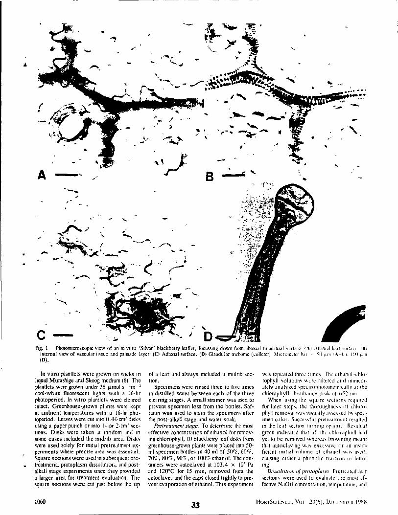
A
-
c
--_ .... -.....
" ,
..
•
- -, ,
. ..:"
.. "" .... -...
.. , " .:
B
1 ... _ .. ............ ---
--_ ... ' "
Fig. 1 Phalomlcroscaplc VICW of an ln Vitro 'SlIvan' blackberry Icaller, focusslng down from JbaxlJI !O JdJXI.!1 .,url.lce 1 \1 .\/1JXIJI k.1f "IlLlLl 1111 Inlernal VICW of vascul.!r rl'isue and pahsJdc 1.lyer (Cl Adaxlal surface. (Dl Glandular trichome (collerer) ~llc.rnmllcr hM = -Il ILnl IA-( " Jill) ~rn (0).
In vitro plantlets were grown on Wlcks rn hqUld Murashige and Skoog medium (6) The plantlets were grown undcr 38 f.Lmol s l'm ~
cool-whlle fluorescent hghts wrth J 16-hr photoperiod. In vrtro plantlets were c1cJred rntact. Greenhouse-grown plJnts were kept at amblent temperatures wuh a 16·hr photoperiod. Leaves were eut rnto 0.44·cm~ drsk'i usrng a paper punch or rnto 1- or ~·cm' ~ecIrons. Dlsks were taken Jt random Jnu ln
sorne cases rncluded the mrdnb area. Dlsk., were used solely for rmtldl pretreJtment experrments whcre precrse area was e~sentlal. Square sectIOns were used rn subsequent pretreatment, protoplasm dlssolutror., and postalkali stage expenments srnce they provlded a larger area for treatment evaluJtlon. The square sections were cut Just below the trp
1060
of J leaf Jnd always rncluded J mldnb ~ec-tian.
Specimens were nnsed three to frve tlmes rn dlstllled water between each of the three cleanng stages. A small strarner was used to prevent specimen loss from the boules. Safranrn was used ta slarn the speCimen, after the post-al kali stage Jnd water SOJk.
Pretreatment stage. To ueterrnme the most effectIve concentrJtlon of ethJnol for removmg chlorophyll, 10 blacI.berry leaf dlsk~ from greenhouse-grown plants were placed mto 50-ml ~peclmen boules m 40 ml of SOl(;, 6OC'c, 70l,{;, 80C:C, 90c.c, or 100~é ethanol. The contarners were autocl.lVeu at 103.4 x 10' PJ and 120°C far 15 mm, removcd from the Jutoclave, and the caps closed tlghtly to prevent evaporatlon of ethanol. ThIs expenment
33
WJ~ repe.lleu three tlnlC\ The CI11.IIlill-L1i1orophyll .,olutlon., \\Ln: tlltered Jllli IfllnJedlJtely Jn.llyzetl 'peLlrophoIOf11ClrlL.lilv .It Ihe chlorophyll JIN'lh.lnce pC.I~ nt h:;~ 11111
When u\lng Ihc "'qu.lre 'CLtIOn., reqllired for IJter SICp ... , Ihe thoroughne ... ., Il! LhltHll' phyll removJI W.I' vl,u.llly .I."e, ... cd hj 'relImen colm. Succe\.,!ul pretre.tlnlCnt re.,ulted ln the k.lt .,eLllon IUPlIng or"lJtll Re\ldll.d grecn Indlc.lled Ih.tt ,dl thl l hloroplivll 11.Il1 yet tn be remllvcu \\IhereJ., br()\~ 11Ing mC.IT1l thJt JutoclJvlng W.I\ eXle\\IVe or .111 II1.,ut· flclent 10111.11 volume ot clhJllol \\"., Il.,~d.
cau~lng clilier .1 phcnolrc rC.ILllon or hurnIng
DIBOIl/tlO/f of pmlOplal/ll l'n:lrl.tlld Ie.t! ~ectJon" were u.,cd III CVJlllJle the mo.,t effective NJOH conccntr.l!lon, tcmpu.tlllre •. Ind
H()RTSU!:~C.[, VOl 23(6), DI (1 \IBl I! I()X/{
(
1
exposure tlme for complete protoplasm removal. NaOH was used al concentrallons of 1%,2%,5%, and 10% al 20°, 55", and 80·C for peflods rangmg from 15 mm 10 1 hr, 10
5-mm Incremenls. The effecl of aUloclavlOg (8) was lesled. The lime for complete protoplasm removal could only be determlOed by mlcroscoplC observatIOn after the phenohc compounds had been bleached 10 the post-alkah stage. The protopla~m dissolu lion slage was deemed complete when mlcroSCOplC observa lion showed the spongy mes· ophyll and the paltsade cells to be free of organelles.
Post·a/ka" stag~. The optImum pretreatment and protoplasm·dlssolutlOn stage treat· menlS were employed usmg new specimens. The most effecllve concentrallon of house· hold blcach (6% NaCIO) for removIOg the brown phenoltc OXldates was determlOed. Concentraltons of 2.5%. 50~, 7SC"'c. ar.d 100% bleach (1.510, 3%, 4.5%, and 6<" '~aCIO) were employed at 20·, 55°, 60·, b5°, and 75°C unlll the sampi es were clear Effec· tlveness of the post·alkah stage was based on the tune needed ta bl:ach specimens from brown ta transparent and upon the mec hanical strength of samples after treatment. A specimen was deemed clear once every spot of brown had been bleached.
Cleared specImens were stamed m aqueous saframn (10 mg·liter· l) and mounted 10
glycerol for seml-permanent mounts. Permanent mounts reqUired dehydratmg cleared. unstamcd matenal through a senes of elhanol concentrallons, stalOmg 10 95% or 100% ethanol. rehydratlOg lOto xylene, and mount· ing 10 Permount.
Pretreatment stage. Spectrophotometnc analysls of chlorophyll extracted from 'SIIvan' leaf dlsks showed that 80% ethanol, wllhm the 15-mm Jutoclavmg mterval, ex· tracted the most chlorophyll. Ethanol concentr:lIIons below 80% removed less chlorophyll durmg thls IOterval. Ethanol at 50% to 70% eventually dlssolved ail of the chlorophyll, but requued longer autoclave limes and a much larger volume of ethanol slOce evaporallon was greater over the extended autoclave mtervals. Ethanol concentratIOns above 80% removed chiorophylliess effecllvely. Chlorophyll removal from IOtact plant lets grown 10 vitrO took 15 to 20 mlO 10
80% ethanol 10 the autoclave. For grecnhouse-grown leaves, chlorophyll removaillme was 25 mm for 'Sllvan' and 35 mm for 'To-
HORTSCIE"CE. VOL. ~3(6), DECE\tOER 1988
tem'. The lime differences for chlorophyll extracllon were probably due to dlfferent amounts of eplcullcular and cUllcular wax (7).
DlSso/ullon ofprotop/asm. Thc most rapld treatment for dlssolvmg the protoplasm was 5% Na OH at 800e for va flOUS penods of lime dependmg on the type of IIssue. Intact plant lets grown 10 vitrO nccded 20 mlO; leaf squares (Jf mature greenhouse·grown 'SIIvan' and 'To!cm' Icaves needed 35 mm and 40 mm, respecllvely. NaOH at 10% caused IIssue dlsmtegrauon, as dld autoclavmg ln NaOH for most samples.
Post·a/ka/I slag~. Immersion m 75% bleach at room temperature for 5 ta 10 mm was the preferred post·alkall trcatment for ln vitrO plant lets. Greenhouse·grown matenal ln 7Se-;, bleach at .55°C becamc transparent ln between 35 to 40 mm. Bleach conccntratlOns below 75t;C took longer to achleve the same results, but 100% bleach or temperatures at or above 60° macerated the tissues. The mldnb and pellole. due to thclr thlckness, were thc last arcas from whlch phcnolic cornpounds were totally bleached.
Il was essentlal to remove die bleach completely by soakmg for a few minutes 10 disIIl1ed water before stalnlOg wllh safranlO because of the destrucllve interactIOn of safranln and sodium molecules. In vttro plantlets statned adequately in only 10 to 30 sec whereas grecnhouse-grown specimens took 1 to 2 mm If superficlal !Issues, such as ham or guard cells, \\-ere of Inter':~l or 510"7 mm If IOternalllssues, such as X) lem, were to be exammed.
Smce ln vllro plantlets were c1eared Intact, mlcroscoplc observatIOn of the external and internai structures of the root, stem, petiole, and leaves was poSSIble. Ali the cell layers froOl ad axial to abaxlal leaf surfaces could be mlcroscoplcally observed (Fig. 1 A, B, and Cl. When mounted abaxlal slde up, the adaxlal epidermal layer was not always qUlle as clear as the upper surface due to follar wldth, but It was usually not difficult ta dlscern cell patterns or stomata. Differences 10
cell shapes and palterns between palisade and spongy mesophyll layers were apparent.
ln the case of greenhouse-grown leaves, secllons mcluding the mldnb were more likely to exceed 2 to 3 mm m wldth. Mounted adaxlal slde up, the lower epldermal layer was usually out of focus due ta the thlck epldermal ce Ils, the many celllayers and the
dlfficulty ln keeplOg the coversllp adhered to the specimen. For optimum examlnallon of both leaf surfaces. specimens were haJved and mounted bath adaxlal and abaxlal side up, examlned and subsequently turned aver, or the mldnb was removed.
ln summary, the cleanng and stalOmg method outhned 10 Table l, wlth mlnor varlallons, gave rapld (maximum tlme of 2 hr) and excellent results for the plants exammed. This c1earmg procedure was parllcularly suc· cessful wllh m VitrO plant lets for whlch organs and celllayers werc made c1early Visible. Due to liS speed and reproduclblhty, thlS procedure may prove to be a use fui laboratorv tool to factlltate stomatal counts and me·asurements. It should be especldlly usdul as a method for showmg the three·dIJTlen· slonal relallonshlps of cell Idyers: vJ5C'JIJr tissues, crystal deposlls. hydathodes. tri·
chomes (Fig ID), Jnd other fohJr struc· tures. We have found the method useful ln obtatnmg a general Vlew of an area that IS to be exammed later uSlOg microtome cuttmgs.
Llteralurt Cited
1. Arnon, H J 19 '9 Leaf cleanng, T Ur.OlC News 37192-19. -
2. Donnelly, D J and W E Vldaver 1984 Lcaf analomy of red raspberry Iransfcrred from cuhurc 10 5011 J. Amer. Soc. Hon_ Scl 109'172-176_
3 Fabbn, A , E. Suner. and S.K Dunston. 1986. Analomlcal changes 10 pc:~lslenr ,,-""cs of tl"UC cul'urcd ,lrJ\I,~crry plan" Jf-:' .:. movallrom culrure SClcn:IJ Horl ~s::: 1-
337 4 Gardner, R D 1975. An OVCrvlCW of boran·
lcal cleanng lechnrque Siam Technol 5099-\05.
5. tchen. C 1952. A mClhud for c1earrng 1U\'cs Ph}1opalhology 42 352
6 Murashlge, T. and F Skoog 1962 A revlscd medium for rapld gro\\<th and blo as· says wnh tobacco tissue cultures Ph\"Slol Plant. 15'473-497 .
7. O'Bncn, T P and \1. McCullv 1981 The study of piani slruclure. prmciplcs and sc· Icclcd mcthods, Tcrmarcarphl Pey ud. Melbourne
8. O'Bnen, T P and 1 von Telchman 19"7J AUloclavrng as an ald ln the c1canng ofpbnt specl.nens. Stam Tcchnol .a9 175-176.
9. RodlO, R J and R E DaVIS 1967 The ~se of paparn m cleanng piani lIssues fur ~ hole mounls. Siam Tcchnol 42 203-:0u
10 Shobc. W R and ~ R Lerslen 1967 A techmque for c1earmg and stalOlOg gymno· spcnn Icavc~. Bot. Gaz. 128 150-t5:
1061
f [ r • t r II " , r ,
Having developed a satisfactory clearing and staining method for observing
tissue-cultured and greenhouse-grown plant material, acclimatization experiments
were initiated in order to 1) detennine the effects of both increased agar
concentrations (6, 9 and 12 g/l) in the culture medium and in vitro rooting
on ex vitro survivaI, growth and stomatal index of micropropagated 'Silvan'
blackberry shoots and 2) detennine the effect of high relative humidity and low
light intensity on the stomatal function of leaves from a) ex vitro plantlets grown
on full and 1/4 strength modified MS (1962) basal medium and b) greenhouse
grown 'Silvan' plants.
35
r
4. Acc:Jimatizaion 01 mJc:ropropalated 'Silvan' blacl.berry
4.1. Introduction
Ex vitro acclimatization can be one of the mos! difficult stages of the
micropropagation process. This is due to the impaired ability of transplants to
control water Joss as a result of poorly fonned epicuticular and cuticular wax
layers (Conner and Conner, 1984, Fabbri et al., 1986, SUUer and Langhans, 1982,
Sutter 1984, 1985), non-functional, open stomata (Brainerd and Fuchigami, 1981,
DonneUy et aL, 1986, Short et al., 1987, Wardle and Short, 1983) and, in sorne
cases, poorly formed root systems (Grout and Aston, 1977).
Generally, plants grown under culture conditions have reduced foliar
epicuticular and cuticular wax. In vitro-formed foliar wax may he different in
arrangement and chemical composition compared with that of greenhouse-grown
plants (Sutter, 1984, 1985). Reducing the relative humidity level in vitro from
100 % to 35 % by llsing a variety of closures for the cuhure containers
increased epicuticular wax fonnation in cauliflower (Bras sica oleracea var botyris)
and chrysanthemum (Chrysanthemum morifolium Ram.) (Short et al., 1987).
Increasing the agar concentration from the usual 6-8 g/l to 15 gI1 resulted in
decreased relative humidity in the culture containers; from 98 % to 89
%. Subculture to medium with an agar concentration of IS g/l 2 weeks prior
to transplantation promoted glaucous leaf production in carnauon (Dianthus
caryophyllus) plantlets and improved transplant survival from Il % to 72 %
(Ziv et al. 1983). Similarly, increasing the agar concentration in the
footing medium from 6 g/l to 14 g/I increased the ex vitro survival of
micropropagated cherry (Prunus cerasus) rootstock from 13 % to 45.3 % (Marin
and Gella, 1987).
Stomata fonned on most in vitro plant leaves did not close in response
to stimuli snch as darkness, abscisic acid, solutions of high osmolarity (mannitol
36
or suerose) or exposur~ to high Ieveis of COz (Brainerd and Fuehigami, 1982,
Wardle and Short, 1983, Wetzstein and Sommer, 1982, Ziv et al., 1987).
Reducing the relative humidity from 95-100 % to 30-40 % in vitro decreased
the time necessary for stomata to close in apple (Malus domestiea (Borkh.)
(Brainerd and Fuehigami, 1981). Since transpiration losses oceur through the
open stomatai pores, researchers have investigated stomatal frequeney or stomatal
index, whieh COlTects for differences in Ieaf cell size, in culture and during
ex vitro acclimatization and eompared it with that of greenhouse-grown control
plant leaves. Stomatal index (S.I.), described by the fonnula: S/(S + E) x
100, where S = number of stomata and E = number of epidermal eells, was
unaffected by micropropagation in both potato (Solaoum laciniatum) (Conner
and Conner, 1984) and rose <&wl muitiflora L.) (Querait, 1988). The S.I.
of Leucaena leucocephala (Lam) De Wit. was lower on leaves from micropropagated
shoots at the end of the multiplication stage (7.21) than on leaves from the
original, mature donor plant (12.02) (Dhawan and Bhojwani, 1987). However,
the S.I. of each successive new leaf following ex vitro transplantation has never
been documented.
Little attention has been paid to the fate of the persistent leaves from
culture during the ex vitro acclimatization period despite the fact that these make
up most of the biomass present at transplantation. It is of interest to know
whether the se le av es contribute to new growth. through photosynthesis Of nutrient
transfer during senescence. or whether they act as sinks (net respirers) using
photosynthates which could otherwise enhance new leaf or root growth. Carbon
fixation of persistent leaves of strawberry (Fra&aria x ananassa Duch) was
insufficient in culture for autotrophic growth and did not increase during the
ex vitro acclimatization period. Persistent leaves of strawberry senesced and
dropped off the transplant almost immediately (Desjardins et al., 1988) to 4
37
wecks after ttansplantation (Grout and Millam, 1985). Grout and Millam
concludcd that strawberry transplants must be dependant upon stored materiaIs
to provide a carbon source for growth and development immediately following
transplantation. Wardle et al. (1983) showed that persistent chrysanthemum
(Cluysaothemum morifolium) leaves acted as storage organs until they senesced;
contributing mineraIs to the new organs developed during ex vitro acclimatization.
Carbon fixation in persistent leaves of red raspberry (Rubus ideaus) was about
one quarter that of control plant levels. After transplantation these leaves
continued to photosynthesize at low levels. However, they became net respirers
and sinks for photoassimilates formed in the new leaves after 1 month ex vitro
(Donnelly and Vidaver, 1984b). The persistent leaves of red raspberry did not
deteriorate rapidly after transplantation and sorne were retained on the transplants
for up to 3 months. The persistent leaves of 'Silvan' blackberry were similar
to those of red raspberry in their retention ex vitro.
There is disagreement in the literature as to the benefits derived from in
vitro rooting. For sorne adventitously propagated plant species, in vitro-formed
roots may not be completely functional after transplantation, possibly because of
incomplete vascular development between the roots and shoots, contributing to
excessive desiccation of transplants on exposure to low relative humidity levels.
Vascular connections were narrow and ill-formed in adventitiously cultured
cauliflower (Brassjca oleracea var. botrytis) restricting acropetal water transfer
(Grout and Aston, 1977). In vitro roots of Gladiolus formed in 0.8 % agar
were abnormally thin, sparse, and lacked root hairs (Logan and Zettler, 1985).
In vitro roots of Acacia 1mB had no root haïrs when grown on agar-solidified
medium and frequently lacked a fully developed vascular system (Skolmen and
Mapes, 1978). In many plant species there is a delay in the initial growth
of in vitro rooted plantlets after transplantation. This too has becn attributed
38
..... to the lack of a functional in vitro root system at transplantation (Debergh and
Maene, 1981, Pierik, 1988). However, initial growth comparisons of shoots and
plandets after transplantation were not reportcd. In some plant species, mots
fonned in vitro died after 2 weeks of growth ex vitro and were replaced by
new ones (Debergh and Maene, 1981). It is also possible that some in vitro
roots are damaged during the planting pracess, increasing the risk of root and!
or stem disease (Debergh and Maene, 1981).
Rooting of shoots in vitro is labour intensive and therefore cosdy due to
the requirement for individual handling of each shoot. For this reason it May
not be economically advantageous to root in vitro, whether the roots are
functional or not (Debergh and Maene, 1981). However, axillary shoot
propagated in vitto-rooted loganberry (Rubus lo&anobaccus Bailey) plandets were
hardier after one month ex vitro compared with shoots that were rooted ex vitro
(Donnelly et al., 1980). Most commercial micropropagation facilities or
companies practice in vitro rooting (Debergh, 1988a). The reason for this is
unc1ear; it is postulated that prejudice against unrooted shoots is due to the
habitual u~e of rooted material for successful transplanting of almost ail seedlings
and vegetatively propagated shoots.
In vitro acclimatization strategies for raspberry have included culturing shoots
on media with reduced (20 %) concentrations of modified MS (1962) macro
elements for 2 weeks prior to transfer (Welander, 1985). This was reported
to increase in vitro rooting from 33 % to 100 % and promote survival (from
63 % to 100 %) 1 month after transplantation. Half-strength modified MS
(1962) was recommended for in vitro moting of apple (Mah{1 domestica L.)
(Zimmerman et al., 1987). Another in vitro acclimatization strategy is the
reduction or elimination of sucrose in the medium. This has been shown to
promote photoautotrophy in cultured plants (Grout and Price, 1987, Kozai, 1988a).
39
The effcet of ~duced macro and micro nutricnts or medium sugar upon certain
aspects of plant physiology, such as stomatal function, has not been invcstigatcd.
The objectivcs of the foUowing cxperiments were to:
1) Detennine the effect of increased medium agar concentration, from the control
levcl of 6 g/l to 9 and 12 g/l, on survival and growth perfonnance (transplant
height, leaf number, longest leaf lellgth) of 'Silvan' shoots and plantlets during
the first 3 weeks ex vitro.
2) Detennine the relationship between the initial (persistent) leaf number or initial
fresh weight with final fresh weight 3 weeks after transplantation.
3) Detennine if the stomatal index differed between leaf populations (persistent,
new leaves fonned during week l, week 2 and week 3) and if il was affected
by different medium agar concentrations or in vitro IOOting during the first 3
weeks ex vitro.
4) Find a suitable method to test the open/closure mechanism of 'Silvan' blackberry
stomata on epidennal peels.
5) Derennine the period of time required after transplantation for stomata ta exhibit
the capacity to open and close and assess this function in persistent and new
leaf populations.
6) Detennine the effect of rugh relative humidity (> 95%) and low Ught levels
(82 pmol sol moZ) on stomatal function of greenho\lSO-amWD 'Silvan' leaves and
ex vitro 'Silvan' plantlet leaves from full and 1/4 strength modified MS (1962)
basal medium.
40
4.2. Materials and Methods
4.2.1. Source Plants
'Silvan' blackberry (Rubus sp.) plants developed from 30 cm long stem
cuttings were placed in a mist chamber for 2 weeks. The rooted-cuttings were
then repotted into 22.S cm standard pots and grown for 6 weeks in the
greenhouse at ambient temperatures (24 ± 5 OC) under natural light supple
mented with cool-white flourescent light, to total 100 pmol S-I m-2 for 16 br
per day. These source plants were grown in PromixR supplemented with 30 g
dolomitic limestone and ISO g osmocote (14:14:14) per 30 cm'.
For tissue culture purposes cuttings (10-30 cm Jong containing 2-3 nodes)
were taken from the source plants and the leaves removed. The cuttings were
washed in distilled water for 10 min and then agitated for 20 min in 10 %
bleach (0.6 % NaCIO). Surface sterilized cuttings were rinsed in sterile distilled
water for 5-10 minutes prior to aseptic shoot-tip excision. In vitro shoots and
plantlets were derived from 1-2 mm long shoot-tip expllll1ts initiated and grown
in 50 ml test tubes containing filter paper wicks and 15 ml of liquid modified
Murashige and Skoog (1 962)(MS) basal medium with additions (Appendix 1).
After 2 subcuItures, at 4 week intervals, shoots were transferred from test tubes
to 400 ml polypropylene containers (10-15 shoots in each) on 50 ml of
the same medium (multiplication) solidified with 6 g/l Difco-Bacto agar.
The shoots were grown under 57 pmol S-I m-2 cool white fluorescent
light with a 16-hr photoperiod at a constant temperature of 25 OC.
41
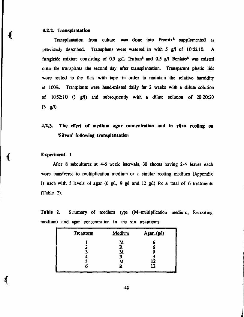
(
(
( ...
4.2.2. Transplantation
Transplantation from culture was done into Prom.ixR supplemented as
prcviously describcd. Transplants wcre watcrcd in with 5 g/l of 10:52:10. A
fungicide mixture consisting of 0.5 g/L TrubanR and 0.5 g/l Benlate· was mistcd
onto the transplants the second day after transplantation. Transparent plastic lids
were sealed to the flats with tape in order to maintain the relative humidity
at 100%. Transplants were hand-misted daily for 2 weeks with a dilutc solution
of 10:52:10 (3 g/l) and subsequently with a dilutc solution of 20:20:20
(3 g/l).
4.2.3. The effe(!t of medium agar concentration and in vitro rootlng on
'Silvan' following transplantation
Experiment 1
After 8 subcultures at 4-6 week intervals, 30 shoots having 2-4 leaves each
were transferred to multiplication medium or a similar rooting medium (Appendix
1) each with 3 levels of agar (6 g/l, 9 gIl and 12 g/l) for a total of 6 treatments
(Table 2).
Table 1. Summary of medium type (M=multiplication medium, R=rooting
medium) and agar concentration in the six treatments.
Treatment
1 2 3 4 S 6
Medium
M R M R M R
42
Aaar (ail>
6 6 9 9
12 12
Twenty shoots or plantlets per treatment, each with 7-8 leaves and
0.8-1.2 cm tall were placed in randomized complete block groupings of 10 and
replicated once in 96-plug plastic gennination flats. Transplants from the lowest
(control) medium agar concentration (6 g/l) had in vitro mots measuring 2-5
cm. In vitro mot length was generally shorter with increasing medium agar
concentrations: 1-3 cm (9 g/l) and 1-2 cm (12 g/l). After 3 weeks the flat
covers were gradually removed over a pcriod of 3 days. Transplants
in the Conviron E-IS growth chamber received 127 ~mol S-I m-2 cool-white
fluorescent light with a 16-hr photoperiod and a constant temperature of 25
± 2 OC. Once the flat covers were removed, transplants were exposed to a
relative humidity of 30-40 %. Leaves formed in culture (persistent) were tagged
on the petiole with a small drop of white Liquid PaperR at the time of
transplantation and thereafter new leaves were tagged on a weekly basis using
different colours of Liquid Paper.
Transplant height was measured from the soil line at transplantation and
from the edge of the flat to the base of the newest leaf from week 1 to
week 5. Height. total number of leaves (both persistent from culture and new)
and the length of the longest leaf were recorded for al1 transplants 1. 2 and
3 weeks after transplantation. Statistical analysis was done separately for each
week. Three randomly chosen transplants per treatrnent were cleared (Chapter
3) each week to determine the foliar stomatal index (S.I.). The S.I. was
calculated from photomicrographs of 60-100 epidennal cells including 15-20
stomata per field of view. Two randomly located fields of view from each
leaf were photographed. The total number of stomata counted for each leaf
type (persistent, week 1, week 2, week 3 and week S) at each weekly time
interval ranged from 40 to 200 (2-10 leaves). The S.I. of gr-.enhouse-grown
plant leaves was determined by clearing two leaves of five source plants
43
(N = 10) and taking photomicrographs of two randomly chosen areas of the
leaves. The total number of stomata photographcd was 110.
Arcsin ttansfonnations of S.I. data were performcd on PC-SAS. Differences
between means at the O.OS level of significance were determined with the
Duncan 's Multiple Range test using the same program.
Experiment l
After 12 subcultures at 4-6 week intervals, 60 shoots having 2-4 leaves
each were ttansferred to multiplication or l'OOting medium as in Experiment 1.
At the end of 3 wecks 10 shoots/plantlets from multiplication and rooting media
with varying agar concentrations (Table 2) were weighed and their leaves counted,
lhen transplanted in a completely randomized design to 36-plug plastic germination
flats. The flat covers were c10sed tightly for the tirst 2 weeks and then
complelely removed. Transplants were grown in a Conviron PGW36
growth chamber under 127 JImol S-I m-z cool white fluorescent Iight with a
16-hr ph010period al a constant lemperature of 25 oC and a relative humidity
level of 80 %. AlI the plants were harvested to determine final fresh weight.
Initial fresh weight originally inc1uded the mot mass (for plantlets). When
harvesting for final fresh weight il was found that, in sorne cases, there was
a large amount of caBus at the base of the stem. This was removed to allow
a better basis for comparison between treatrnents. Flnal fresh weight thus
inc1uded the above-ground biomass (stem and leaves) with any callus removed.
Statistical analysis of all data was done using PC-SAS. Differences between
rneans were te~ted using the Duncan's Multiple Range test and regressions were
done using General Linear Model tests. Correlations between initial fresh weight
and initial leaf number to rmal fresh weight were tested for fit to l:rtear,
quadratic and cubic equations at the 0.05 level of significance.
44
4.2.4. Preliminary tests on stomatal lunction
Leaves of grecnhousc-grown 'Silvan' were covered in aluminium foil 2 br
before removal. Examination of epidennal peels from these severed leaves
showed that most stomata were closed. The peels were then floated on a
phosphate buffer (10-2 M ~HP04 brought to a pH of 6.9 with 10-2 M Kl\P04)
(Will mer and Mansfield, 1969) and placed under a 120 Jlmol S-I m-2 sodium
light. The leaf peels were observed under a light microscope every S min
for 35 min and slomatal apertures we~"e measured directly using an eye-picce
micrometer. After 30 min the majority of stomata were open to some degree.
To detennine the mOSI useful test for slomalal function, greenhouse-grown
'Sil van' epidermal leaf peels on phosphate buffer were exposed to a) ABA (2: 1
10-2 M ABA: phosphate buffer) (Martin et al., 1983), b) light/dark periods or
c) 1 M NaCI treatments. AlI tests were replicated twice.
Five epidennal peels were:
a) floated on the ABA solution white under the sodium light and observed
at 5 min intervals to determine the time necessary for stomatal closure. Once
the stomata had closed the epidermal peels were retloated on the phosphate buffer
under the light and examined every 5 min for stomatal reopening.
b) floated on the phosphate buffer in foil-covered petri dishes and examined
every 5 min to delennine the time necessary for stomalal c1osure. Once stomata
had c1osed. the foil was removed and the petri dishes were placed under the
light. The epidennal peels were examined every 5 min to observe wh en stomalal
reopening occurred.
c) tloated one al a time on the phosphate buffer on a glass microscope
slide and a lM NaCI solution was drawn over them. This was done by
using filter paper to absorb the buffer &olution from one sicle 'Jf the coyer slip
and by adding the NaCI solution via pipette from the other side. Once stomatal
4S
,
l
r j
{ ..
closure was observed through the microscope the lM NaCI solution was replaced
by the buffer by using mter paper to absorb the NaCI solution from one side
of the coyer slip and by adding the buffer solution via pipette from the other
end until stomatal opening was observed.
4.2.5. Evaluation or ex vitro stomatal rundion or 'Silvan' plantlets
Experiment 2
Epidermal peels were always taken from between the second and third
horizontal vein at the base of the leaf. In the 2 and 4 week tests, two
epidermal peels were harvested from each of 2-3 plants/tteatment. In the 3
week test two epidermal peels were harvested from every leaf and 5-6 plantsl
treatment were slùnpled.
Stomatal function was detennined using the lM NaCI test as described in
the previous section. Each field of view/epidermal peel contained between lf)-
20 stomata. Twenty stomata were considered adequately representative of the
total leaf surfl\ce (Meidner, 1981). The total number of stomata measured per
leaf type (leaves which had developed during the fust, second and thini week)
was 40-100 for the 2 and 4 week tests and 150-200 for the 3 week test.
Stomatal apertures were measured directly under the microscope using an eye
piece micrometer in the 2 and 4 week tests and in the 3 week test
photomicrographs of the stomata were taken before and after application of the
lM NaCI solution. Stomatal apertures were subsequently measured in the dark
room by projecting the magnified images of specimen negatives (and a
micrometer sc ale) directly from the negative enlarger onto the bench top.
46
.....
4.2.6. Tbe errects of bigb relative humidity and low light intensity on ex
vitro stomatal lundion 01 'Silvan' plantlets grown on lull and
1/4 strength rooting medium
Experiment 3
Twenty shoots with 2-4 leaves each were transferred from multiplication
medium ta both full strength and 1/4 strength rooting medium (see Appendix
1). Three weeks later the 20 plantlets each from full and 1/4 strength rooting
media were randomly planted into two 36-plug plastic gennination flats and
placed in a dew chamber. The dew chamber had a light intensity of 82.5
pmol sol m02 cool-white fluorescent Iight, a 16 br photoperiod, a constant
temperature of 25 ± 1 OC and a relative humidity of 95-100 %. After 2
weeks in the dew chamber 4 plantlets from each of the in vitro treatments
were harvested, 8 plantIets from each treatment were placed into a growth
chamber and 8 plantlets were left in the dew chamber. The growth chamber
had a light intensity of 90 pmol sol m02 cool-white fluorescent Iight. a 16 br
photoperiod, a constant temperature of 25 oC and a relative humidity which
ranged between 30 - 55 %. After 17 and 21 days ex vitro 4 plantlets were
harvested from each treatment, both from the dew and growth chambers. The
leaves on these transplants were identified as P leaf (persistent leaf from culture),
leaf 1 (flfst new leaf formed after transplantation), leaf 2 (second new leaf),
etc. Two leaves of each type were randomly selected from the four plantlets
from each treatment. Two epidennal peels were removed from each of the
leaves sampled for evaIuation of stomataI function.
StomataI function was detennined using the lM NaCI solution as described
for Experiment 2. Once stomatal closure occurred or if the protoplasm collapsed
an attempt was made ta reopen the stomata by replacing the lM NaCI solution
47
".f \.
with the phosphate buffer. Photomierographs were taken white the epidcnnal
peels were in buffer, after the application of the hypertonie (lM NaCI) solution
and again onee the hypertonie solution was replaeed by the buffer. To ensure
precision in measurement of stomatal aperture a 400-480 magnifieation was used.
Eaeh photomierograph eontained no more than S-8 stomata. The number of
stomata measured for eaeh leaf type of every tteabnent ranged from 5-20 and
was limited because of time considerations and the problem of guard eells going
out of focus white in the hypertonie solution. The stomatal apertures were
measured as in the previous experiment. An aperture of oS 1 pm was considered
closed. This was a eonservative value sinee a stomatal aperture .s 2 pm was
usually considered to be closed (Zelitch, 1961; cited in Brainerd and Fuehigami.
1981).
4.2.7. The efTects of high relative hurnidity and low Iight intensity on
stornatal function of greenhouse-grown 'Silvan' plants.
Six week old greenhouse-grown source plants were placed in the same dew
chamber used for the transplants from culture. At weekly intervals the newly
fonned leaves were tagged. After 4 weeks the original leaves present on the
plant when it was ttansferred to the dew chamber and the new leaves fonned
during the second week in the dew chamber were harvested. Two epidennal
peels were removed from each of these leaves and stomatal function was
determined using the 1 M NaCI test. StatistieaI analysis of aIl data was done
using PC-SAS. Differences between means were tested using the Student's
t-test
48
4.3. RESUL TS AND DISCUSSION
4.3.1. The effect of medium agar concentration and in vitro rooting on
'Silvan' following transplantation.
In experiment 1 transplant ~urvivaI was 98 % during the rust 2 weeks.
AlI of the shoots and plantIets in experiment 2 survived in the 2 week test
and all plantlets survived in the 3 and 4 week tests. However, the survivaI
of shoots was not as good in the 3 (1/30) and 4 (5/15) week tests (Table
3). 'Silvan' shoots were apparentIy less able than plantIets ta adapt ta the
ex vitro environment. The cause for the lack of shoot ~mrvival on Le the covers
were removed is not fully understood but it is postulated that the new roots
formed ex vitro were unable ta compensate for the sudden increase in the
transpiration rate once these covers were removed.
In contra st ta results with carnation (Ziv et al, 1983) and cherry rootstock
(Marin and Gella, 1987) where increasing the medium agar concentration from
6 gIl ta 14-15 g/l increased ex vitro survivaI, 'Silvan' blackberry survival ex
vitro was not affected by the level of agar in the medium. The survival of
rooted treatments was least affected by the early removal of the flat covers
in experiment 3 (Table 3). This suggests that despite possible evidence for
non-functionaI in vitro-formed roots (Grout and Aston, 1977, Logan and Zettler,
1985, Skolmen and Mapes, 1978, Debergh and Maene, 1981). roots of • Silvan'
formed in vitro are probably functional after transplantation. Loganberry plantlets
(Rubus loganobaccus) were also hardier aCter 1 mon th ex vitro than shoots
(Donnelly et al, 1980).
At the time of transplantation mean transplant height was similar for all
treatments (1.1 cm) except treatment 5 (0.9 cm) (Figure 2). The apparent
decrease in height from transplantation (week 0) to week 1 resulted from an
49
undesirable but necessary change in measurement practice implemented to ~uce
handling of the transplants. The plantlets from media containing 6 and
9 g/l agar concentrations were significantly taller than the shoots throughout the
3 week period. This was true for the flfst but not the second and third week
for the highest medium agar concentration (12 g/l). For the fU'St 2 weeks after
transplantation, plantlets from media containing 6 gIl were talle st (2.04 cm) but
the mean height of all rooted transplants were statistically similar by the end
of tne thini week, ranging from 2.84, 2.89 and 2.27 cm for medium agar con
centrations of 6. 9 and 12 gI1 respectively.
Plantlets and shoots all had 7-8 leaves at transplantation (Figure 3). The
initiaI reduction in leaf number from week 0 to week 1 was due to the death
of persistent leaves. At the end of the fl1'st week plantlets had significantly
more leaves than shoots from the same agar concentrations. AU treatments had
a similar mean leaf number at the end of the second (7) and thini week (8-
9) except for treatment 5 which had 6 (second week) and 7 (third week). By
the end of the third week aImost all persistent leaves had abscissed.
One week after transplantation persistent leaves from culture were still the
longest leaves on the transplants and had not grown. The average persistent
longe st leaf lengths were found on plantlets from the 6 and 9 g/l agar con
centrations (1.40 and 1.35 cm) (Figure 4). Shoots from 6 and 9 g/l as weIl
as plantlets from 12 gI1 agar concentrations had similar (ranging from 1.05 -
1.75 cm) average persistent longest leaf lengths al this time, while shoots from
12 g/l agar concentrations (0.98 cm) had the shortest persistent leaves. At the
end of the second and third week plantlets did not differ in average longest
new leaf lengths "tgardless of the agar concentration in the medium from which
they were transferred. However, plants that were rooted al the time of
50
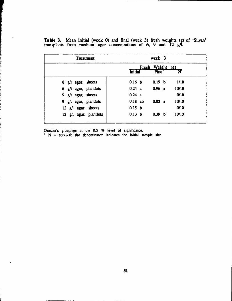
" , ~ ,
Table 3. Mean initial (week 0) and final (week 3) fresh weights (g) of 'Silvan' transplants from medium agar concentrations of 6, 9 and 12 gIl.
Treatment week 3
Fresh Weight (g) Initial Final NI
6 g/1 agar. ~hoots 0.16 b 0.19 b 1/10
6 g/l agar, plantlets 0.24 a 0.96 a 10/10
9 g/l agar, shoots 0.24 a 0/10
9 g/l agar, plantlets 0.18 ab 0.83 a 10/10
12 g/l agar, shoots 0.15 b 0/10
12 g/l agar, planUets 0.13 b 0.39 b 10/10
Duncan's groupings al the O.S % level of significance. • N = survival; the denominator indicates the initial sample size.
SI
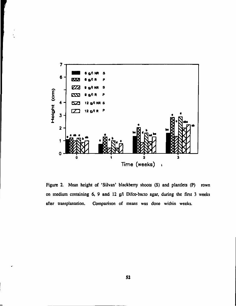
7 - .t/lNA S 6 1!22! .t/lR P
- lZZa .t/lNR S
E t'SSl t t/I R P U ...,
4- fSZJ 12 III NR S .. s:: rz::J 12 t/I R P .~
Q) 3 • l
2 ...
1
0 0 1 2 l
Time (weeks)
Figure 2. Mean height of 'Silvan' blackberry shoots (S) and plantlets (P) rown
on medium containing 6, 9 and 12 gIl Difco-bacto agar, during the fust 3 weeks
after transplantation. Comparison of means was done within weeks.
52
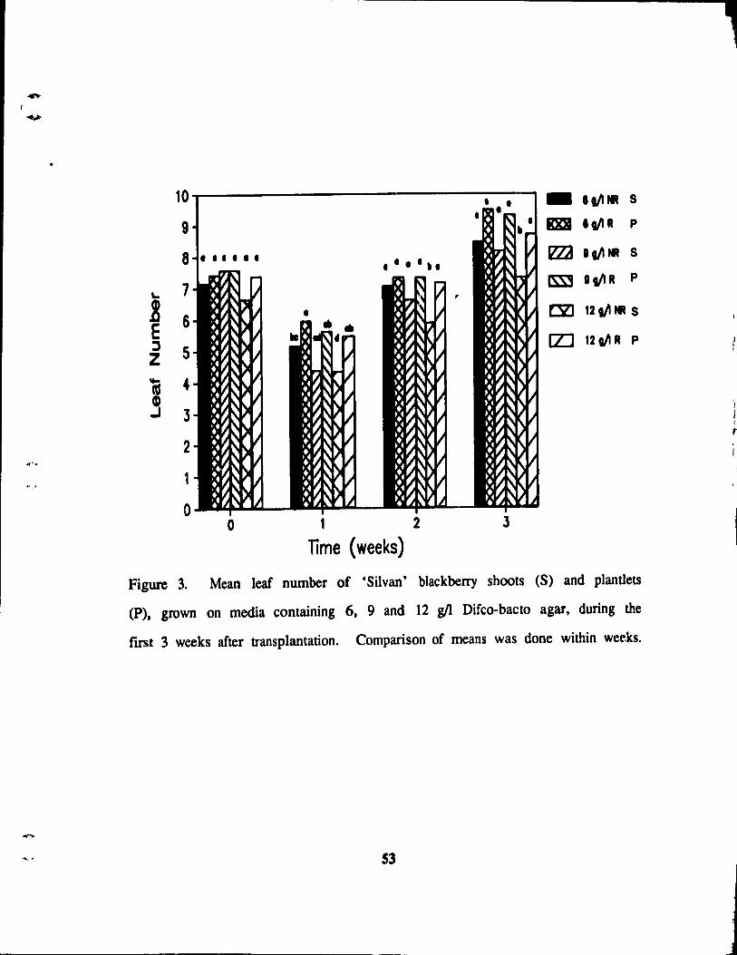
....
10 - 11/1- s
9 • DI II/IR P
8 ~ 11/1- S
L. 7 ~ II/IR P
~ 6 E ~ 5 Z
• CE 121/1- S
[Z] 121/1 R P
i 4 j 3
2
1
0 0 1 2 3
lime (weeks)
Figure 3. Mean leaf number of 'Silvan' blackberry shoots (S) and plantlets
(P), grown on media containing 6, 9 and 12 gIl Difco-bacto agar, during the
fmt 3 weeks after transplantation. Comparison of means was done within weeks.
S3
,
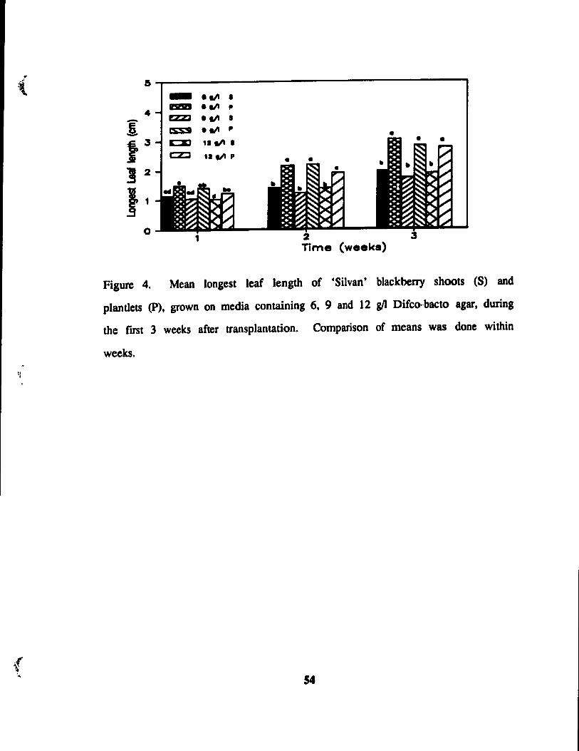
(
( ' ..
a~------------------------------------------~
..
o
- elll • lmD e III p
EZZl • III • • III P
'1 III • '1 iii P
•
Time (weeka)
Figure 4. Mean longest leaf length of 'Silvan' blackberry shoots (S) and
plantlets (P), grown on media containing 6, 9 and 12 g/l Difco-bacto agar, during
the flI'St 3 weeks after transplantation. Comparison of means was donc within
weeks.
S4
transplantation (plantlets) had significantly longer new leaves (2 - 3 cm) than
non-rooted shoots (1.2 - 2 cm).
Ex vitro shoot height, leaf n~!11ber and root length of cauliflower and
chrysanthemum decreased with increasing medium agar con(.entrations. However,
the concentrations of agar used were higher than those used in this experiment
(12. 16 and 20 gIl) (Short et al., 1987). The negative effeet of high agar
concentrations on shoot height, leaf number and root length was attributed to
the decrease of water availability in the culture vessels. Both reduced water
(Debergh, 1983) and minerai (Tanaka, 1981) availability in media occurred
through the decreased pore sile and increased matric potential at high agar con
centrations. The point at which the agar concentration negatively effects plant
growth varies with different brands of agar (Debergh, 1983). It is possible
that increased in viU'O root absorption might counteract the reduced water and
nutrient availability in such cases. This could perhaps explain why in vitro
rooted plantlets, at the highest agar concentration (12 g!l), developed more new
leaves and leaves of longer length after 3 weeks ex vitro than shoots grown
on the same agar concentration (Figures 2-4).
In vitro rooting had a much greater effeet than did medium agar con
centration on perfonnance of ex vitro transplants. 'Silvan° blackberry plantlets
were taller and had greater longe st leaf lengths than shoots during the first 3
weeks ex viU'O (Figure 2 and 4) suggesting that ex vitro plantlets were more
immediately productive than shoots. In vitro-fonned roots of 'Silvan° blackberry
were apparently functional under ex vitro conditions.
55
( 4.3.2. The etrect 01 'rab weilbt at transplantation (initial) and penlstent
leal number on final lresb weight 3 weeks aRer transplantation.
Three weeks after transplantation the mean initial fresh weights of treaunent
2 (0.24 g) and treatment 3 (0.24 g) were significantly greater than treatments
l (0.16 g), 5 (0.15 g) and 6 (0.13 g) but similar to treatment 4 (0.18 g)
(Table 3). At the end of the 3 week period the average fresh weights of
treatments 2 (0.96 g) and 4 (0.83 g) were significantly greater than treatments
1 (0.19 g) and 6 (0.39 g). The net growth increase was statistically similar
between all surviving treatments: 1 (l95 %), 2 (466 %), 4 (499 %) and 6
(308 %) (Table 4). Almost no shoots survived the change in environment once
the covers were removed. This seems to indicate that the shoots were not
as acclirnatized as the plantIets 2 weeks following transplantation.
At the end of 3 weeks ex vitro, treatment 5 had the fewest persistent
leaves (5.6). Treatments 1 (5.7), 5 (5.6) and 6 (5.9) had fewer persistent leaves
than tteatments 2 (7.6) and 4 (7.6). No relationship was observed between
above-ground fresh weight of plants harvested 3 weeks after transplantation and
initial persistent Ieaf number.
Romberger and Tabor (1971) discussed the possibilit: that high agar con
centrations in media may reduce the diffusion rates of enzymes and other large
molecules. This, as weIl as the possibility of lower water potential (Shon et
al., 1987)(Ziv et al., 1983), may have contributed to the negative effeet of high
agar concentrations on initial and final fresh weights of uuliflower and carnation.
Increased agar (Difco-Bacto) concentrations (6 • 8 g/l) significantIy reduced the
fresh weight of Cynara scolymus L.. However, therc were no significant
differences in the fresh weight production of ~ scolymus L. shoots grown on
8 gIl to 1~ g/l agar concentrations (Debergh, 1983).
56
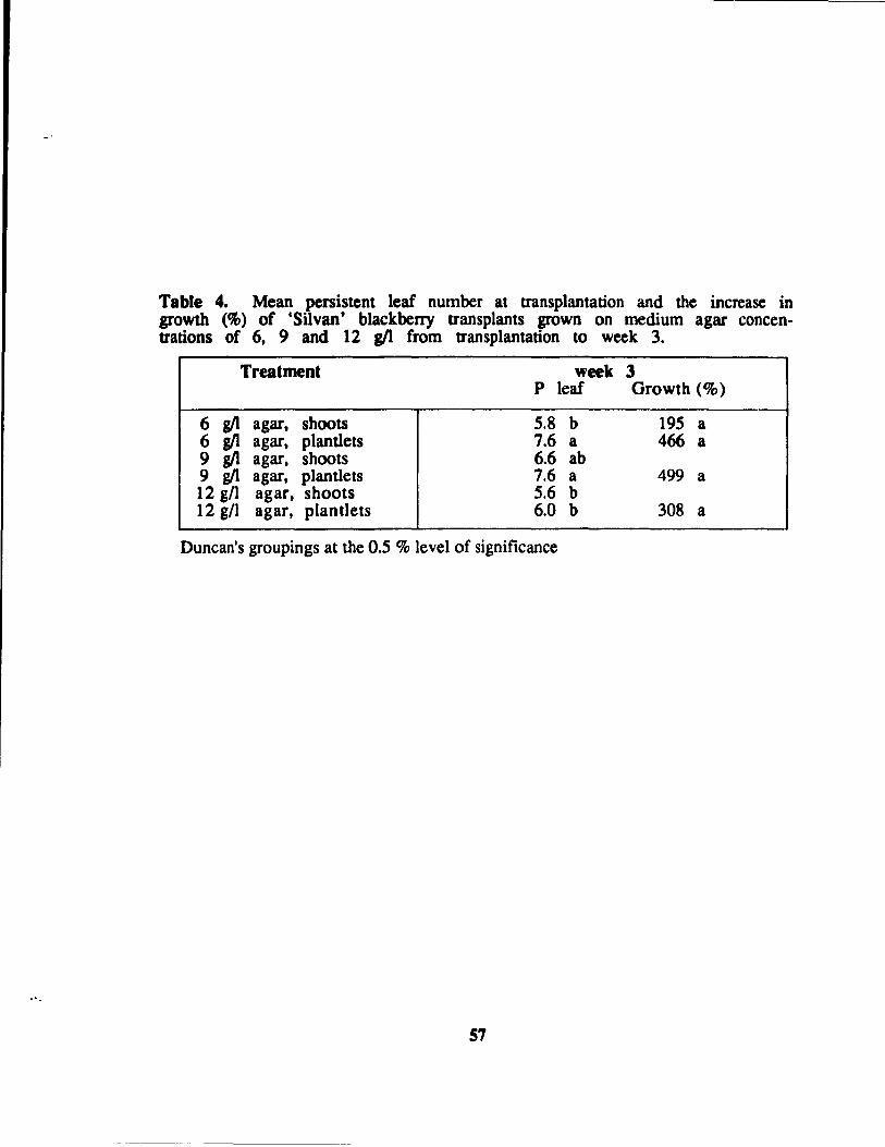
Table 4. Mean persistent Ieaf number al transplantation and the increase in growth (%) of 'Silvan' blackberry transplants grown on medium agar concenttations of 6, 9 and 12 gI1 from ttansplantation lO week 3.
Treatment week 3 p leaf Growth (%)
6 g/l agar, shoots 5.8 b 195 a 6 g/l agar, plantlets 7.6 a 466 a 9 g/l agar, shoots 6.6 ab 9 g/l agar, plantlets 7.6 a 499 a 12 gn agar, shoots 5.6 b 12 gn agar, plantlels 6.0 b 308 a
Duncan's groupings al the 0.5 % level of significance
57
-\.
{ .
No relationship was found between the mean initial and final fresh weights
of plantlets from treatments 2 and 6 of the 3 week test. However, a linear
relationship (r2 = 0.S2) was found between the mean initial and final fresh
weights of plantlets from treatment 4. Despite results from ex vitro rooting
and acclimatization experiments with Pelau:onjum zonale plantlets demonstrating
that plant size (greater than 2 cm in height) and leaf size were very important
factors to consider when transplanting (Aldrufeu, 1987), no relationship was found
between the mean initial Ieaf number and final fresh weight of 'Silvan' plantlets
in any of the treatments. This was surprising since it would he logical to
assume that a greater initial fresh weight at transplantation wou Id provide more
resources in the fonn of stored materials or photosynthetic area for new leaf
growth. Since no relationship was found between initial Ieaf number and final
fresh we.ight there is no substantiation from this work that persistent Ieaves of
'Silvan' blackberry mobilize stored elements for areas of new growth as suggested
for ChQ'santhemum morifolium (Wardle et al., 1983).
In contrast to results with 'Silvan' blackberry, Short et al. (1987) found
lhat increasing the agar concentration decreased Ieaf number in cauliflower and
chrysanthemum plantlets. However, as mentioned previously, the starting point
for their analysis was 12 g/l increasing to 20 g/l. High agar concentrations
(10-20 gII) were shown to reduce the number and Iength of in vitro roots in
jackfruit (Artocarpus hetero.phyllus Lam.) (Rahman and Blake, 1988) and shoot
height and root length in chrysanthemum (Short et al., 1987).
58
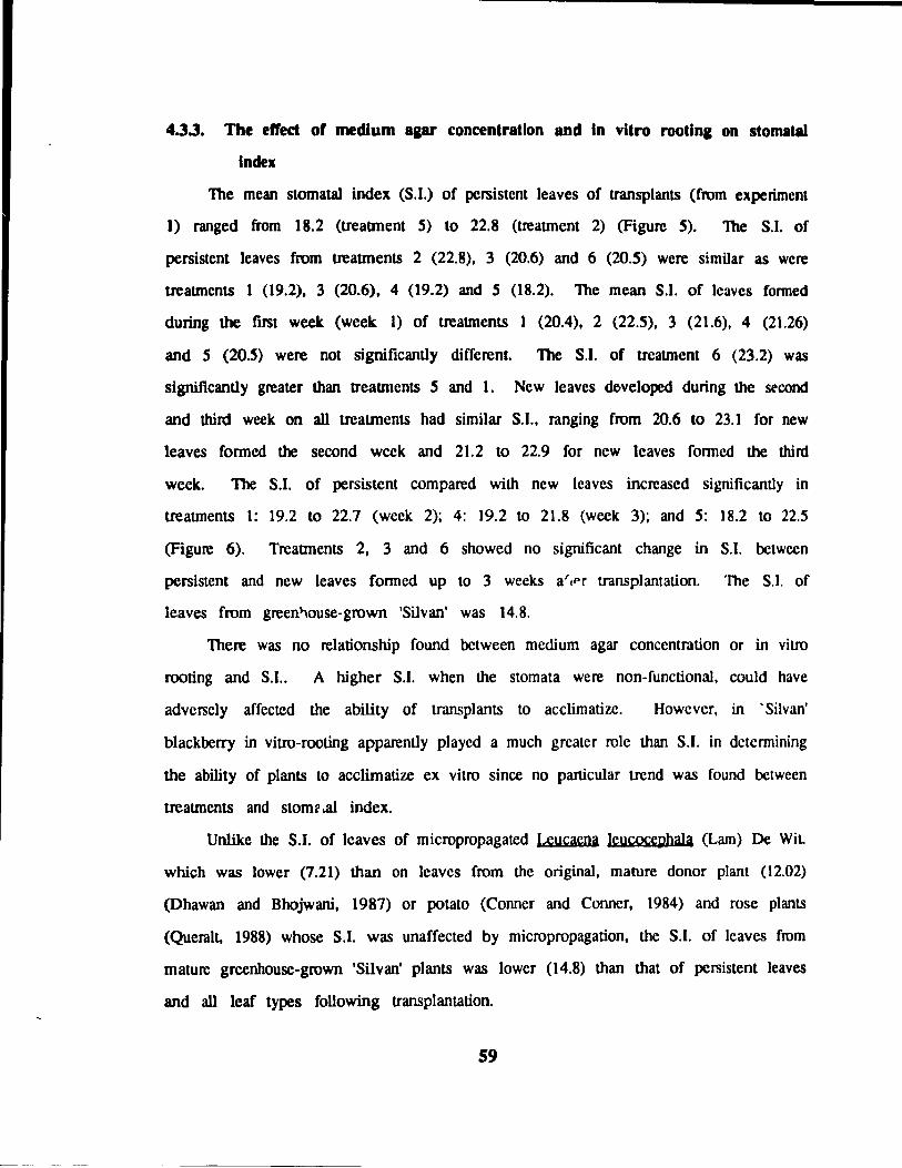
4.3.3. The eft'ect of medium aaar concentration and ln vitro rootina on stomatal
Index
The mean stomatal index (S.I.) of persistent leaves of transplants (from experiment
1) ranged from 18.2 (treattnent 5) 10 22.8 (treatrnenl 2) (Figure 5). The S.I. of
persistent leaves from treatments 2 (22.8), 3 (20.6) and 6 (20.5) were similar as were
treatrnents 1 (19.2), 3 (20.6), 4 (19.2) and 5 (18.2). The mean S.I. of leaves fonned
during the first week (week 1) of treatrnents 1 (20.4), 2 (22.5), 3 (21.6), 4 (21.26)
and 5 (20.5) were not significantly different. The S.I. of treatrnent 6 (23.2) was
significantly greater than treatments 5 and 1. New leaves doveloped du ring the second
and third week on aIl treaunents had similar S.I., ranging from 20.6 10 23.1 for new
leaves fonned the second wcek and 21.2 to 22.9 for new leaves fonncd the third
week. The S.I. of persistent compared with new leaves increased significantly in
treaUllents 1: 19.2 to 22.7 (week 2); 4: 19.2 to 21.8 (week 3); and 5: 18.2 to 22.5
(Figure 6). Treatments 2, 3 and 6 showed no significant change in S.I. bctwcen
persistent and new leaves fonned up to 3 weeks ar,pr transplantation. The S.l. of
leaves from green~ouse-grown 'Silvan' was 14.8.
There was no relationship found bctween medium agar concentration or in vitro
rooting and S.I.. A higher S.I. when the stomata were non-functional, could have
adversely affected the ability of transplants to acclimatize. Howevcr, in 'Silvan'
blackberry in vitro-rooting apparently played a much grcater role than S.I. in dctennining
the ability of plants to acclimatize ex vitro since no particular trend was found bctween
treaUllents and stom~.al index.
Unlike the S.I. of leaves of micropropagated Leucacna leucocçphala (Lam) De WiL
which was lower (7.21) than on leaves from the original, mature donor plant (12.02)
(Dhawan and Bhojwani, 1987) or potato (Conner and Conner, 1984) and rose plants
(QueraIt, 1988) whose S.I. was unaffected by micropropagation, the S.I. of lcaves from
mature grcenhouse-grown 'Silvan' plants was lower (14.8) than that of persistent leaves
and ail leaf types following transplantation.
S9
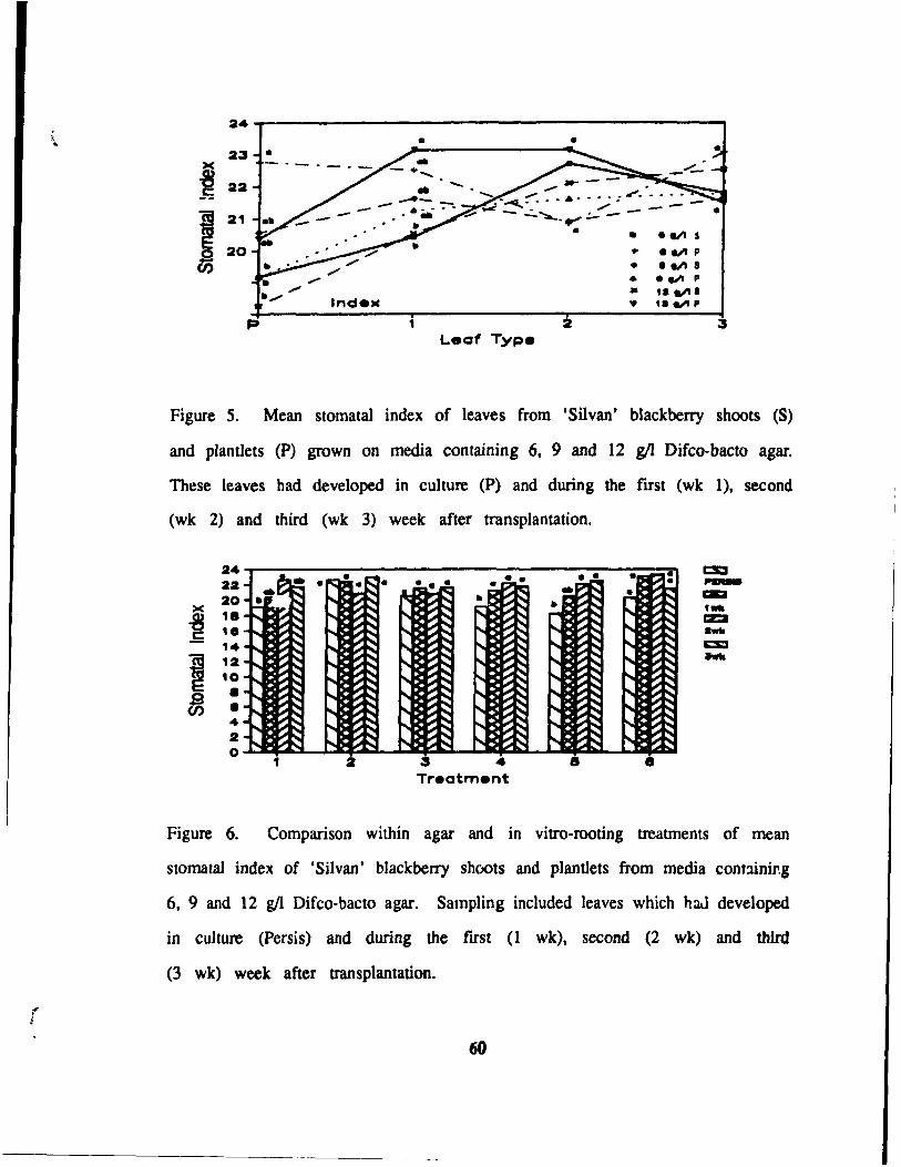
\ 24
•
~ 22 S;
1 21
20 ~
1
• •
- -- --- . - - ... ... " ..... ~ .. ~ ......... ./" - ---
::;;;.0- --....:.,.......... .. --- • --...... -
2 Leat Type
• •
•
.~ S
.~ P
.~ S ........ p
• ,. alla • 1.~P
Figure 5. Mean stomataI index of leaves from 'Silvan' blackberry shoots (S)
and p)antlets (P) grown on media containing 6, 9 and 12 g/l Difco-bacto agar.
These leaves had developed in culture (P) and during the flfst (wk 1), second
(wk 2) and third (wk 3) week after transplantation.
24 22
>< 20
-8 ,.
c: 1. '4
1 12 10
.s • CI) • 4
2 0
.~~ .~ •• s:. P.. .~ • ~: ., ~ II":~ .. ~ .. ~~ ..~~ • ~~
" ~.~ ~~ ~ ;..-~ r;: ~~ . ;..~
;..-~ ;..~ ~~ ;..-~ ~ ~~ ~~
~~ ~~ " ~ ~~ ;..~ ~~ ~ " ;..-~ ~~ ~ ~~ ~ ~~ ~
~~ ~~ ~~ ~~ .... ~~ · ~~ ~~ ..... ~~ t:::~ ~~ ~~
:::~ E::~ ~~ · ~~ ~~ ~~
· ~~ ~~ ~~ ~~ ~~ ~~
· ~~ ~~ ..... ~~ ~~ ~~ ~~ 1""0 ~ ~~ , 2 ... •
Treotment
Figure 6. Comparison within agar and in vitro-rooting treatments of mean
slomalaI index of 'Silvan' blackberry shoots and plantIets from media cont:ùnir.g
6, 9 and 12 g/l Difco-bacto agar. Sampling included leaves which haJ developed
in culture (Persis) and during the fust (l wk), second (2 wk) and thfrd
(3 wk) week after transplantation.
60
4.3.4. Evaluation of ex vitro stomatal function of 'Silvan' plants
The majority of stomata on epidennal peels of greenhouse-grown 'Silvan'
leaves were open after the peels had floated for 30 min on the phosphate buffer
under the sodium light. Stomata closed after exposure to the ABA solution
for 25 min under the sodium light. They did not reopen after 20 min of
floating on the phosphate buffer under light. The stomata closed after 35 min
when the epidennal peels were floated on the phosphate buffer and the petri
dishes darkened with aluminium foil. Subsequent stomatal reopening occurred
within 20 min when the epidennal peels were put back under the sodium light.
The stomata closed within 20 sec when epidermal peels were floated on the
lM NaCI solution, and reopened within 1 min once the lM NaCI solution was
replaced with the phosphate buffer. Due to its rapidity, the lM NaCI treatment
was chosen as the most time-efficient test for the purpose of detennining stomatal
function (open-c1osure mechanism).
Inconsistencies in stomatal aperture (S.A.) measurements occurred when the
phosphate buffer Yt:iS reapplied after the lM NaCI treatment. In sorne cases
the final S.A. were less than the original ones. Perhaps all the NaCI solution
was not completely removed or the epidennal peels had moved so S.A.
measurements were not of the same stomatal populations. The adding and
diluting of the NaCI solution caused stomata to open and close several limes.
However, reopening was impaired after a second lM NaCI application. This
was thought to be due to the incomplete removal of aIl salt when replacing
the NaCI solution with the phosphate buffer.
61
.,. j
4.3.5. Evaluation of stomalal closure of the fint new Jeava of 'Silvan'
plantlets three weeks following transplantation
No stomatal closure occurred on persistent or new leaves in any treatment
at the end of the 2 wee1c test At the end of the 4 week test ali leaves
which were not senescing had stomata which could close in response to the
lM NaCl solution and reopen when floated on buffer (all of the persistent and
many week 1 leaves exhibited senescence).
Original medium agar concentration had no apparent effeet on stomatal
function so all treatments from the 3 week test were pooled. Three weeks
after transplantation persistent leaves had an initial S.A. of 5.2 ± 0.7 pm and
this was basically unchanged after application of the lM NaCI solution where
the mean S.A. was 4.8 ± 1.0 pm (Table 5). The leaves which developed
during the frrst week (week 1) had an initial S.A. of 4.7 ± 1.2 pm. This
S.A. decreased to 2.7 ± 0.6 pm after the 1 M NaCl solution was applied.
The new week 2 leaves had an initial S.A. of 4.0 ± 1.0 pm which completely
closed to 1.5 ± 0.5 pm after the NaCl solution was applied. The new week
3 leaves had an initial stomatal aperture of 4.9 ± 0.8 pm. After the NaCl
solution was applied most stomata completely closed to 0.4 ± 0.3 pm. Stomata
from aU new leaves were able 10 subsequently reopen when the epidermal peels
were floated on the phosphate buffer. The potential for stomata to achieve
complete clos ure was transitional during the fmt 3 weeks of ex vitro accli
matization (fable 6). Each successive new leaf formed under the new envi
ronmental conditions had improved stomatal closure.
A few stomata of the last persistent leaves retained after 3 weeks ex vitro
exhibited Iimited stomatal response 10 the NaCl sulution in that they were able
to close significantly. This occurred in about 40 % of the epidermal peels
evaluated. It is possible that only persistent leaves which were not fully
62
developed al transplantation and whose developmenl overlapped both in vitro and
ex vitro environments were able to develop panial stomatal function. The
persistent leaves which developed last and some new week 1 leaves may he
included in this group.
After severa! days of exposure to relative humidity levels of 30-45 %,
stomata on persistent leaves of apple closed more completely (85 %) 15 min
after excision than persistent leaves exposed to low humidity levels for 4 days
(15 %) or less (5 %) (Brainerd and Fuchigami, 1981). The percent or speed
(rate) of stomatal clos ure of persistent cherry leaves (98 %) was considerably
greater tban in vitro cherry leaves (72 %) when exposed to 45 % relative
humidity levels for 15 min. (Marin et al. 1988). Stomatal conductance of apple
and cherry was significantly less (up to 70 %) on leaves gradually exposed
to relative humidity levels of 65 % over a 12 day period prior to sampling.
Il has been observed in other plant species that the new leaves fonned during
the acclimatization period exhibited improved capacity for stomatal closure when
compared to leaves from culture (Conner and Conner, 1984, Dhawan and
Bhojwani, 1987, Marin et al, 1988, Marin and GeUa, 1988). However, this
was usually determined by measuring tie amount of water lost over time after
leaf detachment not by actual stomatal aperture measurements. Stomatal closure
could have been due to membrane collapse, especially when leaves were exposed
to very low relative humidity levels. The above authors did not verify whether
these stomata could sub~equently reopen.
63
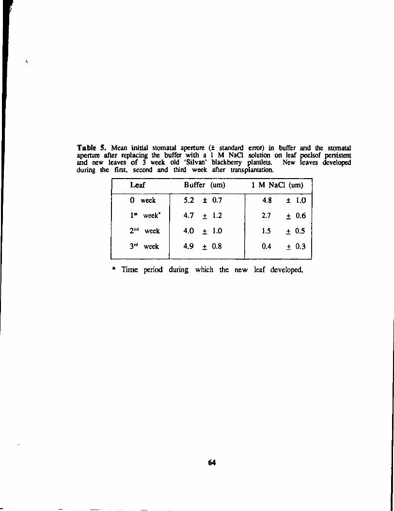
Table 5. Mean initial stomatal apenure (± standard error) in buffer and the stomatal aperture after replacing the buffer with a 1 M NaCl solution on leaf peelsof persistent and new leaves of 3 week old 'Silvan' blackberry plantlets. New leaves developed during the first, second and third week after transplantation.
Leaf Suffer (um) 1 M NaCI (um)
0 week 5.2 ± 0.7 4.8 ± 1.0
I" week· 4.7 ± 1.2 2.7 ± 0.6
2nd week 4.0 ± 1.0 1.5 ± 0.5
3rd week 4.9 ± 0.8 0.4 ± 0.3
• Time period during which the new leaf developed.
64
4.3.6. Tbe ef1'ects or blgb relative bumidity and low IIgbt Intenslty on
ex vitro stornatal runction or 'Silvan' plantlets grown on rull or
114 strengtb rooting media
After 14 days in the dew chamber persistent leaves of the "Full MS"
treatment plants had an initial S.A. of 4.47 pm and the S.A. of the new leaves
varied from 4.61 pm (leaf 1) to 3.38 pm (Ieaf 2) to 3.75 pm (Ieaf 3)
(Table 6). Once the NaCl solution was applied. the fmt new leaf and the
third new leaf showed significant reductions in mean S.A. (:';.27 pm and 1.75
pm. respectively). Only in the third new leaves was stomatal closure complete.
Stomata of persistent and new leaves reopened once the NaCI solution was
replaced with buffer solution. Reopened stomatal apertures of new leaves ranged
from 3.36 pm (leaf 2) to 3.64 pm (leaf 1) and 4.61 pm for persistent leaves.
After 14 days in the dew chamber persistent leaves of the
"1/4 MS" tteatment plants had an initial S.A. of 3.8 pm and the initial S.A.
of the new leaves ranged from 3.89 pm (leaf 2) to 4.11 pm (leaf 3) to 4.92
pm (leaf 1). As in the "Full MS" treatment, when the NaCI solution was
applied only the fIfst new leaves showed a significant reduction in S.A. (3.53
pm). Stomatal closure was incomplete in persistent Jeaves (3.00 pm) and ail
new leaves: 3.29 pm (Ieaf 2) and 3.48 pm (leaf 3). Reopened stomatal apertures
of new leaves ranged from 3.92 pm (leaf 2) to 4.59 pm (leaf 3) and 3.81
pr11 for persistent leaves.
After 14 days under conditions of high humidity stomata of leaves of plants
grown on the "Full MS" treatment appeared to be more functional than stomata
of leaves of plants grown on the "1/4 MS" medium. However, only stomata
from the third new leal' of plants grown on "Full MS" treatment plants was
completely closed (1.75 pm). Stomata of leaves from shoots or plantlets grown
6S
, {
on u 114 MS" tteatmcnt could not generally close beyond 3 pm.
After 17 days in the dew chamber the persistent leaves from plants grown
in the "Full MS" treatment had an initial S.A. of 3.63 pm and the S.A. of
new leaves ranged from 3.80 pm (leaf 2) to 4.46 pm (Ieaf 3) to 6.08 pm
(leaf 1). After application of the NaCI solution ail new leaves had significantly
reduced S.A. which ranged from 2.56 pm (leaf 2) to 2.27 pm (Ieaf 3) to
4.29 pm Oeaf 1) although stomatal closure was incomplete in ail cases. Once
the NaCI solution was replaced with the buffer, the stomata of persistent leaves
(3.93 pm) and the rust (5.73 pm), second (3.63 pm) and third (3.95 pm) new
leavr.~ could ail reopen.
Arter 17 days in the dew chamber the persistent leaves of the "114 MS"
treatment had a mean S.A. of 3.18 pm. The flfst new leaf had an initial
S.A. of 4.75 pm und the second, 4.54 pm. After application of the NaCI
solution stomata from both the tirst (3.14 pm) and second (2.27 pm) new leaves
had significantly reduced apertures. However, complete c10sure was not observed
in any case. After replacement of the NaCI solution with buffer the stomata
of the fust (4.58 pm) and second (4.69 pm) new leaves reopened.
After 14 days in the dew chamber and 3 days in the growth chamber
stomata of new leaves grown on the "Full MS" treatment had initial S.A. which
varied from 4.15 pm (leaf 1) to 2.82 pm (leaf 2) to 2.35 pm (leaf 3). After
application of the NaCI solution, me mean S.A. were significantly reduced on
leaf 1 (3.35 pm) and leaf 3 (1.45 pm) but not on Ieaf 2 (2.09 pm). Stomata
from both the second and third new Ieaves were completely closed. Once the
NaCI solution was replaced by buffer the stomata of the frrst (4.17 pm), second
(3.42 pm) and third (3.05 pm) new leaves reopened.
After 17 days, the initial S.A. of stomata on Ieaves of plants grown on
the "Full MS" tteatment in the growth chamber were smaller, ranging from 2.35
66
pm to 4.15 pm, comparcd with plants from the same "Full MS" treatment in
the dew chamber, where the S.A. ranged from 3.63 pm to 6.08 pm. The
second and third new leaves from plants grown on "Full MS" medium plants
in the growth chamber had smaller S.A. after the NaCl solution was Il'plie<!
than their counterpans in the dew chamber. The stomata of the second
(2.95 pm) and third (1.75 pm) new leaves of plants grown on the "Full MS"
medium and in the dew chamber for 17 days were able to close to a sm aller
aperture than the stomata on the second (3.29 pm) and third (3.48 pm) new
leaves of plants grow,:. on the "1/4 MS" medium. Unfortunately, the data for
day 17 of "1/4 MS" plants in the growth chamber cannot he presented.
After 21 days in the dew chamber the persistent leaves of the "Full MS"
treatment plants had an initial S.A. of 3.78 pm. Stomata on the new leaves
had initial S.A. ranging from 3.27 pm (leaf 3) to 4.31 pm (leaf 2). After
the NaCI solution was applied the S.A. of the persistent leaves was significantly
reduced (2.2 pm) (close to full closure). The S.A. of the new leaves after
the application of the NaCI solution was also significantly reduced ranging from
1.45 pm (leaf 3) to 3.96 (leaf 2). However, only the third new leaf had
stomata which were fully closed (1.45 pm). After replacing the NaCI solution
with buffer, stomata on the persistent leaves reopened (3.93 pm) as did stomata
on the third (4.24 pm) and fourth (2.98 pm) new leaves. Surprisingly, stomata
on the flTSt (3.11 pm) and second (3.36 pm) new leaves closed further.
After 21 days in the dew chamber the initial S.A. of the persistent leaves
of plants grown on the "1/4 MS" medium was 5.45 J.1m. The initial S.A.
of stomata of new leaves ranged from 3.18 pm (leaf 3) to 4.36 pm (leaf 1).
After the NaCl solution application, the stomata of the persistent leaves had a
significantly reduced S.A. of 4.54 pm. Stomata from all the new leaves had
significantly reduced apertures after the NaCI solution was applied, ranging from
67
l 1.74 pm (Ieal 4) to 2.90 pm (Ieaf 1). Only the second and founh leaves
had stomata which could completely close (1.74 pm and 1.81 pm). Once the
NaCI solution was replaced with buffer, stomata of persistent and new leaves
reopencd to apertures which ranged from 2.95 pm (leal 3) to 5.00 pm (P leal)
Twenty-one days after uansplantation, the stomata on the persistent leaves
of plants grown on the "Full MS" medium, which had been in the growth
chamber for 1 week, were initially closed (0.98 pm) but they closed even further
(0.:5 pm) in response to the NaCI solution. The initial S.A. of stomata of
new Icaves ranged from 2.82 pm (lcaf S) to 3.48 pm (leaf 4). Stomata of
most leaves had significantly reduced apertures and most closed after the NaCl
solution was applied. After the NaCI solution was replaced with buffer, stomata
on persistent (2.72 pm), second (3.03 pm), third (2.30 pm), fourth (3.33 pm)
and fifth (2.52 pm) new leaves could reopen but stomata on the
flfst new leaf (1.71 pm) could not.
The stomata of persistent leaves from the "1/4 MS" treatment plants, which
had been in the growth chamber for 1 week, had an initiaI S.A. of 1.29 pm.
The initial S.A. of stomata of new leaves ranged from 2.23 pm (leaf 2) to
2.27 pm (leaf 3). After the application of the NaCI solution, stomata of the
persistent leaves had a significantly reduced S.A. of 0.36 pm (closed). The
stomata on the flfst new Ieaf did not fully close (2.27 pm) but stomata on
the second and third new leaves aIso had significantly reduced apertures and
they closed to 0.82 and 1.01 pm, respectively. Once the NaCI solution was
replaced, stomata on persistent leaves could reopen to sorne extent but the S.A.
was still considered to be closed (1.21 pm). Stomata of the fust (4.54 pm),
second (2.5 pm) and third (2 pm) new Ieaves reopened in response to the
replacement of the NaCI solution but the S.A. of stomata of the third leaf
remained fully closed, and could not reopen.
68
.~.
Stomata of both persistent and new leaves had larger mean initial apertures
when plantlets were grown in the dew chamber for 21 days than when they
were transferred to the growth chamber for 1 week after having grown in the
dew chamber for 2 weeks. Though ail stomata from leaves of plants in the
dew chamber had significanüy reduced apenures in response to the NaCI solution,
the resulting apenure was a1ways greater than that of stomata on leaves from
plants in the growth chamber. These trends were the same for plants from
both "Full MS" and the "114 MS" treatments. Based on the se observations,
it appears that the environ ment, particularly relative humidity in conjunction with
low light intensity, plays an imponant role in determining the capacity for
stomata to function.
The stomata of persistent leaves from the "Full MS" treatment plants in
the growth chamber had a much decreased initial S.A. (0.98 pm) compared with
their counterparts in the dew chamber (3.78 pm). Similarly, after the NaCI
solution had been applied, the S.A. of the persistent leaves from the growth
chamber (0.15 pm) was smaller than that of the persistent leaves from the dew
chamber (2.2 pm). The persistent leaves from the "1/4 MS" treatment in
the growth chamber also had reduced initial S.A. (1.29 pm) (c1osed) when
compared with the persistent leaves from the same medium treatment in the dew
chamber (5.45 pm). This trend held true both after the NaCI was applied
and on ils removal.
The fust new leaf haJ initial S.A. greater than 4 pm for all harvest dates
and treatments except for the treatment placed in the growth chamber for one
week. Dnly the stomala of leaves from 21 day plants which had been in
the growth chamber for 1 week could close after the N aCI solution was applied
(1.70 pm). However, arter removal of the NaCI solution the S.A. did not
increase (1.71 pm). So, the stomata in this case were unable to reopen. There
69
did not seem ta be any ~nd regarding the failure of stomata ta reopen. The
stomata on the f1l'5t new leaves of the "1/4 MS" tteatment plants had reduced
initial apertures when placed in the growth chamber for a week. The majority
of stomata from the fll'st new leaves of aIl treatments had significantly reduced
S.A. even if they could not fully close.
Stomata from the second new leaves of the "Full MS" treatment plants
had initial apertures ranging from 2.82 pm. for the leaves of plants which had
been placed in the growth chamber for 3 days. to 4.31 pm for leaves of plants
which had been left in the dew chamber for 21 days. When comparing the
ability for stomata from the fIfst and second new leaves of the "Full MS"
treatment plants to close in response to the NaCI solution, stomata of the second
new (eaves appeared to respond to the change (decrease) in relative humidity
sooner than stomata of the fU'st new leaves. After 3 days in the growth chamber
stomata of the second new leaves had smaller S.A. (2.09 pm) than their
counterparts Jn the dew chamber (2.56 pm) in response to the NaCI solution.
However, no appreciable difference in S.A. was observed in sTomala of the fmt
new Ieaves under the same conditions. Like the fust new leaves on plants
grown on the "Full MS" medium, stomata of the second new leaves of plants
grown on the "1/4 MS" medium had reduced initial S.A. when plants were
transferred to the growth chamber for 1 week. This trend continued after the
NaCI solution was applied and a1so after it was replaced by the buffer. Only
stomata from the second new leaves of the "Full MS" treatment plants transferred
to the growth chamber and 14 day plants from the "Full MS" and "114 MS"
treatment plants failed to close significantly after application of the NaCI solution.
The third new leaves of the "Full MS" treatment plants in the dew chamber
had initial S.A. which were smaller on day 21 (3.27 pm) than day 17 (4.46
pm). This was also true after the NaCI solution was applied. The same
70
trend was observed for the third new leaves from plants of the same treatments
but transferred to the growth chamber. Stomata on third new leaves of plants
placed in the growth chamber for 3 days and on leaves of plants from the
"Full MS" treatments in the dew chamber for 21 days were able to completely
close (apertures less than 2 pm). The third new leaves on plants grown on
the "1/4 MS" treatment had improved stomataI function when placed in a growth
chamber for a week compared with the third new leaves of plants left in the
dew chamber. The third new leaves of plants from the "1/4 MS" treatment
which were left in the dew chamber were not expanded enough at day 17 to
sample. The plants grown under the " 1/4 MS" treatment were slower in
produCÎllg new leaves ex vitro.
Stomata on each successive new leaf had a greater capacit!' to open and
close both on plants growing in the growth and dew chambers (Table 6). So,
stomatal function was transitional (Figure 7) whether the relative humidity level
was high or not. However, under conditions of low relative humidity (ie
transferred to the growth chamber), stomatal apertures were reduced in ail leave!i
with complete closure occurring in response to lM NaCI in ail leaf typt~s
(persistent to leaf 4-5) regardIess of the media treatment (ie "Full MS" or
"1/4 MS") on which the plantlets were grown. This was in contrast to stomata
on th~ persistent, frrst and second new leaves, developed under high humidity
conditions, which after 21 days cou Id not close. There fore , it appears that ex
vitro environmental factors such as relative humidity and light levels are more
important than in vitro media composition in determining the functional capacity
of stomala after transplantation.
Diluting the basal medium for the "1/4 MS" treatment did not improve
stomatal function of ex vitro 'Silvan' blackberry leaves when compared with
leaves of plantIets grown on the "Full MS" treatment. ": is appears contrary
71
to protocols developed for in vitro acclimatization of red raspberry which l'CC
ommend placing plantlets in 1/S strength medium 2 weeks prior to transplantation
(Welander, 1985). Il is possible that the 3 weeks on 114 strength medium
used in this experiment resulted in nutrient depletion. However, the blackberry
plantlets appeared vigorous and healthy at transplantation. Reducing the sugar
content may promo te photoautotrophy in certain plants (Grout and Price, 1987,
Kozai, 1988a) but it did not appear to improve stomatal function in 'Silvan'
blackberry.
The ability of stomata to function l~ related to their structural or anatomical
features. Guard cell walls of cherry were thinner in vitro than on greenhouse
grown or field-grown plants (Marin et al., 1988). In vitro stomata of cherry
also lacked invaginations in the epidermal ceUs next to the inter-guard cell wall
ends which were evident in acclimatized leaves. The arrangement of microfibrils
of in vitro guard ccli walls did not appear altered when compared to that of
stomata of greenhouse-grown leaves. (Marin et al., 1988). Anatomical changes
which might have occurred in the anatomy of 'Silvan' blackberry stomata were
beyond the scope of this experiment. However, when examining epidennal peels,
it was observed that even if stomata did not close during the application of
the 1 M NaCl solution the guard cell protoplasm collapsed suggesting that the
guard cell membrane was permeable and that the guard cell was physiologically
functional. The protoplasm of in vitro stomata of Clu:ysanthemum morifolium
(Wardle and Short, 1983) and carnation Dianthus. caryophyllus L. (Ziv et al.,
1987) collapsed in a similar way in response to ABA and 0.8 M sucrose.
Wardle and Short (1983) suggested that, during stomatal development, the
deposition of cellulose and microfibrils in the cell walls may dictate the "closed"
position of the guard cells. The closed state of in vitro stomata is partially
71
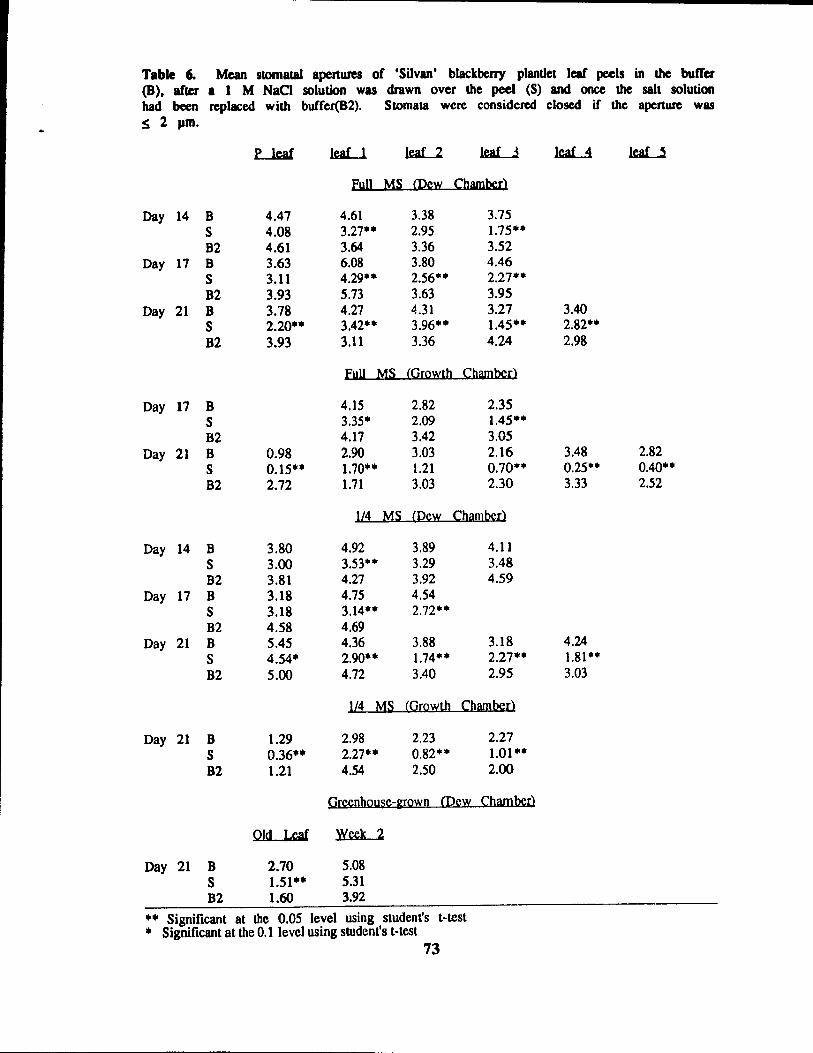
Table 6-(8), afler had becn oS 2 pm.
Mean slOI11atal apertures of 'Silvan' blackberry plantle, leaf peels in the buffet 1 1 M NaCI solution was drawn over the peel (8) and once the sah solution replaced wilh buffer(B2). 8tomata were considered closed if the aperture wu
Day 14 B S B2
Day 17 B 8 B2
Day 21 B S B2
Day 17 B S B2
Day 21 B S B2
Day 14 B 8 B2
Day 17 B S B2
Day 21 B S B2
Day 21 B S B2
Day 21 B S B2
4.47 4.08 4.61 3.63 3.11 3.93 3.78 2.20·· 3.93
0.98 0.15·· 2.72
3.80 3.00 3.81 3.18 3.18 4.58 5.45 4.54· 5.00
1.29 0.36·· 1.21
FuU MS IDew Chamber)
4.61 3.27** 3.64 6.08 4.29·· 5.73 4.27 3.42** 3.11
3.38 2.95 3.36 3.80 2.56·· 3.63 4.31 3.96·· 3.36
3.75 1.75** 3.52 4.46 2.27·· 3.95 3.27 1.45·· 4.24
Full MS (Growth Chamberl
4.15 3.35· 4.17 2.90 1.70·· 1.71
2.82 2.09 3.42 3.03 l.21 3.03
2.35 1.45*· 3.05 2.16 0.70·· 2.30
1/4 MS (Pcw Cham ber)
4.92 3.53** 4.27 4.75 3.14** 4.69 4.36 2.90** 4.72
3.89 3.29 3.92 4.54 2.72··
3.88 l.74·· 3.40
4.11 3.48 4.59
3.18 2.27·· 2.95
1/4 MS (Growth Chamber)
2.98 2.27·· 4.54
2.23 0.82·* 2.50
2.27 1.01·· 2.00
Grccnhouse-grown CDcw Chamberl
OId Leaf Week 2
2.70 1.51·· 1.60
5.08 5.31 3.92
•• Significant at the 0.05 level using student's t-test • Significant at the 0.1 level using sbldent's t-test
73
3.40 2.82·· 2.98
3.48 0.25·· 3.33
4.24 l.81·· 3.03
2.82 0.40·· 2.52
open. Further stomatal opening of in vitro stomala of chrysanthemum and
subsequent reclosure to the original aperture could he induced but further clos ure
could not (Wardle and Short, 1988).
4.3.7. The etTects of high relative humidity and low light intensity on
stomatal fl:!1ction of greenhouse-grown 'Silvan' plants
Examined 4 weeks after the greenhouse-grown 'Silvan' blackberry plants
were placed in the dew chamber, the initial S.A. of leaves present at transfer
(old leaf) was 2.70 ~m. (fable 6). After the 1 M NaCl solution was applied
the mean S.A. was 1.51 ~m. The mean initial S.A. of the leaves tagged 2
weeks (week 2) after transfer to the dew chamber was 5.08 ~m. After the
NaCI solution was applied the S.A. was 5.31 ~m. Only stomata on the leaves
present at transfer were able to close in response to the application of the NaCI
solution. Once the NaCI solution was replaced with buffer the S.A. of the
"old leaf' was 1.60 pm and the "week 2" leaf 3.92 ~m.
Stomata on these newly formed leaves resembled in vitro stomala in that
a) the y would not close in response to the 1 M NaCl solution although the
protoplasm did collapse, and b) their initial S.A. was greater than on leaves
developed under conditions of low relative humidity and high light intensity (in
the greenhouse).
When these greenhouse-grown plants were placed under conditions of low
humidity (25-10 %), leaves which developed in the dew chamber desiccated
quickly des pite the faet that they were growing on plants having a full root
mass. This indicated that the plant eould not balance the water lost through
leaves with non-functional stomata by taking up greater amounts of water through
the roots and subsequently increasing the transpiration rate through these leaves.
74
However, the plants had no problem controlling the transpiration rate in leaves
which had originally developed in the ~reenhouse. This appears to contradict
the idea that the lack of a functional root mass on transplants from culture
is responsible for the lack of evapotranspiration control after transplantation
(Debergh and Maene, 1981, Pierik. 1988). Stomatal function is impaired on
leaves of blackberry which develop under conditions of high relative humidity
and low light levels.
75
1
1 !
Figure 7. Photomicrographs of stomata of a 'Silvan' blackberry leaf, from
plantlets grown on "Full MS", 3 days after removal from the dew chamber
ta the growth chamber. a) while on buffer; b) after application of the lM NaCI
solution; c) after replacement of the NaCI solution with buffer. Arrows indicate
functional or partially functional stamata. Bar = 50 J.lm.
76
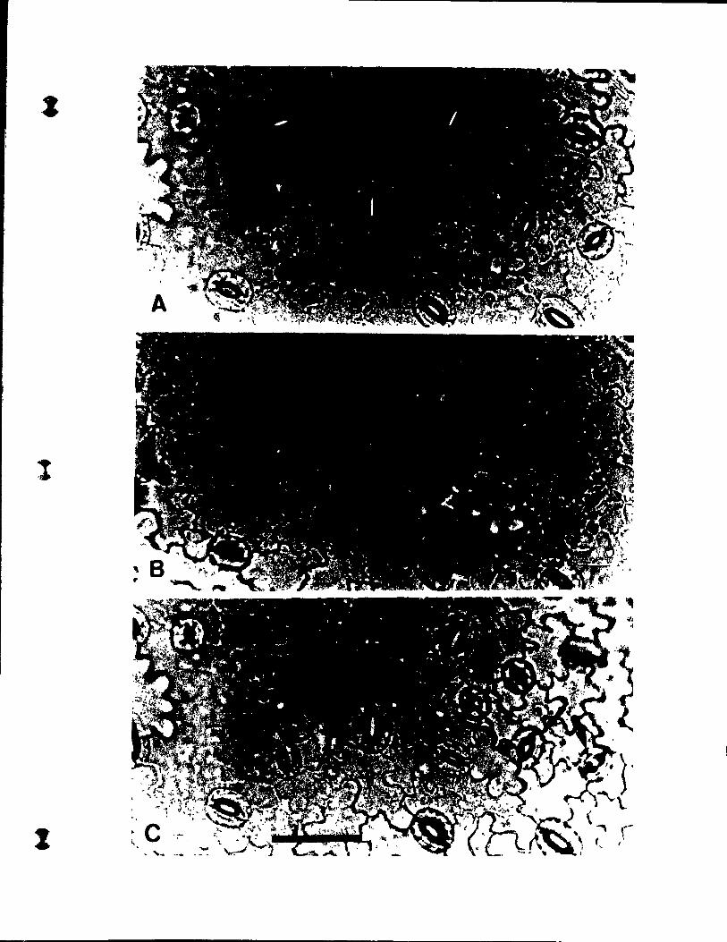
l
1
( 4.4. Conclusions:
Medium agar concentrations of 6, 9 or 12 gIl did not affect ex vitro
survival or shoot height during the frrst 3 weeks ex vitro. However, plantlet
height was reduced by medium agar concentrations of 9 and 12 g!l during the
first 2 weeks after transplantation. By the third week plantlets from all medium
agar concentrations were similar in height (2.84, 2.89 and 2.27 cm for 6, 9
and 12 gIl medium agar concentration respectively). Following transplantation,
leaf number was reduced only when shoots were grown on the highest medium
agar concentration (12 g/l). Plantlets consistently produced longer leaves ex vitro
when compared to shoots. However, agar concentration had no observable effect
on the longest leaf length. At the end of 3 weeks only p!antlets from treatment
4 (plantlets grown on 9 gIl medium agar concentration) showed a significant
relationship between initial and final fresh weight, this was linear (r = 0.52).
No relationship was found between initial and final fresh weight in treatments
2 and 6. Three weeks after transplantation, no relationship was found between
persistent leaf number and final fresh weight of plantlets grown on any medium
agar concentration.
Plantlets of 'Silvan' blackberry performed better than shoots during the fIfSt
three weeks ex vitro. This was demonstrated by the greater transplant height,
longest leaf length and the greater fresh weights in the rooted treatments.
Rooting in vitro appears to override the initially neg.?tive aspects of fmner agar
at a concentration of 12 gIl.
Despite the fact that the S.I. differed between persistent leaves and between
weekly new leaf populations no particular trend could be found. No correlations
were found between stomatal index and agar concentration or in vitro rooting.
A 1 M NaCl solution followed by replacement by buffer was found to
77
be the test of choice for studying stomataI function (clos ure and opening).
Stomata of greenhouse-grown leaves exposed to the 1 M NaCI solution had a
much shorter closure time (1-2 min) when exposed to a 2:1 ratio of 10"2 ABN
buffer or a dark interval of between 25 to 35 min. Some difficulties were
encounte!ed when trying to photograph the epidermal peels after the NaCI
solution was applied. It was sometimes hard to focus since the NaCI solution
caused the removal of water from the cells and made the surface area uneven.
The S.A. which resulted when the NaCI solution was replaced by buffer could
be unreliable due to problems controlling the quantity of salt removed from the
cells and the speed at which it was removed.
In n .. sponse to the hypertonie solution (1 M NaCI), guard cell closure on
epidermal peels improved in each successive new (weekly) leaf population (ex
periment 2) or new leaf (experiment 3) following transplantation. Stomatal func
tion was therefore transitional. That is, sorne but not all stomata were functional
during the Hrst few weeks ex vitro, indicating lhat a change in environmental
parameters during stomatal development may have influenced the ability of
stomata to close. In experiment 2 stomatal function was observed 3 weeks
after transplantation. In experiment 3 stomatal function was observed 2 weeks
after transplantation. However, it was not until 3 weeks after transplantation
that the majority of stomata from new leaves could completely close.
Decreasing the relative humidity from 95 % to appro;:. 50 % 2 weeks
after transplantation, caused stomatal clos ure. Growing the plantlets on "1/4 MS"
medium did not improve ex vitro stomataI function over growing plantlets on
"Full MS" media. The relative humidity level had greater effeet on stomatal
c10sure than did the medium on which the plants were originally grown. The
initial stomatal aperture of persistent leaves from both medium treatments was
reduced when placed in the growth chamber for 1 week (following 2 weeks
78
in the dew chamber). Plant exposure to continued high relative humidity 2
weeks after transplantation appeared to reduce stomatal closu..-e.
Stomata of new leaves of greenhouse-grown 'Silvan' blackberry plants that
developed 2 weeks after transfer to conditions of high relative humidity and
low light intensity (dew rhamber) did not close in response to the application
of the hypertonie solution. Stomata on original leaves (on the plants at transfer)
closcù in response to the NaCI solution after 4 weeks in the dew chamber.
These results indicated that impaired stomataI closure in vitro may be in part
due to environmental conditions such as high humidity levels in the culture
environment
During application of the NaCI solution it was observed that the guard
cell protoplasm of non-funetionaI stomata collapsed even though the guard eell
aperture was not reduced. This indicated that the guard cell membrane was
permeable and that modified anatomical properties of the guard cell waIIs were
most likely responsible for the impaired stomatal function ob:.crved in eultured
plant leaves and leaves from greenhouse-grown plants whieh had developed under
conditions of high relative humidity.
79
5. CONCLUSION
A I1lpid clearing and staining method for tissue-cultured plantlets and
greenhouse-grown leaves was successfully developed for the purpose of observing
stomatal distribution on leaves of varying thickness. This method enables
examination of large numbers of samples of many leaf types as long as they
do not exceed 3 mm in width. A maximum of 2 br are required to complete
the clearing process. ft is particularly useful for the observation of tissue-cultured
shoots and plantlets since the whole plantlet can he cleared without detaching
the stem or roots from the leaves. Disadvantages of the method include the
killing of the celIs during the process and the possible difficulty of focusing
on large samples of uneven thickness.
In vitro' Silvan' blackberry plantlets did not survive better after transplantation
when grown on increased medium agar concentrations (9 and 12 g/l) compared
with the control agar concentration (6 gIl). During the first 2 weeks ex vitro
height was reduced in plant:; from medium agar concentl1ltions over 9 g/l. Leaf
number and length were not affected by medium agar concentration. Plantlets
were generally taller and had longer leaves and a greater fresh weight when
compared with shoots during the fust weeks ex vitro suggesting that in vitro
roots are functional ex vitro. Three weeks after transplantation, a linear
relationship (r = 0.52) was found between initial fresh weight and final frcsh
weight in treatment 4 (plant lets grown on 9 gIl medium agar). However,
treatments 2 (plantlets grown on 6 g/l medium agar) and 6 (plantlets grown
on 12 gIl medium agar) showed no relationship between initial fresh weight and
tinal fresh weight. No relationship was found between initial Ieaf number and
final fresh weight 3 weeks after transplantation for any treatment. Slomatal index
was not affected by medium agar concentration or in vitro rooting. The stomatal
80
index of persistent leaves and all new leaf populations was greatcr than that
of greenhouse-grown 'Silvan'.
Stomatal opening and closure in response to a 1 M NaCI solution was
transitional in that stomata from each new leaf which developed ex vitro showed
greater potential to close. After 2 weeks in a dew chamber, stomata on leaves
of ex vitro plantlets from 1/4 strength rooting medium showed a reduced capacity
to close when compared to those from full strength rooting medium. However,
after this point there was no evident difference in stOmatal function between
the two treatments. Three weeks after transplanting, 'Silvan' plants placed in
the growth ehamber at the end of the second week, had stomata with a greater
eapacity to close in response to the NaCI solution than plants which remained
in the dew chamber. The initial stomatal apenure of persistent leaves was
smaller on plants placed in the growth ehamber than those left in the dew
chamber and only the se stomata (in the growth ehamber) were able to eompletely
close ~ 2 ).lm). The capacity for stomata to reopen after the NaCl solution
remained generally unaltered. Therefore, reduced relative humidity appeared to
promote stomatal clos ure.
Three weeks after transplantation, stomata on new leaves of 'Silvan' plantlets
placed under reduced relative humidity (55 %) for 1 week were functional.
Stomata on leaves of 'Silvan' plantlets maintained under high relative humidity
levels (tOO %) for 3 weeks eould not close completely. Protoplasrn collapse
was observed in response to the NaCI solution even if guard cells did not close
indicating that stomata! non-funclÏon in vitro is most likely due to anatomie al
not physiological abnomlality. New leaves that developed on greenhouse-grown
plants after they had been transferred to a dew ehamber had stomata which
could not close in response to a 1 M NaCI solution. Thus high relative
humidity levels and low light intensity may be largely re'iponsible for impaired
81
stomatal function in culture.
In view of these lesults it would be unwise to raise in vitro medium
agar concentrations above 9 gIl for the purposes of improving the time for and
success of acclimatization. In vitro rooting of 'Silvan' blackberry appeats to
be a beneficial acclimatization strategy. Based on results from direct observations
of stomatal function - blackberry plantlets should be maintained under conditions
of high relative humidity for 2 weeks after transplantation from which point
they should be transferred to conditions of lower relative humidity. The lower
relative humidity levels will promote stomatal function thereby decreasing the time
necessary for transfer to the field or greenhouse.
82
.......
6. SUllestlons lor "artber research
1. Further experiments ta detennine the relationship betwœn initial fresh weight
and final fresh weight after transplantation would help optimize ex vitro
transplant size.
2. Further study of the effeet of high relative humidity and low light levels
on greenhouse-grown plants would increase our understanding of the effects
of these parameters on in vitro plant anatomy.
3. To determine why stomatal function is impaired under conditions of high
relative humidity, il is necessary to complete anatomical studies focusing
on the ultrasttucture of the guard cell membranes and determining differences
in structures which may be related ta environmental conditions.
4. In vitro and ex vitro root function should be evaluated. This could be
done using dyes or radioactive tracers and anatomical observations .
83
{ ...
7. References Cited
Aldrufeu, A. 1987. Rooting and acclimatization of perlargonium plandets. Acta hort. 212. 361-366.
Amou, H.J. 1959. Leaf clearings. Tunox News 37:192-194.
Bin;:hem, R., H.E. Sommer, and C.L. Brown. 1981. Scanning electrOn mi-croscopy of shoot and l'OOt development in sweetgum callus tissue. FOIest Sei. 21:206-212.
Brainerd, K.E. and L.H. Fu'hlgami. t 981. Acclimatization of aseptically cultured apple plants to low relative humid~ty. J. Amer. Soc. Hon. Sei. 106:515-518.
Brainerd, K.E. and L.H. Fuchigami. 1982. Stomatal functioning of in yiJm and green hou se apple leaves in darkness, mannitol, ABA and COz' l. Expt. Bot. 33:388-392.
Brainerd, K.E., L.H. Fuchigami, S. Kwaitkowski, an~ C.S. Clark. 1981. Leaf anatomy and water stress of aseptically cultured 'Pixy' plum grown under different environments. HonSci. 16: 173-175.
Caldwell, J.D. 1984. Blackberry propagation. HonSei. 19(2): 193-195.
Conner, L.N. and A.I. Conner. 1984. Comparative water loss from leaves of Solanum laciniatum plants cultured in vitro and in vivo. Plant Science Leu. 36:241-246.
Conner, A.J. and M.B. Thomas. 1982. Re-establishing plantlets from tissue culture: a review. Proc. Inter. Plant Prop. Soc. 31:342-357.
Converse, R.H. 1981. Relationship of prior heat treatment to growth and fruiting of 'Thomless Oregon Evergreen' blackberry. HortSci. 16(3):312-313.
Converse, R.H. 1984. 19(2): 185-188.
Debergh, P.C. 1983. culture medium.
Blackberry viruses in the United States. HonSci. Vol
Effects of agar brand and concentration of the tissue Physiol. Plant. 59:270-276.
Debergh, P.C. 1986. Micropropagation of herbaceous plants. In: Micro-propagation in horticulture. Practice and commercial problems. Alderson, P.O. and W.M. Dullforce (cds.) p. 27-36 •
84
Debergh, P.C. 1988a. Conttol of in vitro plant propagation. International symposium on plant biotechnology. Piracicaba. Brazit. Oct. 2S-28. 1988.
Debergh, P. 1988b. Improving mass propagation of in vitro plantlets. In: Horticulture in high technology era. Symp. Tokyo, lapIn. May 10-11 p. 47-57.
Debergh, P. 1988e. Mieropropagation of woody species - state of the art on in vitro aspects. Acta Hon. 227:287-295.
Debergh, P.C. and L.J. Maene. 1981. A scheme for commercial propagation of omamental plants by tissue culture. Sei. Hon. 14:335-345.
Desjardins, Y., A. Gosselin and S. Velle. 1987. Acelimatization of ex vitro strawberry plantlets in COz- enriehed environments and supplementary lighting. J. Amer. Soc. Hon. Sei. 112:846-851.
Desjardins, Y., F. Laforge, C. Lussier and A. Gosselin. 1988. Effect of COz enriehment and high photosynthetie photon flux on the development of autotrophy and growth of tisSUf.·-cultured strawberry. raspberry and a~para· gus plants. Aeta Hon. 230:45-53.
Dhawan, V. and S.S. Bhojwalli. 1987. Hardening in vitro and morpho-physiological changes in the leaves during acclimatization of micropropagated plants of Leueaena leueocephala (Lam.) De Wit. Plant Sci. 53:65-72.
Donnelly, DJ. and F.E. Skelton. 1987. Hydathode structure of microprop~6al~d plantlets and greenhouse-grown 'Totem' strawberry plants. J. Amer. Soc. Hon. Sei. 112:755-759.
Donnelly, D.J. and F.E. Skelton. 1989. Comparison of hydathode structure in micropropagated plantlets and greenhou3e-grown 'Queen Elizabeth' rose plants. J. Amer. Soc. Hort. Sci. 114: (In Press).
Donnelly, D.1., F.E. Skelton and H.A. Daubeny. 1986. External leaf features of tissue-cultured 'Silvan' blackberry. HonSci. 21 :306·308.
Donnelly, D.1., F.E. Skelton and J.E. Nelles. 1987. Hydathode anatomy and adaxial water 105S in micropropagated 'Silvan' blackberry. J. Amer. Soc. Hort. Sci. 112:566-569.
85
( Donnelly 0.1., R. Stace-Smith and F.C. Mellor. 1980. In vitro eulture cf
three BlIhlI1 species. Acta Hon. 112:69-75.
Donnelly, D.1. and W.E. Vidaver. 1984a. Leaf anatomy of red raspberry transferred from euhure to soil. J. Amer. Soc. Hort. Sei. 109:172-176.
Donnelly, D.J. and W.E. Vidaver. 1984b. Pigment content and gas exchange of red raspberry in mm and ex ritrg. J. Amer. Soc. Hon. Sci. 109:177-181.
Donnelly, D.J., W.E. Vidaver and K. Colbow. 1984. Fixation of 1~C01 in tissue eultured ~ raspbcrry prior ta and after transfer to sail. Plant Ccli Tissue Organ Culture 3:313-317.
Donnelly, D.J., W.E. Vidaver and K.Y. Lee. 1985. The anatomy of tissue cultured red raspberry prior to and after transfer to soil. Plant Cell Tissue Organ Culture 4:43-50.
Douglas, G.C. 1984. Propagation of ~.ght cultivars of Rhododendron in vitro using agar-solidified and liquid media and direct rooting of shoots in vivo. Sei. Hort. 24:337-347.
Esau, K. 1977. Anatomy of seed plants. 2nd ed. Wiley, New York 500 pp.
Fabbri, A., E. Sutter and S.K. Dunston. 1986. Anatonllcal changes in persistent leaves of tissue-cultured sU'awberry plants after removal from culture. Scientia Hort. 28:331-337.
Fari, M., A. Andrasfalvy and J. Nemeth. 1987. Thin PVC foil covering (TPFC), an efficient method for culture and preacclimatization of in vitro plant cultures. Acta Hort. 212:371-374.
Fuchigami, L.H., T.Y. Cheng and A. Soeldner. 1981. Abaxial transpiration and water loss in aseptically cultured plum. J. Amer. Soc. Hon. Sei. 106:519-522.
Fujiwara, K., T. Kozai and 1. Watanabe. 1987. Fundamental studies on environments in plant tissue culture vessels. (3) Measurements of carbon dioxide gas concentration in stoppered vessels containing tissue cultured plantlets &J'Id estimates of net photosynthetie rates of the plantlets. J. Agr. Met. 43:21-30 [Japanese with English summary).
86
.. Fujiwara, K., T. Kozai and 1. Watanabe. 1988. Development of • photoau-
totrophic tissue culture system for shoots and/or plantlets at rooting and acclimatization stages. Acta Hon. 230:153-166 .
Gardner, R.D. 1975. An overview of botanical clearing technique. Stain Tech. 50:99-105.
Gay A.P. and R.G. Hurd. 1975. The influence of light on stomatal density in the tomato. New. Phytol. 75:37-46.
Grout, B.W.W. 1975. Wax development on leaf surfa,!s of Brassjca oleraceae var. Currawong regenerated from meristem culture. Plant Sei. Len. 5:401-405.
Grout, D.W.W. 1988. Photosynthesis of regenerated planüets in Yilm. and the stresses of transplanting. Acta Hon. 230: 129-135.
Grout, D.W.W. and M.J. Aston. 1977. Transplanting of cauliflower plants regenerated from meristem culture. 1. Water loss and water transfer related to changes in leaf wax and ta xylem regeneration. Hon. Res. 17:1-7.
Grout, B.W.W. and M.J. Aston. 1978. Modified leaf anatomy of cauliflower plantlets regenerated from meristem culture. Ann. Bot. 42:993-995.
Grout, B.W.W. and M.J. Aston. 1978. Transplanting of cauliflower plants regenerated from meristem culture. n. Carbon dioxide fixation and the development of photosynthetic ability. Hon. Res. 17 :65-71.
Grout, D.W.W. and M.E. Donkin. 1987. Photosynthetic activity of cauliflower meristem cultures in vitro and at transplanting into soil. Acta Hon. 212:323-327.
Grout, B.W.W. and S. Millam. 1985. Photosynthetic development of micro-propagated strawberry planüets following transplanting. Ann. Bot. 55: 129-131.
Grout, B.W.W. and F. Priee. 1987. The establishment of photosynthetic independence in strawberry cultures prior to transplanting. Proc. Symp. Plant micropropagation in honicultural industries. Ducate. G., M. Jacobs and A. Simeon (eds). Arlon, Belgium. p. 55-60.
87
Hayashi, M. and T. Kozai. 1987. Development of a facility for accelenting the acclimatization of tissue culturcd plantlets and the perfonnance of test cultivations. Proc. Symp. Plant micropropagation in honicultural industries. Ducate, O., M. Jacobs and A. Simeon. (cds). Arlon, Belgium. p. 123-134.
Hayashi, M., M. Nakayama and T. Kozai. 1988. An application of the acclimatization unit for growth of carnation explants, and for rooting and acclimatization of the plantlets. Acta Hort. 230: 189-194.
Hosier, M.A., G. Flatebo and P.E. Read. 1985. 111 vitro propagation of Lingonbeny. HortSci. 20:364-365.
Ky te, L. 1987. Plants from test tubes. Timber Press. Portland, Oregon. 160 pp.
Kozai, T. 1988a. Autotrophic micropropagation for mass propagation of in-vitro plantlets. Plant biotechnology seminar. Jakarta. 12-13 Dec. 1988.
Kozai, T. 1988b. High technology in protected cutlivation - from environ ment control engineering point of view. In: Horticulture in high technology era. Special Lecture. Tokyo. p. 1-43.
Kozai, T., M. Hayashi, Y. Hirosawa, T. Kodama and 1. Watanabe. 1987. Environmental control for acclimatization of in vitro cultured plantIets. (1) Development of the acclimatization unit for acceleratillg the plantlet growth and the test cultivation. J. Agr. Met. 42:349-358. (Japanese with English summary).
Kozai, r., H. Oki and K. Fujiwara. 1987. Effeets of CO;(enric!Jment and sucrose concentration under high photosynthetic photon fluxes on growth of tissue-cultured C,ymbidium plantlets during the preparation stage. Proc. Symp. Plant micropropagation in horticultural industries. Ducate, G., M. Jacobs and A. Simeon. Arlon, Belgium p. 135-141.
Lakso, A.N., B.I. Reisch, J. Mortensen and M.H. Roberts. 1986. Carbon dioxide enrichment for stimulation of growth of in vitro-propagated grapevines arter transfer from culture. J. Amer. Soc. Hon. Sei. 111:634-638.
Leben, C. 1952. A method for clearing leaves. Phytopath. 42:352.
88
Lee, N., H.Y. Wetzstcin and H.E. Sommer. 1985. Effects of quantum flux density on photosynthesis and chloroplast ultrastructurc in tissue-culturcd plantlets and secdlings of Liquidambar s1)TaciOua L. towards improved ac-climatization and field survival. Plant Physiol. 78:637-641.
Lee, N., H.Y. Wetzstein and H.E. Sommer. 1988. Quantum flux densily eff«ts on the anatomy and surface morphology of in vitro- and in vivo-developcd sweetgum h~aves. J. Amer. Soc. Hon. Sei. 113:167-171.
Leshem, B. 1983. Growth of carnation meristems in li1rg: anatomical structure of abnormal plantIets and the effeet of agar concentration in the medium on their formation. Ann. Bot. 52:413-415.
Lilien-Kipnis H. and M. Kochba. 1987. Mass prùpagation of new gladiolus hybrids. Acta Hort. 212:631-638.
Logan A.E. and F.W. Zettler. 1985. Rapid in vitro propagation of virus-indexed gladioli. Acta Hon. 164: 169-180.
Maene, L. and P. Debergh. 1985. Liquid medium additions to established tissue cultures to improve elongation and rooting in vito. Plant Cell Tissue Organ Culture. 5:23-33.
Maene, L.J. and P.C. De bergh , 1986. Optimization of plant micropropagation. Med. Fac. Landbouww. Gent 51:1479-1488.
Manning, C.E., 0.0. Miller and 1.0. Teare. 1977. Effect of moi sture stress on leaf anatomy and water use efficiency of peas. J. Amer. Soc. Hon. Sci. 102:756-760.
Marin, J.A. and R. Oella. 1987. Acclimatization of the micropropagated cherry rootstock 'Masto de Montanana' (Prunus cerasus L.). Acta Hon. 212:603-
609.
Marin, J.A. and R. Gella. 1988. Is desiccation the cause of the poor survival rate in the acclimatization of micropropagated Prunus cerasys L. Acta Hon. 230: 105-112.
Marin, J.A., R. Gella and M. Herrero. 1988. Stomatal structure and functioning as a response to environmental changes in acclimatized micropropagated Prunys cerasus L. Ann. Bot. 62:663-670.
Martin, E.S., M.E. Donkin and R.A. Stevens. 1983. Stomata. Edward Arnold Lmt. London. England. 61 pp.
89
McGregor, G.R. and K.H. Kroon. 19(5):732-733.
1984. Silvan' blackberry. HortSci.
McPheeters, K.D., R.M. Skirvin and H.K. Hall. 1988. Brambles (Ru bus spp.). Biotechnology in Agriculture and Forestry, Vol 6. Crops II. Edit. Y.P.S. Bajaj. Springer-Verlag Berlin Heidelberg. 104-123.
Meidner, H. 1981. Measurements of stomatal aperture and response to stimuli. pp. 25-49. In Stomatal Physiology. Eds. P.G. Jarvis and T.A. Mansfield. Society for Experimental Botany, Seminar Series 8. Cambridge University Press. Canlbridge. 295 pp.
Moore, J.N. 1984. Blackberry Breeding. Blackberry breeding. HonSci. Vol 19(2):183-185.
Murashige, T. 1974. Plant propagation through tissue culture. Ann. Rev. Plant Physiol. 25:135-166.
Murashige, T and F. Skoog. 1962. A revised medium for rapid growth and bio assays with tobacco tissue cultures. Physiol. Plant. 15:473-497.
O'Brien, T.P. and M. Mc Cully. 1981. The stud.y of plant structure: principles and sel~ted methods. Termarcarphi Pty. Ltd., Melbourne.
O'Brien, T.P. and J. von Teichmann. 1974. Autoclaving as an aid in the clearing of plant specimens. Stain Tech. 49: 175-176.
Pierik, R.L.M. 1988. Handicaps for the large scale commercial application of micropropagation. Acta Hon. 230:63-71.
Poole, R.T. and C.A. Conover. 1983. Establishment and growth of in vitrocultured Dieffenbachia. HonSei. 18: 185-187.
QueraIt, M.C. 1989. HistologicaI and ecophysiological sludy of the changes occurring during the acclimatization of in vitro cultured roses. Ph.D. Thesis. State Univ., Gent, Belgium. 98pp.
Rahman, M.A. and J. Blake. 1988. The effects of medium composition and culture conditions on in vitro rooting and ex vitro establishment of jackfruit (Artocarpus heterophyUus Lam.). Plant CeU Tissue Organ Cult. 13:189-200.
90
.. Rodin, R.J. and R.E. Davis.
for whole mounts. 1967. The use of papain in clearing plant tissues
Stain Tech. 42:203-206 .
Romberger J.A. and C.A. Tabor. 1971. The picea abies shoot apical meristem in culture. 1. Agar and autoclaving effects. Amer. J. Bot. S8: 131-140.
Selvapandiyan, A., J. Subramani, P.N. Bhan and A.R. Mehta. 1988. A simple method for direct transplantation of cultured plants to the field. Plant Sci. S6:ùl-83.
Shimada, N., F. Tanaka and T. Kozai. 1988. Effet~ts of lùw 01 concentration on net photosynthesis of C3 plantlets in Yilm. Acta Hon. 230: 171-187.
Shobe, W.R. and N.R. Lersten. 1967. A technique for clearing and staining gymnosperm leaves. Bot. Gaz. 128:1S0-152.
Shon, K.C., J. Warburton and A.V. Roberts. 1987. ln Yllm hardening of cultured cauliflower and chrysanthemum plantlets to humidity. Acta Hort. 212:329-334.
Skolmen R.G. and M.O. Mapes. 1978. Aftercare procedures required for field survival of tissue culture propagated Acacia kQa. Proc. Inter. Plant. Prop. Soc. 28:156-164.
Smith, M.A.L., J.P. Pal ta and B.H. McCown. 1986. Comparative ~natomy and physiology of (nicrocultured, seedling, and greenhouse-grown Asian white birch. J. Amer. Soc. Hort. Sci. 111:437-442.
Snir, 1. 1988. Red Reaspberry (Rubus idaeus). Biotechnology in Agriculture and Forestry, Vol 6. Crops II. Edit. Y.P.S. Bajaj. Springer-Verlag Berlin Heidelœrg. 124-141.
Sutter, E. 1934. Chemical composItIon of epicuticular wax in cabbage plants grown in Yilm. Cano J. Bot. 62:74-77.
Sutter, E.G. 1985. Morphological, physical and chemical characteri~tics of epicuticular wax on ornarnental plants regenerated in Yllm. Ann. Bot. 55:321-329.
Sutter, E. 1988. Stomatal and cuticular water loss from apple, cherry, and sweetgum plants after removal from in vitro culture. J. Amer. Soc. Hort. Sci. 113:234-238.
91
r .
Sutter, E.O. and M. Huttell. 1984. Use of humidity tents and antitranspirants in the acclimatization of tissue-cultured plants to the greenhouse. Scien-tia Hon. 23:303-312.
Sutter, E. and R.W. Langhans. 1979. Epicuticular wax fonnation on carnation plantlets regenerated from shoot lip cult..are. J. Amer. Soc. Hon. Sci. 104:493-496.
Sutter, E. and R.W. I..anghans. 1982. Formation of epicuticular wax and its effeet on water loss in cabbage plants regenerated from shoot-tip culture. Cano J. Bot. 60:2896-2902.
Sutter, E.O., V. Novella and K. Shackel. 1988. aspects of ',vater stress of cultured plants.
Physiological and anatomical Acta Hon. 230:113-119.
Tanaka, T. 1981. Gels. Sei. Amer. 244(1):124-138.
Timmis. R. and G.A. Richie. 1984. Progress in Douglas rU' tissue culture. Int. Symp. Recent advances in forest biotech. Traverse City, MT 1984. p. 37-46.
Vanderschaeghe, A.M. and P.C. Debergh. 1987. Technical aspects of the control of the relative humidity in tissue culture containers. Proc. Symp. Plant micropropagation in horticultural industries. ducate, G., M. Jacobs and A. Simeon. (eds.) Arlon, Belgium. p. 68-76.
Vandersehaeghe, A.M. and P.C. Debergh. manipulations in the final stages.
1988. Automation of tissue culture Acta Hon. 227:399-401.
Wardle, K., V. Dalsou, 1. Simpkins and K.C. Shon. 1983. Redistribution of rubidium in plants of Chrysanthemum morifoliu~ Ram cv. Snowdon derived from tissue cultures and transferred to soil. Ann. Bot. 51:261-264.
Wardle, K., E.B. Dobbs and K.C. Shon. 1983. aseptically cultured plantlets to hurnidity. 108:386-389.
In Yi1m acclimatization of J. Amer. Soc. Hon. Sci.
Wardle. K., A. Quinlan and 1. Simpkins. 1979. Abscisic &cid and the regulation of watcr 10ss in plantlets of Brassic, oleraçeae L. var. botrytis regenerated through apical meristem culture. Ann. Bot. 43:745-752.
91
1
L_
Wanile, K. and K.C. Shon. 1983. Stomatal response of in vitro cultured plandets. I. Responses in epidermal sb ~ps (lf Cluysanthemum ta
env~ronmenta1 factors and growth regulators. Biochem. Physiol. Pflanzen. 178:619-624.
Welander, M. 1985. In vitro culture of raspberry <Rllblu ideaus) for mass propagation. J. of H\ln. Sei. 60:493-499.
Wetzstein, H.Y. and H.E. Sommer. 1982. Leaf anatomy of tissue culture<! LiQuidambar styraciflua (Hamamelidaceae) during acclimatization. Amer. J. Bot. 69:1579-1586.
Wetzstein, H.Y. and H.E. Sommer. 1983. Scanning electron microscopy of in ï.ilm cultured liQuidambar styraciflua plantlets during aeclimatization. J. Amer. Soc. Hon. Sei. 108:475-480.
Willmer, C.M. and T.A. Mansfield. 1969. A crilical examination of the use of detaehed epidermis in studies of stomatal physiology. New Phytol. 68:363-375.
Zimmennan, R.H., B.W. Yae and 1. Fordham. Comparison of rooting methods for apple cultivars in vitro. Acta Hon. 212:303-310.
Ziv, M. 1986. In Yilm hardening and acc1imatization of tissue culture plants ln: Withers, L.A. and P.G. Alderson (eds.) Plant Tissue Culture and its Agricultural Application. Butterworth. London, 1986, p. 187-196.
Ziv, M., G. Meir and A.H. Halevy. 1983. Factors influencing the production of hardened glaucous carnation plantlets in YitrQ. Plant Cell Tissue Or1;an Culture 2:55-65.
Ziv, M., A. Schwanz and D. Fleminger. 1987. Malfunctioning stomata in vitreous leaves of carnation (Dianthus ~aryQphyllus) plants propagated in vitro; implications for hardening. Plant Sci.52: 127 -134.
93
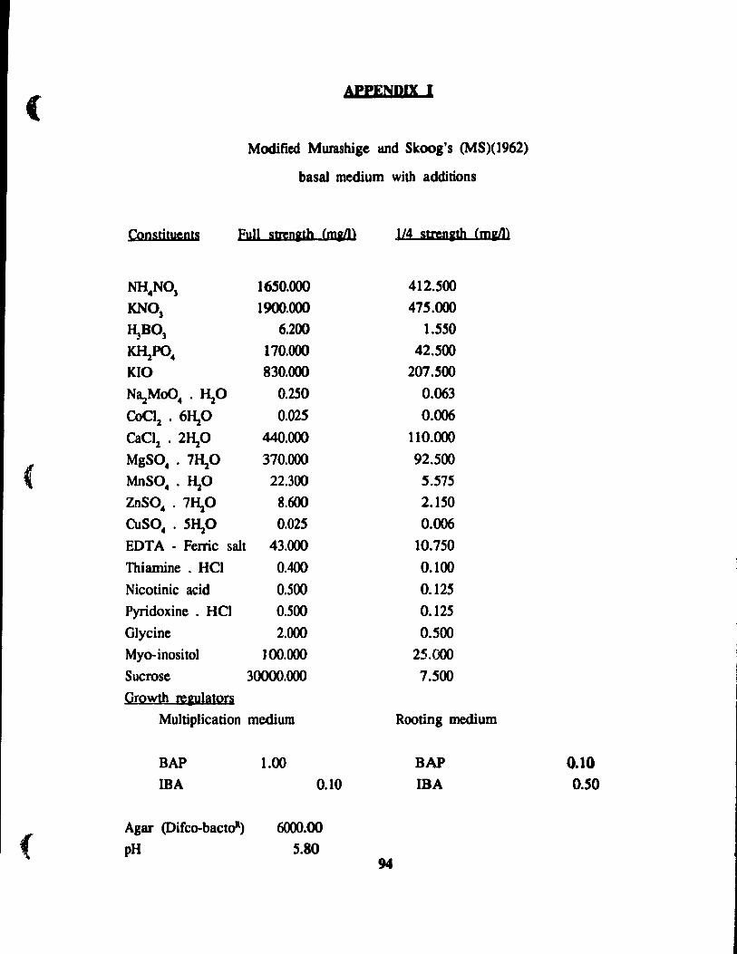
(
(
{
APPENDIX 1
Modifie<! Murashige and Skoog's (MS)(l962)
basal medium with additions
Coostitueots full StteDlth (mail> ,114 stren&th (m&t'1>
NH.NO] 16S0.000 412.S00
KNO] 1900.000 475.000
~BO] 6.200 t.S50
KlI:aPO. t70.000 42.S00
KIO 830.000 207.500
Na..zMoO. . ~O 0.250 0.063
COC~ . 6~0 0.025 0.006
CaClz • 2~O 440.000 110.000
MgSO •. 7~0 370.000 92.500
MoSO •. ~O 22.300 5.575 ZnSO •. 71\0 8.600 2.150
CUSO •. 5~0 0.025 0.006
EDT A - Ferric salt 43.000 10.750
Thiamine . HCI 0.400 0.100
Nicotinic acid 0.500 0.125
Pyridoxine. HCI 0.500 0.125
Glycine 2.000 0.500
Myo-inositol 100.000 25.Cœ
Sucrose 3()()()().OOO 7.500
QrQ~th œ&UhuQrs Multiplication medium Rooting medium
BAP 1.00 BAP
IBA 0.10 IBA
Agar (Difco-bactoR) 6000.00 pH 5.80
94
0.10 0.50