
Presentation1.pptx, radiological anatomy of the abdomen and pelvis.
Upload
claire-jacksonCategory
view
252download
0
ABDOMEN & PELVIS
PATHOLOGY & SCANNING PROTOCOLS

PATHOLOGIES
ABDOMINAL MESENTERIC CYST

ABDOMINAL CYST
An abdominal CT scan revealed a large right upper quadrant cyst measuring 14x17x21 cm ( lateral, anteroposterior and craniocaudal)There was mass effect upon the liver and duodenum. The cyst had a thin smooth wall with internal fluid and high density material consistent with a blood clot.
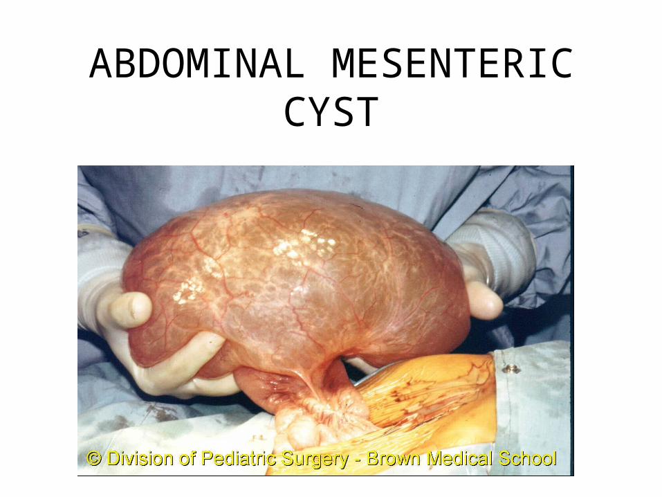
RENAL CYST
NO CONTRAST CONTRAST
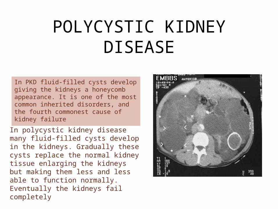
POLYCYSTIC KIDNEY DISEASE
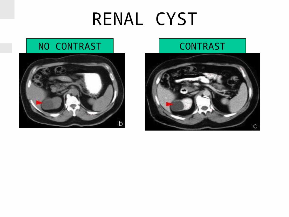
In PKD fluid-filled cysts develop giving the kidneys a honeycomb appearance. It is one of the most common inherited disorders, and the fourth commonest cause of kidney failure.
In polycystic kidney disease many fluid-filled cysts develop in the kidneys. Gradually these cysts replace the normal kidney tissue enlarging the kidneys but making them less and less able to function normally. Eventually the kidneys fail completely
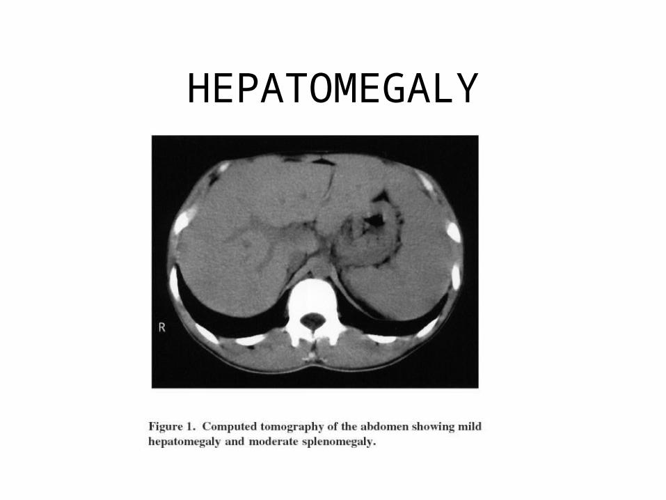
HEPATOMEGALY
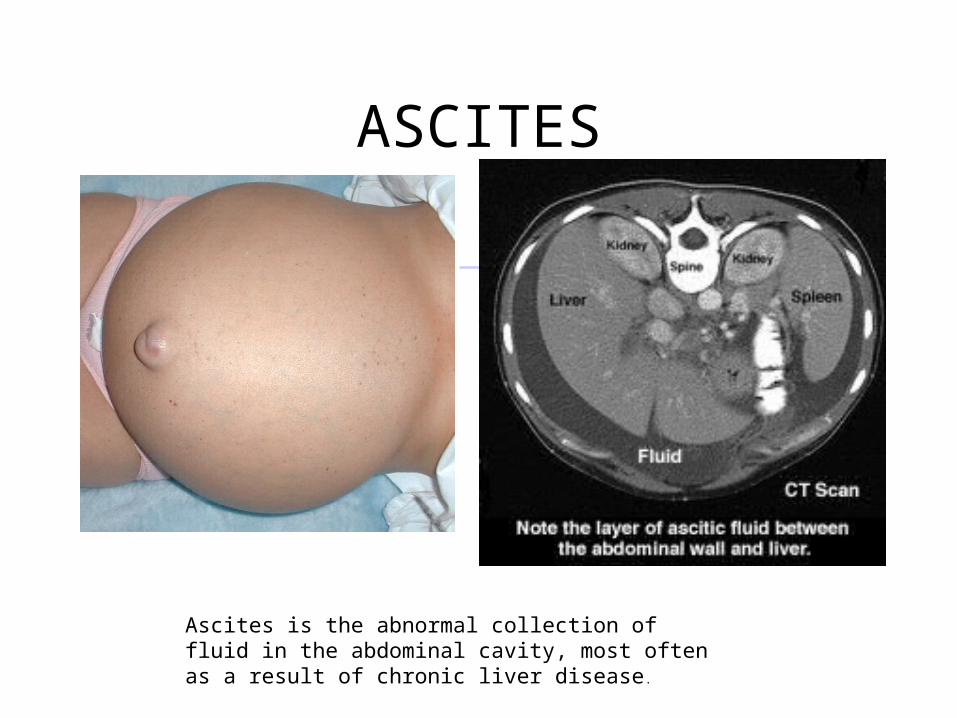
ASCITES
Ascites is the abnormal collection of fluid in the abdominal cavity, most often as a result of chronic liver disease.
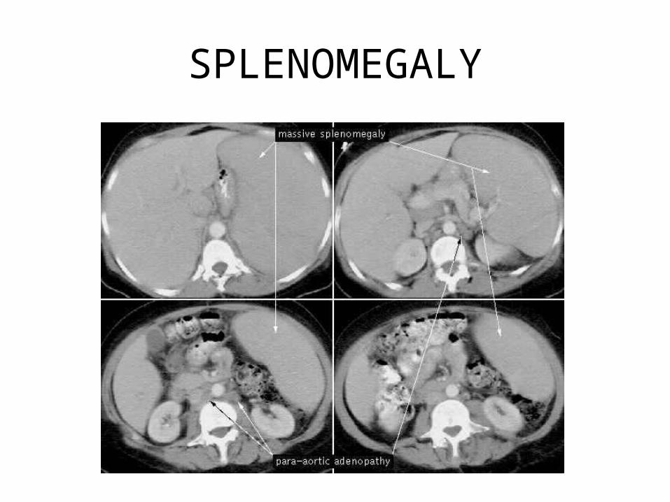
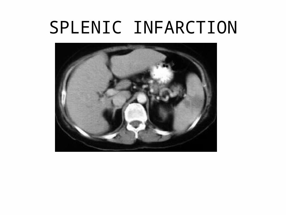
SPLENOMEGALY
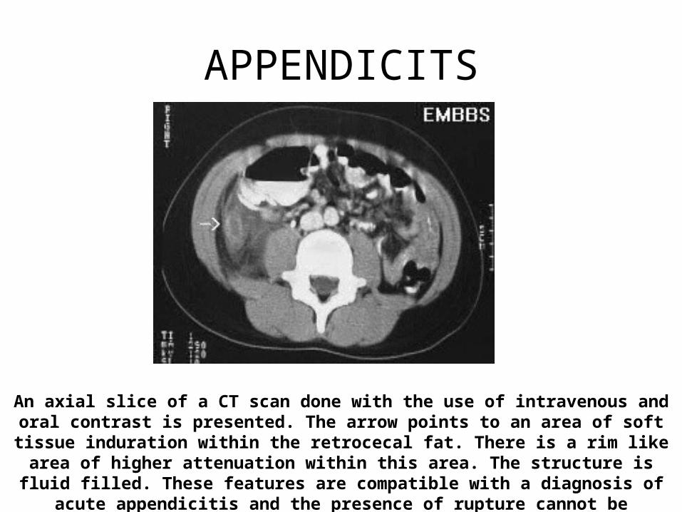
APPENDICITS
An axial slice of a CT scan done with the use of intravenous and oral contrast is presented. The arrow points to an area of soft tissue induration within the retrocecal fat. There is a rim like area of higher attenuation within this area. The structure is fluid filled. These
features are compatible with a diagnosis of acute appendicitis and the presence of rupture cannot be excluded.
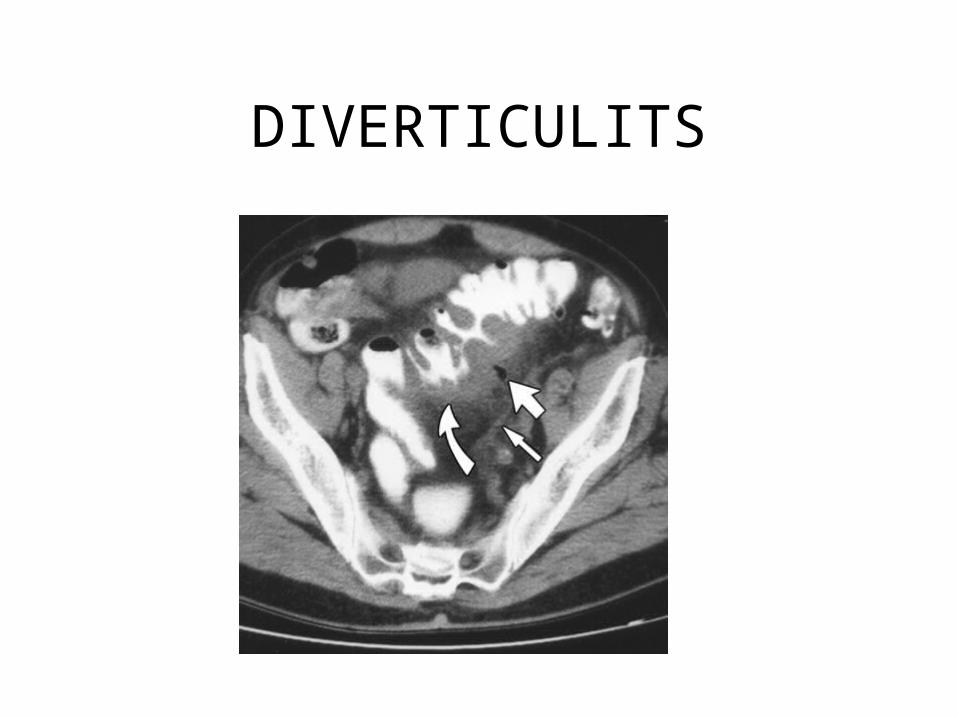
DIVERTICULITS
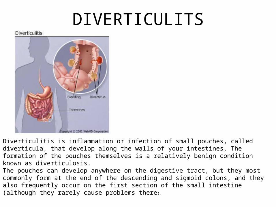
Diverticulitis is inflammation or infection of small pouches, called diverticula, that develop along the walls of your intestines. The formation of the pouches themselves is a relatively benign condition known as diverticulosis. The pouches can develop anywhere on the digestive tract, but they most commonly form at the end of the descending and sigmoid colons, and they also frequently occur on the first section of the small intestine (although they rarely cause problems there).
ABDOMINAL ABSCESS
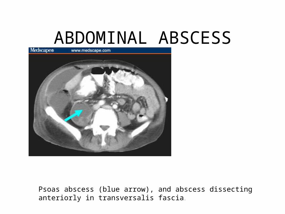
Psoas abscess (blue arrow), and abscess dissecting anteriorly in transversalis fascia.
BOWEL OBSTRUCTION
LIVER METS
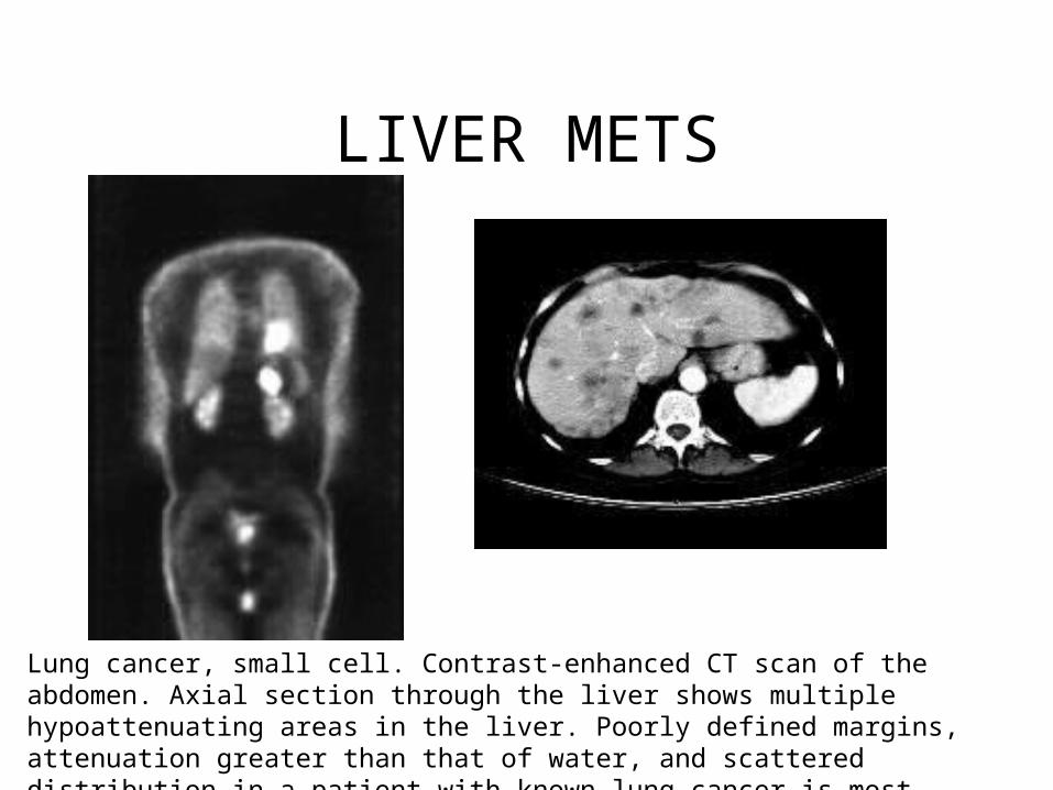
Lung cancer, small cell. Contrast-enhanced CT scan of the abdomen. Axial section through the liver shows multiple hypoattenuating areas in the liver. Poorly defined margins, attenuation greater than that of water, and scattered distribution in a patient with known lung cancer is most consistent with metastatic disease.
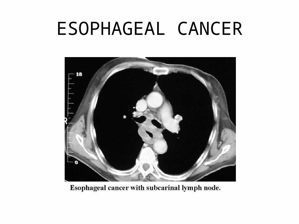
ESOPHAGEAL CANCER
WILMS TUMOR
Wilms tumor, also called nephroblastoma, is a cancer that originates in the kidney. The disease gets its name from a German doctor, Max Wilms, who wrote one of the first medical articles about it in 1899. Ninety percent of all kidney cancers in children are Wilms tumor. The remaining ten percent are rare forms of childhood kidney cancers: clear cell sarcoma of the kidney, malignant rhabdoid tumor of the kidney, and occasionally renal cell carcinoma
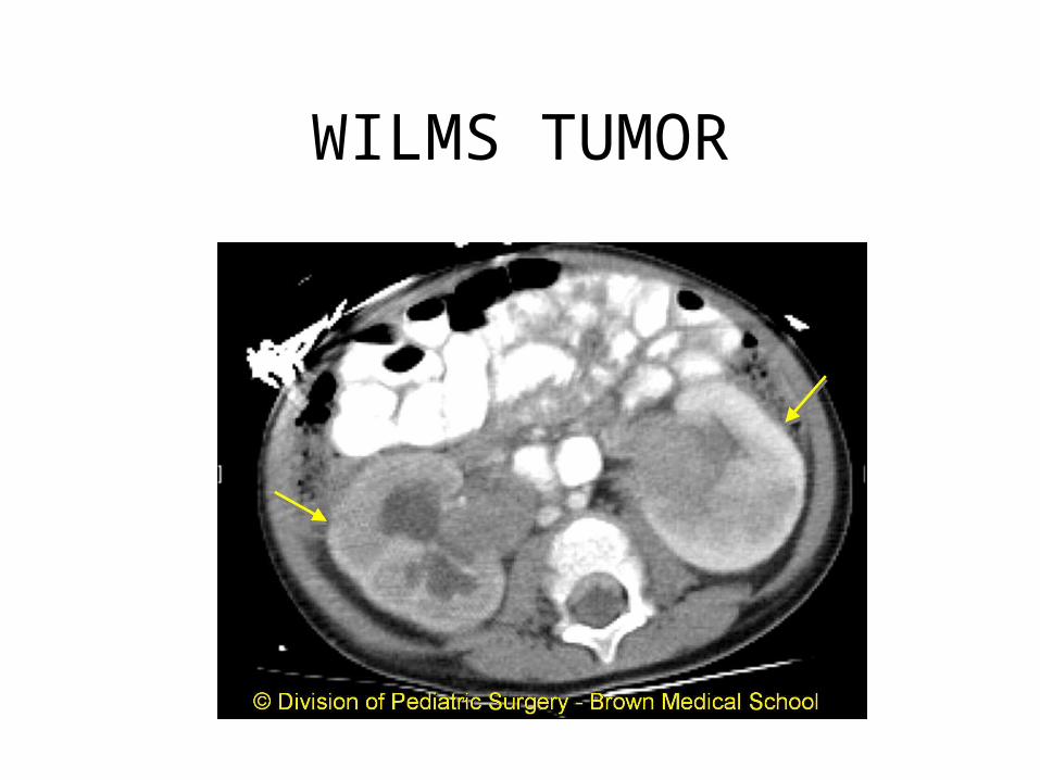
WILMS TUMOR
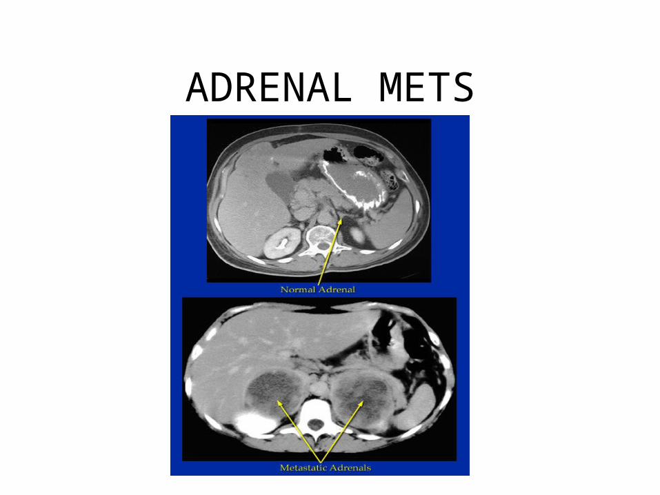
ADRENAL METS
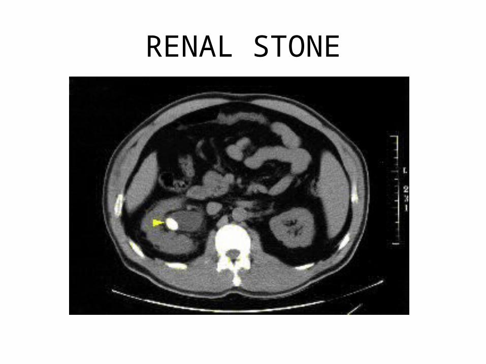
RENAL STONE
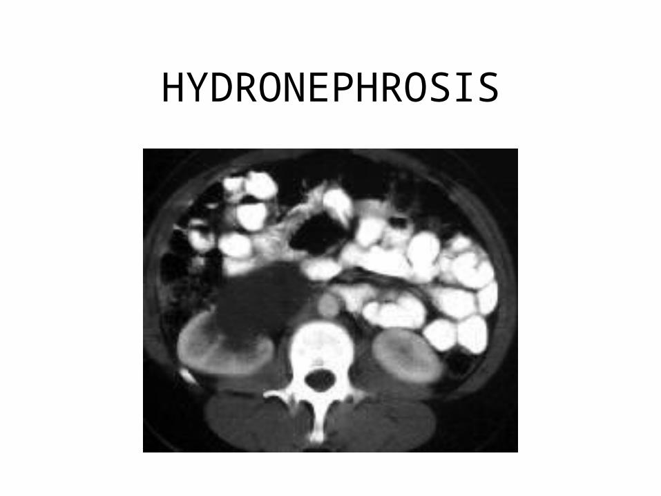
HYDRONEPHROSIS
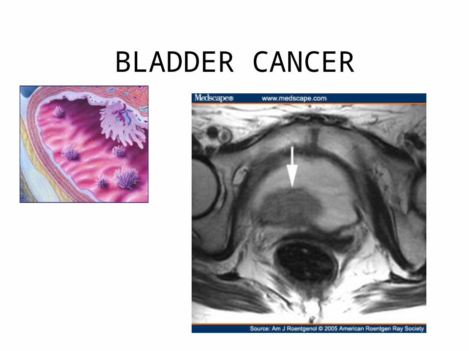
BLADDER CANCER
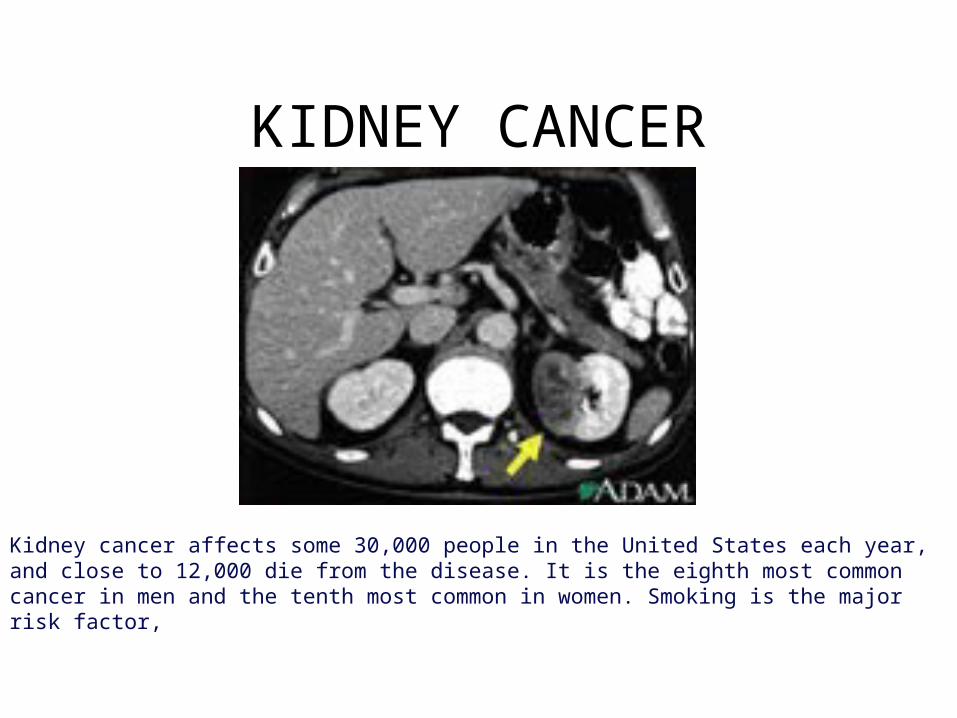
KIDNEY CANCER
Kidney cancer affects some 30,000 people in the United States each year, and close to 12,000 die from the disease. It is the eighth most common cancer in men and the tenth most common in women. Smoking is the major risk factor,
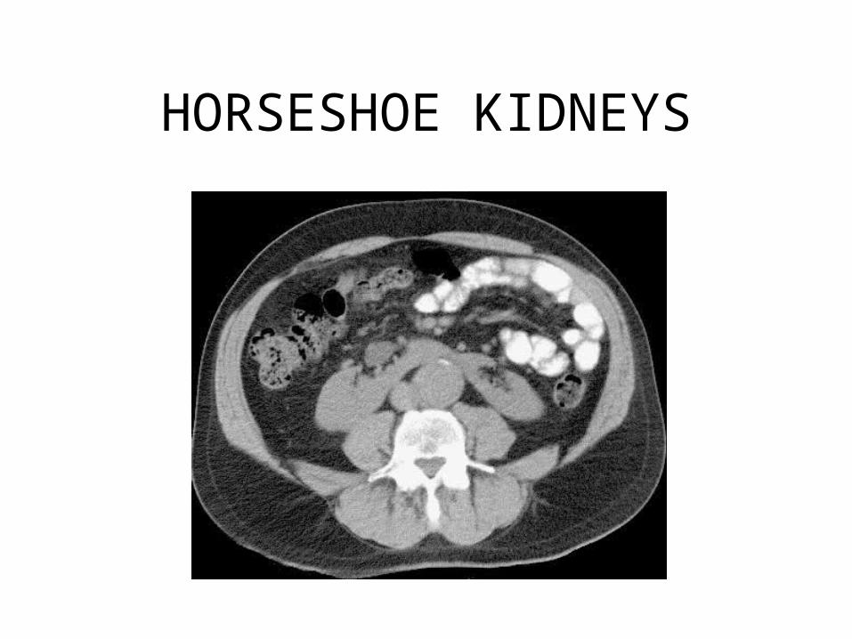
HORSESHOE KIDNEYS
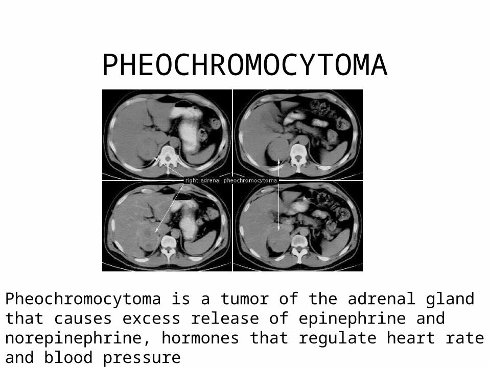
PHEOCHROMOCYTOMA
Pheochromocytoma is a tumor of the adrenal gland that causes excess release of epinephrine and norepinephrine, hormones that regulate heart rate and blood pressure

CIRRHOSIS
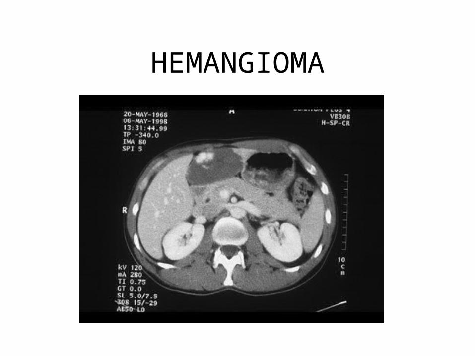
HEMANGIOMA
A cavernous hepatic hemangioma is the most common non-cancerous tumor of the liver. It is believed to be a congenital defect, and is usually not discovered until medical pictures are taken of the liver for some other reason.
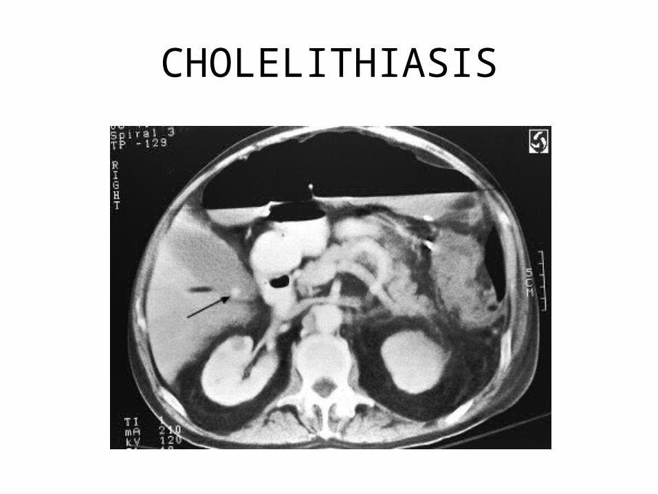
CHOLELITHIASIS
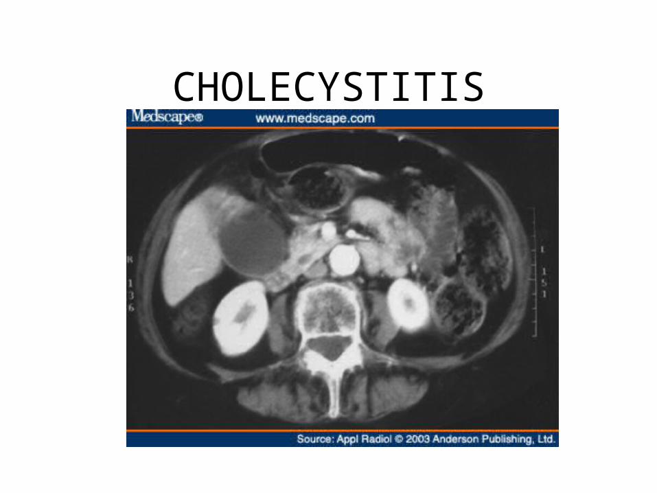
CHOLECYSTITIS

PANCREATIC CANCER
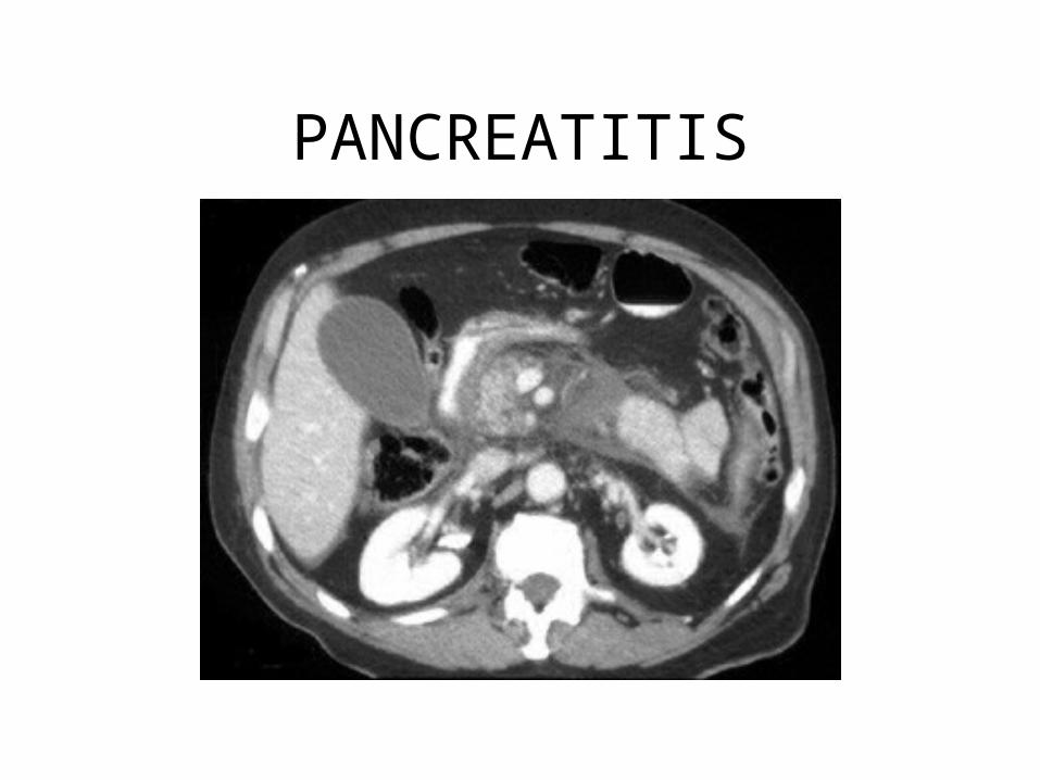
PANCREATITIS
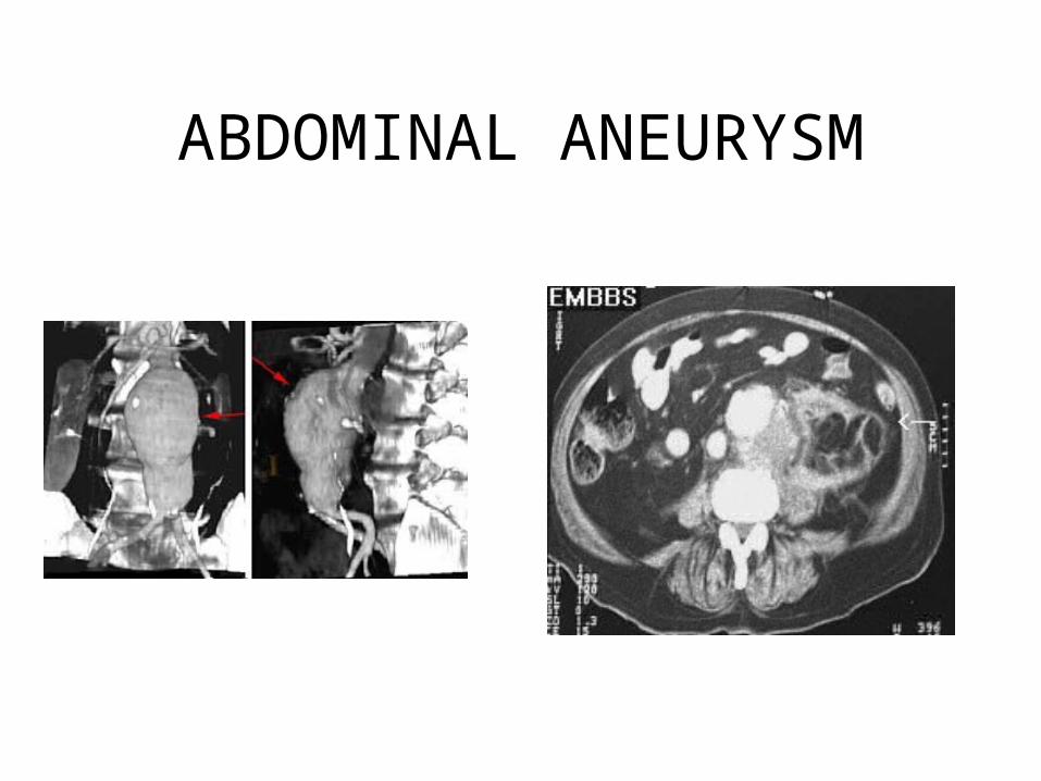
ABDOMINAL ANEURYSM
PROTOCOLS
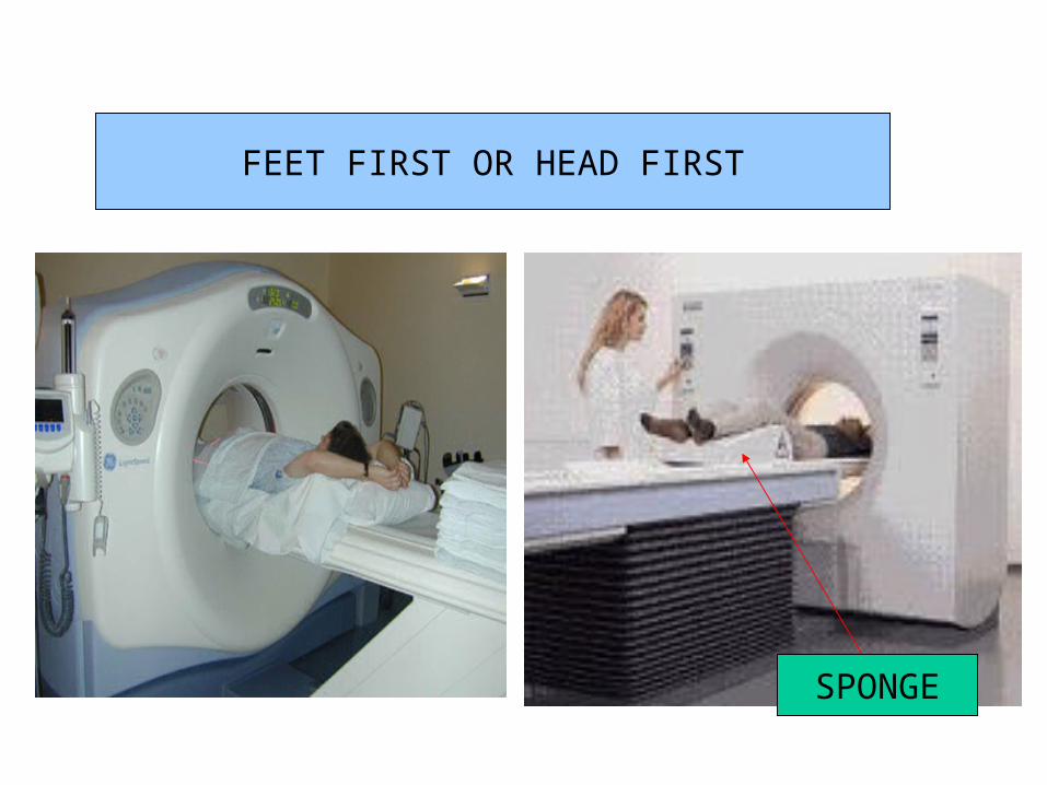
SPONGE
FEET FIRST OR HEAD FIRST
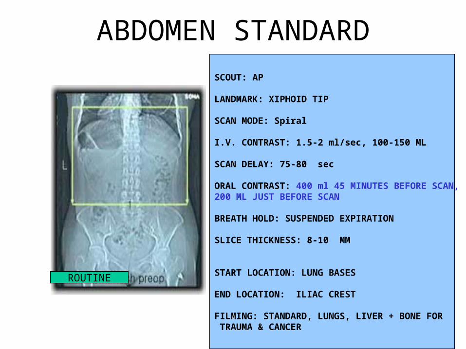
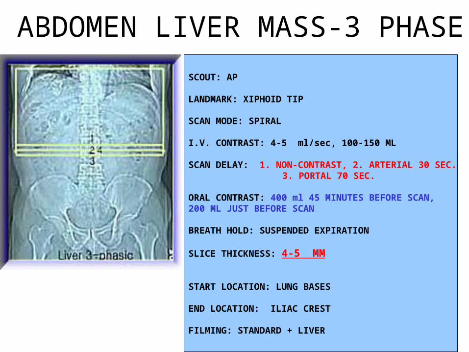
SCOUT: AP
LANDMARK: XIPHOID TIP
SCAN MODE: Spiral
I.V. CONTRAST: 1.5-2 ml/sec, 100-150 ML
SCAN DELAY: 75-80 sec
ORAL CONTRAST: 400 ml 45 MINUTES BEFORE SCAN, 200 ML JUST BEFORE SCAN
BREATH HOLD: SUSPENDED EXPIRATION
SLICE THICKNESS: 8-10 MM
START LOCATION: LUNG BASES
END LOCATION: ILIAC CREST
FILMING: STANDARD, LUNGS, LIVER + BONE FOR TRAUMA & CANCER
ABDOMEN STANDARD
ROUTINE
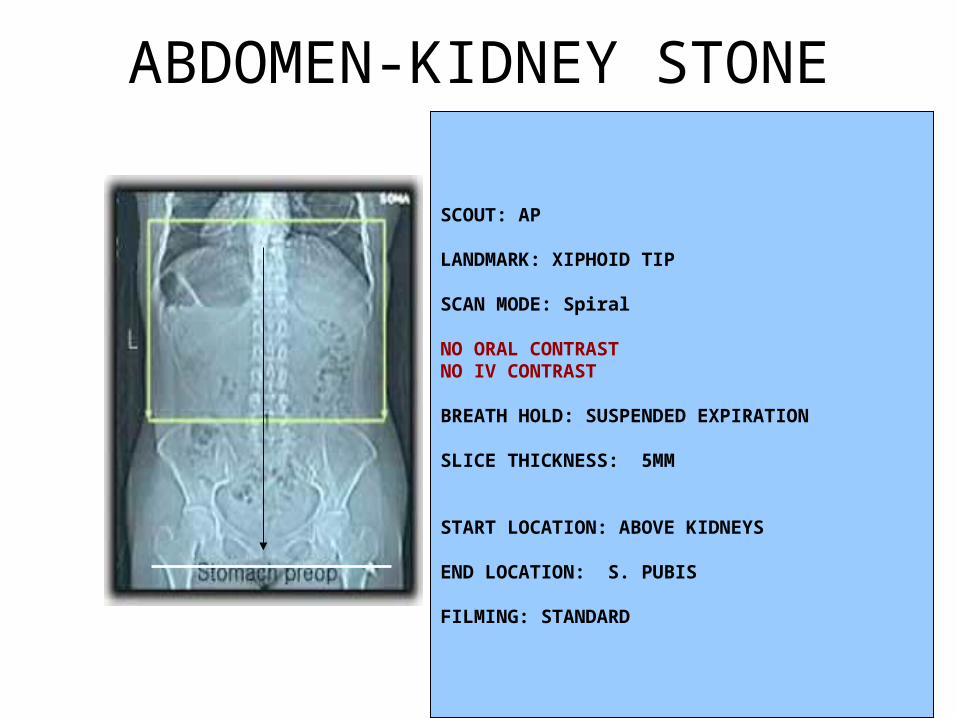
SCOUT: AP
LANDMARK: XIPHOID TIP
SCAN MODE: Spiral
NO ORAL CONTRASTNO IV CONTRAST
BREATH HOLD: SUSPENDED EXPIRATION
SLICE THICKNESS: 5MM
START LOCATION: ABOVE KIDNEYS
END LOCATION: S. PUBIS
FILMING: STANDARD
ABDOMEN-KIDNEY STONE
SCOUT: AP
LANDMARK: XIPHOID TIP
SCAN MODE: SPIRAL
I.V. CONTRAST: 4-5 ml/sec, 100-150 ML
SCAN DELAY: 1. NON-CONTRAST, 2. ARTERIAL 30 SEC. 3. PORTAL 70 SEC.
ORAL CONTRAST: 400 ml 45 MINUTES BEFORE SCAN, 200 ML JUST BEFORE SCAN
BREATH HOLD: SUSPENDED EXPIRATION
SLICE THICKNESS: 4-5 MM
START LOCATION: LUNG BASES
END LOCATION: ILIAC CREST
FILMING: STANDARD + LIVER
ABDOMEN LIVER MASS-3 PHASE
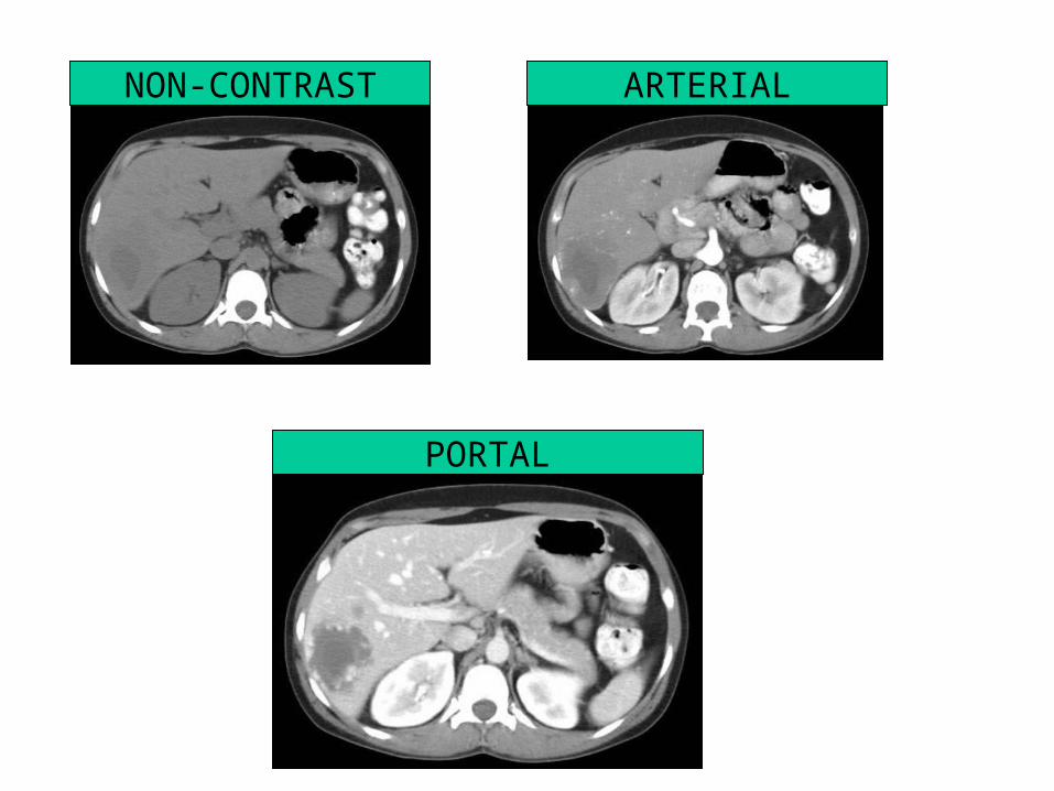
NON-CONTRAST ARTERIAL
PORTAL
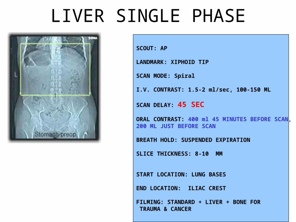
LIVER SINGLE PHASE
SCOUT: AP
LANDMARK: XIPHOID TIP
SCAN MODE: Spiral
I.V. CONTRAST: 1.5-2 ml/sec, 100-150 ML
SCAN DELAY: 45 SEC
ORAL CONTRAST: 400 ml 45 MINUTES BEFORE SCAN, 200 ML JUST BEFORE SCAN
BREATH HOLD: SUSPENDED EXPIRATION
SLICE THICKNESS: 8-10 MM
START LOCATION: LUNG BASES
END LOCATION: ILIAC CREST
FILMING: STANDARD + LIVER + BONE FOR TRAUMA & CANCER
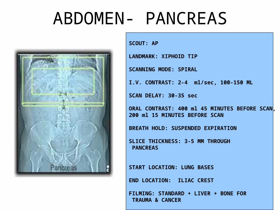
SCOUT: AP
LANDMARK: XIPHOID TIP
SCANNING MODE: SPIRAL
I.V. CONTRAST: 2-4 ml/sec, 100-150 ML
SCAN DELAY: 30-35 sec
ORAL CONTRAST: 400 ml 45 MINUTES BEFORE SCAN, 200 ml 15 MINUTES BEFORE SCAN
BREATH HOLD: SUSPENDED EXPIRATION
SLICE THICKNESS: 3-5 MM THROUGH PANCREAS
START LOCATION: LUNG BASES
END LOCATION: ILIAC CREST
FILMING: STANDARD + LIVER + BONE FOR TRAUMA & CANCER
ABDOMEN- PANCREAS
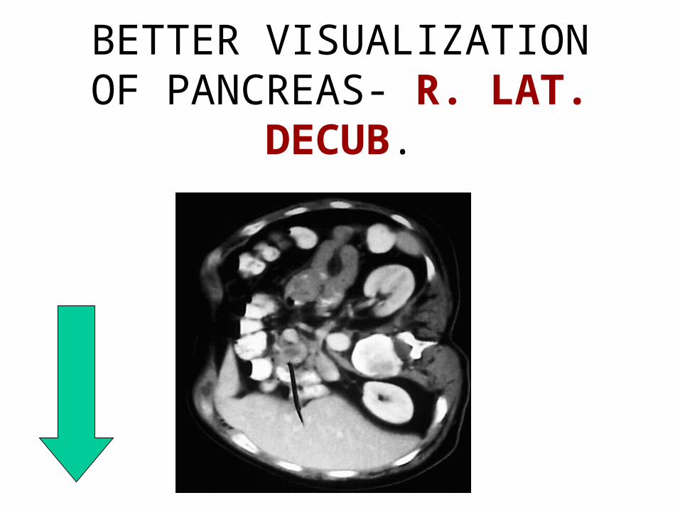
BETTER VISUALIZATION OF PANCREAS- R. LAT. DECUB.
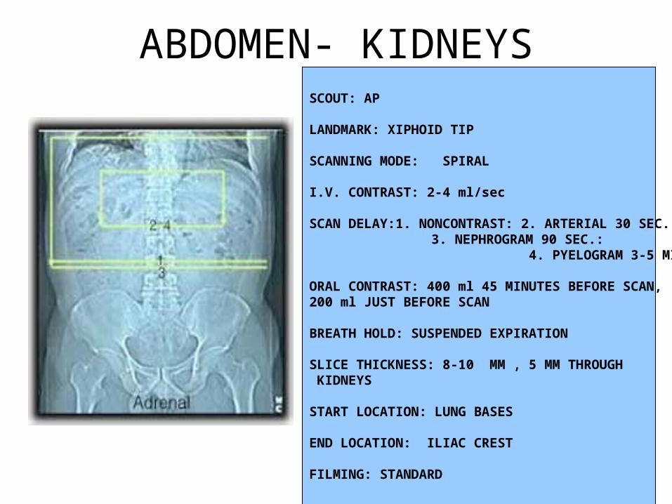
SCOUT: AP
LANDMARK: XIPHOID TIP
SCANNING MODE: SPIRAL
I.V. CONTRAST: 2-4 ml/sec
SCAN DELAY:1. NONCONTRAST: 2. ARTERIAL 30 SEC. 3. NEPHROGRAM 90 SEC.:
4. PYELOGRAM 3-5 MIN.
ORAL CONTRAST: 400 ml 45 MINUTES BEFORE SCAN, 200 ml JUST BEFORE SCAN
BREATH HOLD: SUSPENDED EXPIRATION
SLICE THICKNESS: 8-10 MM , 5 MM THROUGH KIDNEYS
START LOCATION: LUNG BASES
END LOCATION: ILIAC CREST
FILMING: STANDARD
ABDOMEN- KIDNEYS
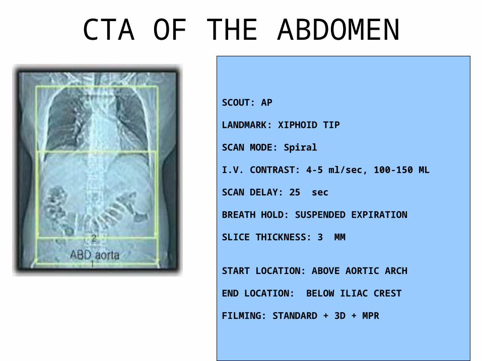
CTA OF THE ABDOMEN
SCOUT: AP
LANDMARK: XIPHOID TIP
SCAN MODE: Spiral
I.V. CONTRAST: 4-5 ml/sec, 100-150 ML
SCAN DELAY: 25 sec
BREATH HOLD: SUSPENDED EXPIRATION
SLICE THICKNESS: 3 MM
START LOCATION: ABOVE AORTIC ARCH
END LOCATION: BELOW ILIAC CREST
FILMING: STANDARD + 3D + MPR
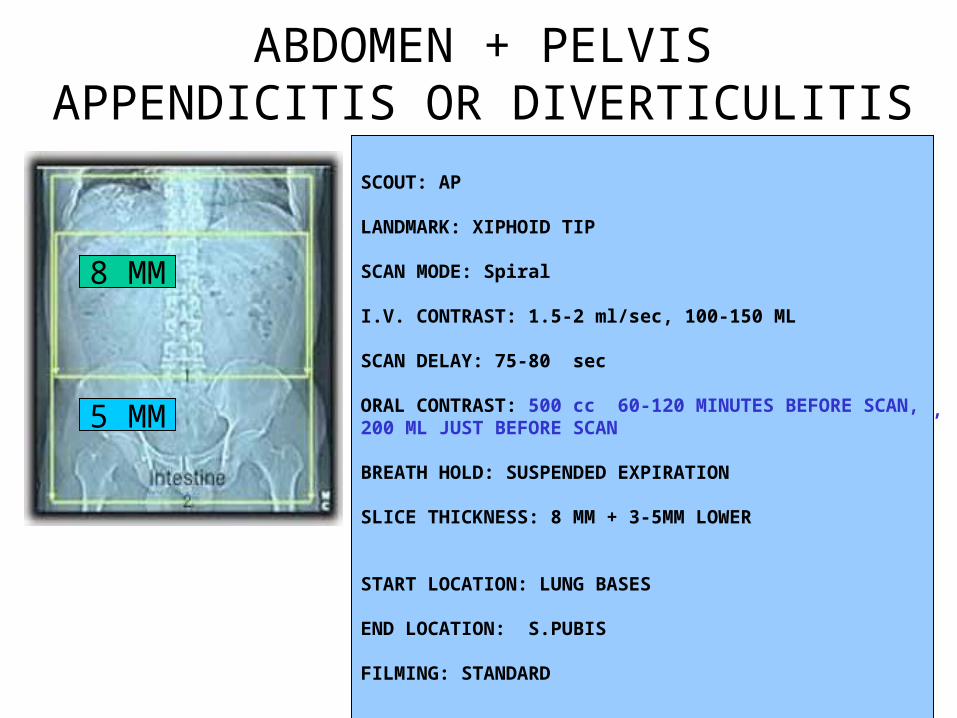
ABDOMEN + PELVISAPPENDICITIS OR DIVERTICULITIS
SCOUT: AP
LANDMARK: XIPHOID TIP
SCAN MODE: Spiral
I.V. CONTRAST: 1.5-2 ml/sec, 100-150 ML
SCAN DELAY: 75-80 sec
ORAL CONTRAST: 400 ml 45 MINUTES BEFORE SCAN, 200 ML JUST BEFORE SCAN
BREATH HOLD: SUSPENDED EXPIRATION
SLICE THICKNESS: 8-10 MM UPPER + 5 MM LOWER
START LOCATION: LUNG BASES
END LOCATION: S. PUBIS
FILMING: STANDARD
8 MM
SCOUT: AP
LANDMARK: XIPHOID TIP
SCAN MODE: Spiral
I.V. CONTRAST: 1.5-2 ml/sec, 100-150 ML
SCAN DELAY: 75-80 sec
ORAL CONTRAST: 500 cc 60-120 MINUTES BEFORE SCAN, 200 ML JUST BEFORE SCAN
BREATH HOLD: SUSPENDED EXPIRATION
SLICE THICKNESS: 8 MM + 3-5MM LOWER
START LOCATION: LUNG BASES
END LOCATION: S.PUBIS
FILMING: STANDARD
5 MM
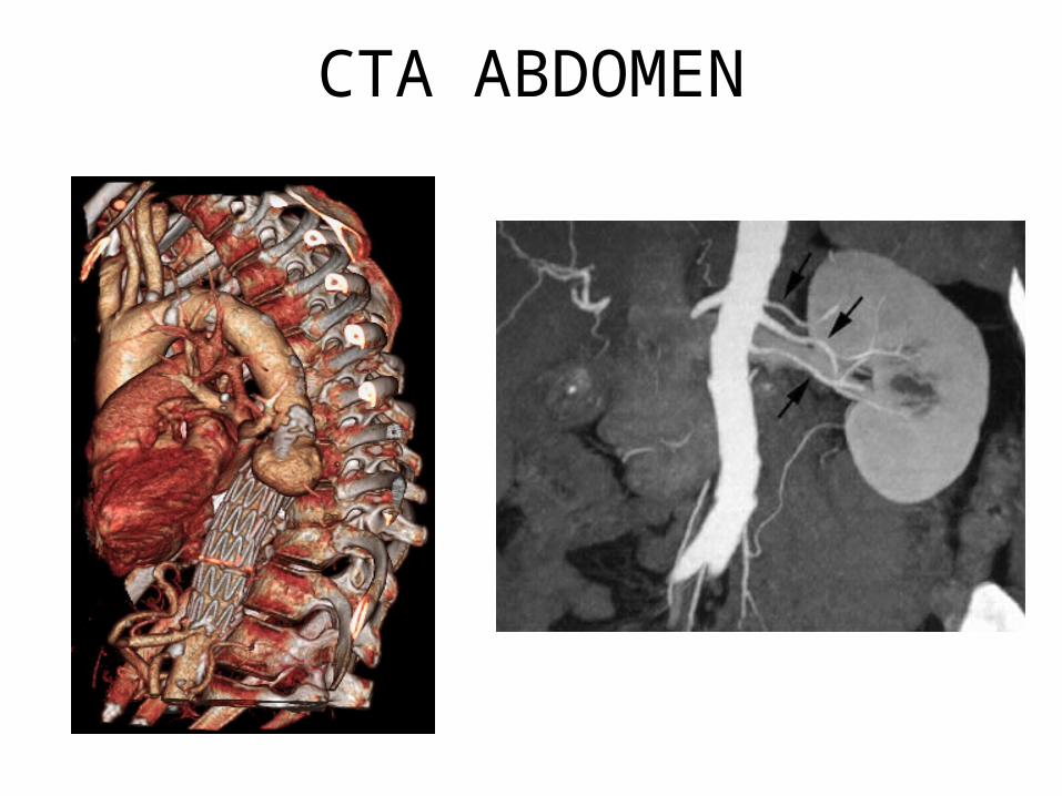
CTA ABDOMEN
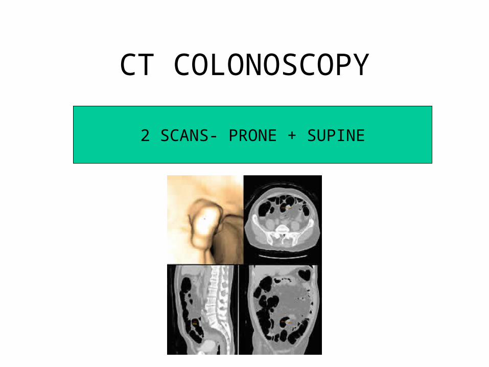
CT COLONOSCOPY
2 SCANS- PRONE + SUPINE
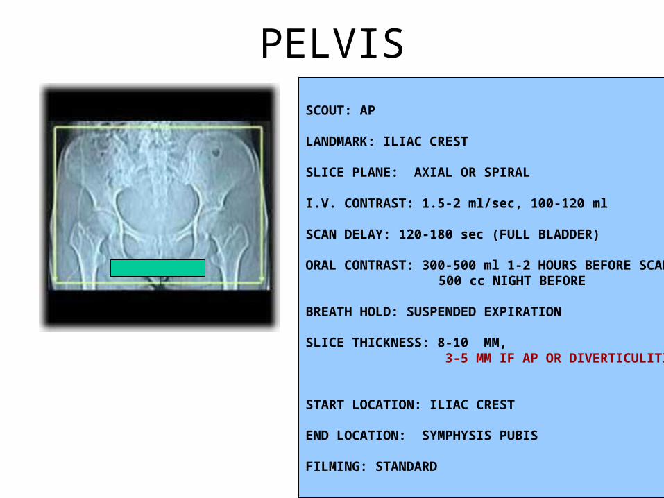
SCOUT: AP
LANDMARK: ILIAC CREST
SLICE PLANE: AXIAL OR SPIRAL
I.V. CONTRAST: 1.5-2 ml/sec, 100-120 ml
SCAN DELAY: 120-180 sec (FULL BLADDER)
ORAL CONTRAST: 300-500 ml 1-2 HOURS BEFORE SCAN500 cc NIGHT BEFORE
BREATH HOLD: SUSPENDED EXPIRATION
SLICE THICKNESS: 8-10 MM, 3-5 MM IF AP OR DIVERTICULITIS
START LOCATION: ILIAC CREST
END LOCATION: SYMPHYSIS PUBIS
FILMING: STANDARD
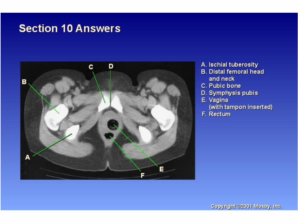
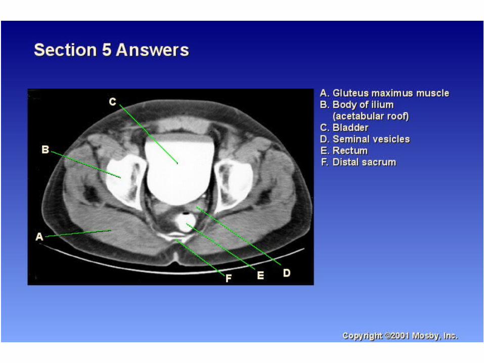
PELVIS
DETECTION OF PROSTATE GLAND AND SEMINAL
VESICLES ABNORMALITIES
BLADDER OPACIFIED
+RECTOSIGMOID COLON AND RECTUM OPACIFIED
VISUALIZATION OF VAGINAL CANAL + CERVIX
AND UTERUS
TAMPON INSERTED IN THE VAGINA DURING CT SCAN OF THE PELVIS