
Adult Abdomen Pelvis Ct
12
Adult Routine Abdomen/Pelvis CT Protocols Version 1.0 10/10/2012 1 The disclaimer on page 1 is an integral part of this document. Copyright © October 10, 2012 by AAPM. All rights reserved. DISCLAIMER: TO THE EXTENT ALLOWED BY LOCAL LAW, THIS INFORMATION IS PROVIDED TO YOU BY THE AMERICAN ASSOCIATION OF PHYCISISTS IN MEDICINE, A NON-PROFIT ORGANISATION ORGANIZED TO PROMOTE THE APPLICATION OF PHYSICS TO MEDICINE AND BIOLOGY, ENCOURAGE INTEREST AND TRAINING IN MEDICAL PHYSICS AND RELATED FIELDS ("AAPM"), 'AS IS' WITHOUT WARRANTIES OR CONDITIONS OF ANY KIND, WHETHER ORAL OR WRITTEN, EXPRESS OR IMPLIED. AAPM SPECIFICALLY DISCLAIMS ANY IMPLIED WARRANTIES OR CONDITIONS OF MERCHANTABILITY, SATISFACTORY QUALITY, NONINFRINGEMENT AND FITNESS FOR A PARTICULAR PURPOSE. SOME JURISDICTIONS DO NOT ALLOW EXCLUSIONS OF IMPLIED WARRANTIES OR CONDITIONS, SO THE ABOVE EXCLUSION MAY NOT APPLY TO YOU. YOU MAY HAVE OTHER RIGHTS THAT VARY ACCORDING TO LOCAL LAW. TO THE EXTENT ALLOWED BY LOCAL LAW, IN NO EVENT WILL AAPM OR ITS SUBSIDIARIES, AFFILIATES OR VENDORS BE LIABLE FOR DIRECT, SPECIAL, INCIDENTAL, CONSEQUENTIAL OR OTHER DAMAGES (INCLUDING LOST PROFIT, LOST DATA, OR DOWNTIME COSTS), ARISING OUT OF THE USE, INABILITY TO USE, OR THE RESULTS OF USE OF THE PROVIDED INFORMATION, WHETHER BASED IN WARRANTY, CONTRACT, TORT OR OTHER LEGAL THEORY, AND WHETHER OR NOT ADVISED OF THE POSSIBILITY OF SUCH DAMAGES. YOUR USE OF THE INFORMATION IS ENTIRELY AT YOUR OWN RISK. THIS INFORMATION IS NOT MEANT TO BE USED AS A SUBSTITUTE FOR THE REVIEW OF SCAN PROTOCOL PARAMETERS BY A QUALIFIED AND CERTIFIED PROFESSIONAL. USERS ARE CAUTIONED TO SEEK THE ADVICE OF A QUALIFIED AND CERTIFIED PROFESSIONAL BEFORE USING ANY PROTOCOL BASED ON THE PROVIDED INFORMATION. AAPM IS NOT RESPONSIBLE FOR A USER'S FAILURE TO VERIFY OR CONFIRM APPROPRIATE PERFORMANCE OF THE PROVIDED SCAN PARAMETERS. SOME JURISDICTIONS DO NOT ALLOW THE EXCLUSION OR LIMITATION OF LIABILITY FOR DAMAGES, SO THE ABOVE LIMITATION MAY NOT APPLY TO YOU.
description
Computer Tomography
Transcript of Adult Abdomen Pelvis Ct
Adult Routine Abdomen/Pelvis CT Protocols Version 1.0 10/10/2012
1 The disclaimer on page 1 is an integral part of this document.
Copyright © October 10, 2012 by AAPM. All rights reserved.
DISCLAIMER: TO THE EXTENT ALLOWED BY LOCAL LAW, THIS INFORMATION IS PROVIDED TO YOU BY THE AMERICAN ASSOCIATION OF PHYCISISTS IN MEDICINE, A NON-PROFIT ORGANISATION ORGANIZED TO PROMOTE THE APPLICATION OF PHYSICS TO MEDICINE AND BIOLOGY, ENCOURAGE INTEREST AND TRAINING IN MEDICAL PHYSICS AND RELATED FIELDS ("AAPM"), 'AS IS' WITHOUT WARRANTIES OR CONDITIONS OF ANY KIND, WHETHER ORAL OR WRITTEN, EXPRESS OR IMPLIED. AAPM SPECIFICALLY DISCLAIMS ANY IMPLIED WARRANTIES OR CONDITIONS OF MERCHANTABILITY, SATISFACTORY QUALITY, NONINFRINGEMENT AND FITNESS FOR A PARTICULAR PURPOSE. SOME JURISDICTIONS DO NOT ALLOW EXCLUSIONS OF IMPLIED WARRANTIES OR CONDITIONS, SO THE ABOVE EXCLUSION MAY NOT APPLY TO YOU. YOU MAY HAVE OTHER RIGHTS THAT VARY ACCORDING TO LOCAL LAW. TO THE EXTENT ALLOWED BY LOCAL LAW, IN NO EVENT WILL AAPM OR ITS SUBSIDIARIES, AFFILIATES OR VENDORS BE LIABLE FOR DIRECT, SPECIAL, INCIDENTAL, CONSEQUENTIAL OR OTHER DAMAGES (INCLUDING LOST PROFIT, LOST DATA, OR DOWNTIME COSTS), ARISING OUT OF THE USE, INABILITY TO USE, OR THE RESULTS OF USE OF THE PROVIDED INFORMATION, WHETHER BASED IN WARRANTY, CONTRACT, TORT OR OTHER LEGAL THEORY, AND WHETHER OR NOT ADVISED OF THE POSSIBILITY OF SUCH DAMAGES. YOUR USE OF THE INFORMATION IS ENTIRELY AT YOUR OWN RISK. THIS INFORMATION IS NOT MEANT TO BE USED AS A SUBSTITUTE FOR THE REVIEW OF SCAN PROTOCOL PARAMETERS BY A QUALIFIED AND CERTIFIED PROFESSIONAL. USERS ARE CAUTIONED TO SEEK THE ADVICE OF A QUALIFIED AND CERTIFIED PROFESSIONAL BEFORE USING ANY PROTOCOL BASED ON THE PROVIDED INFORMATION. AAPM IS NOT RESPONSIBLE FOR A USER'S FAILURE TO VERIFY OR CONFIRM APPROPRIATE PERFORMANCE OF THE PROVIDED SCAN PARAMETERS. SOME JURISDICTIONS DO NOT ALLOW THE EXCLUSION OR LIMITATION OF LIABILITY FOR DAMAGES, SO THE ABOVE LIMITATION MAY NOT APPLY TO YOU.
Adult Routine Abdomen/Pelvis CT Protocols Version 1.0 10/10/2012
2 The disclaimer found on page 1 is an integral part of this document.
Copyright © October 10, 2012 by AAPM. All rights reserved.
ROUTINE ADULT ABDOMEN/PELVIS CT PROTOCOLS
Indications (include but are not limited to) • Evaluation of abdominal, flank, or pelvic pain • Evaluation of known or suspected abdominal or pelvic masses or fluid collections • Evaluation of primary or metastatic malignancies • Evaluation of abdominal or pelvic inflammatory processes • Evaluate infections of the abdomen and pelvis • Evaluate patients with fever or suspected abscess • To identify the cause of bowel obstruction • Evaluation of weight loss • Clarification of findings from other imaging studies or laboratory abnormalities. • For reference, see ACR–SPR Practice Guideline for the Performance of Computed
Tomography (CT) of the Abdomen and Computed Tomography (CT) of the Pelvis Diagnostic Tasks (include but are not limited to)
• Detect soft tissue masses and abnormal fluid collections and determine sizes • Identify abnormal collections of blood • Identify air outside the intestinal tract • Detect nodules or soft tissue masses adjacent to vascular structures • Detect calcifications in abnormal locations or in abdominal and pelvic organs • Characterize soft tissue edema around the organs of the abdomen and pelvis
Key Elements
• Patient positioning is especially important when using tube current modulation. • Scanning should be performed helically whenever possible. • Automatic Exposure Control (AEC): Each manufacturer has unique nomenclature and
operating characteristics for their AEC system(s). Users must become very familiar with how the AEC systems on their particular scanners operate. See Singh et al. Automatic Exposure Control in CT: Applications and Limitations. JACR 2011;8(6):446-449.
Contrast
• Oral: Per radiologist • Injected: Certain indications require administration of intravenous contrast media. • Intravenous contrast enhancement should be performed as directed by the supervising
radiologist using appropriate injection protocols and in accordance with the ACR-SPR Practice Guideline for the Use of Intravascular Contrast Media and the ACR Manual on Contrast Media.
Patient Positioning
• Center the patient within the gantry; this is critical for proper functioning of AEC systems. • Patient supine, typically feet first
Scan Range
• Scan from top of liver to either iliac crest or pubic symphysis, depending on clinical indications. Suspension of Respiration
• Patient should be instructed to hold his/her breath at end of inspiration.
Adult Routine Abdomen/Pelvis CT Protocols Version 1.0 10/10/2012
3 The disclaimer found on page 1 is an integral part of this document.
Copyright © October 10, 2012 by AAPM. All rights reserved.
Additional Image Reconstructions • Certain indications may require that images be reconstructed in coronal and/or sagittal planes. • Very thin images (approximately ≤ 1 mm) may need to be reconstructed to serve as source
images for the sagittal and/or coronal reformatted images. • Creation, use, and archival of these additional images are at the discretion of the supervising
radiologist and/or departmental policy. Very large datasets may result from these additional reconstructions.
Radiation Dose Management
• AEC should be used whenever possible. • Pay careful attention to the values selected to define the desired level of image quality
(eg, Noise Index, Quality Reference mAs, Standard Deviation). • Each manufacturer will have recommendations unique to their systems and system
features. Be sure to work with your CT equipment manufacturer and a qualified medical physicist to ensure safe and appropriate operation of AEC systems.
• If more than one CT localizer radiograph is acquired, AEC systems from different manufacturers can differ with respect to which one is used to determine mA and/or kV settings. Please refer to individual manufacturer protocol instructions.
CTDI measurements and calculations
• Some manufacturers utilize a z-axis “flying focal spot”, in which two unique projections are acquired at the same z-axis table position. When this technique is used, we identify it with **. The CTDIvol on the console accurately accounts for use of this feature.
Adult Routine Abdomen/Pelvis CT Protocols Version 1.0 10/10/2012
4 The disclaimer found on page 1 is an integral part of this document.
Copyright © October 10, 2012 by AAPM. All rights reserved.
Approximate Volume CT Dose Index (CTDIvol) Values
• Approximate values for CTDIvol are listed for three different patient sizes:
Approx. Weight (kg) Approx. Weight (lbs) Approx. CTDIvol (mGy) Small Patient 50-70 110-155 10-17
Average Patient 70-90 155-200 15-25 Large Patient 90-120 200-265 22-35
The approximate CTDIvol values are for reference only and represent a dose to the CT Dose Index phantom under very specific conditions. The CTDIvol displayed on the scanner for a patient of a given size should be similar, but not necessarily an exact match, to those listed in the above table. The provided values are all based on the 32 cm diameter “body” CTDI phantom. It is essential that users recognize that the CTDIvol values reported on the user console prior to acquiring CT localizer radiographs on a particular patient
• Underweight = BMI <18.5
do not represent the CTDIvol that will be delivered during that patient’s scan. CT systems rely on the CT localizer radiograph to 1) estimate the patient’s size, 2) determine the tube current settings for each tube angle and table position that will yield the requested level of image quality, and 3) calculate the average CTDIvol for the patient over the prescribed scan range. Until the CT localizer radiograph is acquired, the reported CTDIvol is not patient-specific, but is based on a generic patient size. The CTDIvol values provided here are approximate, and are intended only to provide reference ranges for the user to consider. They are for a routine CT of an adult’s abdomen/pelvis for the general indictations given at the beginning of this document. Other indications or diagnostic tasks may have different image quality and dose requirements, and hence reasonable ranges of CTDIvol may differ according to those requirements. In this document, a small patient is considered to be approximately 50-70 kg (110-155 lbs), an average patient approximately 70-90 kg (155-200 lbs), and a large patient 90-120 kg (200-265 lbs). However, weight is not a perfect indication of patient size. A person’s height, gender and distribution of weight across the body also must be taken into account. The thickness of the body over the area to be scanned is the best indication of patient size. Bodymass index (BMI) may also be considered:
• Normal weight = BMI of 18.5–24.9 • Overweight = BMI of 25–29.9 • Obesity = BMI of 30 or greater
It is recognized that the median (50th percentile) patient size for adults in the USA is larger than 70 kg. However, the 70 kg patient represents the “Reference Man”, as defined by the International Commission on Radiation Protection (ICRP), upon which AEC systems and tissue weighting factors (used for effective dose estimation) are based.
Adult Routine Abdomen/Pelvis CT Protocols Version 1.0 10/10/2012
5 The disclaimer found on page 1 is an integral part of this document.
Copyright © October 10, 2012 by AAPM. All rights reserved.
INDEX OF ADULT ROUTINE ABDOMEN-PELVIS PROTOCOLS (by manufacturer)
GE
Hitachi
Neusoft
Philips
Siemens
Toshiba
Adult Routine Abdomen/Pelvis CT Protocols Version 1.0 10/10/2012
6 The disclaimer found on page 1 is an integral part of this document.
Copyright © October 10, 2012 by AAPM. All rights reserved.
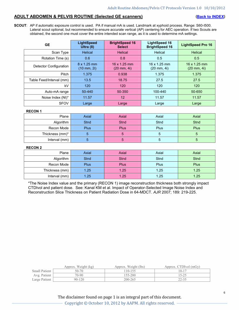
ADULT ABDOMEN & PELVIS ROUTINE (Selected GE scanners) (Back to INDEX) SCOUT: AP if automatic exposure control is used. PA if manual mA is used. Landmark at xyphoid process. Range: S60-I500.
Lateral scout optional, but recommended to ensure accurate vertical (AP) centering for AEC operation. If two Scouts are obtained, the second one must cover the entire intended scan range, as it is used to determine mA settings.
GE LightSpeed Ultra (8)
BrightSpeed 16 Select
LightSpeed 16 BrightSpeed 16 LightSpeed Pro 16
Scan Type Helical Helical Helical Helical Rotation Time (s) 0.6 0.8 0.5 0.5
Detector Configuration 8 x 1.25 mm (10 mm, 2i)
16 x 1.25 mm (20 mm, 4i)
16 x 1.25 mm (20 mm, 4i)
16 x 1.25 mm (20 mm, 4i)
Pitch 1.375 0.938 1.375 1.375 Table Feed/Interval (mm) 13.5 18.75 27.5 27.5
kV 120 120 120 120 Auto-mA range 50-440 50-350 100-440 50-650
Noise Index (NI)* 11.57 12 11.57 11.57 SFOV Large Large Large Large
RECON 1
Plane Axial Axial Axial Axial Algorithm Stnd Stnd Stnd Stnd
Recon Mode Plus Plus Plus Plus Thickness (mm)* 5 5 5 5
Interval (mm) 5 5 5 5
RECON 2
Plane Axial Axial Axial Axial Algorithm Stnd Stnd Stnd Stnd
Recon Mode Plus Plus Plus Plus Thickness (mm) 1.25 1.25 1.25 1.25
Interval (mm) 1.25 1.25 1.25 1.25
*The Noise Index value and the primary (RECON 1) image reconstruction thickness both strongly impact CTDIvol and patient dose. See: Kanal KM et al. Impact of Operator-Selected Image Noise Index and Reconstruction Slice Thickness on Patient Radiation Dose in 64-MDCT. AJR 2007; 189: 219-225.
Approx. Weight (kg) Approx. Weight (lbs) Approx. CTDIvol (mGy) Small Patient 50-70 110-155 10-17 Avg. Patient 70-90 155-200 15-25
Large Patient 90-120 200-265 22-35
Adult Routine Abdomen/Pelvis CT Protocols Version 1.0 10/10/2012
7 The disclaimer found on page 1 is an integral part of this document.
Copyright © October 10, 2012 by AAPM. All rights reserved.
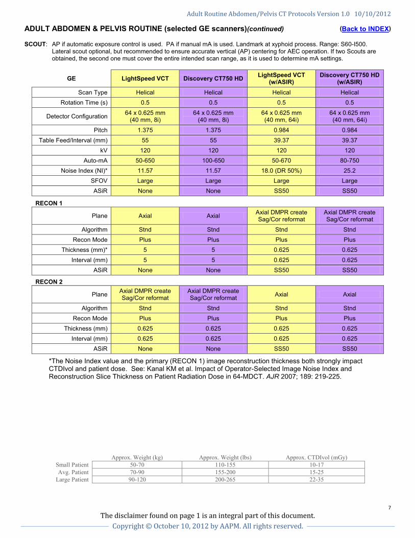
ADULT ABDOMEN & PELVIS ROUTINE (selected GE scanners)(continued) (Back to INDEX) SCOUT: AP if automatic exposure control is used. PA if manual mA is used. Landmark at xyphoid process. Range: S60-I500.
Lateral scout optional, but recommended to ensure accurate vertical (AP) centering for AEC operation. If two Scouts are obtained, the second one must cover the entire intended scan range, as it is used to determine mA settings.
GE LightSpeed VCT Discovery CT750 HD LightSpeed VCT (w/ASIR)
Discovery CT750 HD (w/ASIR)
Scan Type Helical Helical Helical Helical Rotation Time (s) 0.5 0.5 0.5 0.5
Detector Configuration 64 x 0.625 mm (40 mm, 8i)
64 x 0.625 mm (40 mm, 8i)
64 x 0.625 mm (40 mm, 64i)
64 x 0.625 mm (40 mm, 64i)
Pitch 1.375 1.375 0.984 0.984
Table Feed/Interval (mm) 55 55 39.37 39.37 kV 120 120 120 120
Auto-mA 50-650 100-650 50-670 80-750 Noise Index (NI)* 11.57 11.57 18.0 (DR 50%) 25.2
SFOV Large Large Large Large ASiR None None SS50 SS50
RECON 1
Plane Axial Axial Axial DMPR create Sag/Cor reformat
Axial DMPR create Sag/Cor reformat
Algorithm Stnd Stnd Stnd Stnd Recon Mode Plus Plus Plus Plus
Thickness (mm)* 5 5 0.625 0.625
Interval (mm) 5 5 0.625 0.625 ASiR None None SS50 SS50
RECON 2
Plane Axial DMPR create Sag/Cor reformat
Axial DMPR create Sag/Cor reformat Axial Axial
Algorithm Stnd Stnd Stnd Stnd Recon Mode Plus Plus Plus Plus
Thickness (mm) 0.625 0.625 0.625 0.625 Interval (mm) 0.625 0.625 0.625 0.625
ASiR None None SS50 SS50
*The Noise Index value and the primary (RECON 1) image reconstruction thickness both strongly impact CTDIvol and patient dose. See: Kanal KM et al. Impact of Operator-Selected Image Noise Index and Reconstruction Slice Thickness on Patient Radiation Dose in 64-MDCT. AJR 2007; 189: 219-225.
Approx. Weight (kg) Approx. Weight (lbs) Approx. CTDIvol (mGy) Small Patient 50-70 110-155 10-17 Avg. Patient 70-90 155-200 15-25
Large Patient 90-120 200-265 22-35
Adult Routine Abdomen/Pelvis CT Protocols Version 1.0 10/10/2012
8 The disclaimer found on page 1 is an integral part of this document.
Copyright © October 10, 2012 by AAPM. All rights reserved.
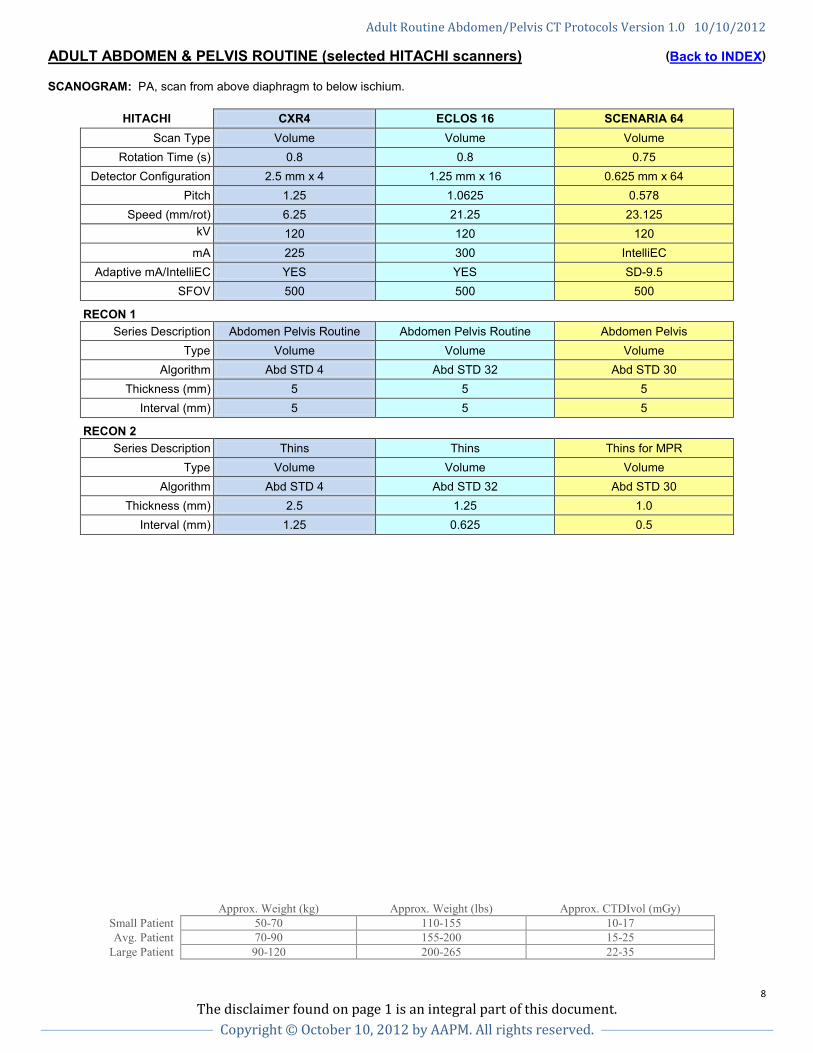
ADULT ABDOMEN & PELVIS ROUTINE (selected HITACHI scanners) (Back to INDEX) SCANOGRAM: PA, scan from above diaphragm to below ischium.
HITACHI CXR4 ECLOS 16 SCENARIA 64 Scan Type Volume Volume Volume
Rotation Time (s) 0.8 0.8 0.75 Detector Configuration 2.5 mm x 4 1.25 mm x 16 0.625 mm x 64
Pitch 1.25 1.0625 0.578 Speed (mm/rot) 6.25 21.25 23.125
kV
120 120 120 mA 225 300 IntelliEC
Adaptive mA/IntelliEC YES YES SD-9.5 SFOV 500 500 500
RECON 1
Series Description Abdomen Pelvis Routine Abdomen Pelvis Routine Abdomen Pelvis Type Volume Volume Volume
Algorithm Abd STD 4 Abd STD 32 Abd STD 30 Thickness (mm) 5 5 5
Interval (mm) 5 5 5
RECON 2
Series Description Thins Thins Thins for MPR Type Volume Volume Volume
Algorithm Abd STD 4 Abd STD 32 Abd STD 30 Thickness (mm) 2.5 1.25 1.0
Interval (mm) 1.25 0.625 0.5
Approx. Weight (kg) Approx. Weight (lbs) Approx. CTDIvol (mGy) Small Patient 50-70 110-155 10-17 Avg. Patient 70-90 155-200 15-25
Large Patient 90-120 200-265 22-35
Adult Routine Abdomen/Pelvis CT Protocols Version 1.0 10/10/2012
9 The disclaimer found on page 1 is an integral part of this document.
Copyright © October 10, 2012 by AAPM. All rights reserved.
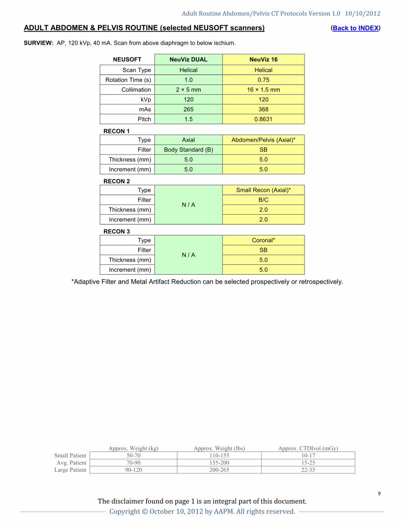
ADULT ABDOMEN & PELVIS ROUTINE (selected NEUSOFT scanners) (Back to INDEX) SURVIEW: AP, 120 kVp, 40 mA. Scan from above diaphragm to below ischium.
NEUSOFT NeuViz DUAL NeuViz 16
Scan Type Helical Helical Rotation Time (s) 1.0 0.75
Collimation 2 × 5 mm 16 × 1.5 mm kVp 120 120
mAs 265 368 Pitch 1.5 0.8631
RECON 1 Type Axial Abdomen/Pelvis (Axial)* Filter Body Standard (B) SB
Thickness (mm) 5.0 5.0 Increment (mm) 5.0 5.0
RECON 2 Type
N / A
Small Recon (Axial)* Filter B/C
Thickness (mm) 2.0 Increment (mm) 2.0
RECON 3 Type
N / A
Coronal* Filter SB
Thickness (mm) 5.0 Increment (mm) 5.0
*Adaptive Filter and Metal Artifact Reduction can be selected prospectively or retrospectively.
Approx. Weight (kg) Approx. Weight (lbs) Approx. CTDIvol (mGy) Small Patient 50-70 110-155 10-17 Avg. Patient 70-90 155-200 15-25
Large Patient 90-120 200-265 22-35
Adult Routine Abdomen/Pelvis CT Protocols Version 1.0 10/10/2012
10 The disclaimer found on page 1 is an integral part of this document.
Copyright © October 10, 2012 by AAPM. All rights reserved.
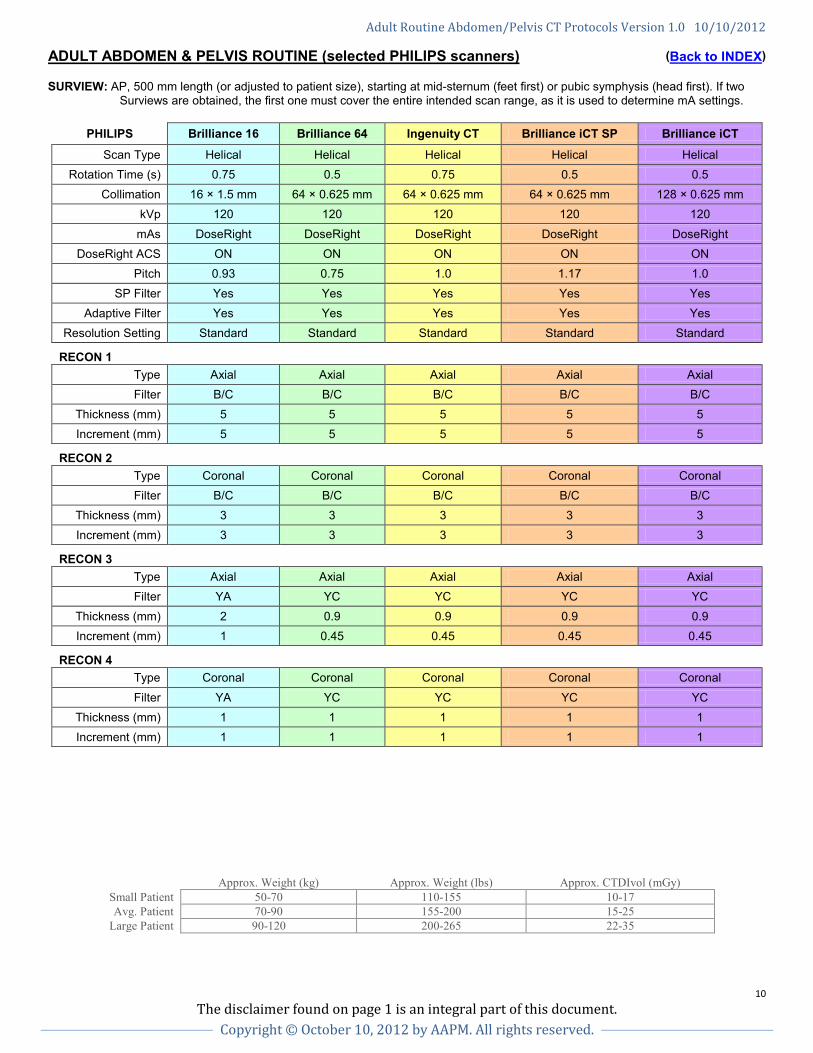
ADULT ABDOMEN & PELVIS ROUTINE (selected PHILIPS scanners) (Back to INDEX) SURVIEW: AP, 500 mm length (or adjusted to patient size), starting at mid-sternum (feet first) or pubic symphysis (head first). If two
Surviews are obtained, the first one must cover the entire intended scan range, as it is used to determine mA settings.
PHILIPS Brilliance 16 Brilliance 64 Ingenuity CT Brilliance iCT SP Brilliance iCT
Scan Type Helical Helical Helical Helical Helical Rotation Time (s) 0.75 0.5 0.75 0.5 0.5
Collimation 16 × 1.5 mm 64 × 0.625 mm 64 × 0.625 mm 64 × 0.625 mm 128 × 0.625 mm kVp 120 120 120 120 120
mAs DoseRight DoseRight DoseRight DoseRight DoseRight DoseRight ACS ON ON ON ON ON
Pitch 0.93 0.75 1.0 1.17 1.0 SP Filter Yes Yes Yes Yes Yes
Adaptive Filter Yes Yes Yes Yes Yes Resolution Setting Standard Standard Standard Standard Standard
RECON 1 Type Axial Axial Axial Axial Axial Filter B/C B/C B/C B/C B/C
Thickness (mm) 5 5 5 5 5 Increment (mm) 5 5 5 5 5
RECON 2 Type Coronal Coronal Coronal Coronal Coronal Filter B/C B/C B/C B/C B/C
Thickness (mm) 3 3 3 3 3 Increment (mm) 3 3 3 3 3
RECON 3 Type Axial Axial Axial Axial Axial Filter YA YC YC YC YC
Thickness (mm) 2 0.9 0.9 0.9 0.9 Increment (mm) 1 0.45 0.45 0.45 0.45
RECON 4 Type Coronal Coronal Coronal Coronal Coronal
Filter YA YC YC YC YC Thickness (mm) 1 1 1 1 1 Increment (mm) 1 1 1 1 1
Approx. Weight (kg) Approx. Weight (lbs) Approx. CTDIvol (mGy) Small Patient 50-70 110-155 10-17 Avg. Patient 70-90 155-200 15-25
Large Patient 90-120 200-265 22-35
Adult Routine Abdomen/Pelvis CT Protocols Version 1.0 10/10/2012
11 The disclaimer found on page 1 is an integral part of this document.
Copyright © October 10, 2012 by AAPM. All rights reserved.
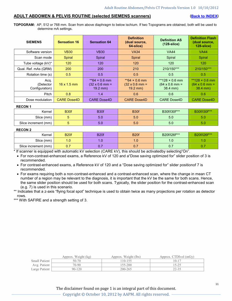
ADULT ABDOMEN & PELVIS ROUTINE (selected SIEMENS scanners) (Back to INDEX) TOPOGRAM: AP, 512 or 768 mm. Scan from above diaphragm to below ischium. If two Topograms are obtained, both will be used to
determine mA settings.
SIEMENS Sensation 16 Sensation 64 Definition
(dual source, 64-slice)
Definition AS (128-slice)
Definition Flash (dual source,
128-slice) Software version VB30 VB30 VA34 VA44 VA44
Scan mode Spiral Spiral Spiral Spiral Spiral Tube voltage (kV)* 120 120 120 120 120
Qual. Ref. mAs (QRM) 200 200 210 210/150*** 210/150*** Rotation time (s) 0.5 0.5 0.5 0.5 0.5
Acq. (Detector
Configuration) 16 x 1.5 mm
**64 × 0.6 mm (32 x 0.6 mm =
19.2 mm)
**64 × 0.6 mm (32 x 0.6 mm =
19.2 mm)
**128 × 0.6 mm (64 x 0.6 mm =
38.4 mm)
**128 × 0.6 mm (64 x 0.6 mm =
38.4 mm)
Pitch 0.8 1.4 0.6 0.6 0.6 Dose modulation CARE Dose4D CARE Dose4D CARE Dose4D CARE Dose4D CARE Dose4D
RECON 1
Kernel B30f B30f B30f B30f/I30f*** B30f/I30f*** Slice (mm) 5 5.0 5.0 5.0 5.0
Slice increment (mm) 5 5.0 5.0 5.0 5.0
RECON 2
Kernel B20f B20f B20f B20f/I26f*** B20f/I26f*** Slice (mm) 1.0 1.0 1.0 1.0 1.0
Slice increment (mm) 0.7 0.7 0.7 0.7 0.7 * If scanner is equipped with automatic kV selection (CARE kV), this should be activatedby selecting“On”. • For non-contrast-enhanced exams, a Reference kV of 120 and a“Dose saving optimized for” slider position of 3 is
recommended. • For contrast-enhanced exams, a Reference kV of 120 and a “Dose saving optimized for” slider positionof 7 is
recommended. • For exams requiring both a non-contrast-enhanced and a contrast-enhanced scan, where the change in mean CT
number of a region may be relevant to the diagnosis, it is important that the kV be the same for both scans. Hence, the same slider position should be used for both scans. Typically, the slider position for the contrast-enhanced scan (e.g. 7) is used in this scenario.
** Indicates that a z-axis “flying focal spot” technique is used to obtain twice as many projections per rotation as detector rows.
*** With SAFIRE and a strength setting of 3.
Approx. Weight (kg) Approx. Weight (lbs) Approx. CTDIvol (mGy) Small Patient 50-70 110-155 10-17 Avg. Patient 70-90 155-200 15-25
Large Patient 90-120 200-265 22-35
Adult Routine Abdomen/Pelvis CT Protocols Version 1.0 10/10/2012
12 The disclaimer found on page 1 is an integral part of this document.
Copyright © October 10, 2012 by AAPM. All rights reserved.
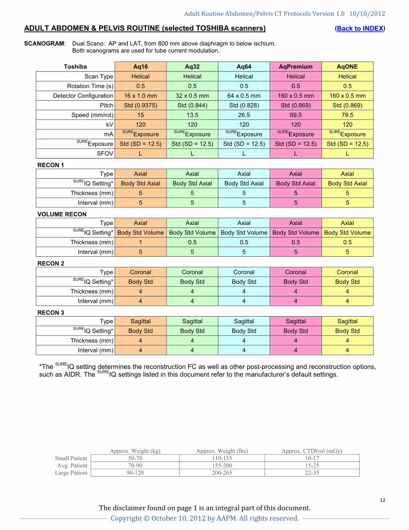
ADULT ABDOMEN & PELVIS ROUTINE (selected TOSHIBA scanners) (Back to INDEX) SCANOGRAM: Dual Scano: AP and LAT, from 800 mm above diaphragm to below ischium.
Both scanograms are used for tube current modulation.
Toshiba Aq16 Aq32 Aq64 AqPremium AqONE Scan Type Helical Helical Helical Helical Helical
Rotation Time (s) 0.5 0.5 0.5 0.5 0.5 Detector Configuration 16 x 1.0 mm 32 x 0.5 mm 64 x 0.5 mm 160 x 0.5 mm 160 x 0.5 mm
Pitch Std (0.9375) Std (0.844) Std (0.828) Std (0.869) Std (0.869) Speed (mm/rot) 15 13.5 26.5 69.5 79.5
kV 120 120 120 120 120 mA SUREExposure SUREExposure SUREExposure SUREExposure SUREExposure
SUREExposure Std (SD = 12.5) Std (SD = 12.5) Std (SD = 12.5) Std (SD = 12.5) Std (SD = 12.5) SFOV L L L L L
RECON 1 Type Axial Axial Axial Axial Axial
SUREIQ Setting* Body Std Axial Body Std Axial Body Std Axial Body Std Axial Body Std Axial Thickness (mm) 5 5 5 5 5
Interval (mm) 5 5 5 5 5
VOLUME RECON Type Axial Axial Axial Axial Axial
SUREIQ Setting* Body Std Volume Body Std Volume Body Std Volume Body Std Volume Body Std Volume Thickness (mm) 1 0.5 0.5 0.5 0.5
Interval (mm) 5 5 5 5 5
RECON 2 Type Coronal Coronal Coronal Coronal Coronal
SUREIQ Setting* Body Std Body Std Body Std Body Std Body Std Thickness (mm) 4 4 4 4 4
Interval (mm) 4 4 4 4 4
RECON 3 Type Sagittal Sagittal Sagittal Sagittal Sagittal
SUREIQ Setting* Body Std Body Std Body Std Body Std Body Std Thickness (mm) 4 4 4 4 4
Interval (mm) 4 4 4 4 4
*The SUREIQ setting determines the reconstruction FC as well as other post-processing and reconstruction options, such as AIDR. The SUREIQ settings listed in this document refer to the manufacturer’s default settings.
Approx. Weight (kg) Approx. Weight (lbs) Approx. CTDIvol (mGy) Small Patient 50-70 110-155 10-17 Avg. Patient 70-90 155-200 15-25
Large Patient 90-120 200-265 22-35


![CT Abdomen and pelvis · ABDOMEN . Patient preparation Oral contrast material to opacity the gastrointestinal tract [gastrographin 38% diluted by water to 4%] - Timing? Not indicated](https://static.fdocuments.in/doc/165x107/5e19a87e3ba4a22e0a18205f/ct-abdomen-and-pelvis-abdomen-patient-preparation-oral-contrast-material-to-opacity.jpg)