A VicRK Signal Transduction System in Streptococcus mutans Affects gtfBCD, gbpB, and ftf
13
JOURNAL OF BACTERIOLOGY, June 2005, p. 4064–4076 Vol. 187, No. 12 0021-9193/05/$08.000 doi:10.1128/JB.187.12.4064–4076.2005 Copyright © 2005, American Society for Microbiology. All Rights Reserved. A VicRK Signal Transduction System in Streptococcus mutans Affects gtfBCD, gbpB, and ftf Expression, Biofilm Formation, and Genetic Competence Development M. Dilani Senadheera, 1 Bernard Guggenheim, 2 Grace A. Spatafora, 3 Yi-Chen Cathy Huang, 1 Jison Choi, 4 David C. I. Hung, 4 Jennifer S. Treglown, 4 Steven D. Goodman, 4 Richard P. Ellen, 1 and Dennis G. Cvitkovitch 1 * Dental Research Institute, University of Toronto, 124 Edward Street, Toronto, Ontario M51G6, Canada 1 ; Institute for Oral Biology, Section for Oral Microbiology and General Immunology, Center for Dental, Oral Medicine and Maxillofacial Surgery, University of Zurich, Zurich, Switzerland 2 ; Department of Biology, Middlebury College, 276 Bicentennial Way, BIH354, Middlebury, Vermont 05753 3 ; and Division of Diagnostic Science, Norris School of Dentistry, University of Southern California, 925 West 34th, Los Angeles, California 90089 4 Received 19 November 2004/Accepted 9 March 2005 Bacteria exposed to transient host environments can elicit adaptive responses by triggering the differential expression of genes via two-component signal transduction systems. This study describes the vicRK signal transduction system in Streptococcus mutans.A vicK (putative histidine kinase) deletion mutant (SmuvicK) was isolated. However, a vicR (putative response regulator) null mutation was apparently lethal, since the only transformants isolated after attempted mutagenesis overexpressed all three genes in the vicRKX operon (Smuvic ). Compared with the wild-type UA159 strain, both mutants formed aberrant biofilms. Moreover, the vicK mutant biofilm formed in sucrose-supplemented medium was easily detachable relative to that of the parent. The rate of total dextran formation by this mutant was remarkably reduced compared to the wild type, whereas it was increased in Smuvic . Based on real-time PCR, Smuvic showed increased gtfBCD, gbpB, and ftf expression, while a recombinant VicR fusion protein was shown to bind the promoter regions of the gtfB, gtfC, and ftf genes. Also, transformation efficiency in the presence or absence of the S. mutans competence-stimu- lating peptide was altered for the vic mutants. In vivo studies conducted using SmuvicK in a specific-pathogen- free rat model resulted in significantly increased smooth-surface dental plaque (Pearson-Filon statistic [PF], <0.001). While the absence of vicK did not alter the incidence of caries, a significant reduction in SmuvicK CFU counts was observed in plaque samples relative to that of the parent (PF, <0.001). Taken together, these findings support involvement of the vicRK signal transduction system in regulating several important physi- ological processes in S. mutans. Among the hundreds of bacterial species that colonize and persist in the oral cavity, Streptococcus mutans is among the few species that have been consistently linked with caries for- mation (31). Under the low-pH conditions that define the plaque environment, S. mutans induces an acid tolerance re- sponse that helps it survive (14, 15, 18). Hence, the ability to adapt to and generate acids at the tooth surface allows S. mutans to predominate within carious lesions by reducing the plaque pH to levels that are inhibitory to other oral microbes (34, 47). In addition, the ability of S. mutans to synthesize extracellular polysaccharides that promote formation of the plaque biofilm also contributes to its pathogenicity (35, 47). As a result, investigations to characterize virulence determinants of this oral pathogen have continued to dominate the caries microbiology field for decades. Two-component signal transduction systems (TCSTS) are among the regulatory networks that are essential for bacterial adaptation, survival, and virulence. These systems function as “molecular switches” to modulate gene expression in response to changes in the external environment (44). Typically, signal transduction is accomplished via two regulatory elements con- sisting of a membrane-associated histidine kinase and a cyto- plasmic response regulator. Upon exposure to an environmen- tal cue, such as pH, osmolarity, or oxidation-reduction potential, the histidine kinase becomes autophosphorylated at a conserved histidine residue. Following the transfer of this phosphate group to a response regulator, the regulator can control the transcription of target genes by binding to their promoter regions. Diverse metabolic processes controlled by TCSTS include chemotaxis, sporulation, quorum sensing, and antibiotic/bacteriocin production in a wide variety of bacteria (3, 8, 30, 43, 45). Previous work conducted in one of our laboratories indicated that genetic competence, biofilm forma- tion, and acid tolerance are mediated by detection of a signal peptide by the comDE TCSTS in S. mutans (27, 28). Genetic competence enables recipient bacteria to inherit heterologous genes that can contribute to the emergence of antibiotic resis- tance, as well as promote genetic variation that can drive over- all fitness and evolution (7). Analysis of the S. mutans UA159 genome database revealed 13 putative TCSTS and at least one independent response regulator (1, 21). The present study describes an investigation into the S. mutans vicRK signal transduction system that en- * Corresponding author. Mailing address: Rm. 449A, Dental Re- search Institute, 124 Edward Street, Toronto ON M51G6, Canada. Phone: (416) 979-4917, ext. 4592. Fax: (416) 979-4936. E-mail: dennis [email protected]. 4064 on December 19, 2018 by guest http://jb.asm.org/ Downloaded from
Transcript of A VicRK Signal Transduction System in Streptococcus mutans Affects gtfBCD, gbpB, and ftf

JOURNAL OF BACTERIOLOGY, June 2005, p. 4064–4076 Vol. 187, No. 120021-9193/05/$08.00�0 doi:10.1128/JB.187.12.4064–4076.2005Copyright © 2005, American Society for Microbiology. All Rights Reserved.
A VicRK Signal Transduction System in Streptococcus mutans AffectsgtfBCD, gbpB, and ftf Expression, Biofilm Formation, and
Genetic Competence DevelopmentM. Dilani Senadheera,1 Bernard Guggenheim,2 Grace A. Spatafora,3 Yi-Chen Cathy Huang,1
Jison Choi,4 David C. I. Hung,4 Jennifer S. Treglown,4 Steven D. Goodman,4Richard P. Ellen,1 and Dennis G. Cvitkovitch1*
Dental Research Institute, University of Toronto, 124 Edward Street, Toronto, Ontario M51G6, Canada1; Institute for OralBiology, Section for Oral Microbiology and General Immunology, Center for Dental, Oral Medicine and Maxillofacial
Surgery, University of Zurich, Zurich, Switzerland2; Department of Biology, Middlebury College, 276 BicentennialWay, BIH354, Middlebury, Vermont 057533; and Division of Diagnostic Science, Norris School of Dentistry,
University of Southern California, 925 West 34th, Los Angeles, California 900894
Received 19 November 2004/Accepted 9 March 2005
Bacteria exposed to transient host environments can elicit adaptive responses by triggering the differentialexpression of genes via two-component signal transduction systems. This study describes the vicRK signaltransduction system in Streptococcus mutans. A vicK (putative histidine kinase) deletion mutant (SmuvicK) wasisolated. However, a vicR (putative response regulator) null mutation was apparently lethal, since the onlytransformants isolated after attempted mutagenesis overexpressed all three genes in the vicRKX operon(Smuvic�). Compared with the wild-type UA159 strain, both mutants formed aberrant biofilms. Moreover, thevicK mutant biofilm formed in sucrose-supplemented medium was easily detachable relative to that of theparent. The rate of total dextran formation by this mutant was remarkably reduced compared to the wild type,whereas it was increased in Smuvic�. Based on real-time PCR, Smuvic� showed increased gtfBCD, gbpB, andftf expression, while a recombinant VicR fusion protein was shown to bind the promoter regions of the gtfB, gtfC,and ftf genes. Also, transformation efficiency in the presence or absence of the S. mutans competence-stimu-lating peptide was altered for the vic mutants. In vivo studies conducted using SmuvicK in a specific-pathogen-free rat model resulted in significantly increased smooth-surface dental plaque (Pearson-Filon statistic [PF],<0.001). While the absence of vicK did not alter the incidence of caries, a significant reduction in SmuvicK CFUcounts was observed in plaque samples relative to that of the parent (PF, <0.001). Taken together, thesefindings support involvement of the vicRK signal transduction system in regulating several important physi-ological processes in S. mutans.
Among the hundreds of bacterial species that colonize andpersist in the oral cavity, Streptococcus mutans is among thefew species that have been consistently linked with caries for-mation (31). Under the low-pH conditions that define theplaque environment, S. mutans induces an acid tolerance re-sponse that helps it survive (14, 15, 18). Hence, the ability toadapt to and generate acids at the tooth surface allows S.mutans to predominate within carious lesions by reducing theplaque pH to levels that are inhibitory to other oral microbes(34, 47). In addition, the ability of S. mutans to synthesizeextracellular polysaccharides that promote formation of theplaque biofilm also contributes to its pathogenicity (35, 47). Asa result, investigations to characterize virulence determinantsof this oral pathogen have continued to dominate the cariesmicrobiology field for decades.
Two-component signal transduction systems (TCSTS) areamong the regulatory networks that are essential for bacterialadaptation, survival, and virulence. These systems function as“molecular switches” to modulate gene expression in response
to changes in the external environment (44). Typically, signaltransduction is accomplished via two regulatory elements con-sisting of a membrane-associated histidine kinase and a cyto-plasmic response regulator. Upon exposure to an environmen-tal cue, such as pH, osmolarity, or oxidation-reductionpotential, the histidine kinase becomes autophosphorylated ata conserved histidine residue. Following the transfer of thisphosphate group to a response regulator, the regulator cancontrol the transcription of target genes by binding to theirpromoter regions. Diverse metabolic processes controlled byTCSTS include chemotaxis, sporulation, quorum sensing, andantibiotic/bacteriocin production in a wide variety of bacteria(3, 8, 30, 43, 45). Previous work conducted in one of ourlaboratories indicated that genetic competence, biofilm forma-tion, and acid tolerance are mediated by detection of a signalpeptide by the comDE TCSTS in S. mutans (27, 28). Geneticcompetence enables recipient bacteria to inherit heterologousgenes that can contribute to the emergence of antibiotic resis-tance, as well as promote genetic variation that can drive over-all fitness and evolution (7).
Analysis of the S. mutans UA159 genome database revealed13 putative TCSTS and at least one independent responseregulator (1, 21). The present study describes an investigationinto the S. mutans vicRK signal transduction system that en-
* Corresponding author. Mailing address: Rm. 449A, Dental Re-search Institute, 124 Edward Street, Toronto ON M51G6, Canada.Phone: (416) 979-4917, ext. 4592. Fax: (416) 979-4936. E-mail: [email protected].
4064
on Decem
ber 19, 2018 by guesthttp://jb.asm
.org/D
ownloaded from

codes a putative histidine kinase (VicK) and a response regu-lator (VicR) (Fig. 1A). This TCSTS in S. mutans was firstdescribed as covRS by Lee et al. (GenBank accession numberAF393849), after its homolog in Streptococcus pyogenes (26).While this system does bear sequence similarity to the covRSsystem of S. pyogenes, it appears more closely related to thevicRK TCSTS of S. pyogenes and Streptococcus pneumoniae. Toclarify this further, the S. pyogenes covR homolog in S. mutanswas named gcrR by Sato et al. (40) and later renamed tarCfollowing its characterization by Idone et al. (21). Figure 1Bclarifies the relationship between various members of theseTCSTS families. Based on these observations, it seems moreappropriate to refer to this TCSTS as vicRK as previously
named in the annotated S. mutans UA159 genome sequenceavailable in the National Center for Biotechnology Informa-tion (NCBI) GenBank database (1). In this paper, we chose tohenceforth refer to the TCSTS as vicRK in accordance with itsdesignation by Ajdic et al. (1).
The vicRK TCSTS family is best characterized in S. pneu-moniae and in Bacillus subtilis (10, 12, 20, 46). In a studyconducted by Wagner et al., overexpression of the vicK gene inS. pneumoniae resulted in a mutant attenuated for virulence ina mouse model (46). Also, a vicK null mutant demonstrated adecrease in transformation efficiency (TE) relative to the wildtype by approximately 3 orders of magnitude (46). Efforts togenerate a deletion mutation in the S. pneumoniae vicR ho-
FIG. 1. (A) Genetic map of the vicRKX operon. Using BlastP searches, putative functions were assigned to genes based on high identity scoresin the NCBI website. Abbreviations: smc, chromosome segregation SMC protein in Streptococcus agalactiae (accession no. NP_687739.1); rnc,RNase III in S. agalactiae (NP_687738.1); vicX, S. pyogenes VicX protein (NP_268804.1), VicX protein in S. pneumoniae (NP_345691.1), andmetallo-�-lactamase superfamily protein in S. agalactiae (NP_687736.1); vicK, S. pyogenes VicK histidine kinase sensor protein (NP_268803.1),VicK in S. pneumoniae (NP_268803), and a sensory box histidine kinase in S. agalactiae (NP_687735.1); vicR, S. pyogenes VicR response regulatorprotein (NP_268802.1) and in S. pneumoniae (NP_606786.1); glnQ to glnP, glutamine transport ATP-binding protein (NP_687733.1), glutaminebinding protein (NP_687732.1), amino acid permease protein (NP_687731.1), and integral membrane protein (NP_687730.1) in S. agalactiae,respectively; endA, putative membrane nuclease EndA in S. pneumoniae (NP_359371.1). (B) BlastP search results using the NCBI website andcomparing various VicR and CovR proteins from various microorganisms (Spn, S. pneumoniae; Sm, S. mutans; Sp, S. pyogenes; Sa, S. aureus; Bs,B. subtilis).
VOL. 187, 2005 CONTROL OF VIRULENCE ATTRIBUTES BY S. MUTANS VicRK 4065
on Decem
ber 19, 2018 by guesthttp://jb.asm
.org/D
ownloaded from

molog (vicR), however, proved unsuccessful. In contrast to therecent publication by Lee et al. (26), in this study we claim thatvicR inactivation in S. mutans is also lethal. Herein we describea link between vicRK signal transduction and S. mutans su-crose-dependent adhesion, biofilm formation, and competencedevelopment. We also report our assessment of the abilities ofan S. mutans vicK-deficient mutant to form plaque and gener-ate caries in a specific-pathogen-free rat model.
MATERIALS AND METHODS
Bacterial strains, plasmids, and media. The bacterial strains, plasmids, andamplicons used in this study are listed in Table 1. The S. mutans UA159 wild-typestrain and its derivatives were routinely maintained on Todd-Hewitt yeast extract(THYE) agar (BBL Becton Dickinson, Cockeysville, MD) containing appropri-ate antibiotics when needed. Antibiotics used for the mutant strains were eryth-romycin (10 �g/ml), spectinomycin (1,200 �g/ml), kanamycin (500 �g/ml), andtetracycline (10 �g/ml). All S. mutans cultures were routinely grown as standingcultures at 37°C in a 5% CO2–95% air mixture.
Construction of S. mutans vicRK mutants. We searched the S. mutans UA159genome database (http://www.genome.ou.edu/smutans.html) for vicR and vicKhomologs using the S. pneumoniae RR02 and HK02 amino acid sequences asqueries, respectively. To delete the vicRK gene pair in S. mutans UA159, we useda ligation-PCR mutagenesis strategy as previously described (25). The resultingputative histidine kinase mutant was termed SmuvicK, while repeated attemptsto generate a vicR null mutant in the NG8 and UA159 backgrounds proved to befutile. In the latter case, all recovered transformants overexpressed the vicRKXgenes and were hence designated Smuvic�. The primers used for mutant con-struction and confirmation are listed in Table 2. Ligation constructs were intro-duced into S. mutans by competence-stimulating peptide (CSP)-induced naturaltransformation (28), and transformants resistant to erythromycin were selectedto confirm appropriate recombination into the chromosome by PCR, followed bynucleotide sequence analysis. The vicRKX expression in the resulting mutantswas monitored using quantitative real-time PCR (rtPCR) and compared withexpression in the UA159 wild-type progenitor.
Growth rates. Growth kinetics were monitored using a Bioscreen microbiologyreader (Bioscreen C Labsystems, Helsinki, Finland). Overnight cultures werediluted 20� in fresh THYE and grown to an optical density at 600 nm (OD600)of approximately 0.4 to 0.5. Twenty microliters of mid-log-phase cells for themutant and wild-type strains were inoculated in triplicate into microtiter platewells containing 400 �l of THYE. Wells containing uninoculated THYE wereused as controls. Using Biolink software (Labsystems), the Bioscreen reader wasprogrammed to monitor OD600s at 37°C every 20 min for 24 h, with moderateshaking every 3 min. OD600 measurements were plotted against time to generategrowth curves.
S. mutans biofilm formation. A modified semidefined minimal medium (SDM)was prepared for biofilm growth experiments as described previously (29, 32).Biofilms were formed in 24-well polystyrene microtiter plates containing 2 ml ofmedium supplemented with 20 mM glucose or 10 mM sucrose. All wells wereinoculated with 20 �l of an overnight cell suspension. In addition to SDM,
SmuvicK and UA159 biofilms were also formed in 0.25� THYE that was sup-plemented with 20 mM glucose or 10 mM sucrose. Following incubation at 37°Cand 5% CO2 for 16 h, the broth was gently removed by aspiration and thebiofilms photographed directly. To closely examine the architecture of the parentand mutant biofilms, we utilized scanning electron microscopy (SEM) as de-scribed previously (28).
RNA preparation and rtPCR analysis. To measure gtfB, gtfC, and ftf expres-sion, total RNA was isolated from bacterial cultures grown in Tryptone yeastextract broth supplemented with 1% sucrose or 1% glucose. To study vicRKexpression, bacterial strains were grown in THYE with or without antibiotics.Overnight cultures were diluted 20� in fresh broth and then grown to mid-logarithmic phase. Cells were harvested by centrifugation and immediately re-suspended in Trizol reagent (Invitrogen) prior to RNA isolation using the Fast-Prep system (Bio 101 Savant) as specified by the manufacturer. To monitor geneexpression, total RNA was subjected to DNase treatment and then reversetranscribed using a first-strand cDNA synthesis kit (MBI Fermentas) in accor-dance with the recommendations of the supplier. Controls for cDNA synthesisincluded a condition with no RNA template and another without reverse tran-scriptase. Finally, the single-stranded cDNAs were incorporated into rtPCRexperiments using a Cepheid Smart Cycler system (Cepheid, Sunnyvale, CA) anda Quantitect SYBR-Green PCR kit (QIAGEN). Each 25-�l reaction mixtureincluded template cDNA, 25 �M each primer, and 2� SYBR-Green mix (con-taining SYBR-Green, deoxynucleoside triphosphates, MgCl2, and Hotstar Taqpolymerase). For maximum efficiency, rtPCR primers were designed to generateamplicons ranging from 100 to 170 bp in size (Table 2). Controls for rtPCRincluded reaction mixtures without template cDNA to effectively rule out thepresence of contaminating DNA and/or the formation of primer dimers. Thecycling conditions were as follows: 95°C for 15 min for the initial denaturation,followed by 35 to 40 cycles of three steps consisting of denaturation at 94°C for15 s, primer annealing at the optimal temperature (Table 2) for 30 s, and primerextension at 72°C for 30 s. For each set of primers, cycle threshold (Ct) values,defined as the first cycle that gave rise to a detectable PCR product above thebackground, were generated. Known genomic DNA concentrations were used togenerate Ct values for specific primer sets. By plotting the DNA concentrationsversus the Ct value, standard curves were generated and used to determinerelative RNA expression levels for the test gene. Results were normalized againstS. mutans gyrA expression that was invariant under the experimental test condi-tions.
Purification of MBP-VicR. The vicR coding sequence was amplified by PCRusing chromosomal DNA derived from S. mutans UA159 with primers oSG241and oSG242. Subsequently, the amplicon was digested with HindIII and EcoRIand ligated to maltose-binding protein (MBP) expression vector pMalc2 (NewEngland Biolabs, Beverly, MA). The resulting plasmid, pSG385, was introducedinto Escherichia coli strain TB1 and selected for resistance to ampicillin. Foroverexpression, E. coli strain TB1 cells containing pSG385 were grown in 1 literof LB supplemented with 2% glucose and 100 �g/ml ampicillin at 37°C withshaking. When cells reached an OD600 of 0.3 to 0.5, expression of the MBP fusedto VicR (MBP-VicR) was induced with 0.3 mM isopropyl-�-D-thiogalactopyr-anoside (IPTG) for 2 h. The cells were harvested by centrifugation (4°C, 5,000 �g, 15 min), resuspended in column buffer (20 mM Tris-HCl, pH 7.4, 200 mMNaCl, 1 mM EDTA), frozen at �20°C overnight, and lysed by sonication. Cells
TABLE 1. Bacterial strains, plasmids, and amplicons used in this study
Strain, plasmid, oramplicon Relevant characteristic(s) Source or reference
S. mutans strainsUA159 Wild type A. S. Bleiweis, University of FloridaSmuvicK UA159 vicK::ermAM Erm This studySmuvic� UA159 derived; vicRKX overexpression; Ermr This study
PlasmidspDL277 E. coli-streptococcal shuttle vector; Specr 4pMal-c2 E. coli expression vector with MBP; Ampr New England BiolabspSG385 E. coli pMal-c2 containing VicR; Ampr This study
AmpliconsPcErm Ermr marker amplified using ermAM cassetteaVicK vicK-5�::PcErm::vicK-3� fragment used for allelic replacement of vicK gene This studyaVicR vicR-5�::PcErm::vicR-3� fragment used for allelic replacement of vicR gene This study
4066 SENADHEERA ET AL. J. BACTERIOL.
on Decem
ber 19, 2018 by guesthttp://jb.asm
.org/D
ownloaded from

that were not lysed along with other debris were removed by centrifugation(9,000 � g, 20 min, 4°C). The cleared cell lysate was applied to an amylose resincolumn (New England Biolabs), preequilibrated with column buffer, and thenwashed with 12 column volumes of column buffer. The protein was eluted withcolumn buffer containing 10 mM maltose. Three-milliliter fractions were col-lected, and fractions containing MBP-VicR, as determined by sodium dodecylsulfate-polyacrylamide gel electrophoresis, were pooled and concentrated usingan Amicon Ultra-15 concentrator (Millipore, Billerica, MA). Briefly, fractionscontaining MBP-VicR were applied to the filter device and centrifuged at 3,000� g and 4°C for 15 min. To reduce the salt concentration, the filter was washedwith modified column buffer (20 mM Tris-HCl, pH 7.4, 50 mM NaCl, 1 mMEDTA) and the purified protein was concentrated to �2 ml. The concentrationof MBP-VicR was determined by using the Bio-Rad Protein Assay (Bio-Rad,Hercules, CA) using bovine serum albumin as the standard. The purified proteinwas stored at 4°C or in glycerol at �80°C until needed.
Mobility shift experiments. The primers oSG181, oSG258, and oSG64 wereend labeled by incubating 1 �M primer with 1 �M [�-32P]ATP, 0.5 �M unlabeledATP, T4 polynucleotide kinase (Promega, Madison, WI), and 1� polynucleotidekinase buffer at 37°C for 30 min. The labeled oSG181 primer was then used togenerate a 194-bp gtfC promoter-containing fragment using oSG137. The labeledoSG258 primer was then used to generate a 215-bp ftf promoter-containingfragment using oSG265. Labeled primer oSG64 and unlabeled primer oSG264were used to generate a 205-bp gtfB promoter-containing fragment. For the
binding reactions, the labeled gtfB (�159 to �36), gtfC wild-type (�89 to �102),and ftf (�141 to �74) promoter-containing fragments were incubated at roomtemperature for 30 min with 0 to 1,000 nM MBP-VicR, reaction buffer (52.5 mMMOPS pH 7.4, 9.5% glycerol, 50 �M EDTA, 50 �g/ml bovine serum albumin),and 50 ng salmon sperm DNA in a volume of 20 �l. Protein-DNA complexeswere separated by nondenaturing gel electrophoresis on 6% acrylamide gels at10 V/cm for 3 h, dried, and visualized with a phosphorimager.
Extracellular polysaccharide synthesis (EPS). Cell cultures at mid-logarithmicphase derived from 1:20 dilutions of overnight cultures were centrifuged for 10min at 4,500 rpm. To measure “released” activity in the culture fluid, the super-natants were filter sterilized and stored at �20°C until further use. EPS assayswere conducted by adding 50 �l of a buffer mixture (100 mM sodium acetatebuffer [pH 5.5], 7 mM sodium fluoride, 0.02% dextran T-10 [average weight,10,000]) to 200 �l of cell-free supernatants. Following their incubation at 37°Cfor 10 min, 0.6 mM [14C]sucrose (11 �Ci/�mol) was added to the mixtures, whichwere then vortexed, and 15 �l of each mixture was spotted in triplicate onto2.3-cm square Whatman 3 MM filter papers. This procedure was repeated aftermixtures were incubated at 37°C for 30 min. Subsequently, the filter papers werewashed three times, 15 min each, in methanol using at least 10 ml of solvent.Filter squares were then dried and radioactivity counted using a liquid scintilla-tion counter. Net EPS activity was measured as the difference in counts at t 30and t 0.
TABLE 2. Primers used for PCR-ligation mutagenesis, rtPCR, VicR cloning, and mobility shift experiments
Primer use and name Nucleotide sequenced Annealing temp (°C),size (bp)
PCR-ligation mutagenesisa
VicK-P1 5� TGGTAAAGCAGTATCTGGCGAGG 3� 53.1, 666VicK-P2 5� GG^CGCGCCATAGTGAGGAAGGCGAAGGGTC 3�VicK-P3 5� GGCCGG^CCCCAGGGACTTGATTCAAACACATTAG 3� 54.1, 886VicK-P4 5� GGCTAAGGAAGGTTATGACACG 3�VicR-P1 5� TCTTTTTCCTGTTCGGTCG 3� 51.4, 615VicR-P2 5� GG^CGCGCCGATGTTACTGTTCGTCGTCTGC 3�VicR-P3 5� GGCCGG^CCCCGTGTCATAACCTTCCTTAGCCA 3� 51.5, 711VicR-P4 5� AGTTCACCAGAGTCAATGGATTCC 3�Erm cst-F 5� GG^CGCGCCCCGGGCCCAAAATTTGTTTGAT 3� 52.3, 876Erm cst-B 5� GGCCGG^CCAGTCGGCAGCGACTCATAGAAT 3�
rtPCR primersVicK-FOR 5�CACTTTACGCATTCGTTTTGCC 3� 52.0, 102VicK-REV 5� CGTTCTTCTTTTTCCTGTTCGGTC 3�VicR-FOR 5� CGCAGTGGCTGAGGAAAATG 3� 53, 157VicR-REV 5� ACCTGTGTGTGTCGCTAAGTGATG 3�VicX-FOR 5� TGCTCAACCACAGTTTTACCG 3� 51.4, 127VicX-REV 5� GGACTCAATCAGATAACCATCAGC 3�Ftf-FOR 5� ATTGGCGAACGGCGACTTACTC 3� 52.8, 103Ftf-REV 5� CCTGCGACTTCATTACGATTGGTC 3�GtfB-FOR 5� ACACTTTCGGGTGGCTTG 3� 49, 127GtfB-REV 5� GCTTAGATGTCACTTCGGTTG 3�GtfC-FOR 5� CCAAAATGGTATTATGGCTGTCG 3� 50.5, 136GtfC-REV 5� TGAGTCTCTATCAAAGTAACGCAG 3�GyrA-FOR 5� ATTGTTGCTCGGGCTCTTCCAG 3� 62.0, 105GyrA-REV 5� ATGCGGCTTGTCAGGAGTAACC 3�
VicR cloningb
oSG241 5� CCGgaattcTATTTTAAGACCATAAGCGAGGTA 3�oSG242 5� CCCaagcttGCTAATAAAATTCGTAAAAATAAGGGAC 3�
Mobility shift productsc
oSG64 (gtfB) 5� GGATATCCCATGGTAGGAACCTCCAAATTTTAAACTG 3�oSG181 (gtfC) 5� AGATACTGTCACCCATCTTTT 3�oSG258 (ftf) 5� CCACCCAAAATTTCCCTTTC 3�oSG264 (gtfB) 5� CAATTAGACTGTTGTTTTTTTG 3�oSG137 (gtfC) 5� CGGGATCCATTTATTATTTTTCTAAAAAA 3�oSG265 (ftf) 5� CTTAATCTAATATGTGAATTTGTT 3�
a AscI restriction sites are in boldface, and FseI restriction sites are underlined.b HindIII cut site is in boldface, and EcoRI cut site is in boldface and underlined.c Underlined italics indicate linker bases.d Circumflexes indicate restriction enzyme cleavage sites.
VOL. 187, 2005 CONTROL OF VIRULENCE ATTRIBUTES BY S. MUTANS VicRK 4067
on Decem
ber 19, 2018 by guesthttp://jb.asm
.org/D
ownloaded from

Competence assay. Overnight cultures of SmuvicK, Smuvic�, and their UA159parent were diluted 20- or 40-fold in prewarmed THYE and incubated at 37°Cuntil an OD600 of approximately 0.3 was reached. Following the incubationperiod, 1 �g of closed circular plasmid DNA, pDL277, Specr (5), was added andthe samples were divided into two aliquots, only one of which was supplementedwith synthetic CSP (sCSP) (Hospital for Sick Children Biotechnology Services,Toronto, Ontario, Canada) at a final concentration of 750 ng/ml (28). To studygenetic competence in the mutant and parent strains, we performed TE assays asdescribed previously (28).
Examination of the vicK deficient strain, in vivo, for plaque formation andcariogenic potential. Specific-pathogen-free, caries-susceptible Osborne-Mendelrats (Center for Dental and Oral Medicine and Cranio-Maxillofacial Surgery,Zurich, Switzerland) were used to investigate in vivo effects of the vicK deletionon smooth-surface dental plaque and smooth and fissure caries, as well as on theestablishment of S. mutans in the oral microbiota. Each experimental groupconsisted of 10 animals. Thirteen days after birth, the animals were transferredto stainless steel screen bottom cages without bedding and fed a finely groundstock diet (diet no. 3433; Provimi Kliba AG). Tap water and food were availablead libitum. On day 20 after birth, the dams were removed and the littermatesdistributed among the treatment groups (10 rats/group). On days 21 and 22, eachrat was infected orally, twice daily, using 200 �l of a heavy bacterial suspensionthat comprised the parent UA159 strain or the vicK deletion mutant. To supportthe implantation of these bacteria, all rats received drinking water containing 2%sucrose and 2% glucose during days 20 to 22, as well as low-cariogenic diet 2000a(consisting of 28% skim milk, 15% powdered sucrose, 49% wheat flour, 5%brewer’s yeast, 2% Gevral protein, and 1% sodium chloride). On day 23 follow-ing the association period, sterilization of the feeding and housing equipmentwas continued. Necessary precautions were taken to avoid cross-contaminationand maintain a clean environment. Five days after association with the teststrains, swabs were taken from the oral cavities of five rats per treatment groupto confirm that the bacteria had become established. Shortly before the end ofthe study, oral swabs were taken from all 20 rats to obtain a final indication of themicrobial status of the animals.
On day 51 (at the end of the 27-day experimental period), the animals weresacrificed. The upper and lower jaws were dissected and immersed in fixative(10% buffered formalin phosphate) for a minimum of 72 h. Erythrosin-stainedmaxillary molars were evaluated for plaque extent using a method describedpreviously (39). Smooth-surface carious lesions were scored according to Keyes(22), and mandibular molars were sectioned and scored for fissure caries asspecified by Konig et al. (24). The data were analyzed by two-way analysis ofvariance and least-significant-difference (LSD) tests using the analysis-of-vari-ance statistics program.
Microbiological analyses of rat samples. The swabs taken from each animalwere immersed in sterile test tubes containing phosphate-buffered saline andthoroughly shaken. An aliquot was used for culture analyses, and the remainingsuspension was immediately frozen. Sample dilutions were plated onto Trypti-case yeast Columbia blood (TYCB) agar using a spiral dilutor to obtain totalfloral counts. Dilutions were also plated onto Trypticase yeast agar supple-mented with 20% sucrose and bacitracin to enumerate S. mutans bacteria. Boththe parental and mutant strains were also plated onto TYCB agar containing 10�g/ml erythromycin as a control for possible contamination.
RESULTS
Structural organization of the S. mutans vicRKX locus. TheS. mutans vicRKX genes span a region of DNA from 1444056to 1446908 bp (NCBI) on the minus strand of the S. mutansUA159 chromosome (Fig. 1A). The vicK coding sequence is 1,350bp in size and encodes a putative histidine kinase sensor proteinwith a predicted mass of 51,686 Da (450 amino acids [aa]). ABlastP search revealed that VicK shares high similarity with the S.pyogenes VicK protein (NP_268803) and with a putative histidinekinase in S. pneumoniae (HK02, NP_358699). The vicR codingsequence is 705 bp in size and encodes a putative response reg-ulator protein with a predicted molecular mass of 26,900 Da (235aa). A BlastP search of VicR revealed that it shared high simi-larity with the S. pyogenes VicR protein (NP_268803.1) and withother response regulators in S. agalactiae (NP_687734) and S.pneumoniae (NP_606786.1). The vicX coding sequence is 801 bp
in size and codes for a hypothetical protein with a predicted massof 29,680 Da (267 aa). A BlastP search indicated that S. mutansVicX shares amino acid similarity with VicX in S. pneumoniae(NP_345691), VicX in S. pyogenes (NP_268804.1), and a metallo-�-lactamase superfamily protein in S. agalactiae (NP_687736).
Confirmation of the S. mutans vic mutants. To characterizethe putative role of vicR and vicK in S. mutans, we constructedmutations within the vicRK coding sequences. The SmuvicKdeletion mutation was confirmed by PCR analysis (results notshown). In contrast, we were unable to isolate a vicR nullmutant. Sequence analysis of transformants revealed the pres-ence of an intact vicR gene, as well as chromosomal integrationof a VicR fragment that was used to mediate allelic exchange5� to the vicRKX operon (Table 1). Hence, instead of theexpected double-crossover event, the presence of the intactvicR gene can be attributed to a Campbell-type crossover eventmediated by the circularized a VicR fragment. To assess vi-cRK-specific expression in SmuvicK and Smuvic� relative tothe wild type, we performed rtPCR experiments, the results ofwhich indicated no vicK-specific expression in the SmuvicKdeletion mutant. rtPCR results for vic gene expression areshown in Fig. 2.
vic mutants have altered growth rates. Growth curve anal-ysis of the SmuvicK and Smuvic� strains revealed alteredgrowth rates compared with that of the UA159 parent strain(Fig. 3). While an overnight culture of UA159 grew as a uni-formly turbid cell suspension, SmuvicK and Smuvic� cells ag-gregated and accumulated at the bottom of the glass tubes(Fig. 4). Notably, a denser cell aggregate was apparent for theSmuvicK mutant compared with that of Smuvic�. This obser-vation was consistent with growth curves derived from each ofthree independent experiments that demonstrated a highergrowth yield for SmuvicK during stationary phase comparedwith the wild-type and Smuvic� strains (Fig. 3). WhileSmuvicK revealed little difference in the exponential-phasegrowth rate (mean [min/generation] standard error, 59.1 2.6) relative to the wild type (50.1 2), the doubling time ofSmuvic� was greatly increased (78.5 1.5).
FIG. 2. S. mutans UA159 and Smuvic� cDNAs were used to studyexpression of the vicRKX genes using quantitative rtPCR. For eachstrain, cDNA samples derived from three independent experimentswere subjected to amplification using vicRKX-specific primers and gyrAprimers (normalizing gene). For mutant and wild-type cDNAs, then-fold increase in vicRKX expression was calculated relative to that ofthe UA159 parent, whose expression was set at user-defined value of1.0.
4068 SENADHEERA ET AL. J. BACTERIOL.
on Decem
ber 19, 2018 by guesthttp://jb.asm
.org/D
ownloaded from
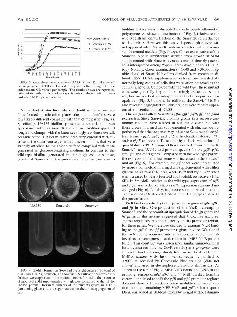
Vic mutant strains form aberrant biofilms. Based on bio-films formed on microtiter plates, the mutant biofilms wereremarkably different compared with that of the parent (Fig. 4).Specifically, UA159 biofilms presented a smooth and evenappearance, whereas SmuvicK and Smuvic� biofilms appearedrough and clumpy, with the latter seemingly less dense overall.As anticipated, UA159 wild-type cells supplemented with su-crose as the sugar source generated thicker biofilms that werestrongly attached to the abiotic surface compared with thosegenerated in glucose-containing medium. In contrast to thewild-type biofilms generated in either glucose or sucrose,growth of SmuvicK in the presence of sucrose gave rise to
biofilms that were easily disrupted and only loosely adherent topolystyrene. As shown at the bottom of Fig. 5, relative to thewild-type strain, only a fraction of the SmuvicK cells attachedto the surface. However, this easily dispersed phenotype wasnot apparent when SmuvicK biofilms were formed in glucose-supplemented medium (Fig. 5, top). Closer examination of theSmuvicK biofilm architecture derived from growth in SDMsupplemented with glucose revealed areas of densely packedcells interspersed among “open” areas devoid of cells (Fig. 5,top). Notably, closer examination (�5,000 and �50,000 mag-nifications) of SmuvicK biofilms derived from growth in di-luted 0.25� THYE supplemented with sucrose revealed ab-normally long chains of cells that were often detached at thecellular junctions. Compared with the wild type, these mutantcells were generally larger and seemingly associated with arougher surface that we interpreted as thicker deposits of ex-opolymer (Fig. 5, bottom). In addition, the Smuvic� biofilmalso revealed aggregated cell clusters that were readily appar-ent at a magnification of �1,000.
The vic genes affect S. mutans gtfB, gtfC, gtfD, ftf, and gbpBexpression. Since SmuvicK biofilms grown in a sucrose-con-taining medium were altered in adherence compared withthose grown in a medium supplemented with glucose, we hy-pothesized that the vic genes may influence S. mutans glucosyl-transferase (gtfB, gtfC, and gtfD), fructosyltransferase (ftf),and/or gbpB expression. To test our hypothesis, we performedquantitative rtPCR using cDNAs derived from SmuvicK,Smuvic�, and UA159 and primers specific for the gtfB, gtfC,gtfD, ftf, and gbpB genes. Compared with the wild-type parent,the expression of all these genes was increased in the Smuvic�
mutant (Fig. 6). For example, the gtf genes were upregulatedby more than fivefold in a medium supplemented with eitherglucose or sucrose (Fig. 6A), whereas ftf and gbpB expressionwas increased by nearly fourfold and twofold, respectively (Fig.6B). In SmuvicK, relative to the wild type, expression of gtfDand gbpB was reduced, whereas gtfC expression remained un-changed (Fig. 6). Notably, in glucose-supplemented medium,expression of gtfB showed 3.7-fold more transcript relative tothe parent strain.
VicR binds specifically to the promoter regions of gtfB, gtfC,and ftf in vitro. Overproduction of the VicR transcript inSmuvic� and the concomitant upregulation of the gtf genes andftf genes in this mutant suggested that VicR, like many re-sponse regulators, might act directly on the promoter regionsfor these genes. We therefore decided to examine VicR bind-ing to the gtfBC and ftf promoter regions in vitro. We clonedthe vicR coding sequence into an expression vector that al-lowed us to overexpress an amino-terminal MBP-VicR proteinfusion. This construct was chosen since similar amino-terminalfusion constructs, like the CovR ortholog in S. pyogenes, wereshown to bind indistinguishably from native CovR (11). TheMBP-S. mutans VicR fusion was subsequently purified by�90% as revealed by Coomassie blue staining (data notshown) and used in electrophoretic mobility shift assays. Asshown at the top of Fig. 7, MBP-VicR bound the DNA of thepromoter regions of gtfB, gtfC, and ftf (MBP purified from thevector alone failed to shift the gtfB and gtfC promoter regions;data not shown). In electrophoretic mobility shift assay reac-tion mixtures containing MBP-VicR and gtfC, salmon spermDNA was added in 100-fold excess by weight without diminu-
FIG. 3. Growth curves of S. mutans UA159, SmuvicK, and Smuvic�
in the presence of THYE. Each datum point is the average of threeindependent OD values per sample. The results shown are represen-tative of two other independent experiments conducted with the mu-tant and UA159 parent strains.
FIG. 4. Biofilm formation (top) and overnight cultures (bottom) ofS. mutans UA159, SmuvicK, and Smuvic�. Significant phenotypic dif-ferences were apparent in the mutant biofilms formed in the presenceof modified SDM supplemented with glucose compared to that of theUA159 parent. Overnight cultures of the mutants grown in THYE(containing glucose as the sugar source) resulted in coaggregation ofcells.
VOL. 187, 2005 CONTROL OF VIRULENCE ATTRIBUTES BY S. MUTANS VicRK 4069
on Decem
ber 19, 2018 by guesthttp://jb.asm
.org/D
ownloaded from

FIG. 5. (Top) SEM of S. mutans UA159, SmuvicK, and Smuvic� developed in SDM supplemented with glucose. (Bottom) SEM of S. mutansUA159 and SmuvicK biofilms developed using 0.25� THYE supplemented with 10 mM sucrose.
4070 SENADHEERA ET AL. J. BACTERIOL.
on Decem
ber 19, 2018 by guesthttp://jb.asm
.org/D
ownloaded from

tion of the shifted complex. There was, however, a hierarchy ofbinding. MBP-VicR bound with the highest affinity to the gtfCpromoter. Although the apparent affinity of MBP-VicR wassimilar for the ftf and gtfB promoters, as judged by the propor-tion of DNA shifted, it was also clear that the shift of the gtfBpromoter was more distinct and clearly visible, similar to the ftf
promoter-containing complex. This suggested a greater sitespecificity of binding. Although we have yet to ascertain theexact sequence that MBP-VicR recognizes, it is likely thatthere are conserved sequence determinants within the givenregions of all three promoter elements. Dubrac and Msadekrecently identified a VicR consensus sequence that seems con-
FIG. 6. (A) Expression of gtfBCD genes in S. mutans UA159, SmuvicK, and Smuvic� strains. Gene expression was monitored by rtPCR usingcDNAs derived from glucose (top)- or sucrose (bottom)-supplemented cultures. Results are the average of three independent experimentsconducted using primers specific for the gtfBCD genes and gyrA (normalizing gene). (B) rtPCR results of S. mutans UA159, SmuvicK, and Smuvic�
cDNAs amplified using S. mutans ftf- and gbpB-specific primers. Each cDNA sample derived from three independent experiments was amplifiedat least twice using ftf, gbpB, and gyrA (normalizing gene) primers.
VOL. 187, 2005 CONTROL OF VIRULENCE ATTRIBUTES BY S. MUTANS VicRK 4071
on Decem
ber 19, 2018 by guesthttp://jb.asm
.org/D
ownloaded from
served across many gram-positive genera (9). As shown at thebottom of Fig. 7, the sequence TGTWAHNNNNNTGTWAH(where W is A or T and H is A, T, or C) was perfectlyconserved in the gtfC promoter, partially conserved in the gtfBpromoter (although there are several possible iterations), andfound perfectly conserved in the ftf promoter but with theimportant caveat that there was an additional 10 bp or helicalrepeat that separated the more conserved hexamers.
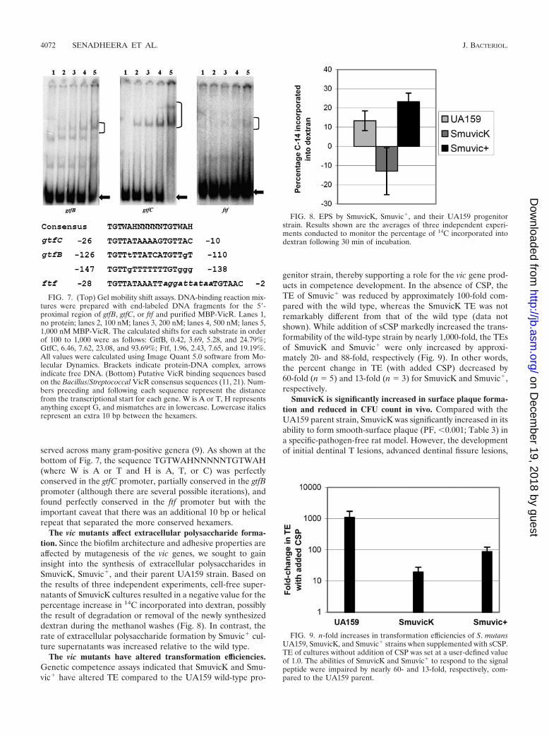
The vic mutants affect extracellular polysaccharide forma-tion. Since the biofilm architecture and adhesive properties areaffected by mutagenesis of the vic genes, we sought to gaininsight into the synthesis of extracellular polysaccharides inSmuvicK, Smuvic�, and their parent UA159 strain. Based onthe results of three independent experiments, cell-free super-natants of SmuvicK cultures resulted in a negative value for thepercentage increase in 14C incorporated into dextran, possiblythe result of degradation or removal of the newly synthesizeddextran during the methanol washes (Fig. 8). In contrast, therate of extracellular polysaccharide formation by Smuvic� cul-ture supernatants was increased relative to the wild type.
The vic mutants have altered transformation efficiencies.Genetic competence assays indicated that SmuvicK and Smu-vic� have altered TE compared to the UA159 wild-type pro-
genitor strain, thereby supporting a role for the vic gene prod-ucts in competence development. In the absence of CSP, theTE of Smuvic� was reduced by approximately 100-fold com-pared with the wild type, whereas the SmuvicK TE was notremarkably different from that of the wild type (data notshown). While addition of sCSP markedly increased the trans-formability of the wild-type strain by nearly 1,000-fold, the TEsof SmuvicK and Smuvic� were only increased by approxi-mately 20- and 88-fold, respectively (Fig. 9). In other words,the percent change in TE (with added CSP) decreased by60-fold (n 5) and 13-fold (n 3) for SmuvicK and Smuvic�,respectively.
SmuvicK is significantly increased in surface plaque forma-tion and reduced in CFU count in vivo. Compared with theUA159 parent strain, SmuvicK was significantly increased in itsability to form smooth-surface plaque (PF, �0.001; Table 3) ina specific-pathogen-free rat model. However, the developmentof initial dentinal T lesions, advanced dentinal fissure lesions,
FIG. 7. (Top) Gel mobility shift assays. DNA-binding reaction mix-tures were prepared with end-labeled DNA fragments for the 5�-proximal region of gtfB, gtfC, or ftf and purified MBP-VicR. Lanes 1,no protein; lanes 2, 100 nM; lanes 3, 200 nM; lanes 4, 500 nM; lanes 5,1,000 nM MBP-VicR. The calculated shifts for each substrate in orderof 100 to 1,000 were as follows: GtfB, 0.42, 3.69, 5.28, and 24.79%;GtfC, 6.46, 7.62, 23.08, and 93.69%; Ftf, 1.96, 2.43, 7.65, and 19.19%.All values were calculated using Image Quant 5.0 software from Mo-lecular Dynamics. Brackets indicate protein-DNA complex, arrowsindicate free DNA. (Bottom) Putative VicR binding sequences basedon the Bacillus/Streptococcal VicR consensus sequences (11, 21). Num-bers preceding and following each sequence represent the distancefrom the transcriptional start for each gene. W is A or T, H representsanything except G, and mismatches are in lowercase. Lowercase italicsrepresent an extra 10 bp between the hexamers.
FIG. 8. EPS by SmuvicK, Smuvic�, and their UA159 progenitorstrain. Results shown are the averages of three independent experi-ments conducted to monitor the percentage of 14C incorporated intodextran following 30 min of incubation.
FIG. 9. n-fold increases in transformation efficiencies of S. mutansUA159, SmuvicK, and Smuvic� strains when supplemented with sCSP.TE of cultures without addition of CSP was set at a user-defined valueof 1.0. The abilities of SmuvicK and Smuvic� to respond to the signalpeptide were impaired by nearly 60- and 13-fold, respectively, com-pared to the UA159 parent.
4072 SENADHEERA ET AL. J. BACTERIOL.
on Decem
ber 19, 2018 by guesthttp://jb.asm
.org/D
ownloaded from

and smooth-surface caries lesions were not significantly alteredin the vicK-deficient mutant compared with the parent (Table3). Although growth of SmuvicK and UA159 in blood agar wassignificantly different at a confidence interval of 95%, therewere no significant differences in the total number of CFUbetween these strains in samples derived from rats (Table 4).While there was no visible growth of the parent strain onTYCB plates supplemented with erythromycin, data collectedfrom TYCB plates without erythromycin revealed a significantreduction in CFU counts for SmuvicK compared with the par-ent strain (PF, �0.001; Table 4).
DISCUSSION
The aim of this study was to investigate the VicRK signaltransduction system and its effects on various virulence at-tributes of S. mutans. Similar to its ortholog in S. pneumoniae,S. mutans VicR seems essential for the viability of this bacte-rium. Based on our findings, the vic gene products appear tomodulate adherence, biofilm formation, and genetic compe-tence development in S. mutans. Notably, they regulate theexpression of several virulence-associated genes affecting syn-thesis and adhesion to polysaccharides, including gtfBCD, ftf,and gbpB. Moreover, studies conducted utilizing the vicK-de-ficient mutant in specific-pathogen-free rats revealed a signif-icant increase in smooth-surface plaque compared with thewild-type UA159 parent, whereas the incidence of dental car-ies was not affected.
The VicRK signal transduction system in S. mutans wasrecently mentioned in a study by Bhagwat et al. in whichconstruction of a vicR knockout mutant proved to be futile (4).We experienced the same outcome when repeated attempts to
construct an S. mutans vicR null mutation in the UA159 andNG8 wild-type strains resulted in loss of viability. Consistentwith this finding is a report by Wagner et al. that indicated aninability to generate a deletion mutation in the S. pneumoniaevicR ortholog (46). Yet, Lee et al. recently published a reportclaiming to have inactivated the S. mutans vicR gene, calledcovR in their paper (26). Upon obtaining this mutant andexamining the cDNAs generated from its RNA pool usingthree different primer sets that flank the vicR coding sequence,we noted transcription of the vicR gene at levels that werecomparable to that of the parent (results not shown). In fact, ifone examines the predicted integration site in the report ofLee et al., it is evident that the map of the locus shows insertionof the mutagenic construct 5� proximal to the wild-type covR(vicR) gene. During our attempted mutagenesis of vicR, wewere unable to demonstrate that vicR is absolutely required forviability. However, it is reasonable to assume that the vicRgene plays a vital role that is essential for the survival of S.mutans under our laboratory conditions. Transformants ob-tained during the mutagenesis of vicR showed overexpressionof the vicRKX genes likely resulting from a promoter duplica-tion caused by a Campbell-type insertion of the circularizedVicR fragment. In contrast, S. mutans viability was not affectedwhen vicK was disrupted. Hence, the phenotypic differencesthat we observed for SmuvicK and Smuvic�, which includeadhesion, biofilm formation, and TE, can probably be attrib-uted to their genotypic differences, although intercellular in-teractions involving cross talk between VicRKX and othersignal transduction systems cannot be discounted.
The ability of S. mutans to colonize teeth is paramount to theinitiation and progression of dental caries. Among the S. mu-tans surface-associated proteins that facilitate adherence andcolonization are glucosyltransferases (GtfB, GtfC, and GtfD)and a fructosyltransferase (Ftf), which catalyze the cleavage ofsucrose to synthesize extracellular glucan and fructan poly-mers, respectively (2, 16, 17, 19, 38, 41). GtfB and GtfC pro-duce water-insoluble glucans, which function as adhesive mol-ecules that anchor bacteria to the tooth pellicle (13, 36). Oralbacterial aggregation is also mediated by interactions betweensurface-associated glucan-binding proteins (Gbp) that adhereto glucans, thereby promoting plaque formation (33). Collec-tively, these enzymes serve an important role in the pathoge-nicity of S. mutans. For instance, rats harboring S. mutansgtfBCD- or ftf-deficient mutants proved to be hypocariogenic(6, 35, 41, 47). Also, systemic or mucosal immunization withGbpB was shown to induce protective immunity against dental
TABLE 3. Mean (per rat) smooth-surface plaque extent, initial and advanced dentinal fissure lesions, and smooth-surface cariesa
Treatment groupb
or parameters Plaque extent ( )Initial dentinalfissure lesions
( )
Advanced dentinalfissure lesions
( )
Smooth-surface caries( )
UA159 1.0 0.00 9.2 1.55 4.4 2.17 0.9 1.29SmuvicK 2.5 0.53c 8.8 1.40d 5.1 1.52d 1.1 1.37d
PF �0.001 �0.001 �0.001 �0.001LSD, 0.001 0.67 1.94 2.78 2.63
a Symbols: , 4 U at risk; , 12 fissures at risk; , 20 U at risk.b There were 10 rats in each treatment group.c LSD, 0.001.d No statistically significant difference.
TABLE 4. Mean (per rat) total flora on Columbia blood agarplates and CFU counts on TYCB plates containing
bacitracin with and without erythromycin
Treatment groupa
or parameter
Total floraon Columbiablood agar
CFU count on TYCB (106)
Witherythromycin
Withouterythromycin
UA159 29.8 18.2 0.0 0.00 15.7 12.80SmuvicK 21.5 25.00b 0.7 0.38 0.7 0.38c
PF �0.05 �0.001LSD, 0.001 12.50
a There were 10 rats per group.b Not statistically significant difference.c LSD, 0.001.
VOL. 187, 2005 CONTROL OF VIRULENCE ATTRIBUTES BY S. MUTANS VicRK 4073
on Decem
ber 19, 2018 by guesthttp://jb.asm
.org/D
ownloaded from

caries in rats, indicating that GbpB may be an important targetfor the development of caries vaccines (42). In this study, weanalyzed biofilms formed by vic mutants using SEM and visualexamination of biofilms grown in microtiter plates. In sucrose-supplemented medium, we noticed that biofilms formed usingUA159 wild-type cells were thicker and firmly attached to thesurface, in contrast to those developed in the presence ofglucose. In contrast, SmuvicK biofilms that formed in the pres-ence of sucrose as the sugar source were loosely attached to theabiotic surface and easily disrupted compared with those de-rived from a glucose-supplemented medium, as well as wild-type biofilms derived from glucose- or sucrose-containing me-dium. Therefore, our findings support vicRK as a regulator ofsucrose-mediated adherence in S. mutans. We henceforth con-ducted quantitative rtPCR experiments to assess the expres-sion of gtfBCD, ftf, and gbpB in SmuvicK and Smuvic� cellsgrown in glucose- and/or sucrose-containing medium. Our re-sults indicated that vicK acts as a positive regulator of ftf, gtfD,and gbpB expression. In the presence of sucrose, increasedexpression of the gtfBC genes was observed only in the vic-overexpressing mutant. The down-regulation of the ftf, gtfD,and gbpB genes can possibly account for the easily detachablebiofilm phenotype of the vicK-deficient mutant as a result of areduction in its rate of total dextran formation. The EPS assayis indicative of the rate of formation of extracellular glucansand fructans produced by the activity of Gtf proteins and Ftf onsucrose but does not, however, differentiate between solubleand insoluble polymers. Repeated measurements of the rate ofdextran formation in SmuvicK resulted in negative values. It ispossible that the type of dextran formed by this mutant isdegraded or easily disturbed and removed during the methanolwashes in the EPS assay protocol. In their publication, Lee etal. reported that CovR (VicR) negatively regulated glucose-and glucuronic acid-containing carbohydrate production (26).Although the CovR (VicR) mutant described by Lee et al.produced a covR transcript, their complementation studiesconducted by supplying the mutant with multiple copies of thegene on a plasmid affected the proportion of glucose- andglucuronic acid-containing EPS, suggesting a relationship be-tween this TCSTS and the type and proportion of EPS pro-duced by S. mutans.
Since studying oral bacteria in their natural mode of growth(biofilms) is of enormous significance to understanding patho-genetic mechanisms, we sought to gain insight into the contri-bution of the vic genes in the formation of S. mutans biofilms.Relative to biofilms formed by the UA159 wild-type parentstrain, the mutant biofilms showed altered architecture asjudged by visual inspection of biofilms on microtiter plates andby SEM. Specifically, compared with wild-type biofilms thatappeared smooth and composed of uniformly distributedstreptococcal chains and intracellular spaces, mutant biofilmsseemed to clump and to form cellular aggregates. SmuvicKbiofilms demonstrated the highest variability, with cellular ag-gregates emerging from relatively large open areas devoid ofcells. Some of the streptococcal chains appeared “curly” (Fig.5, top), the likely result of aberrant cell division or abnormalcarbohydrate polymer deposition at the cell surface. Its chainswere unusually long and seemed disconnected at the cell junc-tions (Fig. 5, bottom), probably contributing to their easilydisruptable biofilm phenotype. Similar to SmuvicK, Smuvic�
exhibited cell aggregates that projected outward in the shapeof circular mounds from an otherwise evenly distributed bio-film architecture. Recently, Ng et al. demonstrated that theVicRK TCSTS in S. pneumoniae positively regulates expres-sion of PcsB (37). PcsB acts as a cell wall hydrolase, anddownregulation of PcsB results in defects in cell separation,synthesis, and morphology (37). Interestingly, the PcsB ho-molog in S. mutans is GbpB, which is positively regulated bythe vic genes. Hence, if gbpB in S. mutans serves a similarfunction, the long streptococcal chains that seemed discon-nected at cell junctions in the vicK-deficient mutant may bepossibly caused by the down-regulation of gbpB in this mutant.Additional studies are warranted not only to understand therole of the vicR and vicX genes in regulating the expression ofgbpB in S. mutans but also to define the role of gbpB as a cellwall hydrolase in S. mutans.
In reference to VicR, the results of the in vitro bindingstudies supported the interaction of MBP-VicR with the pro-moters of gtfB, gtfC, and ftf. While we were able to demonstrateVicR specificity for these regions, we have yet to identify thespecific DNA sequences to which VicR binds. Dubrac andMsadek have described a consensus that accommodates ourhierarchy of binding (9). In addition to providing a consensusconsisting of a conserved hexamer separated by five nonspe-cific nucleotides, they demonstrated that at extremely highconcentrations, the VicR homolog of Staphylococcus aureuscan even bind to a single hexamer. According to our model,each conserved hexamer is recognized by a single VicR mono-mer and hence binding at a site with properly spaced hexamers,like gtfC, actually occurs as a homodimer. In the case of the ftfpromoter sequence, we would argue that the active species isactually a dimer of dimers. Since the hexamers are in directrepeat with the first T’s 11 bp apart, it is possible to havecooperative interactions and hence oligomerization, which hasbeen observed for NtrC (48). Since TCSTS response regulatorsare typically regulated by their cognate histidine kinase, futureexperiments will need to examine the role of the phosphory-lation state of VicR in DNA binding. We do not know the stateof phosphorylation of the MBP-VicR fusion protein, nor do weknow the effects of the presence of the amino-terminal fusionprotein. This leaves us with a conundrum, as the consensussequences actually overlap the promoters themselves. Since wehave seen stimulation of gtfB, gtfC, and ftf transcription in theVicR overproducer, it stands to reason that transcription isenhanced by VicR binding to these promoters. Although themechanism is unclear, there is a precedent. Lantibiotics areoften self-regulated through a TCSTS quorum-sensing system.According to the model, as the lantibiotic concentration in-creases extracellularly, it binds to its cognate histidine kinase,resulting in phosphorylation of its partnered response regula-tor. Similar to our observations, the putative response regula-tor binding sites overlap the promoter regions of select genesjust upstream of the �10 region (23). The specifics of thismechanism are unknown but clearly common among bacteria.Future experiments will focus on finer biochemical analysis ofthe VicR DNA binding interactions, including footprintingexperiments that should identify the binding site.
In accordance with SmuvicK aberrant biofilm formation isthe variant smooth-surface dental plaque content and CFUcount noted for this mutant in vivo relative to the wild type.
4074 SENADHEERA ET AL. J. BACTERIOL.
on Decem
ber 19, 2018 by guesthttp://jb.asm
.org/D
ownloaded from

However, despite an increase in smooth-surface plaque,SmuvicK was not hypercariogenic in a specific-pathogen-freeanimal model relative to the wild type. One possibility is thatthe SmuvicK biofilm was easily disrupted, thereby reducing thevirulence potential usually associated with increased smooth-surface plaque. Supporting this argument is the diminishedSmuvicK viable count observed for this mutant on TYCB agarthat was supplemented with bacitracin. Alternatively, onemight speculate that the SmuvicK adherence defect wasmasked in vivo by the presence of other oral microbes thatcould have “nonspecifically” coaggregated with the mutant,anchoring it to the tooth surface. It is important to note thatthe main factors that affect cariogenicity include the microbialcomposition, the diet, and the nature of the polysaccharidematrix, which determines the diffusion properties of plaque.Hence, in reference to polymer production in SmuvicK, excessplaque extent or volume would not necessarily result in hyper-cariogenicity. Among other phenotypes observed for the vicmutants were alterations in genetic competence developmentin the presence or absence of CSP. The results of our experi-ments indicate that the efficiency with which S. mutans can takeup foreign DNA is indeed affected by the products of the vicgenes. In the absence of CSP, we observed that the TE forSmuvic� was decreased by approximately 100-fold comparedwith the parent strain, whereas the TE was not necessarilyaltered in SmuvicK. The addition of CSP failed to increase theTE of either mutant to levels observed for the wild-type cells(Fig. 9). Previously, we described a comCDE quorum-sensingsystem in S. mutans that induces genetic competence (28). ThecomCDE genes encode the precursor CSP (ComC), its sensorprotein (ComD), and a cognate response regulator (ComE).The absence of any one of these genes compromises TE. Thereliance of the comCDE system on SmuvicK to restore theCSP-dependent wild-type TE levels suggests that the S. mutansvicK-initiated signal transduction system has a distinct regula-tory effect on the competence development pathway. Althoughit is possible that VicK might act as a receptor for CSP inaddition to ComD, this needs to be examined directly to testthis assumption.
In summary, this work provides significant insight into im-portant regulatory functions of the vicRK signal transductionsystem in S. mutans. However, more studies are warranted todefine the downstream target genes that are regulated by thissignaling pathway and the vicX gene. Deciphering the molec-ular mechanism(s) that underlies the vicRK signaling system inthis oral pathogen can foster our understanding of virulencegene regulation in S. mutans and so reveal novel targets fortherapeutics directed against S. mutans cariogenicity.
ACKNOWLEDGMENTS
We thank Robert Chernecky for the SEM, Richard Mair and PeterLau for assistance with bioinformatic analyses, and Song F. Lee forkindly providing the vicR (covR) mutant strain of S. mutans NG8.
This study was supported by NIH grant RO1DE013230 and CIHRgrant MT-15431 to D.G.C., NIH grant R01DE013965 to S.D.G., andNIH grant R15DE014854 to G.A.S. D.G.C. is a recipient of a CanadaResearch Chair. M.D.S. is a CIHR Strategic Training Fellow sup-ported by training grant STP-53877 and a Harron Scholarship.
REFERENCES
1. Ajdic, D., W. M. McShan, R. E. McLaughlin, G. Savic, J. Chang, M. B.Carson, C. Primeaux, R. Tian, S. Kenton, H. Jia, S. Lin, Y. Qian, S. Li, H.
Zhu, F. Najar, H. Lai, J. White, B. A. Roe, and J. J. Ferretti. 2002. Genomesequence of Streptococcus mutans UA159, a cariogenic dental pathogen.Proc. Natl. Acad. Sci. USA 99:14434–14439.
2. Aoki, H., T. Shiroza, M. Hayakawa, S. Sato, and H. K. Kuramitsu. 1986.Cloning of a Streptococcus mutans glucosyltransferase gene coding for insol-uble glucan synthesis. Infect. Immun. 53:587–594.
3. Armitage, J. P. 1999. Bacterial tactic responses. Adv. Microb. Physiol. 41:229–289.
4. Bhagwat, S. P., J. Nary, and R. A. Burne. 2001. Effects of mutating putativetwo-component systems on biofilm formation by Streptococcus mutansUA159. FEMS Microbiol. Lett. 205:225–230.
5. Buckley, N. D., L. N. Lee, and D. J. LeBlanc. 1995. Use of a novel mobilizablevector to inactivate the scrA gene of Streptococcus sobrinus by allelic replace-ment. J. Bacteriol. 177:5028–5034.
6. Burne, R. A., Y. Y. Chen, D. L. Wexler, H. Kuramitsu, and W. H. Bowen.1996. Cariogenicity of Streptococcus mutans strains with defects in fructanmetabolism assessed in a program-fed specific-pathogen-free rat model. J.Dent. Res. 75:1572–1577.
7. Cvitkovitch, D. G. 2001. Genetic competence and transformation in oralstreptococci. Crit. Rev. Oral Biol. Med. 12:217–243.
8. Davies, D. G., M. R. Parsek, J. P. Pearson, B. H. Iglewski, J. W. Costerton,and E. P. Greenberg. 1998. The involvement of cell-to-cell signals in thedevelopment of a bacterial biofilm. Science 280:295–298.
9. Dubrac, S., and T. Msadek. 2004. Identification of genes controlled by theessential YycG/YycF two-component system of Staphylococcus aureus. J.Bacteriol. 186:1175–1181.
10. Fabret, C., and J. A. Hoch. 1998. A two-component signal transductionsystem essential for growth of Bacillus subtilis: implications for anti-infectivetherapy. J. Bacteriol. 180:6375–6383.
11. Federle, M. J., and J. R. Scott. 2002. Identification of binding sites for thegroup A streptococcal global regulator CovR. Mol. Microbiol. 43:1161–1172.
12. Fukuchi, K., Y. Kasahara, K. Asai, K. Kobayashi, S. Moriya, and N. Oga-sawara. 2000. The essential two-component regulatory system encoded byyycF and yycG modulates expression of the ftsAZ operon in Bacillus subtilis.Microbiology 146(Pt. 7):1573–1583.
13. Hamada, S., and H. D. Slade. 1980. Biology, immunology, and cariogenicityof Streptococcus mutans. Microbiol. Rev. 44:331–384.
14. Hamilton, I. R., and N. D. Buckley. 1991. Adaptation by Streptococcus mu-tans to acid tolerance. Oral Microbiol. Immunol. 6:65–71.
15. Hamilton, I. R., and G. Svensater. 1998. Acid-regulated proteins induced byStreptococcus mutans and other oral bacteria during acid shock. Oral Micro-biol. Immunol. 13:292–300.
16. Hanada, N., and H. K. Kuramitsu. 1988. Isolation and characterization ofthe Streptococcus mutans gtfC gene, coding for synthesis of both soluble andinsoluble glucans. Infect. Immun. 56:1999–2005.
17. Hanada, N., and H. K. Kuramitsu. 1989. Isolation and characterization ofthe Streptococcus mutans gtfD gene, coding for primer-dependent solubleglucan synthesis. Infect. Immun. 57:2079–2085.
18. Hanna, M. N., R. J. Ferguson, Y. H. Li, and D. G. Cvitkovitch. 2001. uvrA isan acid-inducible gene involved in the adaptive response to low pH inStreptococcus mutans. J. Bacteriol. 183:5964–5973.
19. Honda, O., C. Kato, and H. K. Kuramitsu. 1990. Nucleotide sequence of theStreptococcus mutans gtfD gene encoding the glucosyltransferase-S enzyme.J. Gen. Microbiol. 136:2099–2105.
20. Howell, A., S. Dubrac, K. K. Andersen, D. Noone, J. Fert, T. Msadek, and K.Devine. 2003. Genes controlled by the essential YycG/YycF two-componentsystem of Bacillus subtilis revealed through a novel hybrid regulator ap-proach. Mol. Microbiol. 49:1639–1655.
21. Idone, V., S. Brendtro, R. Gillespie, S. Kocaj, E. Peterson, M. Rendi, W.Warren, S. Michalek, K. Krastel, D. Cvitkovitch, and G. Spatafora. 2003.Effect of an orphan response regulator on Streptococcus mutans sucrose-dependent adherence and cariogenesis. Infect. Immun. 71:4351–4360.
22. Keyes, P. H. 1958. Dental caries in the molar teeth of rats. Distribution oflesions induced by high-carbohydrate low-fat diets. J. Dent. Res. 37:1077–1087.
23. Kleerebezem, M., and L. E. Quadri. 2001. Peptide pheromone-dependentregulation of antimicrobial peptide production in gram-positive bacteria: acase of multicellular behavior. Peptides 22:1579–1596.
24. Konig, K. G., T. M. Marthaler, and H. R. Muhlemann. 1958. Methodik derkurzfristig erzeugten Rattenkaries. Dtsch. Zahn- Mund- Kieferheilkd. 29:99–127.
25. Lau, P. C., C. K. Sung, J. H. Lee, D. A. Morrison, and D. G. Cvitkovitch.2002. PCR ligation mutagenesis in transformable streptococci: applicationand efficiency. J. Microbiol. Methods 49:193–205.
26. Lee, S. F., G. D. Delaney, and M. Elkhateeb. 2004. A two-component covRSregulatory system regulates expression of fructosyltransferase and a novelextracellular carbohydrate in Streptococcus mutans. Infect. Immun. 72:3968–3973.
27. Li, Y. H., M. N. Hanna, G. Svensater, R. P. Ellen, and D. G. Cvitkovitch.2001. Cell density modulates acid adaptation in Streptococcus mutans: im-plications for survival in biofilms. J. Bacteriol. 183:6875–6884.
28. Li, Y. H., P. C. Lau, J. H. Lee, R. P. Ellen, and D. G. Cvitkovitch. 2001.
VOL. 187, 2005 CONTROL OF VIRULENCE ATTRIBUTES BY S. MUTANS VicRK 4075
on Decem
ber 19, 2018 by guesthttp://jb.asm
.org/D
ownloaded from

Natural genetic transformation of Streptococcus mutans growing in biofilms.J. Bacteriol. 183:897–908.
29. Li, Y. H., P. C. Lau, N. Tang, G. Svensater, R. P. Ellen, and D. G. Cvitko-vitch. 2002. Novel two-component regulatory system involved in biofilmformation and acid resistance in Streptococcus mutans. J. Bacteriol. 184:6333–6342.
30. Li, Y. H., N. Tang, M. B. Aspiras, P. C. Lau, J. H. Lee, R. P. Ellen, and D. G.Cvitkovitch. 2002. A quorum-sensing signaling system essential for geneticcompetence in Streptococcus mutans is involved in biofilm formation. J.Bacteriol. 184:2699–2708.
31. Loesche, W. J. 1986. Role of Streptococcus mutans in human dental decay.Microbiol. Rev. 50:353–380.
32. Loo, C. Y., D. A. Corliss, and N. Ganeshkumar. 2000. Streptococcus gordoniibiofilm formation: identification of genes that code for biofilm phenotypes.J. Bacteriol. 182:1374–1382.
33. Mattos-Graner, R. O., S. Jin, W. F. King, T. Chen, D. J. Smith, and M. J.Duncan. 2001. Cloning of the Streptococcus mutans gene encoding glucanbinding protein B and analysis of genetic diversity and protein production inclinical isolates. Infect. Immun. 69:6931–6941.
34. McDermid, A. S., A. S. McKee, D. C. Ellwood, and P. D. Marsh. 1986. Theeffect of lowering the pH on the composition and metabolism of a commu-nity of nine oral bacteria grown in a chemostat. J. Gen. Microbiol. 132:1205–1214.
35. Munro, C., S. M. Michalek, and F. L. Macrina. 1991. Cariogenicity ofStreptococcus mutans V403 glucosyltransferase and fructosyltransferase mu-tants constructed by allelic exchange. Infect. Immun. 59:2316–2323.
36. Nakano, Y. J., and H. K. Kuramitsu. 1992. Mechanism of Streptococcusmutans glucosyltransferases: hybrid-enzyme analysis. J. Bacteriol. 174:5639–5646.
37. Ng, W. L., K. M. Kazmierczak, and M. E. Winkler. 2004. Defective cell wallsynthesis in Streptococcus pneumoniae R6 depleted for the essential PcsBputative murein hydrolase or the VicR (YycF) response regulator. Mol.Microbiol. 53:1161–1175.
38. Pucci, M. J., K. R. Jones, H. K. Kuramitsu, and F. L. Macrina. 1987.Molecular cloning and characterization of the glucosyltransferase C gene(gtfC) from Streptococcus mutans LM7. Infect. Immun. 55:2176–2182.
39. Regolati, B., and P. Hotz. 1972. Cariostatic effect of glycerophosphate. Helv.Odontol. Acta 16:13–18.
40. Sato, Y., Y. Yamamoto, and H. Kizaki. 2000. Construction of region-specificpartial duplication mutants (merodiploid mutants) to identify the regulatorygene for the glucan-binding protein C gene in vivo in Streptococcus mutans.FEMS Microbiol. Lett. 186:187–191.
41. Schroeder, V. A., S. M. Michalek, and F. L. Macrina. 1989. Biochemicalcharacterization and evaluation of virulence of a fructosyltransferase-defi-cient mutant of Streptococcus mutans V403. Infect. Immun. 57:3560–3569.
42. Smith, D. J., and M. A. Taubman. 1996. Experimental immunization of ratswith a Streptococcus mutans 59-kilodalton glucan-binding protein protectsagainst dental caries. Infect. Immun. 64:3069–3073.
43. Stein, T., S. Borchert, P. Kiesau, S. Heinzmann, S. Kloss, C. Klein, M.Helfrich, and K. D. Entian. 2002. Dual control of subtilin biosynthesis andimmunity in Bacillus subtilis. Mol. Microbiol. 44:403–416.
44. Stock, A. M., V. L. Robinson, and P. N. Goudreau. 2000. Two-componentsignal transduction. Annu. Rev. Biochem. 69:183–215.
45. Strauch, M. A., and J. A. Hoch. 1993. Signal transduction in Bacillus subtilissporulation. Curr. Opin. Genet. Dev. 3:203–212.
46. Wagner, C., A. Saizieu Ad, H. J. Schonfeld, M. Kamber, R. Lange, C. J.Thompson, and M. G. Page. 2002. Genetic analysis and functional charac-terization of the Streptococcus pneumoniae vic operon. Infect. Immun. 70:6121–6128.
47. Yamashita, Y., W. H. Bowen, R. A. Burne, and H. K. Kuramitsu. 1993. Roleof the Streptococcus mutans gtf genes in caries induction in the specific-pathogen-free rat model. Infect. Immun. 61:3811–3817.
48. Yang, X. F., Y. Ji, B. L. Schneider, and L. Reitzer. 2004. Phosphorylation-independent dimer-dimer interactions by the enhancer-binding activatorNtrC of Escherichia coli: a third function for the C-terminal domain. J. Biol.Chem. 279:36708–36714.
4076 SENADHEERA ET AL. J. BACTERIOL.
on Decem
ber 19, 2018 by guesthttp://jb.asm
.org/D
ownloaded from