Streptococcus mutans to Rats - Infection and Immunity - American
8
0019-9567/78/0019-0217$02.00/0 INFECTION AND IMMUNITY, Jan. 1978, p. 217-224 Copyright © 1978 American Society for Microbiology Vol. 19, No. 1 Printed in U.S.A. Effective Immunity to Dental Caries: Dose-Dependent Studies of Secretory Immunity by Oral Administration of Streptococcus mutans to Rats SUZANNE M. MICHALEK, JERRY R. McGHEE,* AND JAMES L. BABB Department of Microbiology and The Institute of Dental Research, University of Alabama in Birmingham, University Station, Birmingham, Alabama 35294 Received for publication 10 June 1977 Rats (COBS/CD) provided Formalin-killed Streptococcus mutans 6715, C211 in their drinking water (108 to 109 equivalent colony-forming units [CFU] per ml) had high levels of specific antibodies in saliva, colostrum, and milk. Rats provided a lower concentration of S. mutans antigen (107 CFU per ml) in water had agglutinin titers in secretions that were similar to those in controls. Gnoto- biotic rats provided S. mutans antigen in food (107 to 108 equivalent CFU per g of diet) manifested a secretory immune response as evidenced by the presence of specific immunoglobulin A antibodies in saliva, colostrum, and milk. Gnoto- biotic rats provided a higher concentration of antigen (109 CFU per g) in food had levels of specific antibodies in their secretions that were similar to those in controls. No significant antibody activity to S. mutans was observed in sera of any group of animals. Furthermore, the presence of specific salivary immuno- globulin A antibodies in gnotobiotic rats correlated with a reduction in the level of plaque, numbers of viable S. mutans in plaque, and levels of S. mutans- induced dental caries. This paper discusses the importance of antigen dosage for induction of a secretory immune response that is protective against S. mutans- induced dental caries. Dental caries is perhaps the most prevalent infectious disease afflicting humans today (32). Streptococcus mutans has been implicated as a principal etiological agent of dental caries in humans (9, 16) and experimental animals (14, 15, 23) because this oral bacterium has the abil- ity to adhere to and produce acid at the tooth surface when certain carbohydrates, especially sucrose, serve as a substrate (9, 36). However, other oral bacterial species probably contribute to this disease (4, 8). Recent investigations have demonstrated that local injection followed by direct instillation of S. mutans antigen into the parotid duct of monkeys induced the production of secretory immunoglobulin A (s-IgA) antibod- ies to S. mutans (6). The presence of these antibodies correlated with a marked reduction in the number of infected sites and the numbers of S. mutans on infected tooth surfaces (7). Results from experimental rodent studies have demonstrated that injection, in the salivary gland region, of either whole, killed S. mutans (18, 34) or glucosyltransferase enzyme prepara- tions of S. mutans (34) induced a local secretory immune response. The presence of salivary s- IgA antibodies correlated with a reduced inci- dence in caries (18, 33, 34). These findings sug- gest an important role for s-IgA antibodies in the control of dental caries. Other investigations have demonstrated that oral or intragastric administration of antigens such as erythrocytes (1), viruses (29), or bacteria (12, 22) results in the appearance of antibodies in external secretions (including saliva) that are predominantly of the s-IgA class (11). The si- multaneous appearance of these s-IgA antibod- ies in external secretions remote from the site of stimulation poses a question as to the origin of the lymphoid cells responsible for this activ- ity. Recently reviewed (30) studies from several laboratories offer a possible explanation. Briefly, this involves antigenic stimulation of lymphoid cells that are located in gut-associated lymphoid tissue. These stimulated lymphoid cells leave the gut-associated lymphoid tissue and migrate through the lymphatics via the mesenteric lymph nodes and enter the blood stream. From the circulation system, these cells home to se- cretory tissues, including mammary, salivary, and lacrymal glands as well as the lamina pro- pria of the gastrointestinal and respiratory tracts. In the environment of these tissues, these lymphocytes further differentiate into mature immunoglobulin A (IgA)-secreting plasma cells 217 Downloaded from https://journals.asm.org/journal/iai on 19 December 2021 by 134.236.29.87.
Transcript of Streptococcus mutans to Rats - Infection and Immunity - American

0019-9567/78/0019-0217$02.00/0INFECTION AND IMMUNITY, Jan. 1978, p. 217-224Copyright © 1978 American Society for Microbiology
Vol. 19, No. 1Printed in U.S.A.
Effective Immunity to Dental Caries: Dose-Dependent Studiesof Secretory Immunity by Oral Administration of
Streptococcus mutans to RatsSUZANNE M. MICHALEK, JERRY R. McGHEE,* AND JAMES L. BABB
Department ofMicrobiology and The Institute ofDental Research, University ofAlabama in Birmingham,University Station, Birmingham, Alabama 35294
Received for publication 10 June 1977
Rats (COBS/CD) provided Formalin-killed Streptococcus mutans 6715, C211in their drinking water (108 to 109 equivalent colony-forming units [CFU] perml) had high levels of specific antibodies in saliva, colostrum, and milk. Ratsprovided a lower concentration of S. mutans antigen (107 CFU per ml) in waterhad agglutinin titers in secretions that were similar to those in controls. Gnoto-biotic rats provided S. mutans antigen in food (107 to 108 equivalent CFU per gof diet) manifested a secretory immune response as evidenced by the presenceof specific immunoglobulin A antibodies in saliva, colostrum, and milk. Gnoto-biotic rats provided a higher concentration of antigen (109 CFU per g) in foodhad levels of specific antibodies in their secretions that were similar to those incontrols. No significant antibody activity to S. mutans was observed in sera ofany group of animals. Furthermore, the presence of specific salivary immuno-globulin A antibodies in gnotobiotic rats correlated with a reduction in the levelof plaque, numbers of viable S. mutans in plaque, and levels of S. mutans-induced dental caries. This paper discusses the importance of antigen dosage forinduction of a secretory immune response that is protective against S. mutans-induced dental caries.
Dental caries is perhaps the most prevalentinfectious disease afflicting humans today (32).Streptococcus mutans has been implicated as aprincipal etiological agent of dental caries inhumans (9, 16) and experimental animals (14,15, 23) because this oral bacterium has the abil-ity to adhere to and produce acid at the toothsurface when certain carbohydrates, especiallysucrose, serve as a substrate (9, 36). However,other oral bacterial species probably contributeto this disease (4, 8). Recent investigations havedemonstrated that local injection followed bydirect instillation of S. mutans antigen into theparotid duct of monkeys induced the productionof secretory immunoglobulin A (s-IgA) antibod-ies to S. mutans (6). The presence of theseantibodies correlated with a marked reductionin the number of infected sites and the numbersof S. mutans on infected tooth surfaces (7).Results from experimental rodent studies havedemonstrated that injection, in the salivarygland region, of either whole, killed S. mutans(18, 34) or glucosyltransferase enzyme prepara-tions of S. mutans (34) induced a local secretoryimmune response. The presence of salivary s-IgA antibodies correlated with a reduced inci-dence in caries (18, 33, 34). These findings sug-
gest an important role for s-IgA antibodies inthe control of dental caries.Other investigations have demonstrated that
oral or intragastric administration of antigenssuch as erythrocytes (1), viruses (29), or bacteria(12, 22) results in the appearance of antibodiesin external secretions (including saliva) that arepredominantly of the s-IgA class (11). The si-multaneous appearance of these s-IgA antibod-ies in external secretions remote from the siteof stimulation poses a question as to the originof the lymphoid cells responsible for this activ-ity. Recently reviewed (30) studies from severallaboratories offer a possible explanation. Briefly,this involves antigenic stimulation of lymphoidcells that are located in gut-associated lymphoidtissue. These stimulated lymphoid cells leavethe gut-associated lymphoid tissue and migratethrough the lymphatics via the mesentericlymph nodes and enter the blood stream. Fromthe circulation system, these cells home to se-cretory tissues, including mammary, salivary,and lacrymal glands as well as the lamina pro-pria of the gastrointestinal and respiratorytracts. In the environment of these tissues, theselymphocytes further differentiate into matureimmunoglobulin A (IgA)-secreting plasma cells
217
Dow
nloa
ded
from
http
s://j
ourn
als.
asm
.org
/jour
nal/i
ai o
n 19
Dec
embe
r 20
21 b
y 13
4.23
6.29
.87.

218 MICHALEK, McGHEE, AND BABB
with antibody specificity to the ingested antigen.These recent studies indicate the induction of
a selective secretory immune response after oralingestion of antigen (10, 19a, 22, 27); however,little infornation is available concerning the in-fluence of the amount of antigen given on theselective induction of a secretory immune re-sponse. Recent results by Andre and co-workers(2) have indicated that intragastric inmuniza-tion of mice with erythrocyte antigen can resultin a condition of hyporesponsiveness; this obser-vation suggests that the induction of immuno-logical tolerance may have occurred. We reporthere that oral administration of various dosesof S. mutans antigen to rats induces a selectivesecretory immune response.
MATERIALS AND METHODSAnimals. Two strains of rats were used in this
study-(i) Sprague-Dawley-derived COBS/CD and(ii) germfree Fischer CD F(344)GN (original breedingcolonies obtained from Charles River Breeding Labo-ratories, Inc., Wilmington, Mass.). The original stockof COBS/CD rats was treated with antibiotics (21)and maintained in a horizontal, double-sided, laminarair-flow unit (Germfree Laboratories, Inc., Miami,Fla.). This colony of rats is free of all indigenouscariogenic bacteria. The COBS/CD rats were housedin sterile plastic cages containing autoclaved hardwoodlaboratory bedding (Northeastern Products Corp.,Warrensbury, N.Y.) in a room maintained at a con-stant temperature (25 + 2°C), 50% relative humidity,and a 12-h light cycle. The germfree rats were main-tained under similar housing conditions in Trexlerplastic isolators (24, 35). Adult breeding rats wereprovided autoclaved Wayne Lab-Blox sterilizable, flu-oride-free, rat chow (Allied Mills, Inc., Chicago, 111.)and sterile dfinking water ad libitum. After parturi-tion, eight to nine pups were assigned to each nursingdam.
Microbiology and immunogen preparation. Inthis study a mutant of S. mutans 6715 wild type,designated as C211, was used. This mutant exhibitsmore glucosyltransferase activity, greater adherenceto glass in vitro, and more virulence in gnotobioticrats than the parent strain (20, 26). Stock cultureswere maintained (4°C) in brain heart infusion agarstabs that contained excess calcium carbonate. Cul-tures of this bacterium were grown in brain heartinfusion broth (37°C, 18 h) in an atmosphere of 95%nitrogen and 5% carbon dioxide immediately beforeinfection of animals. Bacterial immunogen was pre-pared from cultures of S. mutans grown in dialyzedmedia (37°C, 18 to 24 h [18]).
Formalin-killed S. mutans 6715, C211 antigen wasprepared as previously described (20, 22). One portionof the killed cells was washed (five times) in saline(10,000 x g, 30 min), suspended in 0.1% Formalin-saline,and used as antigen for oral immunization ofCOBS/CD rats. The remaining killed cells werewashed in sterile, distilled water (five times), lyophi-lized, and used as antigen for oral immunization of
germfree rats. The number of equivalent colony-form-ing units (CFU) per ml was determined spectropho-tometrically. A standard curve was established from10 separate determinations of the absorbance at 660nm of the bacterial suspension versus the number ofS. mutans 6715, C211 CFU grown anaerobically (95%nitrogen and 5% carbon dioxide) at 37°C on bloodagar and mitis-salivarius agar.
Rats were also challenged with additional antigenpreparations including heat-killed S. mutans 6715,C211 (5 x 10' equivalent CFU per ml) for intravenous(i.v.) immunization and five-times-washed (saline)sheep erythrocytes (SRBC; 50% suspension) for intra-peritoneal (i.p.) immunization.Experimental design. Weanling (age 20 days) spe-
cific pathogen-free COBS/CD rats (25 rats per group)were provided drinking water that contained either107 (group A), 10' (group B), or 109 (group C) equiva-lent CFU per ml and diet #305 (23, 28) ad libitum(Fig. 1). Control offspring (groups D and E, 20 ratsper group) were given sterile drinking water. At age90 days, all rats were mated, and, subsequent to par-turition, litters were reduced to eight pups per dam.Colostrum, milk, serum, and saliva were collected fromeach rat dam at 2- to 5-day intervals throughoutlactation (17) and assayed for immunoglobulin levelsand agglutinin titers to S. mutans.
Germfree weanling rats (age 20 days) were trans-ferred from the isolators to sterile, hooded cages main-tained in a horizontal, double-sided, laminar air-flowunit (Germfree Laboratories, Inc.). Groups of rats (60animals per group) were provided diet #305 containingantibiotics (21) and either 107 (group A), 101 (groupB), or 109 (group C) equivalent CFU per g of diet (10mg, 100 mg, 1 g of lyophilized whole cells per kg ofdiet, respectively) and sterile drinking water ad libi-tum. The appropriate final concentration of antibioticsand lyophilized S. mutans whole cells was initiallyblended with the vitamin mixture and then extensivelymixed with the other dietary ingredients according tothe procedures described by Navia (28). Germfree rats(groups D and E) were provided diet #305 with anti-
EXPERIMENTAL DESIGN
GR sJPc= DY20 DAY 45 DAY 90(moten) LACTATONA 107 4 4 §*44tA !~~~~~~~.........................4...........I.............fB IO8 * .4,4t*Blos !~~~~~~.....................................-..... ......
C 109 i i*4 *4!.................................................
D None .._. _ _ _ _ _ .......... t .L.t.J. .t f
E None t----------. i------- 1--I--LLtII
FIG. 1. Experimental design used in studies of an-tigen-dose dependence for induction of a secretoryimmune response. COBS/CD ratsprovided S. mutansantigen (concentration designated is in equivalentCFU per ml) in drinking water ( ); rats matedor days of colostrum, milk, saliva and serum collec-tion (J); all rats wereprovided diet #305. Gnotobioticrats provided S. mutans antigen (concentration des-ignated is in equivalent CFU per gram) in food(-); rats sacrificed, mated, or days of colostrumand milk collection (1); all rats were provided diet#305.
INFECT. IMMUN.
Dow
nloa
ded
from
http
s://j
ourn
als.
asm
.org
/jour
nal/i
ai o
n 19
Dec
embe
r 20
21 b
y 13
4.23
6.29
.87.

DOSE DEPENDENCE FOR SECRETORY IMMUNE RESPONSE
biotics and sterile drinking water. Rats in groups Athrough D (age 45 days) were challenged orally with50 1d of an 18-h broth culture of S. mutans 6715, C211(5.4 x 10⁶ to 5.8 x 10⁶ CFU). Rats in Group E wereuninfected controls. The day after challenge, individ-ual oral swabs were taken and cultured on blood (brainheart infusion broth base) and mitis-salivarius agarto confirm colonization of mutant C211. The presenceof this bacterium was confirmed in each experimentalanimal at weekly intervals throughout the entire testperiod. No other bacterial species was detected.Some of the animals (35 to 50) from each group
were removed from the experiment at age 90 days.Before sacrifice, saliva and serum were collected fromeach animal. After sacrifice, the mandibles from eachrat were aseptically removed and then defieshed witha sterile scalpel. The right mandible from each animalswas stained and scored for the level of plaque (24).The left mandible was used for microbial analyses(25). Subsequent to plaque and microbial analyses,both mandibles from each animal were stained andscored for canes by the method of Keyes (13). Theremaining rats (10 to 25) in each group were mated,and, after parturition, samples of colostrum and milkwere collected from each rat dam at 2- to 5-day inter-vals throughout lactation (17).At the end of lactation, the lateral tail veins of the
rats (10 to 15 per group) were injected weekly withheat-killed S. mutans 6715, C211 (5 x 10⁶ equivalentCFU per ml) as follows: week 1, 0.75 ml; week 2, 1.5ml; week 3, 3.0 ml. One week after the last injection,rats were test bled from the lateral tail vein, and eachanimal was subsequently injected i.p. with a 50% sus-pension of SRBC (1.0 ml). Four days after injection,rats were exsanguinated by cardiac puncture. All se-rum samples were collected and assayed for antibodyactivity to S. mutans 6715, C211 and to SRBC.Chromatographic fractionations, determina-
tions, and antibody assays. Colostrum and milksamples from gnotobiotic animals were centrifuged at20,000 x g for 2 h at 40C, and whey was collected frombetween the insoluble material and lipid layer anddecaseinated (19). A portion (1.0 ml) ofeach colostrum(five to eight per group) and milk (five to eight pergroup) whey sample was chromatographed on Seph-adex G-200 columns (1.6 by 100 cm, Pharmacia FineChemicals, Inc., Piscataway, N.J.) equilibrated in 1%ammonium bicarbonate buffer (pH 7.0). Peak fractionswere pooled according to their immunoglobulin con-tent and concentrated by negative-pressure dialysisto the original sample volume.The levels of immunoglobulin in saliva, serum,
whey, and whey column fractions were determined bya radial immunodiffusion technique, using anti-rat y,,u, and a heavy-chain sera and serum and colostralwhey standards (17).
All serum, saliva, and whey samples as well as wheyfractions from G-200 were assayed for agglutinin activ-ity to S. mutans by the microtitration technique (3).Saliva and whey fractions from gnotobiotic rats wereassayed for antibody activity to S. mutans before andafter enhancement with optimum concentrations ofeither anti-rat a or anti-rat y heavy-chain sera (17,20). The levels of anti-SRBC antibodies in sera of
immunized rats were determined by microagglutina-tion.
Caries evaluation. Individual pairs of mandibleswere cleaned and then stained with murexide. Afterstaining, the molars were hemisectioned with the aidof a dental drill; buccal, sulcal, and proximal carieswere scored by the method of Keyes (13). The cariesscores from each group of rats were statistically re-duced by computing means, standard deviations, andstandard errors. Differences among means were eval-uated by an analysis of variance and by multiple-meancomparisons, using the Duncan test (5).
RESULTSAntibody response to S. mutans in
COBS/CD rats. Low titers of agglutinin activ-ity to S. mutans were detected in serum samplesfrom COBS/CD rats (provided S. mutans anti-gen in their drinking water) and their controls(Table 1). Rats provided antigen in their drink-ing water at a concentration of either 10' (groupB) or 109 (group C) equivalent CFU per ml hadhigh levels of antibody to S. mutans in theirsaliva. The antibody levels of groups B and Cwere not significantly different. Saliva of ratsprovided the lowest antigen concentration(group A), and their controls exhibited low ag-glutinin activity. Similar patterns of agglutininactivity were observed in colostrum and milkfrom these five groups of animals. The level ofantibodies in the milk of rats in groups B andC decreased slightly during lactation; however,the activity present in milk collected betweendays 15 and 20 was significantly higher thanthat observed in colostrum of animals in groupsA, D, and E. Although colostrum and milk fromrats in group A exhibited higher agglutinin titersthan those from the infected controls (group D),the values were not significantly different.Serum and saliva unoglobulin levels
and agglutinin titers. Each of the majorclaes of immunoglobulins (IgA, -G, and -M)was detected in sera of the five groups of gno-tobiotic rats, and no significant differences inamounts could be discemed (Table 2). Low orundetectable levels of antibodies to S. mutans6715, C211 were observed in sera of these ani-mals. Although similar levels of salivary IgG butno IgM (as reported previously; 22) were ob-served in the five groups, rats provided either107 or 10' S. mutans CFU per g of diet (groupsA and B) displayed approximately three- tofivefold higher levels of salivary IgA than ratsprovided the highest antigen concentration(group C) and controls (groups D and E). Nosignificant difference was observed in the levelsof salivary IgA of groups C, D, and E animals.Saliva of orally immunized animals in groups Aand B exhibited significantly higher levels of
VOL. 19, 1978 219
Dow
nloa
ded
from
http
s://j
ourn
als.
asm
.org
/jour
nal/i
ai o
n 19
Dec
embe
r 20
21 b
y 13
4.23
6.29
.87.

220 MICHALEK, McGHEE, AND BABB
TABLE 1. Agglutinin titers in serum, saliva, colostrum, and milk ofCOBS/CD rats provided increasingamounts of S. mutans 6715, C211 in their drinking water
Agglutinin titerb
Group Concn of im- Colostrum and milk (days of lactation)munogena Serum Saliva
1-3 4-6 7-9 11-12 15-20
A 107 0.3 ± 0.1 0.8 ± 0.2 2.2 ± 0.3 1.8 ± 0.2 1.7 ± 0.5 2.0 ± 0.4 1.2 ± 0.3B 105 0.3 ± 0.1 3.8 ± 0.2 5.8 ± 0.5 5.1 ± 0.5 4.9 ± 0.3 4.0 ± 0.3 3.8 ± 0.1C 109 0.3 ± 0.2 3.5 ± 0.1 4.5 ± 0.5 4.0 ± 0.2 4.3 ± 0.2 3.0 ± 0.1 3.2 ± 0.3D None 0.2 ± 0.1 0.9 ± 0.1 1.3 ± 0.3 1.1 ± 0.2 0.8 ± 0.1 1.1 ± 0.2 0.6 ± 0.3E None 0.3±0.1 0.3±0.2 0.4±0.1 0.2±0.1 0 0 0
a Each group of rats (12 to 15 per group) was provided Formalin-killed S. mutans 6715, C211 whole cells indrinking water. Concentration is in equivalent CFU per milliliter of water. Group D, infected only; group E,noninfected control.bMean values expressed as log2 titer ± standard error.
TABLE 2. Immunoglobulin levels and agglutinin titers in serum and saliva ofgnotobiotic rats givenincreasing amounts of S. mutans 6715, C211 antigen in food and controls
Serum SalivaConcn
Group of im- ImmunoglobulinAgglub Immunoglobulinb Agglutinin titerc
muno- tinin ti- Un-Ani
gena IgM IgG IgA terc IgG IgA treated Anti-a Anti--y
A 107 0.25 ± 0.01 0.88 ± 0.05 0.08 ± 0.01 1.5 ± 0.2 0.14 ± 0.01 0.36 ± 0.05 3.2 ± 0.2 5.7 ± 0.4 2.9 ± 0.2B 108 0.27 ± 0.02 0.73 ± 0.03 0.08 ± 0.01 1.1 ± 0.3 0.13 ± 0.01 0.27 ± 0.02 3.6 ± 0.2 6.0 ± 0.3 2.8 ± 0.2C 109 0.24 ± 0.01 0.74 ± 0.05 0.08 ± 0.01 0.5 ± 0.2 0.10 ± 0.02 0.10 ± 0.01 0.9 ± 0.2 1.5 ± 0.5 1.3 ± 0.4D None 0.25 ± 0.01 0.73 ± 0.04 0.07 ± 0.01 0.9 ± 0.2 0.08 ± 0.01 0.08 ± 0.01 0.3 ± 0.2 0.2 ± 0.2 0.1 ± 0.1E None 0.23 ± 0.05 0.67 ± 0.03 0.07 ± 0.01 0 0.08 ± 0.01 0.06 ± 0.01 0 0 0
aEach group of rats (21 to 24 per group) was provided Formalin-killed S. mutans 6715, C211 whole cells in their diet.Concentration is in equivalent CFU per gram of diet. Group D, infected only; group E, noninfected control.'Mean values expressed in milligrams per milliliter ± standard error. No IgM was detected in saliva." Mean values expressed as log2 titer ± standard error. Agglutinin titer after addition of either anti-rat-a or -y heavy-chain
sera.
anti-S. mutans antibody than did saliva of groupC rats and controls. This agglutinin activity wasenhanced after the addition of optimum concen-trations of anti-rat a heavy-chain sera. No aug-mentation in activity was observed after theaddition of anti-rat y heavy-chain sera. Althoughgroup C rats displayed higher agglutinin titersin saliva than did controls, the values were notsignificantly different.
Colostral and milk whey immunoglobu-lin levels and agglutinin titers. Whey of gno-tobiotic rats obtained during the first 3 days oflactation had more IgA and -G than whey sam-ples obtained after day 4; no immunoglobulincould be detected (Table 3). The levels of IgAin colostral whey of orally immunized rats ingroups A and B were two- to threefold higherthan those in group C rats and controls, whereasan approximately twofold difference was ob-served in the level of IgA in milk obtained fromthese animals. This increased level of IgA inwhey correlated with significant agglutinin ac-tivity to S. mutans 6715, C211. Support thatthis antibody was of the IgA class was found inthe observation that all activity and IgA were
associated with the fraction of whey eluting inthe first peak of a G-200 column. This agglutininactivity was enhanced after the addition of anti-rat a sera. No augmentation of agglutinin activ-ity was observed after the addition of anti-rat -ysera. Low or undetectable agglutinin activitywas observed in whey of rats fed 109 equivalentS. mutans CFU per g of diet and in controlanimals.Effect oforal ingestion ofhigh concentra-
tions of S. mutans antigen on induction ofa secretory immune response. Because ratsfed the highest concentration of S. mutans an-tigen (group C) did not manifest a secretoryimmune response as demonstrated by the pres-ence of specific IgA antibodies in external secre-tions (Tables 2 and 3), the levels of anti-S.mutans and anti-SRBC antibodies were deter-mined in sera of rats after peripheral immuni-zation with the respective antigen (Table 4).Levels of antibodies to either S. mutans orSRBC were either low or undetectable in seraof all five groups of rats before i.v. and i.p.injection of S. mutans 6715, C211 and SRBC,respectively.
INFECT. IMMUN.
Dow
nloa
ded
from
http
s://j
ourn
als.
asm
.org
/jour
nal/i
ai o
n 19
Dec
embe
r 20
21 b
y 13
4.23
6.29
.87.
DOSE DEPENDENCE FOR SECRETORY IMMUNE RESPONSE
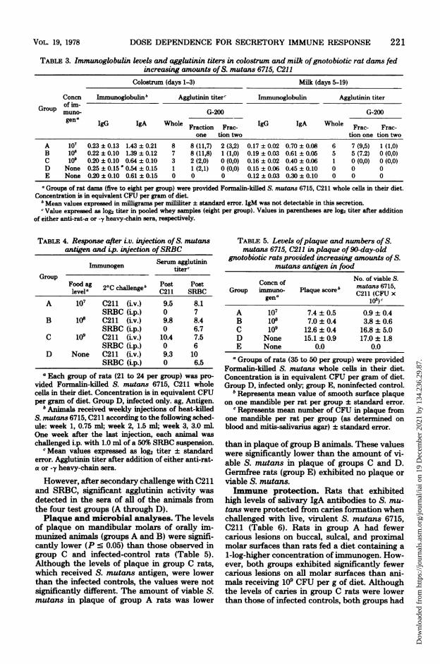
TABLE 3. Immunoglobulin levels and agglutinin titers in colostrum and milk ofgnotobiotic rat dams fedincreasing amounts of S. mutans 6715, C211
Colostrum (days 1-3) Milk (days 5-19)
Concn Immunoglobulinb Agglutinin titer' Immunoglobulin Agglutinin titerof im-
Group muno- G-200 G-200gena IgG IgA Whole Fraction Frac- IgG IgA Whole Frac- Frac-
one tion two tion one tion twoA 107 0.23 ± 0.13 1.43 ± 0.21 8 8 (11,7) 2 (3,2) 0.17 ± 0.02 0.70 ± 0.08 6 7 (9,5) 1 (1,0)B 10i 0.22 ± 0.10 1.39 ± 0.12 7 8 (11,8) 1 (1,0) 0.19 ± 0.03 0.61 ± 0.05 5 5 (7.2) 0 (0,0)C 109 0.20 ± 0.10 0.64 ± 0.10 3 2 (2,0) 0 (0,0) 0.16 ± 0.02 0.40 ± 0.06 1 0 (0,0) 0 (0,0)D None 0.25 ± 0.15' 0.54 ± 0.15 1 1 (2,1) 0 (0,0) 0.15 ± 0.06 0.45 ± 0.10 0 0 0E None 0.20 ± 0.10 0.61 ± 015 0 0 0 0.12 ± 0.03 0.30 ± 0.10 0 0 0
aGroups of rat dam (five to eight per group) were provided Formalin-killed S. mutans 6715, C211 whole celis in their diet.Concentration is in equivalent CFU per gram of diet.
6 Mean values expressed in milligrams per milliliter ± standard error. IgM was not detectable in this secretion.c Value expressed as log2 titer in pooled whey samples (eight per group). Values in parentheses are log2 titer after addition
of either anti-rat-a or -y heavy-chain sera, respectively.
TABLE 4. Response after i.v. injection of S. mutansantigen and i.p. injection ofSRBC
Serum agglutininImmunogen titer'Group
Food ag 2°C ch0engeb Post Postlevela 20Chleg211 SRBC
A 107 C211 (i.v.) 9.5 8.1SRBC (i.p.) 0 7
B i0W C211 (i.v.) 9.8 8.4SRBC (i.p.) 0 6.7
C 109 C211 (i.v.) 10.4 7.5SRBC (i.p.) 0 6
D None C211 (i.v.) 9.3 10SRBC (i.p.) 0 6.5
aEach group of rats (21 to 24 per group) was pro-vided Formalin-killed S. mutans 6715, C211 wholecelia in their diet. Concentration is in equivalent CFUper gram of diet. Group D, infected only. ag, Antigen.
b Animals received weekly injections of heat-killedS. mutans 6715, C211 according to the following sched-ule: week 1, 0.75 ml; week 2, 1.5 ml; week 3, 3.0 ml.One week after the last injection, each animal waschallenged i.p. with 1.0 ml of a 50% SRBC suspension.cMean values expressed as log2 titer ± standard
error. Agglutinin titer after addition of either anti-rat-a or -y heavy-chain sera.
However, after secondary challenge with C211and SRBC, significant agglutinin activity wasdetected in the sera of all of the animals fromthe four test groups (A through D).Plaque and microbial analyses. The levels
of plaque on mandibular molars of orally im-munized animals (groups A and B) were signifi-cantly lower (P ' 0.05) than those observed ingroup C and infected-control rats (Table 5).Although the levels of plaque in group C rats,which received S. mutans antigen, were lowerthan the infected controls, the values were notsignificantly different. The amount of viable S.mutans in plaque of group A rats was lower
TABLE 5. Levels ofplaque and numbers of S.mutans 6715, C211 in plaque of 90-day-old
gnotobiotic rats provided increasing amounts ofS.mutans antigen in food
Concn of No. of viable S.
Group immuno- Plaque score' mutans 6715,gena C211 (CFU x
A 107 7.4 ± 0.5 0.9 ± 0.4B 108 7.0 ± 0.4 3.8 ± 0.6C 109 12.6 ± 0.4 16.8 ± 5.0D None 15.1 ± 0.9 17.0 ± 1.8E None 0.0 0.0
aGroups of rats (35 to 50 per group) were providedFormalin-killed S. mutans whole cells in their diet.Concentration is in equivalent CFU per gram of diet.Group D, infected only; group E, noninfected control.
b Represents mean value of smooth surface plaqueon one mandible per rat per group ± standard error.
' Represents mean number of CFU in plaque fromone mandible per rat per group (as determined onblood and mitis-salivarius agar) ± standard error.
than in plaque of group B animals. These valueswere significantly lower than the amount of vi-able S. mutans in plaque of groups C and D.Germfree rats (group E) exhibited no plaque orviable S. mutans.Immune protection. Rats that exhibited
high levels of salivary IgA antibodies to S. mu-tans were protected from caries formation whenchallenged with live, virulent S. mutans 6715,C211 (Table 6). Rats in group A had fewercarious lesions on buccal, sulcal, and proximalmolar surfaces than rats fed a diet containing a1-log-higher concentration of immunogen. How-ever, both groups exhibited significantly fewercarious lesions on all molar surfaces than ani-mals receiving 109 CFU per g of diet. Althoughthe levels of caries in group C rats were lowerthan those of infected controls, both groups had
221VOL. 19, 1978
Dow
nloa
ded
from
http
s://j
ourn
als.
asm
.org
/jour
nal/i
ai o
n 19
Dec
embe
r 20
21 b
y 13
4.23
6.29
.87.

222 MICHALEK, McGHEE, AND BABB
TABLE 6. Mean caries scores from 90-day-old gnotobiotic rats provided increasing amounts of S. mutans6715, C211 antigen in food
Caries scoreb
Concn of im- Buccal Sulcal ProximalGroup munogena Meanbodywtc
Enamel Dentinal Dentinal Dentinal Dentinal(slight) (slight) (moderate) Enamel (slight)
A 107 7.6 ± 0.3 5.8 ± 0.1 3.4 ± 0.3 2.1 ± 0.3 0.5 ± 0.2 0.0 233.3 ± 13.7B 108 9.5 ± 0.2 7.0 ± 0.3 4.8 ± 0.4 3.0 ± 0.4 1.6 ± 0.3 0.5 ± 0.2 226.6 ± 5.6C 109 19.3 ± 1.0 17.1 ± 1.0 17.7 ± 1.1 15.8 ± 1.2 7.0 ± 0.2 5.0 ± 0.4 221.7 ± 13.2D None 23.8 ± 0.6 18.1 ± 0.9 20.1 ± 0.7 18.1 ± 1.0 7.5 ± 0.3 6.2 ± 0.4 221.1 ± 15.0E None 0.0 0.0 0.0 0.0 0.0 0.0 225.6 ± 8.9
a Groups of rats (35 to 50 per group) were provided Formalin-killed S. mutans whole cells in their diet.Concentration is in equivalent CFU per gram of diet. Group D, infected only; group E, noninfected control.
b Evaluated by the method of Keyes (13). Values represent mean ± standard error of caries on the mandibularmolar surfaces.
c Expressed in grams ± standard error.
extensive decay, and no significant differencewas observed. No differences were observed inthe mean body weights of any of the groupstested (A through E).
DISCUSSIONPrevious investigators have indicated that the
induction of a salivary sIgA immune response,after either local injection of S. mutans antigen(either whole cell [18, 33] or glucosyltransferaseenzyme preparation [34]) or oral ingestion of S.mutans whole-cell antigen (22), correlates withprotection against dental caries in rodents. Thepresent study suggests that the selective induc-tion of a secretory immune response (after oralingestion of S. mutans whole-cell antigen) isdependent upon the antigen dosage. COBS/CDrats provided S. mutans 6715, C211 antigen intheir drinking water (final concentration, 108 tol09 equivalent CFU per ml) exhibited significantlevels of specific antibodies in saliva and mam-mary gland secretions. These findings supportprevious observations concerning gnotobioticrats that were provided a similar antigen prep-aration in their drinking water at a concentrationof 10 equivalent CFU per ml (22). On the otherhand, rats provided 107 equivalent CFU per mlof drinking water exhibited agglutinin activityto S. mutans similar to that observed in controlanimaLs. These results would suggest that theconcentration of antigen presented to these an-imals was insufficient to induce a secretory im-mune response.The settling of high concentrations of antigen
in water presented a problem that was solvedby using food as a carrier. A diet containing 107to 108 equivalent CFU per g of diet elicited asecretory immune response as evidenced by thefinding that treated rats had higher levels of
sIgA in saliva (three- to fourfold), colostrum(two- to threefold), and milk (twofold) than didtheir controls. Furthermore, these animals ex-hibited significant levels of sIgA antibodies toS. mutans in saliva (log2 = 3 and 4, respectively)and milk (log2 = 6 and 5, respectively).Rats provided higher concentrations of anti-
gen in their diet (109 equivalent CFU per g)exhibited levels of immunoglobulin and anti-body similar to those of controls. Because lowlevels of antibody were detected in sera of theseanimals, the results would suggest that evenwith a high concentration of S. mutans, whole-cell antigens did not penetrate the mucosal bar-rier and subsequently induce a systemic immuneresponse. The low levels of detectable antibodyin colostrum, milk, and saliva of these animalssuggest the induction of a state of hyporespon-siveness to S. mutans antigen in secretory tis-sues. Recently, Andre and co-workers (1, 2) re-ported that intragastric administration of largedoses of SRBC to mice rendered them tolerantto subsequent injection of antigen. These ani-mals exhibited circulating antibodies to the an-tigen, and the authors attributed unresponsive-ness to the formation ofimmune complexes afterperipheral administration of SRBC. These re-sults differ from the present findings in two mainregards: (i) Antigen administered in a single,large dose resulted in a serum response (2),whereas continual oral feeding of antigen, asreported here, did not induce circulating anti-bodies. Furthermore, oral administration of bac-terial antigen at optimal concentrations resultedin a secretory immune response. (ii) After oraladministration of antigen, Andre and co-workers(2) observed unresponsiveness when antigen wassubsequently ingested i.p. However, in the pres-ent study, i.v. administration of bacterial antigen
INFECT. IMMUN.
Dow
nloa
ded
from
http
s://j
ourn
als.
asm
.org
/jour
nal/i
ai o
n 19
Dec
embe
r 20
21 b
y 13
4.23
6.29
.87.

DOSE DEPENDENCE FOR SECRETORY IMMUNE RESPONSE
in orally immunized rats resulted in significanttiters of serum antibodies. From our studies itwould appear that the unresponsiveness thatoccurred after feeding large doses of S. mutansantigen was a manifestation of tolerance onlyin the secretory immune compartment. Proof ofthis will require further investigation. Studiesalong these lines are currently underway.
It is noteworthy that COBS/CD rats did notmanifest a secretory immune response whenchallenged with a low antigen concentration, adose which elicited a significant response in gno-tobiotic rats. This finding could be due to theexperimental animal model and/or the methodby which the antigen was delivered. In the for-mer case, a lower concentration of antigen mayhave effectively induced an immune response ingermfree animals, since these animals had pre-viously been exposed to only minimum antigenicstimuli, e.g., food antigens. This could also ex-plain the induction ofa condition ofhyporespon-siveness in gnotobiotic rats when they were pro-vided a concentration of antigen that elicited asignificant response in COBS/CD rats. An alter-native explanation for this difference in the in-duction ofan immune response after oral admin-istration of various antigen concentrations toCOBS/CD and gnotobiotic rats is the methodby which the antigen was delivered. Administra-tion of antigen in food may be more effectivethan water in presenting antigen to the clonesofIgA precursor B-cells located in gut-associatedlymphoid tissue and a subsequent induction ofa selective secretory immune response (10, 19a,22, 27, 30, 31).The direct correlation between the induction
of a selective secretory immune response andcaries immunity was indicated by the demon-stration that gnotobiotic rats, which had specificsIgA antibodies in saliva, colostrum, and milkafter ingestion of S. mutans antigen, had ex-hibited significantly smaller amounts of S. mu-tans in plaque and lower levels of plaque andcaries on all molar surfaces than did the controlanimals or those rats that received antigen butdid not exhibit specific antibodies after oralingestion of S. mutans.The results of this study clearly demonstrate
that the selective induction of a secretory im-mune response is (i) antigen-dose dependent,and (ii) correlated directly (in gnotobiotic rats)with protection against the disease. However,additional investigations are required to deter-mine the mechanism(s) involved in the selectiveinduction of a secretory immune response, in-cluding duration and memory of the immuneresponse, the events that lead to the appearanceof sIgA antibodies in external secretions, andhow these antibodies affect immune protection.
ACKNOWLEDGMENISWe thank Rose Kulhavy for excellent help in fractionation
of samples, Douglas Devenyns for gnotobiotic expertise, andCindy Cox for evaluation of caries lesions. We also thankFrederick W. Kraus and John F. Kearney, University ofAlabama in Birmingham, for their critical review of this work,Catherine Sims for editorial advice, and Jackie Morris fortyping this manuscript.
This work was supported by Public Health Service contractDE-62491 from the National Institute of Dental Researchand grants DE-04217, AI-10854, CA-13148, and DE-02670 fromthe National Institutes of Dental Research and Allergy andInfectious Diseases, the National Cancer Institute, and theNational Institute of Dental Research, respectively.
LITERATURE CMD1. Andre, C., H. Bazin, and J. F. Heremans. 1973. Influ-
ence of repeated administration of antigen by the oralroute on specific antibody-producing cells in the mousespleen. Digestion 9:166-175.
2. Andre, C., J. F. Heremans, J. P. Vaerman, and C. LCambiaso. 1975. A mechanism for the induction ofimmunological tolerance by antigen feeding: Antigen-antibody complexes. J. Exp. Med. 142:1509-1519.
3. Arnold, R. R., J. Mestecky, and J. R. McGhee. 1976.Naturally occurring secretory immunoglobulin A anti-bodies to Streptococcus mutans in human colostrumand saliva. Infect. Immun. 14:355-362.
4. Bowden, G. H., J. M. Hardie, A. S. McKee, P. D.Marsh, E. D. Fillery, and G. L Slack. 1976. Themicroflora associated with developing carious lesionsof the distal surfaces on the upper first premolar in13-14 year old children, p. 223-241. In H. M. Stiles,W. J. Loesche, and T. C. O'Brien (ed.), Microbial as-pects of dental caries. Information Retrieval, Inc.,Washington, D.C.
5. Duncan, D. B. 1955. Range and multiple tests. Biometrics11:142.
6. Emmings, F. G., R. T. Evans, and R. J. Genco. 1975.Antibody response in the parotid fluid and serum ofIrus monkeys (Macacca fascicularis) after local im-munization with Streptococcus mutans. Infect. Immun.12:281-292.
7. Evans, IL T., F. G. Emmings, and RI J. Genco. 1975.Prevention of Streptococcus mutans infection of toothsurfaces by salivary antibodies in Irus monkeys (Ma-cacca fascicularis). Infect. Immun. 12:293-302.
8. Fitzgerald, R. J. 1976. The microbial ecology of plaquein relation to dental caries, p. 849-858. In H. M. Stiles,W. J. Loesche, and T. C. O'Brien (ed.), Microbial as-pects of dental caries. Information Retrieval, Inc.,Washington, D.C.
9. Gibbons, R. J., and J. van Houte. 1975. Dental caries.Annu. Rev. Med. 26:121-136.
10. Goldblum, IL M., S. Ahlstedt, B. Carlsson, L A.Hanson, U. Jodal, G. Lindin-Janson, and A. Sohl-Akerlund. 1975. Antibody-forming cells in human co-lostrum after oral immunization. Nature (London)237:797-799.
11. Heremans, J. F. 1974. Immunoglobulin A, p. 365-522.In M. Sela (ed.), The antigens, vol. 2. Academic Press,Inc., New York.
12. Kaur, J., J. R. McGhee, and W. Burrows. 1972. Im-munity to cholera: the occurrence and nature of anti-body-activity immunoglobulins in the lower ileum ofthe rabbit. J. Immunol. 108:387-395.
13. Keyes, P. H. 1958. Dental caries in the molar teeth ofrats. II. A method for diagnosing and scoring severaltypes of lesions simultaneously. J. Dent. Res.37:1088-1099.
14. Keyes, P. H. 1968. Research in dental caries. J. Am.Dent. Assoc. 76:1357-1373.
VOL. 19, 1978 223
Dow
nloa
ded
from
http
s://j
ourn
als.
asm
.org
/jour
nal/i
ai o
n 19
Dec
embe
r 20
21 b
y 13
4.23
6.29
.87.

224 MICHALEK, McGHEE, AND BABB
15. Krasse, B., and J. Carlsson. 1970. Various types ofstreptococci and experimental caries in hamsters. Arch.Oral Biol. 15:25-32.
16. Loesche, W. J., J. Rowan, L H. Straffon, and P. J.Loos. 1975. Association of Streptococcus mutans withhuman dental decay. Infect. Immun. 11:1252-1260.
17. McGhee, J. R., S. M. Michalek, and V. Ghanta. 1975.Rat immunoglobulins in serum and secretions: purifi-cation of rat IgM, IgA and IgG and their quantitationin serum, colostrum, milk and saliva. Immunochemistry12:817-823.
18. McGhee, J. R., S. M. Michalek, J. Webb, J. M. Navia,A. F. R. Rahman, and D. Legler. 1975. Effectiveimmunity to dental caries: protection of gnotobioticrats by local immunization with Streptococcus mutans.J. Immunol. 114:300-305.
19. Mestecky, J., R. Kulhavy, and F. W. Kraus. 1972.Studies on human secretory immunoglobulin A. II. Sub-unit structure. J. Immunol. 108:738-747.
19a.Mestecky, J., J. R. McGhee, R. Kulhavy, S. M. Mich-alek, S. S. Crago, and R. R. Arnold. 1978. Synthesisof IgA and induction of local immunity. In H. Peeters(ed.), Protides of the biological fluids. Pergamon Press,Inc., Elmsford, N.Y.
20. Michalek, S. M., and J. R. McGhee. 1977. Effectiveimmunity to dental caries: passive transfer to rats ofantibodies to Streptococcus mutans elicits protection.Infect. Immun. 17:644-650.
21. Michalek, S. M., and J. R. McGhee. 1977. Virulence ofStreptococcus mutans: an antibiotic-suppressed ratmodel for studies of pathogenesis. J. Dent. Res.56:205-211.
22. Michalek, S. M., J. R. McGhee, J. Mestecky, R. R.Arnold, and L. Bozzo. 1976. Ingestion of Streptococ-cus mutans induces secretory IgA and caries immunity.Science 192:1238-1240.
23. Michalek, S. M., J. R. McGhee, and J. M. Navia. 1975.Virulence of Streptococcus mutans: a sensitive methodfor evaluating cariogenicity in young gnotobiotic rats.Infect. Immun. 12:69-75.
24. Michalek, S. M., J. R. McGhee, T. Shiota, and D.Devenyns. 1977. Virulence of Streptococcus mutans:cariogenicity of S. mutans in adult gnotobiotic rats.Infect. Immun. 15:466-471.
25. Michalek, S. M., J. R. McGhee, T. Shiota, and D.Devenyns. 197*7. Low sucrose levels promote extensive
Streptococcus mutans-induced dental caries. Infect. Im-mun. 16:712-714.
26. Michalek, S. M., T. Shiota, T. Ikeda, J. M. Navia,and J. R. McGhee. 1975. Virulence of Streptococcusmutans: biochemical and pathogenic characteristics ofmutant isolates. Proc. Soc. Exp. Biol. Med. 150:498-502.
27. Montgomery, P. C., J. Cohn, and E. T. Lally. 1974.The induction and characterization of secretory IgAantibodies, p. 453-462. In J. Mestecky and A. R. Lawton(ed.), The immunoglobulin A system. Plenum Press,New York.
28. Navia, J. M. 1977. Animal models in dental research.The University of Alabama Press, University Station.
29. Ogra, P. L. 1971. The secretory immunoglobulin systemof the gastrointestinal tract, p. 259-279. In P. A. Small,D. H. Dayton, R. M. Channock, H. E. Kaufman, andT. B. Tomasi (ed.), The secretory immunologic system.U. S. Department of Health, Education, and Welfare,Washington, D.C.
30. Parrott, D. M. V. 1976. The gut-associated lymphoidtissues and gastrointestinal immunity, p. 1-32. In A.Ferguson and R. N. M. MacSween (ed.), Immunologicalaspects of the liver and gastrointestinal tract. UniversityPark Press, Baltimore.
31. Rudzik, R., R. L. Clancy, D. Y. E. Perey, R. P. Day,and J. Bienenstock. 1975. Repopulation with IgA-containing cells of bronchial and intestinal lamina pro-pria after transfer of homologous Peyer's patch andbronchial lymphocytes. J. Immunol. 114:1599-1604.
32. Scherp, H. W. 1971. Dental caries: prospects for preven-tion. Science 173:1193-1205.
33. Taubman, M. A., and D. J. Smith. 1974. Effects of localimmunization with Streptococcus mutans on inductionof salivary immunoglobulin A antibody and experimen-tal dental caries in rats. Infect. Immun. 9:1079-1091.
34. Taubman, M. A., and D. J. Smith. 1977. Effect of localimmunization with glucosyltransferase fractions fromStreptococcus mutans on dental caries in rats and ham-sters. J. Immunol. 118:710-720.
35. Trexler, P. C. 1959. The use of plastics in the design ofisolator systems. Ann. N. Y. Acad. Sci. 78:29-36.
36. van Houte, J. 1975. Oral bacterial colonization: mecha-nisms and implications, p. 3-32. In H. M. Stiles, W. J.Loesche, and T. C. O'Brien (ed.), Microbial aspects ofdental caries. Information Retrieval, Inc., Washington,D.C.
INFECT. IMMUN.
Dow
nloa
ded
from
http
s://j
ourn
als.
asm
.org
/jour
nal/i
ai o
n 19
Dec
embe
r 20
21 b
y 13
4.23
6.29
.87.