A Systematic Review of the Normal Sacroiliac Joint Anatomy ...
28
Background: The sacroiliac joint (SIJ) forms a complex joint and has shown to be underappreciated in its involvement with lower back pain. Research efforts have intensified on SIJ anatomy and biomechanics because of its predisposing position to pain and dysfunction in individuals suffering from lower back discomfort. Previous work has focused on SIJ anatomy including bone and joint structure, innervation, as well as biomechanics and the treatment of SIJ pain. However, to date, no review exists describing the range of ‘normal’ anatomic features of the SIJ. Objectives: To describe the normal appearance of the SIJ and adjacent tissues, as opposed to ‘abnormal’ conditions involving SIJ morphology. It will also identify key areas that require further study because of lacking information or disagreement. Study Design: A systematic literature review. Setting: The research took place at the University of Otago, New Zealand. All published research on ‘normal SIJ anatomy’ available from MEDLINE, OVID, Scopus, Web of Science, PubMed, and Science Direct were included, available until December 2018, in English, French, and German. Subject areas included bony landmarks, joint type, bone morphology, ligamentous attachments, muscular and fascial relationships, blood supply, fatty infiltration, and morphologic variation. Methods: Articles met the selection criteria if they contained specific information on SIJ anatomy, including bone morphology and architecture, ligaments, muscle attachments, innervation, vasculature, and the presence of fat. Biomechanics and kinematics related keywords were used as the literature often couples these with the anatomy. Keywords of individual articles were named as ‘structures of interest.’ Results: A total of 88 primary and 101 secondary articles were identified in the time frame from 1851 to 2018. Primary articles provided quantitative data and detailed anatomic descriptions. Secondary articles did not focus specifically on the anatomy of the SIJ. Although research appeared to be in general agreement on bony landmarks, joint type, myofascial attachments, vasculature, and innervation of the SIJ, there was only part consensus on ligament attachments and cartilage structure. Information regarding bone density of the articulating surfaces of the SIJ is lacking. Despite its potential clinical significance, fatty infiltration within the joint lacks research to date. Limitations: Only the given databases were used for the initial search. Keyword combinations used for this review may not have been inclusive of all articles relevant to the SIJ. Work in languages other than the ones listed or work that is not available via the internet may be missing. Conclusions: This study provides an overview of normal SIJ structures, including all neuromusculoskeletal elements related to the joint. There is a lack of knowledge on the SIJ ligaments warranting further investigation. Furthermore, there are discrepancies in relation to the nomenclature, layers, attachment sites, and on the topographical relationships between ligamentous tissues and nerves. Subsequent studies on the quantification of fat and bone density in the SIJ have been suggested. These could be useful radiologic parameters to assess the condition of the joint clinically. This review may provide insight into the clinical signs and abnormal biomechanical features of the joint for the purposes of treating SIJ pain. Key words: Bone density, bony landmarks, fat infiltration, innervation, ligaments morphology, muscles, sacroiliac joint, vasculature Pain Physician 2019: 22:E247-E274 Systematic Review A Systematic Review of the Normal Sacroiliac Joint Anatomy and Adjacent Tissues for Pain Physicians From: 1 Department of Anatomy, University of Otago, Dunedin, New Zealand; 2 School of Medicine, University of Otago, Dunedin, New Zealand; 3 Department of Orthopaedic and Trauma Surgery, University of Leipzig, Leipzig, Germany; 4 Fraunhofer IWU, Dresden, Germany Address Correspondence: Amélie J. Poilliot, PhD cand Department of Anatomy University of Otago 270 Great King Street Dunedin, 9010 New Zealand E-mail: amelie.poilliot@ postgrad.otago.ac.nz Disclaimer: There was no external funding in the preparation of this manuscript. Conflict of interest: Each author certifies that he or she, or a member of his or her immediate family, has no commercial association (i.e., consultancies, stock ownership, equity interest, patent/licensing arrangements, etc.) that might pose a conflict of interest in connection with the submitted manuscript. Manuscript received: 12-08-2018 Revised manuscript received: 01-16-2019 Accepted for publication: 01-22-2019 Free full manuscript: www.painphysicianjournal. com Amelie J. Poilliot, PhD cand 1 , Johann Zwirner, MD 1 , Terence Doyle, MD 2 , and Niels Hammer, MD 1,3,4 www.painphysicianjournal.com Pain Physician 2019; 22:E247-E274 • ISSN 2150-1149
Transcript of A Systematic Review of the Normal Sacroiliac Joint Anatomy ...

Background: The sacroiliac joint (SIJ) forms a complex joint and has shown to be underappreciated in its involvement with lower back pain. Research efforts have intensified on SIJ anatomy and biomechanics because of its predisposing position to pain and dysfunction in individuals suffering from lower back discomfort. Previous work has focused on SIJ anatomy including bone and joint structure, innervation, as well as biomechanics and the treatment of SIJ pain. However, to date, no review exists describing the range of ‘normal’ anatomic features of the SIJ.
Objectives: To describe the normal appearance of the SIJ and adjacent tissues, as opposed to ‘abnormal’ conditions involving SIJ morphology. It will also identify key areas that require further study because of lacking information or disagreement.
Study Design: A systematic literature review.
Setting: The research took place at the University of Otago, New Zealand. All published research on ‘normal SIJ anatomy’ available from MEDLINE, OVID, Scopus, Web of Science, PubMed, and Science Direct were included, available until December 2018, in English, French, and German. Subject areas included bony landmarks, joint type, bone morphology, ligamentous attachments, muscular and fascial relationships, blood supply, fatty infiltration, and morphologic variation.
Methods: Articles met the selection criteria if they contained specific information on SIJ anatomy, including bone morphology and architecture, ligaments, muscle attachments, innervation, vasculature, and the presence of fat. Biomechanics and kinematics related keywords were used as the literature often couples these with the anatomy. Keywords of individual articles were named as ‘structures of interest.’
Results: A total of 88 primary and 101 secondary articles were identified in the time frame from 1851 to 2018. Primary articles provided quantitative data and detailed anatomic descriptions. Secondary articles did not focus specifically on the anatomy of the SIJ. Although research appeared to be in general agreement on bony landmarks, joint type, myofascial attachments, vasculature, and innervation of the SIJ, there was only part consensus on ligament attachments and cartilage structure. Information regarding bone density of the articulating surfaces of the SIJ is lacking. Despite its potential clinical significance, fatty infiltration within the joint lacks research to date.
Limitations: Only the given databases were used for the initial search. Keyword combinations used for this review may not have been inclusive of all articles relevant to the SIJ. Work in languages other than the ones listed or work that is not available via the internet may be missing.
Conclusions: This study provides an overview of normal SIJ structures, including all neuromusculoskeletal elements related to the joint. There is a lack of knowledge on the SIJ ligaments warranting further investigation. Furthermore, there are discrepancies in relation to the nomenclature, layers, attachment sites, and on the topographical relationships between ligamentous tissues and nerves. Subsequent studies on the quantification of fat and bone density in the SIJ have been suggested. These could be useful radiologic parameters to assess the condition of the joint clinically. This review may provide insight into the clinical signs and abnormal biomechanical features of the joint for the purposes of treating SIJ pain.
Key words: Bone density, bony landmarks, fat infiltration, innervation, ligaments morphology, muscles, sacroiliac joint, vasculature
Pain Physician 2019: 22:E247-E274
Systematic Review
A Systematic Review of the Normal Sacroiliac Joint Anatomy and Adjacent Tissues for Pain Physicians
From: 1Department of Anatomy, University of Otago, Dunedin, New
Zealand; 2School of Medicine, University of Otago,
Dunedin, New Zealand; 3Department of Orthopaedic
and Trauma Surgery, University of Leipzig, Leipzig,
Germany; 4Fraunhofer IWU, Dresden, Germany
Address Correspondence: Amélie J. Poilliot, PhD cand
Department of AnatomyUniversity of Otago
270 Great King Street Dunedin, 9010 New Zealand
E-mail: [email protected]
Disclaimer: There was no external funding in the preparation of this
manuscript.Conflict of interest: Each
author certifies that he or she, or a member of his or her immediate family, has
no commercial association (i.e., consultancies,
stock ownership, equity interest, patent/licensing arrangements, etc.) that might pose a conflict of
interest in connection with the submitted manuscript.
Manuscript received: 12-08-2018
Revised manuscript received:01-16-2019
Accepted for publication: 01-22-2019
Free full manuscript:www.painphysicianjournal.
com
Amelie J. Poilliot, PhD cand1, Johann Zwirner, MD1, Terence Doyle, MD2, and Niels Hammer, MD1,3,4
www.painphysicianjournal.com
Pain Physician 2019; 22:E247-E274 • ISSN 2150-1149

Pain Physician: July/August 2019: 22:E247-E274
E248 www.painphysicianjournal.com
detection of SIJ diseases and dysfunction.Our objective was to provide a
systematic review of the literature on the anatomic features of the SIJ. These include the morphologic features of the ilium and sacrum, the ligaments associated with the SIJ, the muscular at-tachments and myofascial relationships, vasculature, innervation, the presence of fat within the joint, and anatomic variation. It will provide insight into the original studies on the various structures from articles published from June 1851 to January 2018 in English, French, and German. This will enable the assessment of the present anatomic knowledge of the SIJ and uncover discrepancies within the literature. Furthermore, it will pro-vide a basis for the ‘normal’ appearance of the structures of the SIJ, while also enabling the comparison of this state with a potentially ‘abnormal’ state of the structures of the SIJ.
Methods
Articles were chosen if they con-tained specific information on the SIJ anatomy including morphology and architecture, ligaments, muscle attach-ments, innervation, vasculature, and presence of fat. Surgical research was included if they had relevant anatomic information on the SIJ that also matched the inclusion criteria. Figure 1 details the method undertaken in this study of relevant articles including the databases searched, keywords, and exclusion crite-ria applied. Biomechanical and mobility-related keywords were used as the lit-erature often couples the anatomy with biomechanics. Keywords of individual articles listed in the following text were named as ‘structures of interest’ (Table 1). These are not specifically linked to the SIJ in most studies but anatomically significant to the joint.
Results
Of the 88 primary sources included in this review, 5 studies had unknown
IIn today’s society, 70%-85% of individuals suffer from pelvic girdle pain (PGP) in their lifetime. Interest has arisen on the subject of the sacroiliac joint (SIJ) because of its direct involvement in PGP
and lumbopelvic pain, but this is still underappreciated as a source of mechanical lower back pain (1-3). Pathological findings have emerged on structural abnormalities, infection, SIJ degeneration, as well as inflammatory and metabolic disorders, with few specifically looking at the joint composition (4,5). Previous review articles on the SIJ have focused on some parts of the descriptive anatomy, diagnosis, and treatment of SIJ syndrome (2,6-13). Other studies have also emerged on the biomechanics of the SIJ, in terms of the structural relationships of the joint (14-21). However, none have indicated its role in pathogenesis. Understanding the ‘normal’ SIJ biomechanics is important when considering changes that may occur in association with pathogenesis around the SIJ. Furthermore, the detection of an ‘abnormal state’ could be clinically significant in relation to the
Fig. 1. Research strategy followed for finding relevant literature, including the databases searched and keywords entered (keywords 1 AND keywords 2) and research exclusion criteria, * refers to the plural.

www.painphysicianjournal.com E249
: The Sacroiliac Joint and Lower Back Pain: A Review
sample sizes (22-26). The most common structures discussed were the ligaments and innervation, with vasculature and fat presence being the minority. Muscle studies were limited as most information was derived from ligament studies and not on the muscle morphology directly. Figure 2 details the strat-egy followed for the screening of the relevant literature.
Bony Landmarks and Joint Characteristics of the SIJ
Typically, the SIJ is described as being composed of 2 distinct parts: the syndesmosis and articular parts (Fig. 3) (27-29).
Iliac and Sacral SIJ-Related Landmarks
The bony landmarks of the sa-crum and ilium are generally well described in the literature (22,30-49). Figures 4, 5, and 6 recount SIJ-related landmarks.
Classification of the SIJThe SIJ classification differs
from it being a diarthrosis, an amphiarthrosis, or presenting features of both as a ‘diarthro-amphiarthrosis’ (Table 2) (22,25,30-
Table 1. Muscles, ligaments, and fascia with a direct relation to the SIJ. These are the ‘structures of interest’.
Fig. 2. PRISMA flow chart for the methodology undertaken for the screening of relevant literature based on Moher et al. (191). Primary papers (14,16,17,20,22-26,28-33,38,42,43,50-57,69,71,73,74,78-84,86-97,99,100,102,105-108,110-117,121,126,130,132,133,136,137,146-157,169-172,179), secondary papers (1-13,15,18,19,21,27,34-37,39-41,44-49,58-68,70,72,75-77,85,98,101,103,104,109,118-120,122-125,127-129,131,134,135,138-145,155,158-168,173-178,180-183,185-190).

Pain Physician: July/August 2019: 22:E247-E274
E250 www.painphysicianjournal.com
Fig. 3. Transverse sections of a pelvis at L5 (A), at mid sacrum S2 (B), and at S4 (C) with corresponding E12-plastinated slices (right). Darker gray regions highlight the cartilaginous (synovial) portion, the lighter gray regions show the ligamentous (syndesmotic) portion with the ISL. A, anterior; L, left; P, posterior; R, right.
Fig. 4. Landmarks of the medial ilium based on Otter, (41) and Postacchini et al., (31). AIIS: anterior inferior iliac spine, ASIS: anterior superior iliac spine, PSIS: posterior superior iliac spine, PIIS: posterior inferior iliac spine, Ridge: constant ridge from iliac crest to auricular surface, GSN: greater sciatic notch, OF: obturator foramen. A: anterior, P: posterior, S: superior, I: inferior.

www.painphysicianjournal.com E251
: The Sacroiliac Joint and Lower Back Pain: A Review
Fig. 5. Landmarks of the medial iliac surface. Gray area: auricular surface, blue area: iliac tuberosity, green asterisks: smooth narrow depression, red asterisks: pre-auricular sulcus. A, anterior; I, inferior; P, posterior; S, superior.
Fig. 6. Landmarks of
the posterior (left) and
lateral sacrum (right). PSF1-
4: posterior sacral foramina
1-4, cranial depression
(shaded blue), middle depression (shaded yellow),
caudal depression (shaded green),
LSC (black asterisk on
each tubercle), intermediate
sacral crest (red asterisk on each tubercle). A, anterior; I,
inferior; L, left; P, posterior; R,
right; S, superior.

Pain Physician: July/August 2019: 22:E247-E274
E252 www.painphysicianjournal.com
32,50-57). Most argue that the joint is diarthrodial as it presents the classic features of a synovial joint (25,30,32,50,51,55,58-60). Two studies describe the joint as being an amphiarthrosis (42,61). One study suggests that because of its shape it should be classified as a condylarthrosis. Arguably, it is not a symphysis, nor a synarthrosis, because of the presence of the interosse-ous sacroiliac ligament (ISL) in the dorsal aspect of the joint. It is not an amphiarthrosis as the cartilage does not continue throughout the whole joint space (53). However, research indicates that SIJ changes through time. Embryologically, the anterior SIJ forms in the same manner as other diarthrodial joints, although cav-itation arises later (10th week in utero) and progresses less rapidly until the seventh month of gestation. The joint does not cavitate uniformly as it occurs in other diarthrodial joints (22,62). Children and young adults reflect characteristics of a diarthrodial joint, whereas older individuals present amphiarthrodial character-istics. This is because older individuals gradually de-crease in mobility in regions that present features of a diarthro-amphiarthrosis (41,52,55). The classification is sometimes based on movement rather than structure (30,51,53). The study by Walker (63) provides a more encompassing definition: it is a diarthrodial joint with an interosseous portion. This definition however does not account for age-related changes in structure and movement (173).
Cartilage Type and Morphometry of the Articular Surfaces
There is disagreement in the distribution and morphology of the cartilage (64). Some state that the iliac side is composed of fibrocartilage and the sacral side is composed of hyaline cartilage (22,58,61,65-68). Others report a mixture of both cartilages on both sides (22,25,48,54,55,57,59). The sacral cartilage ap-pears smooth, light-gray, and ‘glistening’ that rough-ens and becomes yellow with age (25,32,51,55,69,70). The iliac side has a blueish, dull, striated, or striped appearance that also roughens and yellows with age (25,32,51,55,69-71). The iliac cartilage is consistently 2 to 3 times thinner than the sacral with this thickness remaining constant during ageing (25,32,51,58,72). The sacral cartilage is thicker in women than in men, with no significant difference on the iliac side (69). Histologi-cally, degenerative changes arise earlier in the SIJ than in other diarthrodial joints already noticeable in infants. The sacral cartilage remains largely unaltered until old age. The iliac cartilage however, presents osteoarthritic changes (73). It is composed of a dense fibrous network or collagen bundles in infancy and gradually becomes hyaline cartilage over time (54).
There are no significant differences in the values in SIJ articular space width between both genders or between sides (68,74,75). However, there is a reduc-tion in articular width specifically seen after the age
Table 2. Summary of results of joint type assessment from the literature. aA type of diarthrodial joint; *no data available.

www.painphysicianjournal.com E253
: The Sacroiliac Joint and Lower Back Pain: A Review
of 12 (74,75). The auricular surfaces present signs of degeneration such as erosion and irregularities that are noticeable in individuals as young as 20 years old, more prominently on the iliac side (21,32,55,56,76,77). A morphometry study of the articular surfaces in adults revealed that the mean area of the joint was approxi-mately 13 cm2 (33). Sexual dimorphism was only present for 2 features: the joint is more inferior in men relative to the anterior superior iliac spine and the surface area is smaller in women (33,78).
Bone Density of the Iliac and Sacral Articular Surfaces
The pelvic bone is made up of low-density trabecu-lar bone covered by a layer of high-density cortical bone. This enables the pelvis to be a low weight structure capable of withstanding high loads. One study tested the theory of muscle forces on the entire pelvic bone. They discovered a higher strain in the subchondral and trabecular bone than that of the cortical shell (79). The distribution of the subchondral bone density shows a consistent pattern with the highest density along the auricular surfaces, whereas centrally there are lower density grades in the region of the ‘degenerative zones’ (80). Vogler et al (81) reported the appearance of bone density in healthy individuals and found that it devel-ops in a ‘non-uniform’ and ‘ill-defined’ manner on the iliac side versus a ‘uniform’ and ‘well-defined’ manner on the sacral side.
Regarding the internal trabecular bone arrange-ment of the sacrum, cortical thickness did not vary between genders nor with age, ranging from 0.5 to 2.25 mm thickness with specification of the zones with higher density at the anterior aspect of the S1-S2 portion of the sacrum (82,83). One study specifically examined the bone densities of the sacral and iliac ar-ticulating counter parts. They found that the sacral side is approximately half as dense as its iliac counterpart at the anterior, central, and posterior portions (84). Re-garding the subchondral bone architecture of the iliac and sacral counterparts, the sacral subchondral bone plate is thin with trabecular spongiosa inserting into it at a right angle. The iliac bone however is thicker, with the trabecular spongiosa inserting into the bone plate obliquely (73).
Ligaments of the SIJThe SIJ is composed of intrinsic and extrinsic liga-
ments sometimes referred to as ‘capsular’ and ‘acces-sory ligaments’ (85,86). The intrinsic ligaments are the
anterior sacroiliac ligament (ASL), the ISL, the posterior sacroiliac ligament (PSL), and the long posterior sacro-iliac ligament (LPSL), and the extrinsic ligaments are the iliolumbar ligament (ILL), the sacrotuberous ligament (STL), and the sacrospinous ligament (SSL) (85).
The ASLThe ASL is a thickening of the anterior and inferior
parts of the joint capsule. It is a thin ligament, passing from the anterior aspect of the sacrum to the antero-medial border of the ilium (Fig. 7) (17,42,54,55,86). It crosses the SIJ anteroinferior from the sacrum to insert onto the periosteum of the ilium near the auricular margins (28,54). It is shown to be a single structure in 3 studies (28,54,55).
It has also been described as consisting of the up-per, middle, and lower parts spreading in a fan-like shape from the anterior sacrum to the wing of the ilium (17,42,57,86). Weisl (86) reports the superior part of the ASL arising from the ventral half of the lateral border of the sacral ala and continues to the adjacent medial border of the iliac fossa. The thickness and width of the ASL increase at the pelvic brim while spreading along the ilium for 20 mm mingling with fibers from the ILL superiorly. On the anterior aspect, inferior fibers of the ASL attach laterally in a line at the first 3 sacral foramina converging together dorsolaterally (86). The inferior part of the ASL is continuous with a number of structures, namely the PSL, SSL, and STL (Fig. 8) (20). Figures 9 and 10 detail a summary of attachment sites of the ASL.
The Posterior Sacroiliac Ligamentous Complex
There is difficulty describing the fascicles and parts of the PSL as its fibers merge with other structures like the ISL, LPSL, SSL, and STL (55,86). The LPSL attaches in-feriorly to the lateral sacral crest (LSC) of S3-S5, whereas the PSL has variable attachment sites on the sacrum (Fig. 11) (86,87). The ISL is deep to the PSL, however the LSPL is lateral to the PSL with some fibers mixing with it.
The Interosseous (Axial) LigamentThe ISL, which is sometimes called the ‘axial ligament’
or ‘vague ligament’ (8,57,88), fills the syndesmotic joint space. This space is posterior to the auricular surfaces as the deepest layer (Figs. 11 and 12). The ISL attaches to the iliac tuberosity and is only present in the cranial part of the joint (17,38,50,55,57,86). The fibers are described as having a dorsolateral (cranial group) and cranial (caudal

Pain Physician: July/August 2019: 22:E247-E274
E254 www.painphysicianjournal.com
Fig. 7. Photographs of a dissected superior anterior sacroiliac joint. (A) the anterior sacroiliac ligament shaded in yellow on each side according to Jaovisidha et al., (28), Puhakka et al., (54) and Sashin (55). (B) the anterior sacroiliac ligament as separate entities, blue shading according to Gerlach and Lierse (42); Right side red shading according to Weisl, (86). S: superior, I: inferior, L: left, R: right.
Fig. 8. Anterior-inferior view of the pelvic outlet on a dry specimen. The inferior portion of the ASL is highlighted in red. I, inferior; L, left; R, right; S, superior.
Fig. 9. Attachment sites of ASL on the anterior sacrum. Darkest zones are referenced the most in the literature (more than 3 articles). AF1-4, anterior sacral foramina 1-4; I, inferior; L, left; R, right; S, superior; S1-5, sacral segments 1-5.

www.painphysicianjournal.com E255
: The Sacroiliac Joint and Lower Back Pain: A Review
Fig. 10. Attachment sites of ASL on the medial ilium. Darker zones are referenced the most in the literature with a higher degree of consensus between authors. A, anterior; AIIS, anterior inferior iliac spine; ASIS, anterior superior iliac spine; GSN, greater sciatic notch; I, inferior; OF, obturator foramen; P, posterior; PIIS, posterior inferior iliac spine; S, superior.
Fig. 11. Superior view of the pelvic inlet and ligament attachment site of the ISL. Transverse section at the level of S1. The dark gray region is the ISL, the lighter gray shading is the PSL, the red shading is the LPSL.
Fig. 12. E12-plastinated slice at the level of L5 at a superior view. The interosseous portion filled with the ISL is highlighted in blue, the ASL in red, and the PSL in yellow. A, anterior; L, left; P, posterior; R, right.

Pain Physician: July/August 2019: 22:E247-E274
E256 www.painphysicianjournal.com
group) direction from their sacral attachments. Each group has approximately 13 bundles of parallel fibers with the caudal ones being longer and flatter than the cranial ones. Bundles insert into the retro-auricular re-gions of the sacrum and ilium. Dorsally and cranially they reach the LSC border of the sacral ala and run up to the crest and medial border of the iliac fossa (86). Sashin (55) stated that these layers interweave together.
The PSL The PSL is located posterior to the articular space
(17). The PSL ‘cranial group’ runs dorsolaterally and the PSL ‘caudal group’ runs cranially. One part of the cranial group passes dorsolaterally from the lateral edge of the dorsal half of the sacral ala to the ridge on the ilium. It extends between the crest and auricular surface. The fibers of the PSL merge with the ASL superiorly. Posteriorly, it is sometimes a separate entity from the rest of the PSL. The caudal group forms the true PSL. Some fibers pass dorsolaterally as a single, sometimes split band arising from the iliac tuberosity to the first sacral segment, lateral sacral tubercle, and base of the articular process. Other fibers run cranially from the LSC of S2-S3 and insert on the inferior inner lip of iliac crest and posterior superior iliac spine (PSIS) (86). One study
describes the PSL as being composed of various fibrous bands originating from 3 centers all intermingling as the PSL layer. To summarize, the layer attaches from the LSC to the first sacral depression and to the ilium on the PSIS and iliac tuberosity, attaching to the adjacent bony structures in between these sites (Fig. 13) (42).
The most superficial layer PSL can contain up to 4 fibrous bands arising from the PSIS to the respective posterior medial sacral tubercles arranged in a combi-nation of various patterns (Fig. 14) (57). Figures 15 and
Fig. 13. Posterior articulated pelvis with the PSL highlighted in red. Note that in this figure it is composed of 2 sections. I, inferior; L, left; R, right; S, superior.
Fig. 14. Patterns of the posterior sacroiliac ligaments according to Hakim, (57). (A) On the left, a rectangular structure found in 64% of specimens from the posterior superior iliac spine (PSIS) to the S1-S2 levels of the intermediate sacral crest. The right side displays the two (sometimes three) bands bifurcations from the PSIS to S1, S2 and S3 posterior medial sacral tubercles found in 63% of individuals, three bands found in 53%. (B) The four bands pattern from the posterior superior iliac spine (PSIS) to the intermediate sacral crest found in 50% of individuals is on the left. The fan shape arrangement on the right was found in 36% of specimens. S: superior, I: inferior, L: left, R: right.

www.painphysicianjournal.com E257
: The Sacroiliac Joint and Lower Back Pain: A Review
16 detail a summary of attachment sites for the PSL in the literature.
The LPSLThe LPSL is a fibrous sheet running from the PSIS
to the third or fourth lateral sacral tubercles (Fig. 17) (86,87,89,90). There is no consensus if the LPSL is a single structure or a composition of various ligamentous parts (86,87,89). The LPSL cranial fibers form a fascial sheet of weak fascicles passing dorsolaterally from the superior articular process and from each of the posterior sacral tubercles to the inner lip of the iliac crest. The caudal fibers compose the so-called LPSL (86). It is formed of
parallel fibers flattened dorsoventrally and it is sepa-rated from the caudal fibers of the PSL by the lateral branches of the dorsal sacral rami (86,89,90). Figures 18 and 19 detail a summary of the attachment sites of the LPSL in the literature.
The ILLThe ILL is comprised of one to 4 different ligament
bands (8,91). Some state that the ILL is one band origi-nating from the transverse process of L5 to the poste-rior part of the iliac crest (42,88).
The ILL is often described to be formed of 2 bands (92-99). An anterior band arises from the tip of the
Fig. 15. Attachment sites of the PSL on the posterior sacrum. Darkest zones are referenced the most in the literature with a higher degree of consensus between authors. I, inferior; L, left; PSF1-4, posterior sacral foramina 1-4; R, right; S, superior.
Fig. 16. Attachment sites of the PSL on the medial ilium. Darker zones are referenced the most in the literature with a higher degree of consensus between authors. A, anterior; AIIS, anterior inferior iliac spine; ASIS, anterior superior Iliac spine; GSN, greater sciatic notch; I, inferior; OF, obturator foramen; P, posterior; PIIS, posterior inferior iliac spine; S, superior.

Pain Physician: July/August 2019: 22:E247-E274
E258 www.painphysicianjournal.com
transverse process of L5 to the periosteum on the anterior margin of the iliac crest (92-94,100). It has a torsional inferolateral direction in its course from L5 to the ilium (96). In one study, it has an origin on the transverse process of L4 in approximately 17% of the specimens (97). The posterior band runs from L5 to the posterior margin of the iliac crest in a ‘torsion-like manner’ (92,95,96). Another study found that the posterior and anterior bands were 2 distinct structures in 69% of their specimens. In the remaining 31%, the bands coursed together as one (100).
The ILL has been described as 4 individual parts: a dorsal band, a ventral band, a lumbosacral band, and a sacroiliac part (Fig. 20) (91,101). Although previously there was no mention of the sacroiliac part, unless the lumbosacral band was defined as a conjunction of the two (57). The ‘lumbosacral band’ of the ILL has also
Fig. 18. Attachment sites of the LPSL on the posterior sacrum. Darkest zones are referenced the most in the literature with a higher degree of consensus between authors. I, inferior; L, left; PSF1-4, posterior sacral foramina 1-4; R, right; S, superior.
Fig. 17. Posterior view of an E12-plastinated hemipelvis. The LPSL (black section) is blending with the course of the STL inferiorly (green section). I, inferior; L, lateral; M, medial; S, superior.
Fig. 19. Attachment sites of LPSL on the medial ilium. Darker zones are referenced the most in the literature with a higher degree of consensus between authors. A, anterior; AIIS, anterior inferior iliac spine; ASIS, anterior superior Iliac spine; GSN, greater sciatic notch; I, inferior; OF, obturator foramen; P, posterior; PIIS, posterior inferior iliac spine; S, superior.
been reported as the ‘lumbosacral ligament’ (57,102-106). It is present in all specimens bilaterally, running from the body of the first sacral vertebrae to meet with the ILL or connect directly to the transverse process of L5 (103,105,106).

www.painphysicianjournal.com E259
: The Sacroiliac Joint and Lower Back Pain: A Review
An imaging study determined the interindi-vidual variation that occurs for the ILL. The ligament ranges from being a thick to thin, broad, small or even long band, and has various directions and at-tachment sites within the same gender (98). In indi-viduals of African descent, it was found that the ILL was composed of one single thicker band in contrast with 2 bands in Caucasian individuals (107). Figures 21 and 22 detail a summary of the attachment sites of the ILL.
The STL and the SSL The SSL and STL prevent the sacrum from tilt-
ing when forces are applied (Fig. 23) (35,108-110). The SSL arises from the anterolateral surface of the last 2 sacral segments and the first coccygeal seg-ment to the ischial spine (42,55). The sacral fibers are mingled with the ASL superiorly and the ischial fibers merge with the STL (42,111).
Medially, the STL arises along a line running from the posterior inferior iliac spine down to the
Fig. 20. The ILL on a dry specimen, anterior/superior view (left) with shaded components of the ILL (red), superior-lateral view (right). (1) From the transverse process of L5 is a dorsal band running to the cranial part of the iliac tuberosity on the medial part of the iliac crest. (2) The ventral band running from the ventrocaudal part of the transverse process of L5 to the ventrocaudal part of the iliac tuberosity on the sacropelvic surface. (3) A lumbosacral band running from the ventrolateral aspect of the vertebral body of L5 and ventromedial part of the transverse process of the middle ventrolateral part of the base of the sacrum near the SIJ. (4) A sacroiliac part running from the cranial surface of the ala of sacrum directly caudal to the transverse process of L5 to the ventromedial part of the iliac tuberosity together with the ventral band. A, anterior; I, inferior; L, left; P, posterior; R, right; S, superior.
Fig. 21. Attachment sites of the ILL on the medial ilium. Darker zones are referenced the most in the literature with a higher degree of consensus between authors. A, anterior; AIIS, anterior inferior iliac spine; ASIS, anterior superior Iliac spine; GSN, greater sciatic notch; I, inferior; OF, obturator foramen; P, posterior; PIIS, posterior inferior iliac spine; S, superior.

Pain Physician: July/August 2019: 22:E247-E274
E260 www.painphysicianjournal.com
border of the iliac bones to the caudal edge of the sacrum and coccyx (C1-C2). It also attaches to the LSC on a level with S3-S4 and PSL on this line (42,55,112). It inserts on the ischial tuberosity inferiorly (16,42,113). The fibers run in a twisting course inferiorly obliquely from the sacrum crossing the posterior surface of the
SSL (96,108). The STL also shares attachments with vari-ous muscles and fascia of the pelvis, thigh, and back. These include the erector spinae (ES) muscles, piriformis muscle, gluteus maximus (GMax) muscle, the biceps femoris (BF) muscle, and the thoracolumbar fascia (TLF) (14,87,108,114-116).
Further Ligaments Related to the SIJ Additional SIJ ligaments have been reported. A
‘polar anteroinferior ligament’ is located anteriorly near the lip separating the second and third sacral fora-men and inserts on the sacral anteroinferior angle of the auricular surface on the sacrum to the homologous iliac surface. It is described as a short, resistant, oblique band running anteriorly from the iliac surface to the sacral surface. It is formed by 2 fascicles, one anterior and one posterior, separated by a small neurovascular bundle (117). It has a similar location as the ‘horizon-tal ligament on S3’ as described by Steinke et al (20) located ventrally covering the third sacral vertebra between the second and third sacral foramen. ‘Illi’s ligament’ has been referred to with many studies dis-agreeing on its presence, whereas few have proven its existence (88, 118-121). It is a small but strong band of tissue originating below the superior part of the SIJ and slightly posterosuperior to the margin of the ar-ticulating surface of the sacrum. It is also 1.5 cm above the superior border of the articulating surface of the
Fig. 23. Sacrotuberous and SSLs on plastinated prosections of hemipelvis, (A) anterior view, (B) lateral view, (C) posterior view. In green is the STL, in red the SSL. A, anterior; I, inferior; L, lateral; M, medial; P, posterior; S, superior.
Fig. 22. Attachments of the ILL on the anterior pelvis and lumbar vertebrae. Darker zones are referenced the most in the literature with a higher degree of consensus between authors. AIIS, anterior inferior iliac spine; ASIS, anterior superior iliac spine; I, inferior; L, left; R, right; S, superior.
www.painphysicianjournal.com E261
: The Sacroiliac Joint and Lower Back Pain: A Review
ilium. One author revised this research and found a dense fibrous band of connective tissue coursing from the posterosuperior attachment on ilium (blending with the ISL) to the anteroinferior attachments on the sacrum (histologically found to insert into the hyaline cartilage). It was argued that this structure may be an anterior extension of the ISL, but it is usually a distinct band (121). Gairdner and Barlow (24) described a group of ligaments as ‘sacro-sciatic ligaments’ presumed to be the PSLs. From these, one ligament named ‘Zaglas’ is mentioned as one of the bands of the PSL. It is also referred to in the Le Blanche et al (88) study with no further description as to its components. Albee (50) references a ‘round ligament’ attached to the posterior irregular part of the ilium deduced as being the iliac tuberosity with no mention as to the other attachment site.
Myofascial Relationships of the SIJVarious muscles cross the SIJ, but none are thought
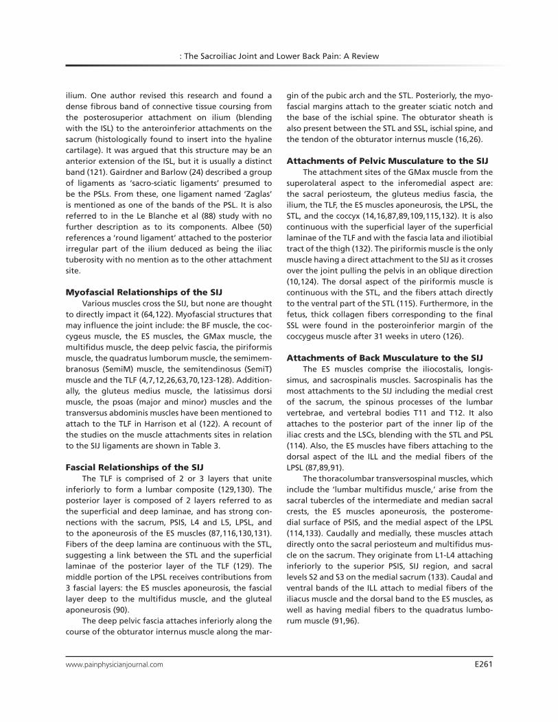
to directly impact it (64,122). Myofascial structures that may influence the joint include: the BF muscle, the coc-cygeus muscle, the ES muscles, the GMax muscle, the multifidus muscle, the deep pelvic fascia, the piriformis muscle, the quadratus lumborum muscle, the semimem-branosus (SemiM) muscle, the semitendinosus (SemiT) muscle and the TLF (4,7,12,26,63,70,123-128). Addition-ally, the gluteus medius muscle, the latissimus dorsi muscle, the psoas (major and minor) muscles and the transversus abdominis muscles have been mentioned to attach to the TLF in Harrison et al (122). A recount of the studies on the muscle attachments sites in relation to the SIJ ligaments are shown in Table 3.
Fascial Relationships of the SIJThe TLF is comprised of 2 or 3 layers that unite
inferiorly to form a lumbar composite (129,130). The posterior layer is composed of 2 layers referred to as the superficial and deep laminae, and has strong con-nections with the sacrum, PSIS, L4 and L5, LPSL, and to the aponeurosis of the ES muscles (87,116,130,131). Fibers of the deep lamina are continuous with the STL, suggesting a link between the STL and the superficial laminae of the posterior layer of the TLF (129). The middle portion of the LPSL receives contributions from 3 fascial layers: the ES muscles aponeurosis, the fascial layer deep to the multifidus muscle, and the gluteal aponeurosis (90).
The deep pelvic fascia attaches inferiorly along the course of the obturator internus muscle along the mar-
gin of the pubic arch and the STL. Posteriorly, the myo-fascial margins attach to the greater sciatic notch and the base of the ischial spine. The obturator sheath is also present between the STL and SSL, ischial spine, and the tendon of the obturator internus muscle (16,26).
Attachments of Pelvic Musculature to the SIJThe attachment sites of the GMax muscle from the
superolateral aspect to the inferomedial aspect are: the sacral periosteum, the gluteus medius fascia, the ilium, the TLF, the ES muscles aponeurosis, the LPSL, the STL, and the coccyx (14,16,87,89,109,115,132). It is also continuous with the superficial layer of the superficial laminae of the TLF and with the fascia lata and iliotibial tract of the thigh (132). The piriformis muscle is the only muscle having a direct attachment to the SIJ as it crosses over the joint pulling the pelvis in an oblique direction (10,124). The dorsal aspect of the piriformis muscle is continuous with the STL, and the fibers attach directly to the ventral part of the STL (115). Furthermore, in the fetus, thick collagen fibers corresponding to the final SSL were found in the posteroinferior margin of the coccygeus muscle after 31 weeks in utero (126).
Attachments of Back Musculature to the SIJThe ES muscles comprise the iliocostalis, longis-
simus, and sacrospinalis muscles. Sacrospinalis has the most attachments to the SIJ including the medial crest of the sacrum, the spinous processes of the lumbar vertebrae, and vertebral bodies T11 and T12. It also attaches to the posterior part of the inner lip of the iliac crests and the LSCs, blending with the STL and PSL (114). Also, the ES muscles have fibers attaching to the dorsal aspect of the ILL and the medial fibers of the LPSL (87,89,91).
The thoracolumbar transversospinal muscles, which include the ‘lumbar multifidus muscle,’ arise from the sacral tubercles of the intermediate and median sacral crests, the ES muscles aponeurosis, the posterome-dial surface of PSIS, and the medial aspect of the LPSL (114,133). Caudally and medially, these muscles attach directly onto the sacral periosteum and multifidus mus-cle on the sacrum. They originate from L1-L4 attaching inferiorly to the superior PSIS, SIJ region, and sacral levels S2 and S3 on the medial sacrum (133). Caudal and ventral bands of the ILL attach to medial fibers of the iliacus muscle and the dorsal band to the ES muscles, as well as having medial fibers to the quadratus lumbo-rum muscle (91,96).

Pain Physician: July/August 2019: 22:E247-E274
E262 www.painphysicianjournal.com
Attachments of Thigh Musculature to the SIJThe BF has muscular attachments to structures of
the SIJ, like the STL (108,113,115,133). The BF muscle attaches to the lower part of the STL superficial fibers, with some of its lateral deep fibers also connecting with it (108). Vleeming et al (115) also determined that in half of their cadavers, the BF tendon was connected to the STL, and in some this was completely fused with the ligament (60). Another study compared the hamstring attachments in patients with pain and those without pain. Results showed that the STL was continuous with BF muscle and SemiT muscle in all of their specimens
without pain and in 88% of the patients with pain. The STL, however, was not continuous with SemiM muscle in any of the patients (113). In addition, references re-counted in Woodley et al (109) describe the STL having attachments with the piriformis muscle, the obturator internus muscle, SemiM muscle, and SemiT muscle.
Vasculature of the SIJThe common iliac vessels and the internal iliac
vessels lie superior to the SIJ at the levels of L5-S2 an-teriorly. Posteriorly, the superior gluteal vessels lie an-terior to the SIJ at the levels of S2 to S3 (134). The SIJ is
Table 3. Muscle and fascial connections to the ligaments of the SIJ from the literature.

www.painphysicianjournal.com E263
: The Sacroiliac Joint and Lower Back Pain: A Review
supplied by the median sacral artery and the lateral sacral branches of the internal iliac artery. These arise from the posterior sacral iliac blood supply from the gluteal arteries (43,135). A nutrient artery provides blood to the anterior SIJ region originating from the iliolumbar artery (136). It is shown to arise variably from the common iliac artery or the internal iliac artery to course across the SIJ and enter the nutrient foramen of the ilium (43,137,138). Histologically, there is vascular tissue present in the posterior part at the junction between the articular joint space and where the ISL arises (54). Vascular branches originate from the inferior gluteal artery that travels close to the SSL and STL, penetrating the STL close to its origin on the sacrum (139). This occurred via one artery in 69% of specimens and 2 arteries in 8% (16). Lai et al (140) showed variation in both the origin and distribution of the blood supply to the STL. Branches from the inferior and superior gluteal arteries enter the ligament close to the ischial tuberosity and sacrum in a variety of patterns. These could be a com-bination of one to 4 branches of the inferior gluteal artery entering the STL with one to 2 branches from the superior gluteal artery (Fig. 24) (140). The subchondral bone plate of the SIJ on both the iliac and sacral sides is penetrated by multiple blood vessels that are in close to the articular cartilage. How-ever, the origin of these small blood vessels is not described (54,73,141).
The venous drainage is said to be analo-gous to the arterial anatomy, mainly from branches of the internal iliac veins (43). The venous drainage of the SIJ was stated to occur from tributary veins from the median and lateral sacral veins forming part of the sacral venous plexus anterior to the sacrum (135,142). The Batson ‘pre-sacral’ plexus or ‘vertebral venous plexus’ found within the vertebrae and sacrum is involved in the ve-nous drainage of the SIJ (143-145).
Innervation of the SIJThe anterior aspect of the SIJ is inner-
vated by segments L4-S2, sometimes L3, and the sacral plexus, and in one study, even the superior gluteal nerve (Table 4) (70,146-148).
Fig. 24. Dorsal view of the blood supply of the posterior sacroiliac joint and the sacrotuberous ligament (STL) based on Lai et al., (140). The gluteus maximus muscle (GMax) is reflected to expose the STL and piriformis muscles. (A) Example of pattern 1 with 4 penetrating branches from the inferior gluteal artery and one branch from the superior gluteal artery. (B) Example of pattern 1 with 2 perforating branches into the superior part of the STL and one branch from the inferior gluteal artery into the caudal part of the ligament. (C) Example of pattern 1 with 2 branches from the inferior gluteal artery and 2 branches from the superior gluteal artery penetrating the STL. (D) Example of pattern 2 with one branch perforating into the superior part of the STL from the superior gluteal artery. The superior gluteal artery in this case also crosses over the piriformis muscle providing a branch to the STL inferiorly. The inferior gluteal artery splits in 2 and perforates the caudal STL with 2 branches. S: superior, I: inferior, P: posterior, A: anterior

Pain Physician: July/August 2019: 22:E247-E274
E264 www.painphysicianjournal.com
The posterior aspect of the SIJ is innervated by a nerve plexus formed by the lateral branches of posterior rami of L5 to S4 (Table 5) (23,70,147-154). The posterior in-nervation was noted to be asymmetrical between the left and right sides of the body, and interindividual dif-ferences were found (23,146-156). Two groups stated the involvement of the sacral plexus in SIJ innervation, when other groups did not mention its contribution. Ikeda (148) stated that the sacral plexus is involved in SIJ innervation, but Grob et al (150) explicitly excluded its contribution. In more detail, the upper anterior portion of the SIJ is innervated by the ventral ramus of L5 and the lower portion by S2 or branches of sacral plexus (148). Two studies found contributions of L4 and L5 (146,156).
Fatty Infiltration of the SIJThe presence of fat in the SIJ is barely investi-
gated in the literature, and the reason for its presence within the SIJ is not clear to date. Nine anatomy articles
mention the presence of ‘areolar connective tissue’ within the posterior SIJ, which is presumed to be fat (54,55,86,89,91,92,96,152,157). The tissue is found between the meshes of the ISL or between the PSL (54,55,86). Fat is observed posteriorly where the lateral branches of the dorsal sacral rami can be identified between the ISL, the sacroiliac part of the ILL, and the middle of the LPSL (57,89,91,92). Histologically, loose connective tissue with vessels interrupting the ASL was found in the center of the SIJ, with fatty tissue poste-riorly within the ligaments (54). Also ‘loose connective tissue’ or ‘fat’ surrounding neurovascular structures was found travelling through the deep portion of the LPSL in the posterior part of the joint (141,152).
Magnetic resonance imaging (MRI) scans revealed fat metaplasia (an effect of inflammatory products on normal fat metabolism) producing an accumulation of fat within the subchondral bony areas (158). Fat infil-tration within the SIJ has been shown to be an inde-pendent predictor of ankyloses. The fat presence was
Table 5. Innervation of the posterior SIJ from the literature. Shaded sections are the contributing branches found in each study. Dark sections are the most common contributions, light gray sections are for the occasional contribution.
Table 4. Innervation of the anterior SIJ from the literature. Shaded sections are the contributing branches found in each study. Dark sections are the most common contributions, light gray sections are for the occasional contribution.

www.painphysicianjournal.com E265
: The Sacroiliac Joint and Lower Back Pain: A Review
higher in cases of ankylosing spondylitis (AS) than in patients presenting no pathology (159,160).
Muscle degeneration is a common feature associ-ated to nonspecific lower back pain. The infiltration of adipose tissue into skeletal muscle is indicative of muscle atrophy commonly associated with a consider-able loss in muscle strength and function (161,162). This can compromise joint stability and lead to further injury and lower back pain (163). Previous literature demon-strates that in certain lumbopelvic pain-associated con-ditions like hip osteoarthritis, lateral hip pain, and other nonspecific lower back conditions, there is a decrease in muscle volume accompanied by fatty infiltration within the muscles (161,163-166). Fatty infiltration has been assessed in the gluteal (167) and lumbar musculature (164,168) and may have a direct impact in SIJ-related biomechanics associated with muscle atrophy in this area.
Morphologic Variations of the SIJ and Accessory SIJs
Accessory SIJs (ASIJs) and morphologic varia-tions of the SIJ have been reported in the literature (22,29,59,157,169-179). ASIJs are additional articular facets within the syndesmotic SIJ, covered with hyaline or fibrocartilage in their own capsule and synovial membrane (Fig. 25) (22,72,157,169,170,175,177). They are generally observed in 8%-40% of specimens (63). A higher frequency of ASIJs has been reported in older specimens (29,63,157,169,174,179). ASIJs appear more commonly in men (25%) than women (21%) (174), and more frequently on the right side of the body (177). There was a higher frequency of ASIJs in obese patients and women with 3 or more childbirths (179). The major-ity of ASIJ cases revealed only one or 2 articular facets (29,169,171,172). Some found up to 3 ASIJs in 34% of investigated cadavers (22). The facets can range from 2 mm to 1 cm in diameter, and are located in the following areas: posterior to the articular surface (in region of the LSC at the level of the first or second sacral foramen), on the sacrum, on the medial surface of the PSIS, and on the iliac tuberosity (22,29,169-171,178). One study reports the presence of an ‘axial SIJ’ in 92% of cadavers located slightly caudal to the second sacral foramen with the concavity in the sacral side (157). They describe it as an ’extracapsular junction’ in both genders, and therefore, suggest that this should be included in the anatomic description of the SIJ. In their study, the ‘axial SIJ’ is a different type of ASIJ and it is more common (63,157).
The following morphologic variations were reported in patients without SIJ-related problems (29,172,179). The iliosacral complex (Fig. 25C): an iliac projection inserting cranioposteriorly into a sacral re-cess at the level of the first and second sacral foramen in the interosseous region or the inferior articular por-tion. It occurred more frequently in women, mostly bilaterally (172). Bipartite appearances (Fig. 25D) reflect dysmorphic posterior iliac changes, which are more frequent in women for one study (29), when this was found to be more common in men in another (179). These occurred in approximately 30% unilaterally with 70% bilaterally, respectively (172). However, this was less common in another study with a result of only 6% unilaterally (179). A crescent-like articular surface (Fig. 25E) describes a bulged sacral surface occurring in ap-proximately 4% of patients, bilaterally and unilaterally. It was also more common in women with no relation to age (29,179). Single semicircular defects (Fig. 25F) in the articular surface occurred in approximately 4% of individuals, mostly bilaterally (29,179). These are more common in women at the posterior superior aspect of the ISL, with all cases found only on the sacral side (172). Small ossification centers of sacral wings (Fig. 25G) ap-pearing as triangular osseous bodies in the superior posterior region were found in 3 individuals aged <30 years. These just occurred in approximately 1% of pa-tients (29,179). Isolated synostosis (Fig. 25H) were noted as unilateral, involving the middle third of the right SIJ at the level of the first sacral foramen occurring in <1% of specimens. Finally, dysmorphic changes were more common in women located below the second sacral foramen unilaterally and bilaterally occurring in 16% of patients (172).
discussion
This review covers all SIJ-related anatomic struc-tures including less investigated ones, such as bone density, vasculature, and fat presence. The accurate clarification of these properties potentially contributes to the understanding of the biomechanical behavior of the SIJ.
There is a general agreement regarding bony landmarks, joint type, myofascial attachments, vasculature, and innervation of the SIJ.
The standard terminology of the landmarks of the sacrum and ilium is in agreement. The majority of authors classify the joint as a diarthrosis. Knowing its

Pain Physician: July/August 2019: 22:E247-E274
E266 www.painphysicianjournal.com
Fig. 25. Examples of transverse sections of ilium and sacrum as can be found between S1 and S4 showing variation in joint morphology, these may occur in conjunction with one another. (A) ‘normal sacroiliac joint’ as seen in the majority of cases, (B) accessory sacroiliac joint, (C) iliosacral complex, (D) bipartite appearance/dysmorphic posterior iliac changes, (E) semi-circular defects, (F) crescent articular surface, (G) isolate synostosis (bilateral), (H) sacral wing ossification centers. Percentages represent the occurrence of these characteristic from the studies by Prassopoulos et al., (29), Demir et al., (179) and El Rafei et al., (172). A: anterior, P: posterior, L: left, R: right.

www.painphysicianjournal.com E267
: The Sacroiliac Joint and Lower Back Pain: A Review
specific composition is clinically significant to enable to predict the SIJ behavior with regard to pathology and biomechanics over time. The existing disagreements depend on what features are used for the assessment, such as structure, the amount of movement, or even the type of study (180). The term diarthro-amphiarthrosis is a better term, as it encompasses both its reduction of movement through time and its diarthrodial structural characteristics (66). Furthermore, the amphiarthrosis classification is used when movement reduction in the SIJ arises (61). A possible age-related movement reduc-tion can be a consequence of a natural degeneration that negatively impacts the SIJ mobility or the response of the SIJ to the decreased movement of the elderly, or a combination of the 2.
The SIJ is central within the pelvis and transmits forces from the axial skeleton through the legs and spine (8,181). Understanding the muscular relation-ships, which directly impact the ligamentous structures stabilizing the joint, can help elucidate pain problems that arise secondary to muscular/fascial forces. This is beneficial for physiotherapists who treat patients with SIJ pain to find adequate treatment exercises. Although there is general consensus on the attachment sites of most myofascial structures, further research on the piriformis muscle and a reassessment of the ES muscles would be beneficial to complete the understanding of the attachment sites of these structures, as there is limited published research on these.
The vasculature of the SIJ is crucial for surgeons who must know the relationships of the joint with neighboring structures as they could be damaged dur-ing an intervention (136). Further in-depth research on the smaller branches of the common iliac, internal iliac, sacral iliac, and gluteal vessels can be advantageous to improve this knowledge, especially with respect to its interindividual variation within the SIJ and its ligaments.
Innervation of the SIJ is neurologically relevant for the attribution of pain to certain spinal segments. Knowing specific branches is important clinically for di-agnosis, treatment, and even surgical intervention into the SIJ. The treatment of pain of SIJ in inflammatory diseases or SIJ dysfunction has had considerable atten-tion. Radiofrequency ablation of the nerves around the SIJ is one of the common options for the treatment of pain (151,153,154,182). The literature on this topic is in general agreement: that the anterior portion of the SIJ is innervated by branches from L4-S2 and the posterior portion by branches from L5-S4.
There is part consensus concerning the cartilage composition and the attachment sites of the ligaments of the SIJ.
The cartilaginous composition of the articular SIJ is not in complete agreement, although general con-sensus is that hyaline cartilage is present on the sacral side and fibrocartilage on the iliac side. Knowing the composition of the cartilage is important for the under-standing of how the joint may evolve with age or with certain common pathologies like osteoarthritis and AS, especially if certain diseases tend to involve a certain type of cartilage. This can help predict which area of the SIJ would be targeted and help in the prevention and subsequent treatment options for a condition.
Literature demonstrates that it is difficult to dif-ferentiate the layers of ligaments involved with the SIJ, especially in the posterior region. The anatomy of these ligaments reflects the difficulty in understanding the associated biomechanics of the posterior SIJ. Bio-mechanics are useful for understanding SIJ pain that may arise from the forces applied to the SIJ through these ligaments. Generally, literature describes the same attachment areas with only slight differences. Furthermore, there may be interindividual differences and pathological states that may influence the results of these studies. Therefore, it may be interesting to re-search the frequency of the interindividual variation in the ‘normal’ general population similar to the research by Hakim (57). This research may potentially help de-termine if a certain ligament pattern tends to be more commonly associated with SIJ pain than others.
The multiple names used for the classification of the SIJ ligaments creates the impression as if ‘additional ligaments’ exist, which may be underrepresented in the literature. For example, the ‘Zaglas’ ligament is only mentioned in 2 articles (24,88). This ligament could have been overseen, or it represents a specific variation of the PSL. The most likely scenario is that the ligament is a part of the PSL and was named differently by sub-sequent authors. Otherwise, the ‘sub capsular ligament’ has similar attachment sites as the inferior portion of the ASL and could have been determined as a separate entity (117). Similarly, the ‘round ligament’ could pos-sibly be a part of the ISL, although they differentiate the two (50). This could be owing to the dissimilarity of the course of fibers in the round ligament and the ISL. It is also possible that the ligament reflects ‘Illi’s liga-ment’ or the posterior part of the ILL. These additional ligaments should be investigated further to classify all SIJ ligaments with the same nomenclature.

Pain Physician: July/August 2019: 22:E247-E274
E268 www.painphysicianjournal.com
Further research is needed regarding the presence of fat in the SIJ
Although literature reports fat in their findings, there are no studies attempting to quantify fat in the SIJ, nor are there any studies looking into its function. Clinically and biomechanically, this could be useful information as perhaps fat within the joint is an age-related ‘normal’ characteristic, sign of degeneration, or pathology. If fat is a ‘normal’ characteristic, it could be functionally useful as a cushion for surrounding ‘hard structures’ such as bones and cartilages when forces are transmitted from the spine to the legs. Therefore, fatty tissue could fulfil a shock absorbing function, as seen in other locations of the human body like the heel pad (183). As stated for the functional fat pad of the heel, it may be directly related to weight (184). Furthermore, the presence of fat in the SIJ could be a developmental relict both embryologically and evolutionary.
Understanding the appearance of fat metaplasia and its appearance within the SIJ would allow clinicians to prepare for a condition to provide the adequate treatment as early as fat is detected. Some studies have shown that there is often fat metaplasia in the bony structures of the SIJ in cases with AS (158-160,185-188). It is an ongoing effect of inflammatory products on normal fat metabolism, producing an accumulation of fat within the subchondral bony areas (158,189). Maksymowych et al (160) determined that fat infiltra-tion within the SIJ is an independent predictor of anky-loses. This was supported by Guo et al (159), in which fat presence was higher in cases of AS than in patients presenting no pathological conditions. Fat metaplasia was tested on healthy patients to find a ‘baseline result’ for a comparison between patients with AS and those without. Results determined that fat metaplasia was very common and increased with age. This provides us with a range of fat metaplasia values for the general population (186).
Surrounding structures of the SIJ have been re-ported to contain fat, such as the ILL fibers after the fifth decade of life, making fat related to the natural degeneration of tissues (92). Similar findings were de-scribed by Hammer et al (96), in which adipose tissue was present within the space between the 2 bands of the ILL in tissues of cadavers aged >60 years. This could be a potential first step in determining whether fat is related to age and degeneration or whether it is a path-ological occurrence. Moreover, fatty infiltration within the muscles is commonly associated with a decrease in muscle strength and atrophy, which influences the joint
biomechanics and generates pain (161,162,164). Fat within skeletal muscle and ligaments may be the cause of ‘abnormal’ biomechanics of a structure because it replaces the healthy fibers that may impact its func-tion. This would suggest a potential ‘fatty infiltration’ of the ligamentous portion of the SIJ has the potential of generating pain because of its relation to functional impairment.
Further research is needed with regard to the specific bone density of the articulating sacral and iliac counterparts of the SIJ
Loading properties of the pelvis may be related to the distribution of bone density of the articular surfaces. This was previously reported in which a higher incidence of bone osteoporosis was seen in patients suffering from chronic lower back pain owing to their poor mobility, which effectively ‘demineralized’ the bone (181,190). Clinically, an abnormal increase in bone density is termed ‘sclerosis’ and is often associated to pathogenesis of some kind that is often seen as a developing change on computed tomography (CT) scans. Interestingly, a higher bone density is seen more commonly developing on the iliac side rather than the sacral side in cases of AS, and is termed ‘sclerosis’ by clinicians (81,84). This begs the question of whether this change in bone structure is related to the pathology or if it is a ‘normal’ or de-generative occurrence or a combination of these. Tuite (127) described the sclerosis pattern development of AS as more ill-defined than that of osteoarthritis, suggest-ing that depending on the condition, sclerosis develops in a specific manner and could be a diagnostic tool for conditions like SIJ dysfunction. To date, there is no range of bone density values of healthy subjects that could be characterized as a ‘normal’ bone density of the iliac and sacral articular counterparts, nor has the distribution of the bone density of the joint been assessed. Such base-line data would be valuable for the radiologist assessing the joint clinically. These values could then be compared with bone densities seen in pathological states of the joint. Conclusions could be drawn on whether a particu-lar bone density value is part of a ‘normal range’ or is suspicious for the subsequent development of defined pathologies. Similarly, determining the bone density ar-rangement within the joint may provide insight in the development of certain pathologies or changes in loads applied to the SIJ in relation to SIJ syndrome. Ideally, medical interventions could be started at an early state of the disease avoiding vast functional impairments of the SIJ.

www.painphysicianjournal.com E269
: The Sacroiliac Joint and Lower Back Pain: A Review
ASIJs and the anatomic variation of the joint are well-described morphologically; biomechanical influence of these variations should be looked at in depth
It has been demonstrated that morphologic varia-tions are likely to occur in the general population. Con-sidering the high percentage of people suffering from lower back pain during their lifetime, these morpho-logic differences may be responsible for back pain, and should be considered during diagnosis and treatment and could be investigated using imaging technology like MRI and CT. In addition, ASIJs, although well de-fined, are often not recognized as a ‘true articular part’ of the joint that may be problematic for identifying cer-tain pathologies and preventing these in the long run. Considering certain study results, ASIJs are the most common morphologic variation of the SIJ reported and should therefore also be considered as possible pain generating sites.
Limitations of the StudyThis review has researched the literature to date
on the anatomic properties of the SIJ using a set of
guidelines and inclusion/exclusion criteria. All attempts were made to encompass all the relevant literature to date, but some studies may have been missed as only certain databases were used for the initial search. Ad-ditionally, keyword combinations used for this review may not have been inclusive of all articles relevant to the SIJ. Also, only English, German, and French articles were included in this review.
conclusions
There was a part consensus on the ligament attach-ments and cartilage structure of the joint. Furthermore, although ASIJs and anatomic variation are well-de-scribed morphologically, the biomechanical influence of these variations should be investigated in more depth. Clinically relevant areas lacking research identi-fied in this review include the bone density distribution of the SIJ iliac and sacral articulating counterparts, as well as fat presence within the joint space. These are areas that should be investigated further to advance the understanding of the anatomy of the SIJ.
RefeRences1. Youssef P, Loukas M, Chapman JR, Osk-
ouian RJ, Tubbs RS. Comprehensive anatomical and immunohistochemical review of the innervation of the human spine and joints with application to an improved understanding of back pain. Childs Nerv Syst 2016; 32:243-251.
2. Cohen SP, Chen Y, Neufeld NJ. Sacroili-ac joint pain: A comprehensive review of epidemiology, diagnosis and treatment. Expert Rev Neurother 2013; 13:99-116.
3. Schwarzer AC, Aprill CN, Bogduk N. The sacroiliac joint in chronic low back pain. Spine (Phila Pa 1976) 1995; 20:31-37.
4. Calvillo O, Skaribas I, Turnipseed J. Anat-omy and pathophysiology of the sacro-iliac joint. Curr Rev Pain 2000; 4:356-361.
5. Fortin JD, Dwyer AP, West S, Pier J. Sac-roiliac joint: Pain referral maps upon applying a new injection/arthrogra-phy technique. Part 1: Asymptomatic volunteers. Spine (Phila Pa 1976) 1994; 19:1475-1482.
6. Paci M. Structure, physiopathology and rehabilitation of the sacroiliac joint. Eura
Medicophys 1999; 35:207-218.7. Forst SL, Wheeler MT, Fortin JD, Vilensky
JA. The sacroiliac joint: Anatomy, physiol-ogy and clinical significance. Pain Physi-cian 2006; 9:61-68.
8. Vleeming A, Schuenke MD, Masi AT, Carreiro JE, Danneels L, Willard FH. The sacroiliac joint: An overview of its anato-my, function and potential clinical impli-cations. J Anat 2012; 221:537-567.
9. Attias N, Arzani S, Duncan G, Taber KH, Hayman LA. Sectional imaging anatomy: Pelvic ring ligaments. J Comput Assist Tomo 2001; 25:975-979.
10. Alderink G. The sacroiliac joint: Review of anatomy, mechanics, and function. J Orthop Sports Phys Ther 1991; 13:71-84.
11. Doncker D, Thyes A, Van Gaver P. L’articulation sacro-iliaque. Acta Orthop Belg 1953; 19:7-41.
12. Robert R, Salaud C, Hamel O, Hamel A, Philippeau JM. Anatomie des douleurs de l’articulation sacro-iliaque. Rev Rhum 2009; 76:727-733.
13. Chou LH, Slipman CW, Bhagia SM, Tsaur L, Bhat AL, Isaac Z, Gilchrist R, El Abd OH, Lenrow DA. Inciting events ini-tiating injection-proven sacroiliac joint syndrome. Pain Med 2004; 5:26-32.
14. Barker PJ, Hapuarachchi KS, Ross JA, Sambaiew E, Ranger TA, Briggs CA. Anatomy and biomechanics of gluteus maximus and the thoracolumbar fascia at the sacroiliac joint. Clin Anat 2014; 27:234-240.
15. Philippeau JM, Hamel O, Pecot J, Robert R. Are sacrospinal and sacrotuberal liga-ments involved in sacro-iliac joint sta-bility? Anatomy and an original biome-canical experimental study. Morphologie 2008; 92:16-30.
16. Hammer N, Steinke H, Slowik V, Josten C, Stadler J, Bohme J, Spanel-Borowski K. The sacrotuberous and the sacrospi-nous ligament: A virtual reconstruction. Ann Anat 2009; 191:417-425.
17. Steinke H, Hammer N, Slowik V, Stadler J, Josten C, Bohme J, Spanel-Borowski

Pain Physician: July/August 2019: 22:E247-E274
E270 www.painphysicianjournal.com
K. Novel insights into the sacroiliac joint ligaments. Spine (Phila Pa 1976) 2010; 35:257-263.
18. Sichting F, Rossol J, Soisson O, Klima S, Milani T, Hammer N. Pelvic belt effects on sacroiliac joint ligaments: A compu-tational approach to understand thera-peutic effects of pelvic belts. Pain Physi-cian 2014; 17:43-51.
19. Buford WL, Moulton DL, Gugala Z, Lindsey RW. The sacroiliac spine: Com-puter simulation of motion and model-ing of the ligaments. Conf Proc IEEE Eng Med Biol Soc 2010; 2010:5117-5120.
20. Steinke H, Hammer N, Lingslebe U, Höch A, Klink T, Böhme J. Ligament-induced sacral fractures of the pelvis are possible. Clin Anat 2014; 27:770-777.
21. Weisl H. The articular surfaces of the sacro-iliac joint and their relation to the movements of the sacrum. Acta Anat (Basel) 1954; 22:1-14.
22. Schunke B, Bernard G. The anatomy and development of the sacroiliac joint in man. Anat Rec 1938; 72:313-331.
23. Bradley KC. The anatomy of backache. Aust N Z J Surg 1974; 44:227-232.
24. Gairdner W, Barlow J. Mechanism of the pelvic articulations. Month J Med Sci 1851; 21:289-292.
25. von Luschka H. Die kreuzdarmbeinfu-ge und die schambeinfuge des men-schen. Virchows Arch F Path Anat 1854; VII:299-316.
26. Cameron J. The fascia of the pelvis. J Anat Physiol 1907; 42:112-125.
27. Bellamy N, Park W, Rooney PJ. What do we know about the sacroiliac joint? Se-min Arthritis Rheum 1983; 12:282-313.
28. Jaovisidha S, Ryu KN, De Maeseneer M, Haghighi P, Goodwin D, Sartoris DJ, Resnick D. Ventral sacroiliac ligament: Anatomic and pathologic consider-ations. Invest Radiol 1996; 31:532-541.
29. Prassopoulos PK, Faflia CP, Volouda-ki AE, Gourtsoyiannis NC. Sacroiliac joints: Anatomical variants on CT. J Comput Assist Tomo 1999; 23:323-327.
30. Brooke R. The sacro-iliac joint. J Anat 1923; 58:299-305.
31. Postacchini R, Trasimeni G, Ripani F, Sessa P, Perotti S, Postacchini F. Mor-phometric anatomical and CT study of the human adult sacroiliac region. Surg Radiol Anat 2017; 39:85-94.
32. Macdonald GR, Hunt TE. Sacroiliac joints; observations on the gross and histological changes in the various age groups. Can Med Assoc J 1952; 66:157-163.
33. Rana SH, Farjoodi P, Haloman S, Dut-
ton P, Hariri A, Ward SR, Garfin SR, Chang DG. Anatomic avaluation of the sacroiliac joint: A radiographic study with implications for procedures. Pain Physician 2015; 18:583-592.
34. Waldrop JT, Ebraheim NA, Yeasting RA, Jackson WT. The location of the sac-roiliac joint on the outer table of the posterior ilium. J Orthop Trauma 1993; 7:510-513.
35. Hatfield KD. The preauricular sulcus. Australas Radiol 1971; 15:168-169.
36. Gulekon IN, Turgut HB. The preau-ricular sulcus: Its radiologic evidence and prevalence. Kaibogaku Zasshi 2001; 76:533-535.
37. Dee PM. The preauricular sulcus. Radi-ology 1981; 140:354.
38. Rosatelli AL, Agur AM, Chhaya S. Anat-omy of the interosseous region of the sacroiliac joint. J Orthop Sports Phys Ther 2006; 36:200-208.
39. Wilder DG, Pope MH, Frymoyer JW. The functional topography of the sac-roiliac joint. Spine (Phila Pa 1976) 1980; 5:575-579.
40. Beal MC. The sacroiliac problem: Review of anatomy, mechanics, and diagnosis. J Am Osteopath Assoc 1982; 81:667-679.
41. Otter R. A review study of the differ-ing opinions expressed in the litera-ture about the anatomy of the sacroiliac joint. Eur J Chiropr 1985; 33:221-242.
42. Gerlach U, Lierse W. Functional con-struction of the sacroiliac ligamen-tous apparatus. Acta Anat (Basel) 1992; 144:97-102.
43. Esses SI, Botsford DJ, Huler RJ, Raus-chning W. Surgical anatomy of the sa-crum. A guide for rational screw fixation. Spine (Phila Pa 1976) 1991; 16:S283-288.
44. Ellis H. Anatomy for anaesthetists. 5. The lumbar spine and sacrum. Anaes-thesia 1962; 17:238-246.
45. Cheng JS, Song JK. Anatomy of the sa-crum. Neurosurg Focus 2003; 15:1-4.
46. Xu R, Ebraheim NA, Gove NK. Surgi-cal anatomy of the sacrum. Am J Orthop (Belle Mead NJ) 2008; 37:E177-E181.
47. Whelan MA, Gold RP. Computed to-mography of the sacrum: 1. Normal anatomy. AJR Am J Roentgenol 1982; 139:1183-1190.
48. Cyteval C, Sarrabère-Baron MP, De-coux E, Larroque G. Sacral bone-coc-cyx sacroiliac joint. Normal aspect and x-ray technique. EMC-Radiologie 2005; 2:87-102.
49. Deveneau NE, Greenstein M, Maha-lingashetty A, Herring NR, Lipetskaia
L, Azadi A, Ostergard DR, Francis SL. Surface and boney landmarks for sacral neuromodulation: A cadaveric study. Int Urogynecol J 2015; 26:263-268.
50. Albee FH. A study of the anatomy and the clinical importance of the sacroiliac joint. JAMA 1909; LIII:1273-1276.
51. Bowen V, Cassidy JD. Macroscopic and microscopic anatomy of the sacro-iliac joint from embryonic life until the eighth decade. Spine (Phila Pa 1976) 1981; 6:620-628.
52. Delmas A. The sacro-iliac joint and the statics of the body. Rev Rhum 1950; 17:475-481.
53. Dieulafé L, Saint-Martin M. Le type ar-ticulaire sacro-iliaque. C R Assoc Anat 1912; 14:95-109.
54. Puhakka KB, Melsen F, Jurik AG, Boel LW, Vesterby A, Egund N. MR imaging of the normal sacroiliac joint with corre-lation to histology. Skeletal Radiol 2004; 33:15-28.
55. Sashin D. A critical analysis or the anat-omy and the pathologic changes of the sacro iliac joints. J Bone Joint Surg 1930; 12:891-910.
56. Vleeming A, Stoeckart R, Volkers A, Snijders C. Relation between form and function in the sacroiliac joint. Part 1: Clinical anatomical aspects. Spine (Phila Pa 1976) 1990; 15:130-132.
57. Hakim M. Recherches sur l’articulation sacro-iliaque chez l’homme et les an-thropoïdes. In: Faculté de médecine de Paris, PhD thesis, Université de Paris, 1937:1-96.
{58. Cassidy JD. The pathoanatomy and clini-cal significance of the sacroiliac joints. J Manipulative Physiol Ther 1992; 15:41-42.
59. Dietrichs E. Anatomy of the pelvic joints: A review. Scand J Rheumatol Suppl 1991; 88:4-6.
60. Vora A, Doerr K, Wolfer L. Functional anatomy and pathophysiology of axial low back pain: Disc, posterior elements, sacroiliac joint, and associated pain generators. Phys Med Rehabil Clin N Am 2010; 21:679-709.
61. Christ B, Günther J, Frölich E, Huang R, Flöel H. Morphological basis of Sell’s irritation point of the sacroiliac joint. Manuelle Medizin 2001; 39:241-245.
62. Okumura M, Ishikawa A, Aoyama T, Ya-mada S, Uwabe C, Imai H, Matsuda T, Yoneyama A, Takeda T, Takakuwa T. Car-tilage formation in the pelvic skeleton during the embryonic and early-fetal period. PLoS One 2017; 12:e0173852.
63. Walker J. The sacroiliac joint: A critical

: The Sacroiliac Joint and Lower Back Pain: A Review
www.painphysicianjournal.com E271
review. Phys Ther 1992; 72:903-916.64. Grieve GP. The sacro iliac joint. Physio-
therapy 1976; 62:384-400.65. Resnick D, Niwayama G, Goergen TG.
Degenerative disease of the sacroiliac joint. Invest Radiol 1975; 10:608-621.
66. Paquin JD, Vanderrest M, Marie PJ, Mort JS, Pidoux I, Poole AR, Roughley PJ. Biochemical and morphologic stud-ies of cartilage from the adult human sacroiliac joint. Arthritis Rheum 1983; 26:887-895.
67. Benneman R. Untersuchungen am ilio-sacralgelenk des menschen. Verh Anat Ges 1979; 73:187-190.
68. Brower A. Disorders of the sacroiliac joint. Surg Rounds Orthop 1989; 13:47-54.
69. Salsabili N, Valojerdy MR, Hogg DA. Variations in thickness of articular car-tilage in the human sacroiliac joint. Clin Anat 1995; 8:388-390.
70. Cohen SP. Sacroiliac joint pain: A com-prehensive review of anatomy, diagno-sis and treatment. Anesth Analg 2005; 101:1440-1453.
71. Dijkstra PF, Vleeming A, Stoeckart R. Complex motion tomography of the sacroiliac joint: An anatomical and roentgenological study. Fortschr Rontg Neuen 1989; 150:635-642.
72. Cole AJ, Dreyfuss P, Stratton SA. The sacroiliac joint: A functional approach. Crit Rev Phys Rehabil Med 1996; 8:125-152.
73. Kampen WU, Tillmann B. Age-related changes in the articular cartilage of hu-man sacroiliac joint. Anat Embryol (Berl) 1998; 198:505-513.
74. Sgambati E, Stecco A, Capaccioli L, Brizzi E. Morphometric analysis of the sacroiliac joint. Ital J Anat Embryol 1997; 102:33-38.
75. Kalenderer O, Turgut A, Bacaksiz T, Bil-gin E, Kumbaraci M, Akkan HA. Evalu-ation of symphysis pubis and sacroiliac joint distances in skeletally immature patients: A computerized tomography study of 1020 individuals. Acta Orthop Traumatol Turc 2017; 51:150-154.
76. Nishi K, Saiki K, Imamura T, Okamoto K, Wakebe T, Ogami K, Hasegawa T, Mori-uchi T, Sakamoto J, Manabe Y, Tsurumo-to T. Degenerative changes of the sac-roiliac auricular joint surface-validation of influential factors. Anat Sci Int 2017; 92:530-538.
77. Nishi K, Tsurumoto T, Okamoto K, Oga-mi-Takamura K, Hasegawa T, Moriuchi T, Sakamoto J, Oyamada J, Higashi T, Manabe T, Saiki K. Three-dimensional morphological analysis of the human
sacroiliac joint: Influences on the de-generative changes of the auricular sur-faces. J Anat 2018; 232:238-249.
78. Valojerdy MR, Hogg DA. Sex differences in the morphology of the auricular sur-faces of the human sacroiliac joint. Clin Anat 1989; 2:63-67.
79. Dalstra M, Huiskes R. Load transfer across the pelvic bone. J Biomech 1995; 28:715-724.
80. Putz R, Müller-Gerbl M. Anatomische besonderheiten des beckenrings. Un-fallchirurg 1992; 95:164-167.
81. Vogler JB 3rd, Brown WH, Helms CA, Genant HK. The normal sacroiliac joint: A CT study of asymptomatic patients. Radiology 1984; 151:433-437.
82. Peretz AM, Hipp JA, Heggeness MH. The internal bony architecture of the sacrum. Spine (Phila Pa 1976) 1998; 23:971-974.
83. Ebraheim N, Sabry FF, Nadim Y, Xu R, Yeasting RA. Internal architecture of the sacrum in the elderly. An anatomic and radiographic study. Spine (Phila Pa 1976) 2000; 25:292-297.
84. McLauchlan GJ, Gardner DL. Sacral and iliac articular cartilage thickness and cellularity: Relationship to subchondral bone end-plate thickness and cancel-lous bone density. Rheumatology (Ox-ford) 2002; 41:375-380.
85. Cuppett M, Paladino J. The anatomy and pathomechanics of the sacroiliac joint. Athl Ther Today 2001; 6:6-14.
86. Weisl H. The ligaments of the sacroiliac joint examined with particular reference to their function. Acta Anat (Basel) 1954; 20:201-213.
87. Vleeming A, Pool-Goudzwaard A, Ham-mudoghlu D, Stoeckart R, Snijders C, Mens J. The function of the long dor-sal sacroiliac ligament: Its implication for understanding low back pain. Spine (Phila Pa 1976) 1996; 21:556-562.
88. Le Blanche AF, Mabi C, Bigot JM, Rous-seau J, Trêves R, Outrequin G, Dupuy JP, Caix M. The sacroiliac joint: Ana-tomical study in the coronal plane and MR correlation. Surg Radiol Anat 1996; 18:215-220.
89. McGrath MC, Nicholson H, Hurst P. The long posterior sacroiliac ligament: A histological study of morphological re-lations in the posterior sacroiliac region. Joint Bone Spine 2009; 76:57-62.
90. Moore AE, Jeffery R, Gray A, Stringer MD. An anatomical ultrasound study of the long posterior sacro-iliac ligament. Clin Anat 2010; 23:971-977.
91. Pool-Goudzwaard AL, Kleinrensink GJ, Snijders CJ, Entius C, Stoeckart R. The sacroiliac part of the iliolumbar liga-ment. J Anat 2001; 199:457-463.
92. Luk KDK, Ho HC, Leong JCY. The ilio-lumbar ligament: A study of its anatomy, development and clinical-significance. J Bone Joint Surg Br 1986; 68:197-200.
93. Hanson P, Sonesson B. The anatomy of the iliolumbar ligament. Arch Phys Med Rehabil 1994; 75:1245-1246.
94. Basadonna PT, Gasparini D, Rucco V. Iliolumbar ligament insertions. In vivo anatomic study. Spine (Phila Pa 1976) 1996; 21:2313-2316.
95. Rucco V, Basadonna PT, Gasparini D. Anatomy of the iliolumbar ligament: A review of its anatomy and a magnetic resonance study. Am J Phys Med Rehabil 1996; 75:451-455.
96. Hammer N, Steinke H, Bohme J, Stadler J, Josten C, Spanel-Borowski K. Descrip-tion of the iliolumbar ligament for com-puter-assisted reconstruction. Ann Anat 2010; 192:162-167.
97. Uhthoff HK. Prenatal development of the iliolumbar ligament. J Bone Joint Surg Br 1993; 75:93-95.
98. Hartford J, McCullen M, Harris R, Brown C. The iliolumbar ligament: Three-dimensional volume imaging and computer reformatting by magnetic resonance: A technical note. Spine (Phila Pa 1976) 2000; 25:1098-1103.
99. Zoccali C, Skoch J, Patel AS, Walter CM, Avila MJ, Martirosyan NL, Demitri S, Baaj AA. The surgical anatomy of the lumbosacroiliac triangle: A cadaveric study. World Neurosurg 2016; 88:36-40.
100. Fujiwara A, Tamai K, Yoshida H, Kuri-hashi A, Saotome K, An HS, Lim TH. Anatomy of the iliolumbar ligament. Clin Orthop Relat R 2000; 380:167-172.
101. Pool-Goudzwaard A, Hoek van Dijke G, Mulder P, Spoor C, Snijders C, Stoeckart R. The iliolumbar ligament: Its influence on stability of the sacroiliac joint. Clin Biomech (Bristol, Avon) 2003; 18:99-105.
102. Leong JCY, Luk KDK, Chow DHK, Woo CW. The biomechanical functions of the iliolumbar ligament in maintaining sta-bility of the lumbosacral junction. Spine (Phila Pa 1976) 1987; 12:669-674.
103. Briggs C, Chandraraj S. Variations in the lumbosacral ligament and associated changes in the lumbosacral region re-sulting in compression of the fifth dor-sal root ganglion and spinal nerve. Clin Anat 1995; 8:339-346.
104. Nathan H, Weizenbluth M, Halperin

Pain Physician: July/August 2019: 22:E247-E274
E272 www.painphysicianjournal.com
N. The lumbosacral ligament (LSL), with special emphasis on the “lumbo-sacral tunnel” and the entrapment of the 5th lumbar nerve. Int Orthop 1982; 6:197-202.
105. Hanson P, Sorensen H. The lumbosacral ligament: An autopsy study of young black and white people. Cells Tissues Or-gans 2000; 166:373-377.
106. Olsewski JM, Simmons EH, Kallen FC, Mandel FC. Evidence from cadavers suggestive of entrapment of the fifth lumbar spinal nerve by lumbosacral ligaments. Spine (Phila Pa 1976) 1991; 16:336-347.
107. Hanson P, Magnusson SP, Sorensen H, Simonsen EB. Differences in the il-iolumbar ligament and the transverse process of the L5 vertebra in young white and black people. Acta Anat (Basel) 1998; 163:218-223.
108. van Wingerden JP, Vleeming A, Snijders CJ, Stoeckart R. A functional-anatomical approach to the spine-pelvis mecha-nism: Interaction between the biceps femoris muscle and the sacrotuberous ligament. Eur Spine J 1993; 2:140-144.
109. Woodley S, Kennedy E, Mercer S. Anat-omy in practice: The sacrotuberous liga-ment. NZ Journal of Physiotherapy 2005; 33:91-94.
110. Varga E, Dudas B, Tile M. Putative pro-prioceptive function of the pelvic liga-ments: Biomechanical and histological studies. Injury 2008; 39:858-864.
111. Florian-Rodriguez ME, Hare A, Chin K, Phelan JN, Ripperda CM, Corton MM. Inferior gluteal and other nerves associ-ated with sacrospinous ligament: A ca-daver study. Am J Obstet Gynecol 2016; 215:646.e1-646.e6.
112. Loukas M, Louis RG Jr, Hallner B, Gup-ta AA, White D. Anatomical and surgi-cal considerations of the sacrotuberous ligament and its relevance in pudendal nerve entrapment syndrome. Surg Ra-diol Anat 2006; 28:163-169.
113. Bierry G, Simeone FJ, Borg-Stein JP, Clavert P, Palmer WE. Sacrotuberous ligament: Relationship to normal, torn, and retracted hamstring tendons on MR images. Radiology 2014; 271:162-171.
114. Bogduk N. A reappraisal of the anatomy of the human lumbar erector spinae. J Anat 1980; 131:525-540.
115. Vleeming A, Stoekart R, Snijders C. The sacrotuberous ligament: A conceptual approach to its dynamic role in stabi-lizing the sacroiliac joint. Clin Biomech (Bristol, Avon) 1989; 4:201-203.
116. Vleeming A, Pool-Goudzwaard AL, Stoeckart R, Van Wingerden JP, Snij-ders CJ. The posterior layer of the tho-racolumbar fascia: Its function in load-transfer from spine to legs. Spine (Phila Pa 1976) 1995; 20:753-758.
117. Bonjean P, Dejussieu U, Bisch A. Un ligament non encore decrit de l’artli-culation sacro-iliaque, le ligament po-laire infero-anterieur. Journal de Medi-cine de Bordeaux et du Sud-Ouest 1955; 132:597-599.
118. Illi F. The mechanism of the vertebral column and the pelvis. In: The Vertebral Column, Life-Line of the Body. Chicago, Illinois, National College of Chiroprac-tic, 1951: pp. 12-16.
119. Heine KH. Anatomie und gelenkme-chanick der Iliosakralregion. Hippokrates 1957; 28:110-114.
120. McGregor M, Cassidy JD. Post-surgical sacroiliac joint syndrome. J Manipulative Physiol Ther 1983; 6:1-11.
121. Freeman M, Fox D, Richards T. The su-perior intracapsular ligament of the sa-croiliac joint: Presumptive evidence for confirmation of Illi’s ligament. J Manipu-lative Physiol Ther 1990; 13:384-390.
122. Harrison D, Harrison D, Troyanovich S. The sacroiliac joint: A review of anatomy and biomechanics with clinical implica-tions. J Manipulative Physiol Ther 1997; 20:607-617.
123. Porterfield J, DeRosa C. The sacroiliac joint. In: Gould JA (ed). Orthopaedic and Sports Physical Therapy. St. Louis, CV Mosby Company, 1990: pp. 553-573.
124. Oldreive WL. A critical review of the literature on the anatomy and biome-chanics of the sacroiliac joint. J Man Ma-nip Ther 1996; 4:157-165.
125. Vleeming A, Snijders C, Stoeckart R, Mens J. A new light on low back pain. In: Vleeming A, Mooney V, Snijders C, Dorman T (eds). Interdisciplinary World Congress on Low Back Pain the Integrated Function of the Lumbar Spine and Sacro-iliac Joints. San Diego, University of Cali-fornia, 1995: pp. 9-11.
126. Hayashi S, Kim JH, Rodriguez-Vazquez JF, Murakami G, Fukuzawa Y, Asamoto K, Nakano T. Influence of developing ligaments on the muscles in contact with them: A study of the annular liga-ment of the radius and the sacrospinous ligament in mid-term human fetuses. Anat Cell Biol 2013; 46:149-156.
127. Tuite MJ. Sacroiliac joint imaging. Semin Musculoskel R 2008; 12:72-82.
128. Mcgill SM. A biomechanical perspective
of sacro-iliac pain. Clin Biomech (Bristol, Avon) 1987; 2:145-151.
129. Willard FH, Vleeming A, Schuenke MD, Danneels L, Schleip R. The tho-racolumbar fascia: Anatomy, function and clinical considerations. J Anat 2012; 221:507-536.
130. Bogduk N, Macintosh JE. The applied anatomy of the thoracolumbar fascia. Spine (Phila Pa 1976) 1984; 9:164-170.
131. Barker PJ, Briggs CA. Attachments of the posterior layer of lumbar fascia. Spine (Phila Pa 1976) 1999; 24:1757-1764.
132. Stecco A, Gilliar W, Hill R, Fullerton B, Stecco C. The anatomical and function-al relation between gluteus maximus and fascia lata. J Bodyw Mov Ther 2013; 17:512-517.
133. Cornwall J, Stringer MD, Duxson M. Functional morphology of the thoraco-lumbar transversospinal muscles. Spine (Phila Pa 1976) 2011; 36:E1053-E1061.
134. Zoccali C, Skoch J, Patel A, Walter CM, Maykowski P, Baaj AA. The surgical neu-rovascular anatomy relating to partial and complete sacral and sacroiliac re-sections: A cadaveric, anatomic study. Eur Spine J 2015; 24:1109-1113.
135. Bernard TN, Cassidy JD. The sacroiliac joint syndrome: Pathophysiology, diag-nosis and management. In: Frymoyer JW (ed). The Adult Spine: Principles and Practice. New York, Raven Press, 1991: pp. 2107-2130.
136. Alla SR, Roberts CS, Ojike NI. Vascular risk reduction during anterior surgical approach sacroiliac joint plating. Injury 2013; 44:175-177.
137. Rusu MC, Cergan R, Dermengiu D, Curca GC, Folescu R, Motoc AGM, Jianu AM. The iliolumbar artery—anatomic considerations and details on the com-mon iliac artery trifurcation. Clin Anat 2010; 23:93-100.
138. Ebraheim NA, Lu J, Biyani A, Yang H. Anatomic considerations of the princi-pal nutrient foramen and artery on in-ternal surface of the ilium. Surg Radiol Anat 1997; 19:237-239.
139. Thompson JR, Gibb JS, Genadry R, Bur-rows L, Lambrou N, Buller JL. Anatomy of pelvic arteries adjacent to the sacro-spinous ligament: Importance of the coccygeal branch of the inferior gluteal artery. Obstet Gynecol 1999; 94:973-977.
140. Lai J, du Plessis M, Wooten C, Gielecki J, Tubbs RS, Oskouian RJ, Loukas M. The blood supply to the sacrotuberous liga-ment. Surg Radiol Anat 2017; 39:953-959.
141. Egund N, Jurik AG. Anatomy and histol-

: The Sacroiliac Joint and Lower Back Pain: A Review
www.painphysicianjournal.com E273
ogy of the sacroiliac joints. Semin Mus-culoskel R 2014; 18:332-339.
142. Zeit RM, Cope C. Anatomy of the sacral venous plexus. AJR Am J Roentgenol 1983; 140:143-144.
143. Groen RJ, Groenewegen HJ, van Alphen HA, Hoogland PV. Morphology of the human internal vertebral venous plex-us: A cadaver study after intravenous Araldite CY 221 injection. Anat Rec 1997; 249:285-294.
144. Nathoo N, Caris EC, Wiener JA, Mendel E. History of the vertebral venous plex-us and the significant contributions of Breschet and Batson. Neurosurgery 2011; 69:1007-1014.
145. Gonzales GR, Payne R. Analgesia using vertebral venous plexus (Batson’s plex-us) shunting of rectally administered opioids and anesthetics to the central nervous system. J Pain Symptom Manage 1992; 7:189-190.
146. Cox M, Ng G, Mashriqi F, Iwanaga J, Alonso F, Tubbs K, Loukas M, Oskouian RJ, Tubbs RS. Innervation of the anterior sacroiliac joint. World Neurosurg 2017; 107:750-752.
147. Solonen KA. The sacroiliac joint in the light of anatomical, roentgenological and clinical studies. Acta Orthop Scand Suppl 1957; 27:1-127.
148. Ikeda R. Innervation of the sacroiliac joint. Macroscopical and histological studies. Nihon Ika Daigaku Zasshi 1991; 58:587-596.
149. Horwitz M. The anatomy of (A) the lum-bosacral nerve plexus—its relation to variations of vertebral segmentation, and (B), the posterior sacral nerve plex-us. Anat Rec 1939; 74:91-107.
150. Grob K, Neuhuber W, Kissling R. Inner-vation of the human sacroiliacal joint. Z Rheumatol 1995; 54:117-122.
151. Yin W, Willard F, Carreiro J, Dreyfuss P. Sensory stimulation-guided sacroiliac joint radiofrequency neurotomy: Tech-nique based on neuroanatomy of the dorsal sacral plexus. Spine (Phila Pa 1976) 2003; 28:2419-2425.
152. McGrath MC, Zhang M. Lateral branch-es of dorsal sacral nerve plexus and the long posterior sacroiliac ligament. Surg Radiol Anat 2005; 27:327-330.
153. Cox RC, Fortin JD. The anatomy of the lateral branches of the sacral dorsal rami: Implications for radiofrequency ablation. Pain Physician 2014; 17:459-464.
154. Roberts S, Burnham R, Ravichandiran K, Agur A, Loh E. Cadaveric study of sac-
roiliac joint innervation: Implications for diagnostic blocks and radiofrequen-cy ablation. Reg Anesth Pain Med 2014; 39:456-464.
155. Fortin JD, Kissling RO, O’Connor BL, Vilensky JA. Sacroiliac joint innervation and pain. Am J Orthop (Belle Mead NJ) 1999; 28:687-690.
156. Szadek KM, Hoogland PV, Zuurmond WW, de Lange JJ, Perez RS. Nocicep-tive nerve fibers in the sacroiliac joint in humans. Reg Anesth Pain Med 2008; 33:36-43.
157. Bakland O, Hansen JH. The “axial sacro-iliac joint”. Anat Clin 1984; 6:29-36.
158. Bredella MA, Steinbach LS, Morgan S, Ward M, Davis JC. MRI of the sacroiliac joints in patients with moderate to se-vere ankylosing spondylitis. Am J Roent-genol 2006; 187:1420-1426.
159. Guo RM, Lin WS, Liu WM, Zhou WY, Cao SE, Wang J, Li QL. Quantification of fat infiltration in the sacroiliac joints with ankylosing spondylitis using IDEAL sequence. Clin Radiol 2017; 73:231-236.
160. Maksymowych WP, Wichuk S, Chiow-chanwisawakit P, Lambert RG, Pedersen SJ. Fat metaplasia and backfill are key intermediaries in the development of sacroiliac joint ankylosis in patients with ankylosing spondylitis. Arthritis Rheu-matol 2014; 66:2958-2967.
161. Zacharias A, Pizzari T, English DJ, Kapa-koulakis T, Green RA. Hip abductor muscle volume in hip osteoarthritis and matched controls. Osteoarthritis Carti-lage 2016; 24:1727-1735.
162. Goutallier D, Postel JM, Bernageau J, La-vau L, Voisin MC. Fatty muscle degen-eration in cuff ruptures. Pre- and post-operative evaluation by CT scan. Clin Orthop Relat Res 1994; 304:78-83.
163. D’Hooge R, Cagnie B, Crombez G, Vanderstraeten G, Dolphens M, Dan-neels L. Increased intramuscular fatty infiltration without differences in lum-bar muscle cross-sectional area during remission of unilateral recurrent low back pain. Man Ther 2012; 17:584-588.
164. Battaglia PJ, Maeda Y, Welk A, Hough B, Kettner N. Reliability of the Goutal-lier classification in quantifying muscle fatty degeneration in the lumbar mul-tifidus using magnetic resonance im-aging. J Manipulative Physiol Ther 2014; 37:190-197.
165. Grimaldi A, Richardson C, Durbridge G, Donnelly W, Darnell R, Hides J. The association between degenerative hip joint pathology and size of the gluteus
maximus and tensor fascia lata muscles. Man Ther 2009; 14:611-617.
166. Danneels LA, Vanderstraeten GG, Cam-bier DC, Witvrouw EE, De Cuyper HJ. CT imaging of trunk muscles in chronic low back pain patients and healthy con-trol subjects. Eur Spine J 2000; 9:266-272.
167. Engelken F, Wassilew GI, Kohlitz T, Brockhaus S, Hamm B, Perka C, Died-erichs uG. Assessment of fatty degen-eration of the gluteal muscles in pa-tients with THA using MRI: Reliability and accuracy of the Goutallier and quar-tile classification systems. J Arthroplasty 2014; 29:149-153.
168. Niemelainen R, Briand MM, Battie MC. Substantial asymmetry in paraspinal muscle cross-sectional area in healthy adults questions its value as a marker of low back pain and pathology. Spine (Phila Pa 1976) 2011; 36:2152-2157.
169. Trotter M. A common anatomical varia-tion in the sacro-iliac region. J Bone Joint Surg 1940; 22:293-299.
170. Ehara S, Elkhoury GY, Bergman RA. The accessory sacroiliac joint: A common anatomic variant. Am J Roentgenol 1988; 150:857-859.
171. Valojerdy MR, Hogg DA. Anatomical note: The occurrence of accessory sac-roiliac joints in man. Clin Anat 1990; 3:257-260.
172. El Rafei M, Badr S, Lefebvre G, Machu-ron F, Capon B, Flipo R, Cotte A. Sacro-iliac joints: Anatomical variations on MR images. Eur Radiol 2018; 28:5328.
173. Miller AN, Routt ML Jr. Variations in sacral morphology and implications for iliosacral screw fixation. J Am Acad Or-thop Surg 2012; 20:8-16.
174. Seligman S. Articulatio sacro-iliaca ac-cessoria. Anat Anz Bd 1935; 79:225-241.
175. Jazuta K. Die nehengelenkflächen am kreuz und hüftbein. Anat Anz Bd 1929; 68:137-144.
176. Schwegel A. Knochenvarietäten. Ztschr F Rat Med 1859; 5:283-330.
177. Petersen O. Über artikulationsflächen an der unterfläche des os sacrum. Anat Anz 1905; 26:521-524.
178. Douglas E, Derry M. Note on accesso-ry articular facets between the sacrum and ileum and their significance. J Anat Physiol 1911; 45:202-210.
179. Demir M, Mavi A, Gumusburun E, Bayram M, Gursoy S, Nishio H. Ana-tomical variations with joint space mea-surements on CT. Kobe J Med Sci 2007; 53:209-217.

Pain Physician: July/August 2019: 22:E247-E274
E274 www.painphysicianjournal.com
180. Cichoke A. Anatomy and function of the sacroiliac articlulations: A review of the literature. Chiropractic 1989; 2:65-70.
181. Foley B, Buschbacher R. Sacroiliac joint pain: Anatomy, biomechanics, diagno-sis, and treatment. Am J Phys Med Reha-bil 2006; 85:997-1006.
182. Aeschbach A, Mekhail NA. Common nerve blocks in chronic pain manage-ment. Anesthesiol Clin North Am 2000; 18:429-459.
183. Cichowitz A, Pan WR, Ashton M. The heel: Anatomy, blood supply, and the pathophysiology of pressure ulcers. Ann Plast Surg 2009; 62:423-429.
184. Riddiford-Harland DL, Steele JR, Baur LA. Medial midfoot fat pad thickness and plantar pressures: Are these relat-ed in children? Int J Pediatr Obes 2011; 6:261-266.
185. Weber U, Pedersen SJ, Zubler V, Rufi-bach K, Chan SM, Lambert RGW, Os-tergaard M, Maksymowych WP. Fat in-
filtration on magnetic resonance imag-ing of the sacroiliac joints has limited diagnostic utility in nonradiographic ax-ial spondyloarthritis. J Rheumatol 2014; 41:75-83.
186. Ziegeler K, Eshkal H, Schorr C, Sieper J, Diekhoff T, Makowski MR, Hamm B, Hermann KGA. Age- and sex-depen-dent frequency of fat metaplasia and other structural changes of the sac-roiliac joints in patients without axial spondyloarthritis: A retrospective, cross-sectional MRI study. J Rheumatol 2018; 45:915-921.
187. Krohn M, Braum LS, Sieper J, Song IH, Weiß A, Callhoff J, Althoff CE, Hamm B, Hermann KGA. Erosions and fatty lesions of sacroiliac joints in patients with axial spondyloarthritis: Evaluation of different MRI techniques and two scoring methods. J Rheumatol 2014; 41:473-480.
188. Sawicki LM, Lutje S, Baraliakos X, Braun
J, Kirchner J, Boos J, Heusch P, Ruhl-mann V, Herrmann K, Umutlu L, Quick HH, Antoch G, Buchbender C. Dual-phase hybrid F-18-fluoride positron emission tomography/MRI in ankylos-ing spondylitis: Investigating the link between MRI bone changes, regional hyperaemia and increased osteoblas-tic activity. J Med Imag Radiat On 2018; 62:313-319.
189. Ahlström H, Feltelius N, Nyman R, Häll-gren R. Magnetic resonance imaging of sacroiliac joint inflammation. Arthritis Rheum 1990; 33:1763-1769.
190. Gaber TA, McGlashan KA, Love S, Jen-ner JR, Crisp AJ. Bone density in chronic low back pain: A pilot study. Clin Rehabil 2002; 16:867-870.
191. Moher D, Liberati A, Tetzlaff J, Altman DG; PRISMA Group. Preferred reporting items for systematic reviews and meta-analyses: The PRISMA statement. Ann Intern Med 2009; 151:264-269, W64.