THE ANATOMY, TAPHONOMY, TAXONOMY AND SYSTEMATIC AFFINITY ...
24
THE ANATOMY, TAPHONOMY, TAXONOMY AND SYSTEMATIC AFFINITY OF MARKUELIA : EARLY CAMBRIAN TO EARLY ORDOVICIAN SCALIDOPHORANS by XI-PING DONG* , STEFAN BENGTSON , NEIL J. GOSTLING§ à , JOHN A. CUNNINGHAM§ , THOMAS H. P. HARVEY – , ARTEM KOUCHINSKY , ANATOLY K. VAL’KOV** , JOHN E. REPETSKI , MARCO STAMPANONI àà §§ , FEDERICA MARONE à and PHILIP C. J. DONOGHUE§ –– *School of Earth and Space Science, Peking University, Beijing 100871, China Department of Palaeozoology, Swedish Museum of Natural History, Box 50007, SE-104 05 Stockholm, Sweden àDepartment of Biological Sciences, SUNY Oswego, 133 Piez Hall, Oswego, NY 13126, USA §School of Earth Sciences, University of Bristol, Wills Memorial Building, Queen’s Road, Bristol BS8 1RJ, UK –Department of Earth Sciences, University of Cambridge, Downing Street, Cambridge, CB2 3EQ, UK **Institute of Geology of Diamonds and Noble Metals, Siberian Branch of the Russian Academy of Sciences, 39 Prosp. Lenina, Yakutsk, 677891 Russia US Geological Survey, MS 926A, National Center, Reston, VA 20192, USA ààSwiss Light Source, Paul Scherrer Institut, 5232 Villigen, Switzerland §§Institute for Biomedical Engineering, University of Zu ¨rich and ETH Zu ¨rich, Zu ¨rich, Switzerland ––Author for correspondence: [email protected] Typescript received 8 July 2009; accepted in revised form 10 November 2009 Abstract: Markuelia is a vermiform, annulated introvertan animal known as embryonic fossils from the Lower Cambrian to Lower Ordovician. Analysis of an expanded and revised dataset for Introverta shows that the precise position of Markuelia within this clade is dependent on the taxa included. As a result, Markuelia is assigned to the scalidophoran total group to reflect uncertainty as to whether it is a stem-scalid- ophoran or a stem-priapulid. The taxonomy of the genus is revised to provide an improved taxonomic framework for material assigned to Markuelia. Five species are recognized: M. secunda Val’kov, M. hunanensis Dong and Donoghue, M. lauriei Haug et al., M. spinulifera sp. nov. and M. wal- oszeki sp. nov. Finally, the preservation of Markuelia is evalu- ated in the light of both the taphonomy of the fossil embryos themselves and the experimental taphonomy of the priapulid Priapulus caudatus, which has been proposed as both a close relative and an anatomical analogue of Markuelia. Key words: embryos, taphonomy, preservation, Priapulida, Scalidophora, Introverta, taxonomy. E mbryos make for rare fossils, and, hence, their recog- nition as elements of the fossil record has arrived very late. Among the first fossil embryos to be described as such were representatives of Markuelia (Bengtson and Yue, 1997), described originally by Val’kov (1983) as globular microfossils. Without access to a scanning elec- tron microscope, Val’kov was not able to make out the structural details of this organism, but he later reported an outer layer with a smooth surface and an inner one with a complex pattern of parallel double-walled septa (Val’kov 1987). Bengtson and Yue (1997) found that the globular fossils were in fact late-developmental embryonic stages of a segmented worm-like organism, coiled up within a spherical outer membrane. The anatomy of Markuelia has subsequently been elucidated more fully, and phylogenetic analysis has revealed Markuelia to be closely related to the living scalidophoran phyla (Kin- orhyncha, Loricifera, Priapulida) (Dong et al. 2004; Dong et al. 2005; Donoghue et al. 2006b). Markuelia has been shown to have a broad stratigraphical and geographical distribution, extending from the Lower Cambrian of Sibe- ria, through the Middle Cambrian of China and Australia, to the Lower Ordovician of the United States (Donoghue et al. 2006a). Our aim is to provide an improved taxonomic frame- work for the material currently referred to Markuelia, which exhibits anatomical variation that is currently poorly delineated by the existing species concepts. We also seek to establish whether the anatomical characteris- tics used in resolving the affinity of M. hunanensis are [Palaeontology, Vol. 53, Part 6, 2010, pp. 1291–1314] ª The Palaeontological Association doi: 10.1111/j.1475-4983.2010.01006.x 1291
Transcript of THE ANATOMY, TAPHONOMY, TAXONOMY AND SYSTEMATIC AFFINITY ...
THE ANATOMY, TAPHONOMY, TAXONOMY AND
SYSTEMATIC AFFINITY OF MARKUELIA: EARLY
CAMBRIAN TO EARLY ORDOVICIAN
SCALIDOPHORANS
by XI-PING DONG* , STEFAN BENGTSON� , NEIL J. GOSTLING§� ,
JOHN A. CUNNINGHAM§ , THOMAS H. P. HARVEY– ,
ARTEM KOUCHINSKY� , ANATOLY K. VAL’KOV** , JOHN E. REPETSKI�� ,
MARCO STAMPANONI��§§ , FEDERICA MARONE� and
PHILIP C. J. DONOGHUE§––*School of Earth and Space Science, Peking University, Beijing 100871, China
�Department of Palaeozoology, Swedish Museum of Natural History, Box 50007, SE-104 05 Stockholm, Sweden
�Department of Biological Sciences, SUNY Oswego, 133 Piez Hall, Oswego, NY 13126, USA
§School of Earth Sciences, University of Bristol, Wills Memorial Building, Queen’s Road, Bristol BS8 1RJ, UK
–Department of Earth Sciences, University of Cambridge, Downing Street, Cambridge, CB2 3EQ, UK
**Institute of Geology of Diamonds and Noble Metals, Siberian Branch of the Russian Academy of Sciences, 39 Prosp. Lenina, Yakutsk, 677891 Russia
��US Geological Survey, MS 926A, National Center, Reston, VA 20192, USA
��Swiss Light Source, Paul Scherrer Institut, 5232 Villigen, Switzerland
§§Institute for Biomedical Engineering, University of Zurich and ETH Zurich, Zurich, Switzerland
––Author for correspondence: [email protected]
Typescript received 8 July 2009; accepted in revised form 10 November 2009
Abstract: Markuelia is a vermiform, annulated introvertan
animal known as embryonic fossils from the Lower Cambrian
to Lower Ordovician. Analysis of an expanded and revised
dataset for Introverta shows that the precise position of
Markuelia within this clade is dependent on the taxa included.
As a result, Markuelia is assigned to the scalidophoran total
group to reflect uncertainty as to whether it is a stem-scalid-
ophoran or a stem-priapulid. The taxonomy of the genus is
revised to provide an improved taxonomic framework for
material assigned to Markuelia. Five species are recognized:
M. secunda Val’kov, M. hunanensis Dong and Donoghue,
M. lauriei Haug et al., M. spinulifera sp. nov. and M. wal-
oszeki sp. nov. Finally, the preservation of Markuelia is evalu-
ated in the light of both the taphonomy of the fossil embryos
themselves and the experimental taphonomy of the priapulid
Priapulus caudatus, which has been proposed as both a close
relative and an anatomical analogue of Markuelia.
Key words: embryos, taphonomy, preservation, Priapulida,
Scalidophora, Introverta, taxonomy.
E mbryos make for rare fossils, and, hence, their recog-
nition as elements of the fossil record has arrived very
late. Among the first fossil embryos to be described as
such were representatives of Markuelia (Bengtson and
Yue, 1997), described originally by Val’kov (1983) as
globular microfossils. Without access to a scanning elec-
tron microscope, Val’kov was not able to make out the
structural details of this organism, but he later reported
an outer layer with a smooth surface and an inner one
with a complex pattern of parallel double-walled septa
(Val’kov 1987). Bengtson and Yue (1997) found that the
globular fossils were in fact late-developmental embryonic
stages of a segmented worm-like organism, coiled up
within a spherical outer membrane. The anatomy of
Markuelia has subsequently been elucidated more fully,
and phylogenetic analysis has revealed Markuelia to be
closely related to the living scalidophoran phyla (Kin-
orhyncha, Loricifera, Priapulida) (Dong et al. 2004; Dong
et al. 2005; Donoghue et al. 2006b). Markuelia has been
shown to have a broad stratigraphical and geographical
distribution, extending from the Lower Cambrian of Sibe-
ria, through the Middle Cambrian of China and Australia,
to the Lower Ordovician of the United States (Donoghue
et al. 2006a).
Our aim is to provide an improved taxonomic frame-
work for the material currently referred to Markuelia,
which exhibits anatomical variation that is currently
poorly delineated by the existing species concepts. We
also seek to establish whether the anatomical characteris-
tics used in resolving the affinity of M. hunanensis are
[Palaeontology, Vol. 53, Part 6, 2010, pp. 1291–1314]
ª The Palaeontological Association doi: 10.1111/j.1475-4983.2010.01006.x 1291
peculiar to this species or whether they are features of the
genus as a whole. We recognize five species: M. secunda
Val’kov, M. hunanensis Dong and Donoghue, M. lauriei
Haug et al., M. spinulifera sp. nov. and M. waloszeki sp.
nov., which share features of gross anatomy, scalid
arrangement and tail morphology. Finally, we consider
the prevalence of embryos of Markuelia in the light of
empirical evidence of taphonomy from the fossils them-
selves and in the light of an experimental taphonomy
study of priapulids, which have been proposed as close
living relatives and anatomical analogues of Markuelia.
MATERIALS AND METHODS
Localities
The fossil embryos were obtained from the Lower Cam-
brian Pestrotsvet Formation at localities in the Aldan
River Basin, Siberia; from the Middle Cambrian Inca
Shale at North Rogers Ridge, and the Beetle Creek and
Monastery Creek formations at Mount Murray, Georgina
Basin, northern Australia; from the Middle Cambrian
Huaqiao Formation and Late Cambrian Bitiao Formation
of Wangcun, Hunan Province, south China; and from the
Lower Ordovician Vinini Formation of Battle Mountain,
Carlin, Nevada, USA. Further locality details are provided
by Donoghue et al. (2006a).
Fossil recovery and analysis
The fossil materials on which this study has been based
were recovered through use of buffered acetic acid (6–
10 per cent) or buffered formic acid (c. 10 per cent). The
embryos were recovered subsequently by manually sorting
through the acid insoluble residue under a binocular
microscope. Fossils were then coated with gold-palladium
for examination using a scanning electron microscope or
remained uncoated for examination using an environ-
mental scanning electron microscope. Some of the speci-
mens were also examined by synchrotron radiation X-ray
tomographic microscopy (SRXTM) (Donoghue et al.
2006b) on the X04SA and X02DA (TOMCAT) beamlines
at the Swiss Light Source, Paul Scherrer Institute, Villigen,
Switzerland.
Experimental taphonomy
Adults of the priapulid Priapulus caudatus were trawled
from Gullmarsfjorden, Sweden. The ovaries were dissected
from the females and macerated to liberate the eggs,
which were then mixed with sperm collected from the
males. Excess sperm was washed off after 30 min to pre-
vent polyspermy. The developing embryos were kept in
the dark at 6�C with a daily water change. Fertilizations
were set up at different time periods, so that different
stages of development would be present and included in
the lysing procedure for the taphonomy experiment. Lysis
was achieved through exposure of the embryos to anoxic
seawater for a period of 72 h. The embryos were subse-
quently placed in vials with oxic or anoxic artificial sea-
water, as appropriate. These vials were then maintained at
15�C, and a sample was taken from the parallel sets at
weekly intervals to 6 weeks to analyse the progressive deg-
radation of the embryos and larvae. Each sample was
fixed in glutaraldehyde and prepared for thin sectioning
and scanning electron microscopy.
THE ANATOMY OF MARKUELIA
Gross anatomy
Markuelia is known only from spherically to sub-
spherically enrolled embryos, frequently encased in their
fertilization envelope, preserved by replication and ⁄ or
replacement of soft tissues by calcium phosphate. These
embryos range in size from a minimum diameter of
275 lm (M. lauriei) to over 400 lm (M. hunanensis,
M. secunda, M. spinulifera). At this stage of development,
Markuelia is a densely annulated, bilaterally symmetrical
vermiform metazoan, tightly coiled in either an S-shaped
loop or an inverted S-shaped loop. A distinct head and
spine-bearing tail occur terminally at opposing ends of
the principal body axis but, in the coiled state of the
embryo, the head and tail are juxtaposed and orientated
in opposing directions. The trunk is oblate in cross-
sectional profile and c. 150 lm in width, becoming
increasingly dorsoventrally compressed at the anterior
and posterior extremities. It is possible that dorsoventral
compression is an artefact of the close-packing of the
embryo into a spherical fertilization envelope.
Head
The anterior end of the embryo, as observed in some spe-
cies, is characterized by a terminal mouth cone sur-
rounded by circumferential rings of spines, termed
scalids, where the position of the scalids intercalates those
of the preceding and proceeding rings. The precise num-
ber of rings varies between the different species of Marku-
elia, from 3 to 8. In the species in which it can be
determined, namely M. hunanensis and M. lauriei, 25 sca-
lids comprise the three rings closest to the mouth cone:
eight in the ring adjacent to the mouth cone, eight in the
1292 P A L A E O N T O L O G Y , V O L U M E 5 3
second ring, and nine scalids in the third. The scalids
show evidence of post mortem collapse, indicating that
they were lightly cuticularized and that the cuticular enve-
lope collapsed to two-dimensions upon decay of the
underlying tissues. The scalids are of simple spine-like
morphology in all species of Markuelia, where they can
be observed, except M. spinulifera, where the surface of
the scalid is itself covered by smaller scale spines.
The position of the mouth cone varies within taxa,
reflecting an everted position where it occurs at the ante-
rior extreme of the larva, flanked by externally positioned
scalids, and an inverted position where it is enclosed
within a terminal lumen. This portion of the larva repre-
sents the introvert, and it is devoid of the annulation that
is so conspicuous of the conjoining trunk.
Trunk
The trunk is vermiform and annulated, and although it
maintains an approximately constant width throughout
much of its extent, it tapers towards the tail end, becom-
ing dorsoventrally flattened. We have used the non-
committal term annulation in describing the metameric
nature of the trunk cuticle in Markuelia; however, as
noted previously (Dong et al. 2004; Dong et al. 2005), we
do not exclude the possibility that the anatomy of the
trunk was more universally metameric or segmented. Evi-
dential support for metamerism may be found in the
septa that extend internally from the cuticle wall (Dong
et al. 2004; Dong et al. 2005) and, further, that there are
internal anatomical structures preserved in register with
the annulation in two specimens of M. secunda (Bengtson
and Yue, 1997).
The precise number of annuli is known for seven speci-
mens, because only these specimens preserve both the
head and the tail. The majority of these specimens are
representatives of M. hunanesis, for which annulation
counts of 56, 60, 64, 68 have been obtained. The one
specimen of M. secunda for which annuli could be
counted possessed 75 annulations, and two specimens of
M. lauriei possessed 72 and 86 annulations, respectively.
Although sample size is very small, the degree of variation
encountered in M. hunanensis could indicate a mode of
growth in which annuli are formed sequentially from a
growth zone. Sequential segment addition is considered
plesiomorphic for arthropods, in contrast to the derived
mode of simultaneous segment specification known from
long germ band insects (e.g. Peel 2004). Taking such a
model for Markuelia, the various specimens could repre-
sent different growth stages – although intraspecific varia-
tion might also be a factor, as it is in some extant
myriapod arthropods (see Fusco 2005) – while the
absence of preserved embryos with smaller numbers of
annuli could reflect a comparatively late timing of the
development of a robust cuticle. Alternatively, embryonic
annulus development in Markuelia might have been more
similar to that in the extant nematode Caenorhabditis ele-
gans, where a variable number of annulus furrows appear
to be formed more or less simultaneously via the con-
striction of epidermal actin fibre bundles during embryo
elongation (see Costa et al. 1997).
Some species of Markuelia possess posteriorly directed
spines projecting from the annuli. These are most exten-
sively developed in M. secunda, where as many as five
may be present on each annulus, aligned such that the
spines of succeeding annuli intercalate the position of
those of the preceding annulus, and the spines on every
third annulus are in register. In M. lauriei, the spines are
present, but much fewer in number, and in M. hunanensis
such spines are absent.
Tail
All species of Markuelia possess three bilaterally arranged,
nested pairs of terminal spines which are recurved to
varying degrees. The three pairs of spines are arranged
around a terminal lumen that has been traced extending
into the trunk in tomographic sections and interpreted as
the anus and digestive tract (Donoghue et al. 2006b). One
pair of spines is positioned dorsally with respect to the
anus and is shorter than the other two pairs of spines in
some species. The second pair of spines lies ventrally with
respect to the anus; these spines are typically more nar-
rowly based than the other two pairs and are only
recurved close to the tip. The third pair is positioned lat-
erally with respect to the anus. The consistency in num-
ber and position, and overall form of these tail spines
strongly suggests that there is homology between the
spines of the Markuelia species; we therefore propose a
scheme of homology whereby the three pairs of spines are
termed the dorsal, ventral and lateral pairs, respectively.
The morphology of homologous spines varies between
species – they are more elongate in M. hunanensis and
M. spinulifera, while they exhibit a comparatively squat
morphology in M. lauriei, M. waloszeki and M. secunda.
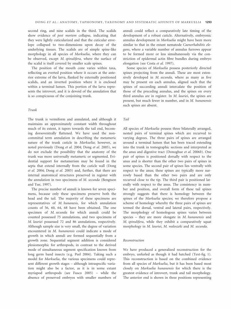
Reconstruction
We have produced a generalized reconstruction for the
embryo, unfurled as though it had hatched (Text-fig. 1).
This reconstruction is based on the combined evidence
from all species of Markuelia, but it has been based most
closely on Markuelia hunanensis for which there is the
greatest evidence of introvert, trunk and tail morphology.
The anterior end is shown in three positions representing
D O N G E T A L . : A N A T O M Y , T A P H O N O M Y , T A X O N O M Y A N D S Y S T E M A T I C A F F I N I T Y O F M A R K U E L I A 1293
the cycle of eversion of the introvert; a cut away view of
the inverted position is also shown. In the inverted posi-
tion, the scalids point anteriorly, but lie against the body
and point posteriorly as they are everted, a character that
serves to distinguish scalids from pharyngeal teeth
(Nielsen 2001, p. 332).
THE AFFINITY OF MARKUELIA
When Bengtson and Yue (1997) identified Markuelia as a
fossil embryo for the first time, they considered its affinity
to annelids and lobopods, while Conway Morris (1998)
speculated that Markuelia might be an embryonic hal-
kieriid. The description of more complete material con-
strained further speculation, leading to the conclusion
that Markuelia was a member of Introverta. Within this
framework, initial cladistic analyses including fossil and
living ecdysozoans indicated that Markuelia is a stem-
priapulid (Dong and Donoghue 2002), but subsequent
analyses suggested a stem-scalidophoran affinity (Dong
et al. 2004; Dong et al. 2005). This conclusion was robust
to the elucidation of the composition and arrangement of
the introvert scalids (Donoghue et al. 2006b), but these
phylogenetic conclusions have always been sensitive to the
fact that the anatomy of Markuelia is not only incom-
pletely known, but is known only from embryonic mate-
rial. Sensitivity analysis of the dataset has shown that the
phylogenetic position of Markuelia is sensitive to the sets
of taxa included in analyses, in some instances resolved as
stem-priapulids and, more precisely, as a sister taxon to
palaeoscolecids (Cobbett et al. 2007). This may provide
some support for Huang’s speculative suggestion that
Markuelia species are embryos of palaeoscolecids (Huang
et al. 2006). Indeed, the presence of bilaterally symmetri-
cal pairs of perianal spines in both Markuelia and some
palaeoscolecids potentially provides evidence of a close
relationship. However, the numbers and proportions of
scalids in the head, and spines in the tail of Markuelia
indicate that character homology and close kinship are
dubious (see Harvey et al., 2010). Given the sensitivity of
the dataset to taxon inclusion, and the recent description
of a greater number of scalidophoran taxa, we compiled a
revised and expanded cladistic dataset for cycloneuralians,
including representative fossil and living panarthropods,
and Gastrotricha as a root. This dataset has been
described elsewhere (Harvey et al. 2010), but it has been
augmented with codings for Markuelia based on the ana-
tomical information we present here (Table 1).
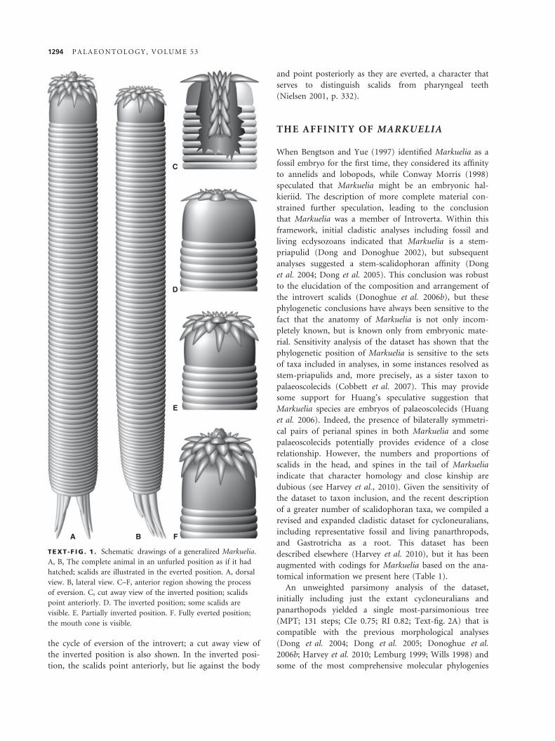
An unweighted parsimony analysis of the dataset,
initially including just the extant cycloneuralians and
panarthopods yielded a single most-parsimonious tree
(MPT; 131 steps; CIe 0.75; RI 0.82; Text-fig. 2A) that is
compatible with the previous morphological analyses
(Dong et al. 2004; Dong et al. 2005; Donoghue et al.
2006b; Harvey et al. 2010; Lemburg 1999; Wills 1998) and
some of the most comprehensive molecular phylogenies
A B F
E
D
C
TEXT -F IG . 1 . Schematic drawings of a generalized Markuelia.
A, B, The complete animal in an unfurled position as if it had
hatched; scalids are illustrated in the everted position. A, dorsal
view. B, lateral view. C–F, anterior region showing the process
of eversion. C, cut away view of the inverted position; scalids
point anteriorly. D. The inverted position; some scalids are
visible. E. Partially inverted position. F. Fully everted position;
the mouth cone is visible.
1294 P A L A E O N T O L O G Y , V O L U M E 5 3
(e.g. Dunn et al. 2008); it disagrees with some molecular
phylogenies, but these have been based on very little data
(e.g. Park et al. 2006; Sørensen et al. 2008).
Inclusion of Markuelia again yields a single MPT (136
steps; CIe 0.72; RI 0.80; Text-fig. 2B) in which it is
resolved as the sister taxon to Scalidophora. Inclusion of
representative stem-arthropods and a putative stem-ony-
chophoran results in 16 MPTs (143 steps; CIe 0.70; RI
0.80) that differ only in terms of the relative relationships
of the extinct and extant panarthropods. Tardigrada is
solely responsible for the conflict between the MPTs, and
the exclusion of this leaf results in a single MPT (138
steps; CIe 0.72; RI 0.80; Text-fig. 2C); the stem-scalidoph-
oran position of Markuelia remains stable throughout.
Inclusion of a large number of extinct introvertans results
in 276 MPTs (209 steps; CIe 0.54; RI 0.72), the strict con-
sensus of which is not well resolved, but unequivocally
identifies Markuelia as a member of the priapulid total
group (Text-fig. 2D). Reweighting according to the
rescaled consistency indices of the unweighted analysis
produced two MPTs at 89.47 steps CIe 0.73; RI 0.86 com-
patible with two trees from among 276 in unweighted
analysis that differ only in the relative relations of Priapu-
lites and Priapulopsis (the strict consensus of these two
MPTs is shown in Text-fig. 2E). However, this scheme of
relationships is sensitive to the taxon set, and the exclu-
sion of just a single taxon can lead to a radically different
solution. For instance, exclusion of Cricocosmia produces
26 MPTs, the strict consensus of which resolves palaeo-
scolecids as stem-nematomorphs, and Markuelia together
with a rump of extinct introvertans (Tylotites, Scoleco-
furca, Louisella, Ottoia, Corynetis, Selkirkia, Maotianshania
and Tabelliscolex) as stem-scalidophorans (208 steps; CIe
0.54; RI 0.72; Text-fig. 2F).
These analyses demonstrate that a precise resolution of
the phylogenetic affinity is contingent upon the tests of
character polarity and congruence provided by the wealth
of other fossil introvertans. Unfortunately, however, the
anatomy of the fossil introvertans is not well resolved,
particularly with regard to the anatomy and scalid
arrangement of the introvert itself. This does not occur
because of the vagaries of fossilization, but because spe-
cialists have failed to resolve and describe these characters
from the original material (Conway Morris 1977; Maas
et al. 2007b are notable exceptions), and it is not possible
to glean this information from figures alone. Until this
situation is revised, it will not prove possible to resolve
the phylogenetic position of Markuelia or the remaining
fossil introvertans more precisely. Traditionally, these taxa
would be assigned to the scalidophoran stem-group. This
is precisely in accordance with the original utility of the
stem-group as it was formulated by Hennig as a dustbin
for fossil taxa that failed to show the necessary character-
istics diagnostic of the respective crown-groups, but for
which it was not possible to distinguish whether those
characters were primitively absent (they had not yet
evolved) or whether they had merely rotted away and so
were not preserved (Donoghue 2005; Hennig 1981).
However, the stem-group has since acquired an exclu-
sively phylogenetic meaning, eschewing its use as a quali-
fication of systematic classification (Donoghue 2005).
Thus, in such instances, it is most appropriate to assign
taxa such as Markuelia to the scalidophoran total group
(Donoghue and Purnell 2009) to better express equi-
vocation over its systematic classification given that it
may be a stem-scalidophoran or a stem-priapulid.
THE PRESERVATION OF MARKUELIA
Specimens of Markuelia are preserved at various stages of
decay. In a few specimens, the entire anatomy of the
animal, including the head, is preserved (Text-fig. 3A).
The head is consistently less well preserved than the other
parts of the animal’s external anatomy; where it is pres-
ent, it invariably shows signs of decay-related collapse
(Text-fig 3C). More commonly, the head is absent from
otherwise well-preserved embryos (Text-fig. 3B), indicat-
ing that it is the first part of the organism to be destroyed
by decay.
Embryos that have decayed to a greater extent exhibit a
continuum of increasing collapse and shrinkage (Text-
fig. 3D–F). As this process continues, the annuli become
increasingly indistinct, and the interior of the embryo
may shrink to c. 20 per cent of its original diameter. In
some cases, the shrunken embryo is attached to the cho-
rion by phosphatic filaments. These filaments may repre-
sent fungal hyphae or filamentous bacteria (see discussion
in Xiao and Knoll (1999); these features will be termed
‘filaments’ hereafter).
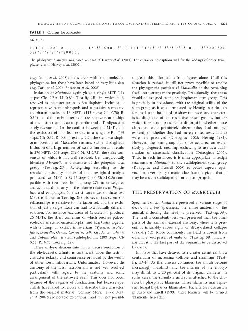
TABLE 1 . Codings for Markuelia.
Markuelia
1 1 1 0 1 1 1 0 0 0 - 0 - - - - - - - - - - 1 2 ? ? ? 0 0 0 0 - - ? ? 0 0 ? 1 1 1 1 ? 1 ? 1 ? ? ? ? ? ? ? ? ? ? ? ? ? ? 1 0 - - - ? ? ? ? 0 0 0 ? 0 0
0 ? ? ? ? ? ? ? ? ? ? ? ? ? ? 0 0 1 1 0
The phylogenetic analysis was based on that of Harvey et al. (2010). For character descriptions and for the codings of other taxa,
please refer to Harvey et al. (2010).
D O N G E T A L . : A N A T O M Y , T A P H O N O M Y , T A X O N O M Y A N D S Y S T E M A T I C A F F I N I T Y O F M A R K U E L I A 1295
GastrotrichaPeripatus† MicrodictyonTardigrada† Aysheaia† KerygmachelaNematomorphaNematoda† Fieldia† AncalagonKinorhynchaLoricifera† Palaeopriapulites† SicyophorusMaccabeusHalicryptusPriapulopsis† PriapulitesAcanthopriapulusPriapulusMeiopriapulus
Tubiluchus† Xiaoheiqingella† Yunnanpriapulus† Selkirkia† Corynetis† Maotianshania
E
† Markuelia† Tylotites† Cricocosmia† Tabelliscolex† Palaeoscolecida† Ottoia† Louisella† Scolecofurca
GastrotrichaPeripatusTardigradaNematomorphaNematodaKinorhynchaLoriciferaMeiopriapulusTubiluchusMaccabeusHalicryptusPriapulopsisAcanthopriapulusPriapulus
AGastrotrichaPeripatusTardigradaNematomorphaNematoda† MarkueliaKinorhynchaLoriciferaMeiopriapulusTubiluchusMaccabeusHalicryptusPriapulopsisAcanthopriapulusPriapulus
BGastrotricha† MicrodictyonPeripatus† Aysheaia† KerygmachelaNematomorphaNematoda† MarkueliaKinorhynchaLoriciferaMeiopriapulusTubiluchusMaccabeusHalicryptusPriapulopsisAcanthopriapulusPriapulus
C
GastrotrichaPeripatus† MicrodictyonTardigrada
† Aysheaia† KerygmachelaNematomorphaNematoda† Fieldia† AncalagonKinorhynchaLoricifera† Selkirkia† Corynetis† Maotianshania† Markuelia† Tylotites† Cricocosmia† Tabelliscolex† Louisella† Ottoia† Palaeoscolecida† Scolecofurca† Palaeopriapulites
† SicyophorusMeiopriapulusTubiluchus† Xiaoheiqingella† YunnanpriapulusMaccabeusHalicryptusPriapulopsis† PriapulitesAcanthopriapulusPriapulus
DGastrotrichaPeripatus† MicrodictyonTardigrada† Aysheaia† KerygmachelaNematoda† Palaeoscolecida
Nematomorpha† Markuelia† Tylotites† Scolecofurca† Louisella† Ottoia† Corynetis† Selkirkia† Maotianshania† Tabelliscolex† Fieldia† AncalagonKinorhynchaLoricifera
† Palaeopriapulites† SicyophorusMeiopriapulusTubiluchus† Xiaoheiqingella† YunnanpriapulusMaccabeusHalicryptus† PriapulitesPriapulopsisAcanthopriapulusPriapulus
F
TEXT -F IG . 2 . Cladograms arising from phylogenetic analyses. A, MPT from analysis of extant cycloneuralians and panarthropods
only. B, MPT from analysis of taxa analysed in (A) plus Markuelia. C, MPT from analysis of taxa included in (B) plus representative
stem-arthropods and a putative stem-onychophoran, but excluding tardigrades. D, Strict consensus tree from analysis of taxa in (C)
plus a large number of extinct introvertans. E, Strict consensus tree from the reweighted analysis of taxa in (D). F, Strict consensus
tree from analysis of taxa in (D) with Cricocosmia excluded.
1296 P A L A E O N T O L O G Y , V O L U M E 5 3
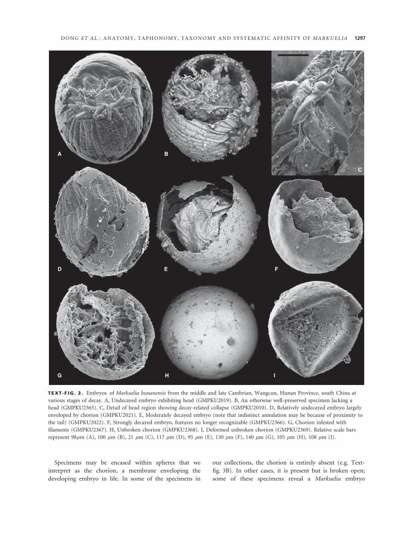
Specimens may be encased within spheres that we
interpret as the chorion, a membrane enveloping the
developing embryo in life. In some of the specimens in
our collections, the chorion is entirely absent (e.g. Text-
fig. 3B). In other cases, it is present but is broken open;
some of these specimens reveal a Markuelia embryo
A
D
G H I
E F
B
C
TEXT -F IG . 3 . Embryos of Markuelia hunanensis from the middle and late Cambrian, Wangcun, Hunan Province, south China at
various stages of decay. A, Undecayed embryo exhibiting head (GMPKU2019). B, An otherwise well-preserved specimen lacking a
head (GMPKU2365). C, Detail of head region showing decay-related collapse (GMPKU2010). D, Relatively undecayed embryo largely
enveloped by chorion (GMPKU2021). E, Moderately decayed embryo (note that indistinct annulation may be because of proximity to
the tail) (GMPKU2022). F, Strongly decayed embryo, features no longer recognizable (GMPKU2366). G, Chorion infested with
filaments (GMPKU2367). H, Unbroken chorion (GMPKU2368). I, Deformed unbroken chorion (GMPKU2369). Relative scale bars
represent 98lm (A), 100 lm (B), 21 lm (C), 117 lm (D), 95 lm (E), 130 lm (F), 140 lm (G), 105 lm (H), 108 lm (I).
D O N G E T A L . : A N A T O M Y , T A P H O N O M Y , T A X O N O M Y A N D S Y S T E M A T I C A F F I N I T Y O F M A R K U E L I A 1297
within (Text-fig. 3D), while others are shown to be
empty, and further specimens contain filaments (Text-
fig. 3G). Our collections also contain a large number of
unbroken spheres, which are most probably chorions, and
these may or may not be deformed (Text-fig. 3H, I).
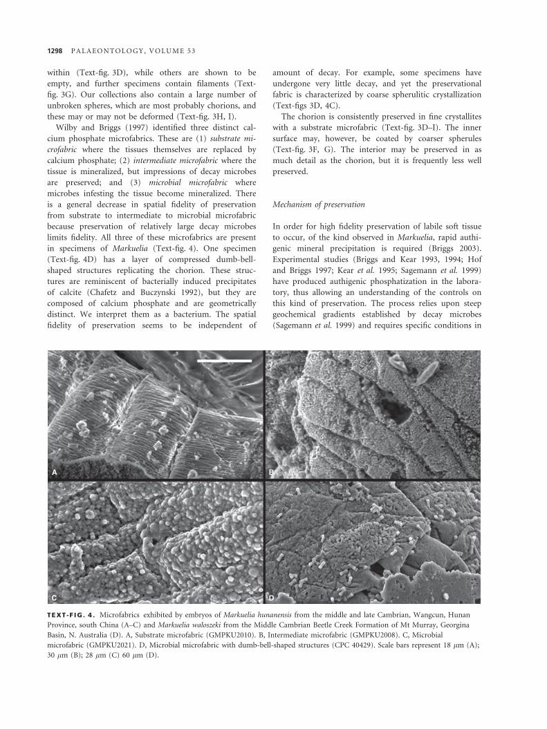
Wilby and Briggs (1997) identified three distinct cal-
cium phosphate microfabrics. These are (1) substrate mi-
crofabric where the tissues themselves are replaced by
calcium phosphate; (2) intermediate microfabric where the
tissue is mineralized, but impressions of decay microbes
are preserved; and (3) microbial microfabric where
microbes infesting the tissue become mineralized. There
is a general decrease in spatial fidelity of preservation
from substrate to intermediate to microbial microfabric
because preservation of relatively large decay microbes
limits fidelity. All three of these microfabrics are present
in specimens of Markuelia (Text-fig. 4). One specimen
(Text-fig. 4D) has a layer of compressed dumb-bell-
shaped structures replicating the chorion. These struc-
tures are reminiscent of bacterially induced precipitates
of calcite (Chafetz and Buczynski 1992), but they are
composed of calcium phosphate and are geometrically
distinct. We interpret them as a bacterium. The spatial
fidelity of preservation seems to be independent of
amount of decay. For example, some specimens have
undergone very little decay, and yet the preservational
fabric is characterized by coarse spherulitic crystallization
(Text-figs 3D, 4C).
The chorion is consistently preserved in fine crystallites
with a substrate microfabric (Text-fig. 3D–I). The inner
surface may, however, be coated by coarser spherules
(Text-fig. 3F, G). The interior may be preserved in as
much detail as the chorion, but it is frequently less well
preserved.
Mechanism of preservation
In order for high fidelity preservation of labile soft tissue
to occur, of the kind observed in Markuelia, rapid authi-
genic mineral precipitation is required (Briggs 2003).
Experimental studies (Briggs and Kear 1993, 1994; Hof
and Briggs 1997; Kear et al. 1995; Sagemann et al. 1999)
have produced authigenic phosphatization in the labora-
tory, thus allowing an understanding of the controls on
this kind of preservation. The process relies upon steep
geochemical gradients established by decay microbes
(Sagemann et al. 1999) and requires specific conditions in
A B
C D
TEXT -F IG . 4 . Microfabrics exhibited by embryos of Markuelia hunanensis from the middle and late Cambrian, Wangcun, Hunan
Province, south China (A–C) and Markuelia waloszeki from the Middle Cambrian Beetle Creek Formation of Mt Murray, Georgina
Basin, N. Australia (D). A, Substrate microfabric (GMPKU2010). B, Intermediate microfabric (GMPKU2008). C, Microbial
microfabric (GMPKU2021). D, Microbial microfabric with dumb-bell-shaped structures (CPC 40429). Scale bars represent 18 lm (A);
30 lm (B); 28 lm (C) 60 lm (D).
1298 P A L A E O N T O L O G Y , V O L U M E 5 3
order for calcium phosphate, rather than calcium carbon-
ate, to be precipitated. This is the calcium carbonate –
calcium phosphate switch (Briggs and Wilby 1996). The
key conditions needed to set the switch to calcium phos-
phate are (1) sufficient concentration of phosphate to
inhibit calcite or aragonite precipitation, (2) reduced pH,
and (3) a closed system (Briggs and Kear 1993, 1994;
Briggs and Wilby 1996). While experimental studies
(Briggs and Kear 1993, 1994) have shown that pH is a
major control in calcium phosphate precipitation, it is
insufficient to induce calcium phosphate precipitation if
the phosphorous concentration is too low (Briggs and
Wilby 1996). Indeed, in the absence of a significant exter-
nal source of phosphorous, experimental studies have
been able to yield calcium phosphate precipitation only in
crustaceans (Briggs and Kear 1993, 1994; Hof and Briggs
1997) and (to a lesser extent) squid (Kear et al. 1995),
where the large carcasses could act as a phosphorous
source. This would suggest that an external source of
phosphorous must be invoked to explain the preservation
of embryos, which obviously lack a large, phosphate-rich
carcass.
Experiments on lobster eggs provide evidence that
relates specifically to the preservation of eggs and
embryos. At the time of writing, these experiments have
failed to produce calcium phosphate precipitation. How-
ever, mineralization of the chorion in calcium carbonate
and calcium phosphate has been observed (Martin et al.
2003, 2005), albeit in coarse spherules rather than the
substrate microfabric seen in the chorions of fossils. In
another series of experiments (Martin et al. 2004), sedi-
ment particles became attached to the chorion. While the
experiments have yielded no information on preservation
of the interior, either of these mechanisms may represent
an essential first stage in preservation that is needed to
prevent collapse (Martin et al. 2003).
The mechanisms of preservation described above can
account for the variation observed in our collections. As
the chorion is consistently preserved in crystals at least as
fine as those of the interior, and because its amount of
decay seems unrelated to that of the interior, it is sug-
gested that the chorion is mineralized in a distinct, initial
stage of replacement. The interior of the embryo may be
preserved later; the length of time between these two
phases of mineralization may be an important control on
the amount of decay observed in the interior. Interior
mineralization may preserve filaments that grow during
or after decay or may not occur at all, the latter resulting
in empty chorion. Gostling et al. (2008) have shown that
the rates of decay of the chorion and embryo are not
linked and Raff et al. (2008) have shown that distinct
microbial communities infest the embryo and chorion.
This explains the presence of chorions without embryos
or embryos without chorions in fossil assemblages.
Comparative taphonomy
While it may be possible to explain what is fossilized of
the embryos of Markuelia in terms of replication of
organic substrates by various modes of bacterially medi-
ated authigenic mineralization, what it does not explain
is why these stages of embryonic development are pre-
served in preference to earlier embryonic and post-
embryonic development. There are few candidate fossils
representing earlier stages of the embryology of Markuelia
(Dong et al. 2004; Dong et al. 2005), and their character-
istics are so generic that they are uninformative. Further-
more, the security with which they are associated with
Markuelia amounts to little more than co-association
(Donoghue and Dong 2005). Experimental investigation
into the relationship between embryology and taphonomy
has shown that preservation potential varies with devel-
opment, and, in some instances, the earliest stages of
embryology have the greatest preservation potential (Gos-
tling et al. 2009; Gostling et al. 2008; Raff et al. 2006).
However, in Markuelia it is the late embryonic stage that
evidently had the highest fossilization potential given that
it is the only fossil embryo known from a broad geo-
graphical and stratigraphical range of localities, extending
from the Lower Cambrian of Siberia, through Middle
Cambrian of Australia and China, Late Cambrian of
China, to Early Ordovician of the United States
(Donoghue et al. 2006a). This has been explained as an
artefact of the precocious development of a cuticle in the
embryo, the cuticle serving as a suitable substrate for
mineralization associated with exceptional preservation,
the effect of the fertilization envelope in establishing a
geochemical microenvironment necessary for the initia-
tion of mineralization (Donoghue et al. 2006a). It
remains possible, however, that cuticle is relatively decay
resistant and, thus, that it is available as a substrate for
replication by nucleating mineral crystals long after tis-
sues of other composition have decayed away (Gostling
et al. 2009), perhaps providing the ions necessary for
mineralization to occur.
To determine the relative preservation potential of cuti-
cle in an embryo such as Markuelia, we undertook a
decay experiment based on the priapulid Priapulus cauda-
tus, which is both an extant relative and an analogue of
the embryo of Markuelia. In fresh larvae, where the intro-
vert was retracted, the cuticle bearing larval spines is posi-
tioned internally, just as it is in Markuelia (Text-fig. 5A).
However, as decay progressed, the visceral portion of the
head became detached from its surrounding cuticle, until
the muscles of the introvert were retracted, while the
spines remained everted (Text-fig. 5B). As decay pro-
gressed, the muscle decayed to nothing until all that
remained was cuticle. Eventually, even the cuticle was
subject to decay collecting a thick mantle of bacteria
D O N G E T A L . : A N A T O M Y , T A P H O N O M Y , T A X O N O M Y A N D S Y S T E M A T I C A F F I N I T Y O F M A R K U E L I A 1299
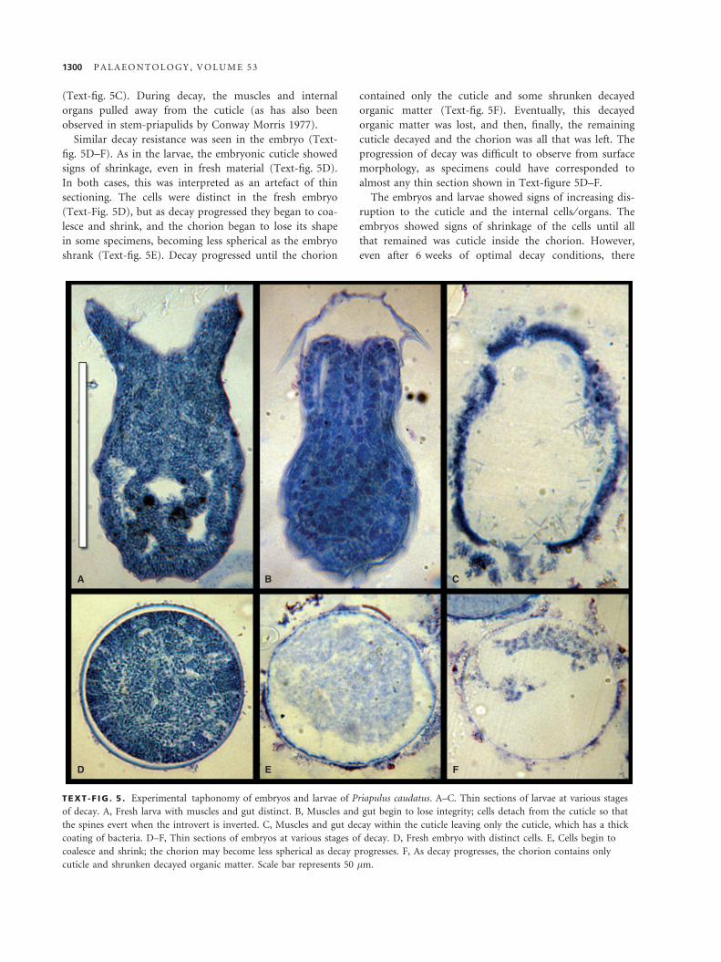
(Text-fig. 5C). During decay, the muscles and internal
organs pulled away from the cuticle (as has also been
observed in stem-priapulids by Conway Morris 1977).
Similar decay resistance was seen in the embryo (Text-
fig. 5D–F). As in the larvae, the embryonic cuticle showed
signs of shrinkage, even in fresh material (Text-fig. 5D).
In both cases, this was interpreted as an artefact of thin
sectioning. The cells were distinct in the fresh embryo
(Text-Fig. 5D), but as decay progressed they began to coa-
lesce and shrink, and the chorion began to lose its shape
in some specimens, becoming less spherical as the embryo
shrank (Text-fig. 5E). Decay progressed until the chorion
contained only the cuticle and some shrunken decayed
organic matter (Text-fig. 5F). Eventually, this decayed
organic matter was lost, and then, finally, the remaining
cuticle decayed and the chorion was all that was left. The
progression of decay was difficult to observe from surface
morphology, as specimens could have corresponded to
almost any thin section shown in Text-figure 5D–F.
The embryos and larvae showed signs of increasing dis-
ruption to the cuticle and the internal cells ⁄ organs. The
embryos showed signs of shrinkage of the cells until all
that remained was cuticle inside the chorion. However,
even after 6 weeks of optimal decay conditions, there
A B C
D E F
TEXT -F IG . 5 . Experimental taphonomy of embryos and larvae of Priapulus caudatus. A–C. Thin sections of larvae at various stages
of decay. A, Fresh larva with muscles and gut distinct. B, Muscles and gut begin to lose integrity; cells detach from the cuticle so that
the spines evert when the introvert is inverted. C, Muscles and gut decay within the cuticle leaving only the cuticle, which has a thick
coating of bacteria. D–F, Thin sections of embryos at various stages of decay. D, Fresh embryo with distinct cells. E, Cells begin to
coalesce and shrink; the chorion may become less spherical as decay progresses. F, As decay progresses, the chorion contains only
cuticle and shrunken decayed organic matter. Scale bar represents 50 lm.
1300 P A L A E O N T O L O G Y , V O L U M E 5 3
were individuals that were recognizable as embryos. This
is virtually identical to the condition we observe in
Markuelia, where all that appears to be preserved is cuti-
cle within a chorion. Hence, it appears that the fossiliza-
tion of Markuelia is not perhaps comparable to the
preservation of cleavage and gastrula-stage embryos from
deposits such as the Ediacaran Doushantuo Formation
(Hagadorn et al. 2006; Xiao et al. 1998) and the Cam-
brian Kuanchuanpu Formation (Bengtson and Yue 1997;
Steiner et al. 2004). Rather, it reflects the mineral replica-
tion of cuticle that is more comparable to ‘Orsten’-style
fossilization of the 3D cuticles of microscopic arthropods
(Maas et al. 2006), though preservation potential may
have been increased because of the enclosure of the larva
within a chorion facilitating the development of a geo-
chemical microenvironment of concentrated ions and
microbial activity necessary for diminishing autolysis and
enhancing mineral replication of cuticle (Donoghue et al.
2006a; Gostling et al. 2009; Gostling et al. 2008; Raff et al.
2008; Raff et al. 2006).
SYSTEMATIC PALAEONTOLOGY
The specimens figured in this paper are located in the follow-
ing repositories: Geological Museum of Peking University,
China (GMPKU), National Museum of Natural History,
Smithsonian Institution, Washington DC, USA (USNM), Swed-
ish Museum of Natural History (NRM), Commonwealth
Palaeontological Collections, Bureau of Mineral Resources,
Canberra (CPC) and Institute of Palaeontology, Bonn Univer-
sity (UB).
NEPHROZOA Jondelius, Ruiz-Trillo, Baguna and Riutort,
2002
ECDYSOZOA Aguinaldo, Turbeville, Linford, Rivera, Garey,
Raff and Lake, 1997
INTROVERTA Nielsen, 1995
SCALIDOPHORA Lemburg, 1995
Genus MARKUELIA Val’kov, 1983 sedis mutabilis
Type species. Markuelia secunda Val’kov, 1984.
Stratigraphical range and distribution. Lower Cambrian of Sibe-
ria, Middle Cambrian of China and Australia, Late Cambrian of
China, Lower Ordovician of the United States.
Emended diagnosis. Wormlike animal with profusely ann-
ulated (about 56–86 annulations) trunk that coiled in an
inverted S-shaped or S-shaped loop into a sphere, with
anterior and posterior ends juxtaposed laterally. The pos-
terior end with three pairs of bilaterally arranged, termi-
nal spines.
Remarks. Markuelia secunda was first described as a fossil
embryo by Bengtson and Yue (1997), although it had ear-
lier been figured and briefly described as chambered
organism of unknown affinities by Val’kov (1983) and
subsequently figured several times by Val’kov and other
Russian scientists (Bengtson and Yue 1997). The descrip-
tion of M. secunda by Bengtson and Yue (1997) stated
that the chambers are segments of a wormlike animal that
is tightly looped into a sphere. The two ends of the body
form an inverted S-shaped double loop on the opposite
hemisphere. The anterior end is not well preserved in
the available specimens. The posterior end has modified
segments, each with two symmetrically placed processes
that join into a kind of posterior comb. The (trunk) seg-
ments carry short conical spines. Typically, a spine recurs
in similar position on every third segment. Additional
specimens of Markuelia demonstrate that the trunk may
coil in both directions, resulting in mirror-imaged forms
(see Donoghue et al. 2006a). The arrangement where the
head (when viewed pointing upwards) is to the left of the
tail is here defined as the L-form (equivalent to an
S-shaped loop); the opposite arrangement is the R-form
(equivalent to an inverted S-shaped loop). Furthermore,
SRXTM studies show that M. secunda has another un-
exposed pair of spines, giving it a total of three pairs of
terminal spines, the same as in the other species
of Markuelia (see Donoghue et al. 2006b). The hundreds
of specimens of Markuelia found from Middle and Late
Cambrian in western Hunan, China (Dong et al. 2004;
Dong et al. 2005), from Middle Cambrian in Georgina
Basin, northern Australia and from Tremadocian, in
Nevada, USA (Donoghue et al. 2006a) all lack broad con-
ical trunk spines of this kind, except for M. waloszeki. We
therefore conclude that these are the character of the spe-
cies M. secunda rather than that of the genus Markuelia.
We assign Markuelia to Scalidophora sedis mutabilis to
reflect the uncertainty as to whether it represents a stem-
scalidophoran or a stem-priapulid as discussed above.
Markuelia secunda Valkov, 1984
Text-figure 6
v.1983 Markuelia cf. prima Val.; Khomentovsky et al.,
p. 28 [nomen nudum].
v.1983 Markuelia secunda Val.; Khomentovsky et al.,
p. 29 [nomen nudum].
v.1983 Markuelia prima Valkov; Val’kov, pl. fig. 14
[nomen nudum].
v.1983 Markuelia secunda Valkov; Val’kov, pl. fig. 15–17
[nomen nudum].
v*1984 Markuelia secunda Valkov, 1983; Val’kov and
Karlova, pp. 23–24, pl. 2, fig. 15.
v.1984 Markuelia prima Valkov, 1983; Val’kov and
Karlova, pl. 2, fig. 15 [nomen nudum].
D O N G E T A L . : A N A T O M Y , T A P H O N O M Y , T A X O N O M Y A N D S Y S T E M A T I C A F F I N I T Y O F M A R K U E L I A 1301
A
D
H
I J
E
F
G
B C
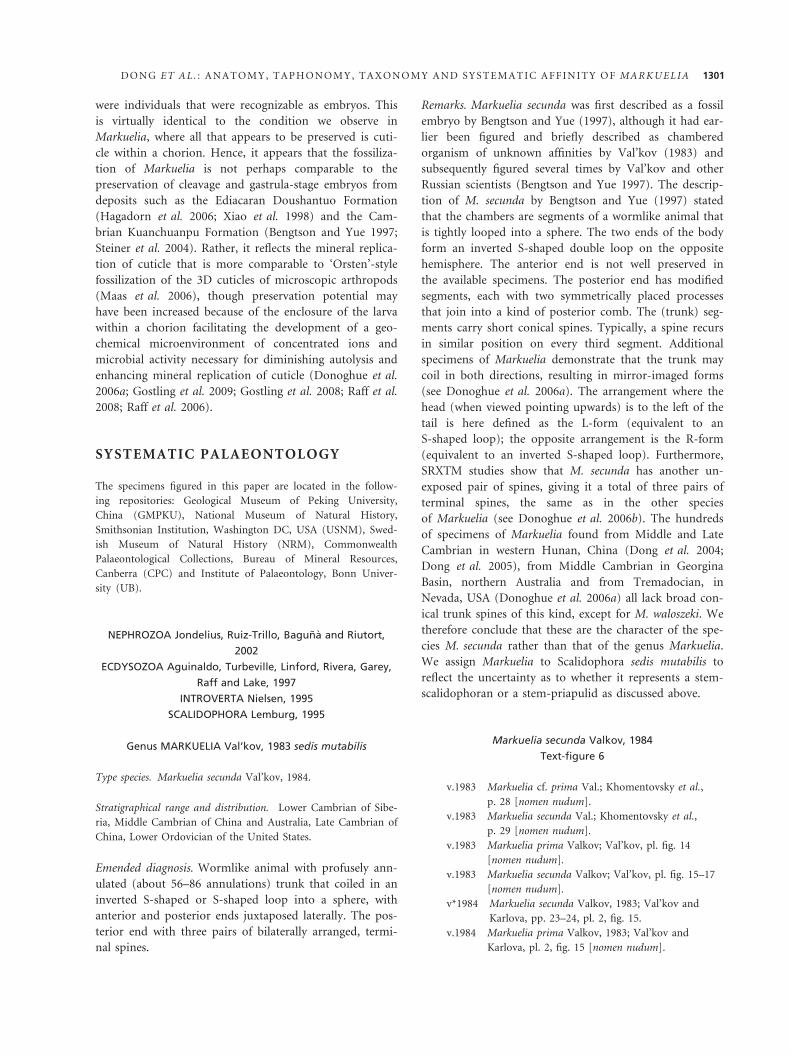
TEXT -F IG . 6 . Two embryos of Markuelia secunda Valkov, 1984 (L-forms), from section ‘Dvortsy’, basal part of the Pestrotsvet
Formation, Yakutia, sample Sib73-15-SB. SEM (A–C, G–J) and SRXTM (D–F) images. A–G, NRM X2240 (also figured by Bengtson
and Yue 1997, fig. 2b; Dong et al. 2005, fig. 2i; and Donoghue et al. 2006b, fig. 2i, j). A, View with tail end at upper middle. B, View
with head end at upper middle. C, Detail of B, showing apatite-encrusted radiating spokes. D, Orientation like A, volume-texture
rendering of whole body plus surface rendering of tail spines. E, Separate rendering of tail spines. F, Exploded view of E; top = dorsal
pair; middle = lateral pair; bottom = ventral pair. G, Detail of loop side, showing conical spines. H–J, NRM X2239 (also figured by
Bengtson and Yue 1997, fig. 2a; and Dong et al. 2005, fig. 2h). H. View of loop side. I, Detail of H, showing interfingering marginal
spines from adjacent trunk sides. J, Detail of H, showing apatite-encrusting radiating spokes. Scale bars represent 100 lm (A, B, D,
H); 80 lm (E, F); 70 lm (G); 30 lm (I); 26 lm (J); 22 lm (C).
1302 P A L A E O N T O L O G Y , V O L U M E 5 3
v.1987 Markuelia secunda Valkov, 1983; Val’kov, p. 116,
pl. 14, figs 11–13, text-fig. 16.
. 1989 Markuelia prima Val.; Missarzhevsky, p. 211,
pl. 30, fig. 4.
v.1990 Markuelia secunda Valkov, 1987; Khomentovsky
et al., pp. 30–31, pl. 4, figs 3–6.
1993 Markuelia prima; Khomentovsky and Karlova,
p. 40, fig. 10.
1993 Markuelia secunda; Khomentovsky and Karlova,
p. 33, fig. 3.
v.1997 Markuelia secunda Valkov, 1987; Bengtson and
Yue, pp.1647–1648, fig. 2a, b.
v.1998 Markuelia secunda; Conway Morris, fig. 2a, b.
v.2005 Markuelia secunda; Dong et al., p. 472, fig. 2h, I.
v.2005 Markuelia secunda; Donoghue and Dong,
pp. 90–91, fig. 4d, e.
v.2006a Markuelia secunda; Donoghue et al., pp. 233–235,
fig. 1f–j.
v.2006b Markuelia secunda; Donoghue et al., pp. 681–682,
fig. 2i, j.
v.2007 Markuelia; Barton et al., fig. 10.8.
Holotype. Val’kov and Karlova 1984, pl. 2, fig. 15, No. 762 ⁄ 37
(see under ‘Remarks’).
Type locality and horizon. Height 1291 m, River Gonam, Aldan
River Basin, southern Yakutia, Siberia. Base of the Pestrotsvet
Formation, Lower Tommotian, Lower Cambrian.
Additional material. NRM X2239, NRM X2240, NRM X3803 (all
three from the base of the Pestrotsvet Formation, Dvortsy sec-
tion, left bank of the Aldan river, 4–5 km upstream of the
mouth of the Dyalkhakh river); NRM X3801, NRM X3802 (both
from the base of the Pestrotsvet Formation, at a height of
1291 m, on the right side of the Gonam river, 30 km from its
mouth), NRM X3804, NRM X3805 (both from the base of the
Pestrotsvet Formation, left bank of the Aldan river, 7 km
upstream of the mouth of the Ulakhan-Sulugur river).
Emended diagnosis. Markuelia species with short, conical
spines on trunk annuli and a relatively long central pair
of tail spines.
Description. The embryos are all spherical, except where sec-
ondarily broken. The diameter is 480–550 lm. They are pre-
served through encrustation with a 5- to 7-lm thin layer
consisting of fibrous apatite growing normal to the encrusted
surface (Text-fig. 6C). In outer appearance, the embryos are
either smooth and featureless or show the surface of the convo-
luted segmented body, more or less effaced. We interpret the
completely smooth ones as embryos within a fertilization
envelope. This is also suggested by the partial presence in some
specimens of a smooth outer cover over a segmented body
(Donoghue et al. 2006a, fig. 1h).
In the developed embryos, the body is clearly divisible into a
head region (Text-fig. 6A, B, D), a thorax with about 75 trans-
verse annulations, and a tail with posteriorly pointing spines
(Text-fig. 6A, D–F).
The annuli are beset with short, conical spines, 50–60 lm long,
that narrow into a sharp point distally (Text-fig. 6G). Annuli may
have 0–2 spines at irregular intervals, and there is a tendency for a
spine position to repeat itself on every three segments. Occasion-
ally, spines that are slightly longer (80 lm) and more slender than
the more medially placed ones are visible in lateral positions
(Text-fig. 6H, I). The tail end bears two pairs of spines visible on
the surface of the enrolled embryo (dorsal and lateral pairs), and
another pair (ventral pair) beneath them (Text-fig. 6D–F).
Internally, there are spoke-like structures corresponding in
spacing to the external annulations (Text-fig. 6C, J). In the speci-
men in Text-figure 6C, they are separate from the body wall. As
this specimen also contains numerous other encrusted filaments
of less regular appearance (e.g. Text-fig. 6B, upper right), it can-
not be excluded that these particular spokes are fortuitous effects
of diagenesis. The specimen in Text-figure 6J, however, shows
spokes that are regularly arranged, and the two lower spokes are
partly oblique to the body annulations. This supports the inter-
pretation that the spokes are separate from the body wall and
that, therefore, the annulations correspond to an internal seg-
mentation. The spokes most likely represent an organ system that
is not part of the body wall or mesentries, although the exact nat-
ure of this system remains unclear (see Bengtson and Yue 1997).
Remarks. The 1983 references to M. secunda and M. prima
are all nomina nuda, as they were not accompanied by any
description or definition. In 1984, Val’kov and Karlova
gave a description of M. secunda Valkov, 1983, with refer-
ence to one figured specimen, No. 762 ⁄ 37 (pl. 2, fig. 15). In
the plate caption, the same specimen is referred to as
M. prima, Valkov 1983. Still lacking a description or defini-
tion, M. prima thus remained a nomen nudum and would
otherwise have entered into objective synonymy with
M. secunda, as Val’kov and Karlova (1984) named the only
figured specimen M. secunda in the main text and
M. prima in the figure caption. The figured specimen, No.
762 ⁄ 37, is the holotype by monotypy, as none other was
figured or mentioned by Val’kov and Karlova (1984). This
is in spite of a later designation by Val’kov (1987) of
another specimen as holotype for M. secunda.
Khomentovsky et al. (1990, pl. 4, fig. 7) recorded
Markuelia from 25–30 m below the Pestrotsvet Formation
at Dzhanda, but the figured specimen is nothing more
than a featureless globule. Khomentovsky and Karlova
(1993, 2005) record Markuelia, but without illustration,
from the Ust’-Yudoma Formation that underlies the Pes-
trotsvet Formation in southeastern Siberian Platform and
is therefore of Nemakit–Daldynian age.
Markuelia hunanensis Dong and Donoghue in Dong et al.,
2004
Text-figure 7
v.*2004 Markuelia hunanensis Dong and Donoghue in
Dong et al., pp. 237–40, figs 1a–f, 2a–f.
D O N G E T A L . : A N A T O M Y , T A P H O N O M Y , T A X O N O M Y A N D S Y S T E M A T I C A F F I N I T Y O F M A R K U E L I A 1303
A
D
H
M N O
I J K
L
E
F
G
B C
TEXT -F IG . 7 . Markuelia hunanensis Dong and Donoghue, 2002, (L-forms) from the Middle and Late Cambrian Bitiao Formation of
Wangcun, Hunan Province, south China. SEM (A, L, M) and SRXTM (B–K, N, O) images. A–K, GMPKU2205 showing the introvert in
the inverted position (also figured by Donoghue et al. 2006b fig. 2a–f). A, Complete specimen. B, Complete specimen with scalids
highlighted. C, Specimen rotated through 90 degrees from (B). D–K, Volume rendering of the scalids in lateral (D–G) and anterior
(H–K) views. D, H, Eight scalids in the first row. E, I, 16 scalids in the first two rows. F, J, 25 scalids in the first three rows. G, K, All
scalids. L, GMPKU2010 (holotype; also figured by Dong et al. 2004, fig. 1f, 2a–f; Dong et al. 2005, fig. 1i, 2a; and Peng and Dong 2008,
fig. 3b) showing the everted introvert with spines pointing posteriorly. M. GMPKU2017 (also figured by Dong et al. 2005, fig. 2b)
showing the six posterior spines. N, O, GMPKU2011 (also figured by Dong et al. 2004, fig. 1e; Donoghue et al. 2006b, fig. 2g–h; and
Peng and Dong 2008, figs 2d–f, 3a, 4a–c). N, Reconstruction of posterior region with tail spines shown in yellow. O, Section showing
the position of the digestive tract in red. Scale bars represent 60 lm (A–C); 50 lm (D–K); 39 lm (L); 19 lm (M); 50 lm (N, O).
1304 P A L A E O N T O L O G Y , V O L U M E 5 3
A
D E
F
HG
B C
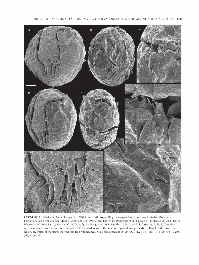
TEXT -F IG . 8 . Markuelia lauriei Haug et al., 2009 from North Rogers Ridge, Georgina Basin, northern Australia; Monastery
Formation, late Templetonian, Middle Cambrian; CPC 39946 (also figured by Donoghue et al. 2006a, fig. 1a; Maas et al. 2006, fig. 6b;
Webster et al. 2006, fig. 1c; Maas et al. 2007a, b, fig. 7a; Haug et al. 2009, figs 1b, 2d, 5a–d, 6a–b; R-form). A, B, D, E, Complete
specimen viewed from various orientations. C, F, Detailed views of the anterior region showing scalids. G, Detail of the posterior
region. H, Detail of the trunk showing slender protuberances. Scale bars represent 50 lm (A, B, D, E); 11 lm (C); 5 lm (F); 19 lm
(G); 11 lm (H).
D O N G E T A L . : A N A T O M Y , T A P H O N O M Y , T A X O N O M Y A N D S Y S T E M A T I C A F F I N I T Y O F M A R K U E L I A 1305
v.2005 Markuelia hunanensis; Donoghue and Dong,
pp. 90–91, fig. 4f–i.
v.2005 Markuelia hunanensis; Dong et al., pp. 468–482,
fig. 1a–i, 2a–e, g.
v.2006a Markuelia hunanensis; Donoghue et al.,
pp. 233–235.
v.2006b Markuelia hunanensis; Donoghue et al.,
pp. 681–682, fig. 2a–h.
v.2007 Markuelia hunanensis; Dong, pp. 930–934,
figs 1a–l, 2a–i.
v.2008 Markuelia hunanensis; Peng and Dong, figs 1–4.
v.2009 Markuelia hunanensis; Dong, pp. 431–434, figs 2–5.
v.2009 Markuelia elegans Dong, p. 434, fig. 6.
Holotype. GMPKU2010.
Type locality and horizon. Wangcun section, Yongshun County,
western Hunan, South China. Bitiao Formation, Furongian,
Upper Cambrian.
Additional material. GMPKU2007-19, 2021, 2205, 2213, 2214,
2218, 2222-5, 2227-32, 2237, 2238. All from the Wangcun section,
Yongshun County, western Hunan, South China. All but GMPKU
2021 from Bitiao Formation, Furongian, Upper Cambrian.
GMPKU 2021 from Middle Cambrian Huaqiao Formation.
Emended diagnosis. Markuelia species in which the embryo
possesses few if any trunk spines, and in which the
recurved pairs of tail spines are robust and proportionally
elongate with respect to the width of trunk annuli.
Description. Markuelia hunanensis has the most simple body
form among known species of Markuelia. It has unornamented
scalids and no trunk spines in any of the specimens examined.
The material is well preserved (showing features of the head,
trunk and tail), to poorly preserved (with poorly defined annula-
tions, and neither a head, nor a tail). The trunk cuticle has fine
striations that are truncated at the boundaries between the annu-
li (Text-fig. 4A). Embryos are all spheroidal to sub-spheroidal
and are preserved in apatite. All of the pre-hatched larval speci-
mens are coiled tightly within their eggs.
The anterior end has up to 40 scalids surrounding the mouth
cone, but the first three rows around the mouth cone show the
Markuelia scalid formula of 8, 8 and 9 scalids (Text-fig. 7D–K).
The introvert may be inverted, with few scalids visible on the
surface (Text-fig. 7A–C), to fully everted showing some, or all,
of the scalids (Text-fig. 7L). The scalids are arranged in 5–8
rows. When the introvert is inverted the scalids point anteriorly
(Text-fig. 7A–K), but when everted, the scalids lie against the
body and point posteriorly (Text-fig. 7L). The direction in
which the scalids point provides no taxonomic information.
However, the arrangement of the scalids and the resulting sym-
metry does. It is possible to count the number of annulations in
the trunk of four specimens; these have 56, 60, 64 and 68 annuli.
The tail bears three pairs of tail spines; the dorsal pair are
shorter than the other two pairs of spines (Text-fig. 7M). The
preservation of specimens, in the best instances, preserves body
openings, allowing the gut to be analysed (Text-fig. 7N, O).
Remarks. The original diagnosis of Markuelia hunanensis
Dong and Donoghue, 2004 is that of a species of Marku-
elia with six terminal, posterior spines arranged radially
and away from a central depression or opening, lacking
trunk spines in all examined specimens and showing at
least three overlapping rows of posteriorly directed cir-
cum-oral scalids (Dong et al. 2004). Based on additional
well-preserved embryos, the posterior spines were revealed
to be arranged bilaterally rather than radially, and that
two of them (the dorsal pair) are smaller than the others
(Dong et al. 2005).
Recently, in the light of synchrotron-radiation X-ray
tomographic microscopy (SRXTM), the circum-oral
scalids of one specimen were found to be within the
embryo and situated on the inverted introvert, showing
that Markuelia hunanensis was able to invert the introvert
(Donoghue et al. 2006b). This indicates that the orienta-
tion of the circum-oral scalids is consistent with retraction
of the introvert. The posteriorly directed circum-oral sca-
lids only represent the late pre-hatching stage of Markuelia
hunanensis, when the introvert was everted with the
mouth cone and scalids fully exposed. Accordingly, the
orientation of the circum-oral scalids should not be
included in the diagnosis of Markuelia hunanensis.
Markuelia lauriei Haug et al., 2009
Text-figures 8, 9
v.2006a Markuelia n. sp.; Donoghue et al., pp. 233–235,
fig. 1a.
v.2006 Markuelia sp.; Maas et al., fig. 6a–b.
v.2006 Markuelia sp.; Webster et al., fig. 1c.
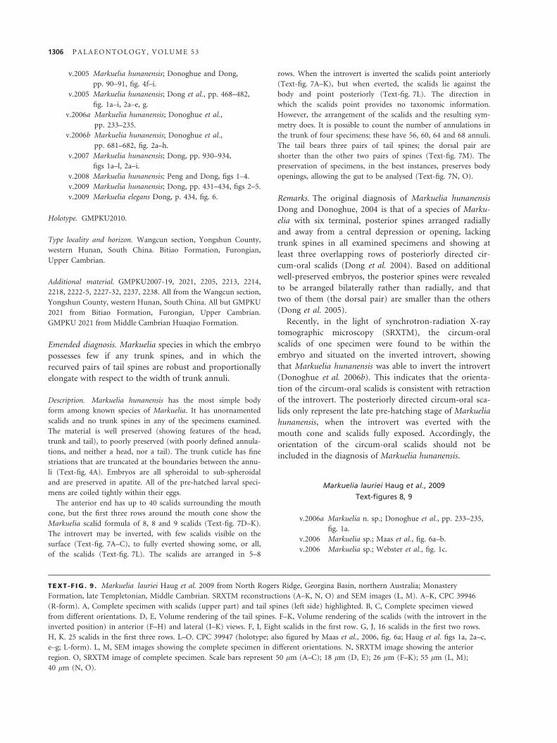
TEXT -F IG . 9 . Markuelia lauriei Haug et al. 2009 from North Rogers Ridge, Georgina Basin, northern Australia; Monastery
Formation, late Templetonian, Middle Cambrian. SRXTM reconstructions (A–K, N, O) and SEM images (L, M). A–K, CPC 39946
(R-form). A, Complete specimen with scalids (upper part) and tail spines (left side) highlighted. B, C, Complete specimen viewed
from different orientations. D, E, Volume rendering of the tail spines. F–K, Volume rendering of the scalids (with the introvert in the
inverted position) in anterior (F–H) and lateral (I–K) views. F, I, Eight scalids in the first row. G, J, 16 scalids in the first two rows.
H, K. 25 scalids in the first three rows. L–O. CPC 39947 (holotype; also figured by Maas et al., 2006, fig. 6a; Haug et al. figs 1a, 2a–c,
e–g; L-form). L, M, SEM images showing the complete specimen in different orientations. N, SRXTM image showing the anterior
region. O, SRXTM image of complete specimen. Scale bars represent 50 lm (A–C); 18 lm (D, E); 26 lm (F–K); 55 lm (L, M);
40 lm (N, O).
1306 P A L A E O N T O L O G Y , V O L U M E 5 3
A
D
L
M
N O
E F G H
KJI
B C
D O N G E T A L . : A N A T O M Y , T A P H O N O M Y , T A X O N O M Y A N D S Y S T E M A T I C A F F I N I T Y O F M A R K U E L I A 1307
A
D E F
IHG
J K
B C
1308 P A L A E O N T O L O G Y , V O L U M E 5 3
v.2007a Markuelia; Maas et al., fig. 7a.
v*2009 Markuelia lauriei Haug et al. pp. 306–312,
figs 1a–b, 2a–g, 5a–d, 6a–b.
Holotype. CPC 39947.
Type locality and horizon. North Rogers Ridge, Georgina Basin,
northern Australia. Monastery Formation, late Templetonian,
Middle Cambrian.
Additional material. CPC 39946 (from the type locality).
Diagnosis. Markuelia species with slender protuberances
on trunk annuli.
Description. Like all other species of Markuelia the embryo is
sub-spherical to discoidal and coiled in either an S-shaped or an
inverted S-shaped loop with the head and tail juxtaposed (Text-
figs 8A, B, D, E, 9L, M). The thorax is composed of 86 and 72
transverse annulations, in the specimens CPC 39946 and CPC
39947 respectively. The thorax is flattened dorsoventrally and
tapers towards the posterior end. The trunk is 125 lm in diame-
ter. Some of the segments bear protuberances (Text-fig. 8H), but
they are slender and unlike the short, broad spines of M. secunda,
with which they may be homologous. The trunk cuticle has fine
striations that appear to be continuous across the boundaries
between annuli (Text-fig. 8H). The embryo is preserved in apatite.
SRXTM revealed that the head bears scalids with the same
number and symmetry in the first three scalid rows; eight in the
first circumoral ring, eight in the second and nine in the third
(Text-fig. 9F–K). In specimen CPC 39946, a total of 35 hollow
scalids have been identified in total in five rings. The scalids
point anteriorly when in the inverted position (Text-fig. 9A–K)
and point posteriorly and lay flat to the body as they are everted
(Text-fig. 8C, D, F). One spine in CPC 39946 preserves two
accessory spines, which were termed ‘spinules’ by Haug et al.
(2009) (Text-fig. 8F; see also Haug et al. 2009, fig. 5c). SRXTM
analysis also revealed that the head is preserved, but obscured by
glue, in the holotype; the scalids are everted in this specimen
(Text-fig. 9N, O).
The tail bears six dorsoventrally recurved, equally sized, spines
in three pairs at the terminal pole of the embryo (Text-figs 8G,
9D–E). The posterior spines are c. 100 lm long and 30 lm at
their widest point.
Remarks. There are four specimens of Markuelia known
from Australia: CPC 39946 (specimen lost), CPC 39947,
CPC AAAAA and CPC BBBBB. We consider CPC 39946
and CPC 39947 to belong to M. lauriei and CPC 40429
and CPC 40430 to belong to M. waloszeki. There is some
confusion because these specimens were assigned specimen
numbers while they were temporarily housed in Bonn,
and there are inconsistencies in the way these numbers
have been used in the literature. Maas et al. (2006) figured
specimen CPC 39946 as UB W 133 and CPC 39947 as UB
W 132; both specimens were assigned to Markuelia sp.
However, Donoghue et al. (2006a) figured CPC 39946 as
UB W 132, CPC 40429 as UB W 133 and CPC 40430
without giving a specimen number; all three specimens
were assigned to Markuelia n. sp. Haug et al. (2009) estab-
lished the name Markuelia lauriei assigning CPC 39947 as
the holotype and listing CPC 39946 as an additional speci-
men belonging to this species. In their synonomy list for
M. laurei, Haug et al. listed ‘v 2006 Markuelia n. sp. Don-
oghue et al., figs 1A–D (CPC 39946, 39947)’. As figure 1B,
C of Donoghue et al. (2006a) shows CPC 40429 and 1D
shows CPC 40430, it is unclear which specimens Haug
et al. consider to be synonyms of M. lauriei.
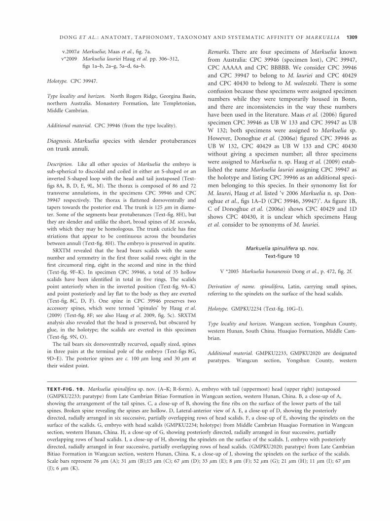
Markuelia spinulifera sp. nov.
Text-figure 10
V *2005 Markuelia hunanensis Dong et al., p. 472, fig. 2f.
Derivation of name. spinulifera, Latin, carrying small spines,
referring to the spinelets on the surface of the head scalids.
Holotype. GMPKU2234 (Text-fig. 10G–I).
Type locality and horizon. Wangcun section, Yongshun County,
western Hunan, South China. Huaqiao Formation, Middle Cam-
brian.
Additional material. GMPKU2233, GMPKU2020 are designated
paratypes. Wangcun section, Yongshun County, western
TEXT -F IG . 10 . Markuelia spinulifera sp. nov. (A–K; R-form). A, embryo with tail (uppermost) head (upper right) juxtaposed
(GMPKU2233; paratype) from Late Cambrian Bitiao Formation in Wangcun section, western Hunan, China. B, a close-up of A,
showing the arrangement of the tail spines. C, a close-up of B, showing the fine ribs on the surface of the lower parts of the tail
spines. Broken spine revealing the spines are hollow. D, Lateral-anterior view of A. E, a close-up of D, showing the posteriorly
directed, radially arranged in six successive, partially overlapping rows of head scalids. F, a close-up of E, showing the spinelets on the
surface of the scalids. G, embryo with head scalids (GMPKU2234; holotype) from Middle Cambrian Huaqiao Formation in Wangcun
section, western Hunan, China. H, a close-up of G, showing posteriorly directed, radially arranged in four successive, partially
overlapping rows of head scalids. I, a close-up of H, showing the spinelets on the surface of the scalids. J, embryo with posteriorly
directed, radially arranged in four successive, partially overlapping rows of head scalids. (GMPKU2020; paratype) from Late Cambrian
Bitiao Formation in Wangcun section, western Hunan, China. K, a close-up of J, showing the spinelets on the surface of the scalids.
Scale bars represent 76 lm (A); 31 lm (B);15 lm (C); 67 lm (D); 33 lm (E); 8 lm (F); 52 lm (G); 21 lm (H); 11 lm (I); 67 lm
(J); 6 lm (K).
D O N G E T A L . : A N A T O M Y , T A P H O N O M Y , T A X O N O M Y A N D S Y S T E M A T I C A F F I N I T Y O F M A R K U E L I A 1309
Hunan, South China. Bitioa Formation, Furongian, Upper
Cambrian.
Material. Three well-preserved specimens.
Occurrence. As for the type locality. Middle Cambrian Huaqiao
Formation and Upper Cambrian (Furongian) Bitiao Formation,
western Hunan, South China.
Diagnosis. Markuelia species in which the scalids are orna-
mented with numerous densely packed spinelets.
Description. Only the embryos of the pre-hatching stage are
preserved in our collection. The embryos are sufficiently tightly
coiled into an inverted S-shaped loop that the lateral margins of
the trunk are directly juxtaposed (Text-fig. 10A, D, G). The
trunk varies in width from 138 to 263 lm. Transverse annula-
tions (Text-fig. 10A, B, D, G) range in anterior–posterior length
from c. 21 to 32 lm. The posterior pole of the embryos is char-
acterized by a terminal spine-bearing region surrounding a cen-
tral opening. There are a total of three pairs of hollow spines
surrounding the opening (Text-fig. 10A–D). Their long axes are
parallel to, and concave margins and tips are directed away
from, the anterior–posterior axis of the animal. The two nar-
rower, straight spines (ventral pair) were positioned within the
terminal opening and arranged bilaterally. The four larger
curved spines (dorsal and lateral pairs), 75 lm in length, 22 lm
in maximum width, are curved in approximately the same direc-
A
E
F
G H
B C
D
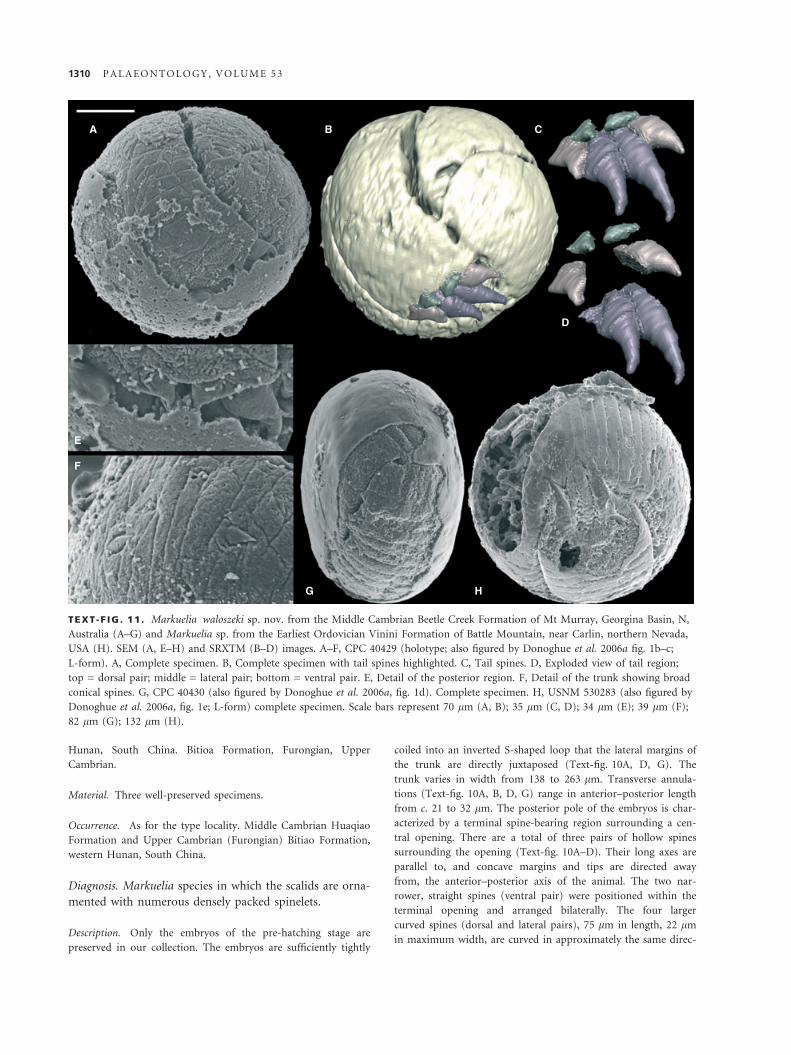
TEXT -F IG . 11 . Markuelia waloszeki sp. nov. from the Middle Cambrian Beetle Creek Formation of Mt Murray, Georgina Basin, N,
Australia (A–G) and Markuelia sp. from the Earliest Ordovician Vinini Formation of Battle Mountain, near Carlin, northern Nevada,
USA (H). SEM (A, E–H) and SRXTM (B–D) images. A–F, CPC 40429 (holotype; also figured by Donoghue et al. 2006a fig. 1b–c;
L-form). A, Complete specimen. B, Complete specimen with tail spines highlighted. C, Tail spines. D, Exploded view of tail region;
top = dorsal pair; middle = lateral pair; bottom = ventral pair. E, Detail of the posterior region. F, Detail of the trunk showing broad
conical spines. G, CPC 40430 (also figured by Donoghue et al. 2006a, fig. 1d). Complete specimen. H, USNM 530283 (also figured by
Donoghue et al. 2006a, fig. 1e; L-form) complete specimen. Scale bars represent 70 lm (A, B); 35 lm (C, D); 34 lm (E); 39 lm (F);
82 lm (G); 132 lm (H).
1310 P A L A E O N T O L O G Y , V O L U M E 5 3
tion (towards the embryo; Text-fig. 10A–D). The surface of the
upper parts, including the tips, of the tail spines are smooth,
whereas there are fine transverse ribs on the surface of the lower
parts of the tail spines (Text-fig. 10C). The anterior pole is char-
acterized by a terminal spine-bearing region. The spines, ranging
36–61 lm in length and 15–18 lm in width, are posteriorly
directed and arranged radially in four to six successive, partially
overlapping rows. Broken scalids reveal that all the spines are
hollow, and they are flattened in cross-sectional profile, oriented
with the long axis at a tangent to the surface of the trunk
(Text-fig. 10A, D–K). There are numerous spinelets on the sur-
face of all the scalids. The spinelets range 1–6 lm in length and
0.8–1.2 lm in maximum width.
Remarks. The present species is characterized by the den-
sely packed spinelets on the surface of the head scalids.
This character is the essential difference from previously
described species of Markuelia with known scalids. The
spinelets in this species are far more numerous and den-
sely packed than the structures described as spinules in
M. lauriei. Although accessory structures are rare on the
scalids of Cambrian taxa, spinelets have also been
observed on the scalids of Ottoia prolifica by Conway
Morris (1977, pl. 4, fig. 4).
Markuelia waloszeki sp. nov.
Text-figure 11A–G
v*2006a Markuelia n. sp. Donoghue et al., pp. 233–235;
fig. 1b–d.
Derivation of name. Names in honour of Prof. Dieter Waloszek
who collected and recovered the specimens.
Holotype. Specimen CPC40429 (Text-fig. 11A–F).
Type locality and horizon. Middle Cambrian Beetle Creek For-
mation of Mt Murray, Georgina Basin, N. Australia.
Additional material. One specimen from the type locality.
Occurrence. As for type locality.
Diagnosis. Markuelia species with broad conical spines on
the trunk annuli and a tail in which the dorsal pair of tail
spines is less than half the length of the ventral and lateral
pairs.
Description. The embryo is sub-spherical to discoidal, and the
body is tightly coiled into an S-shaped loop (Text-fig. 11A, G).
The anterior region is not preserved in either specimen. The
annuli bear broad conical spines (Text-fig. 11E, F). One speci-
men preserves the posterior, which bears three pairs of
recurved spines (Text-fig. 11B–D). The outer lateral and ventral
pairs are relatively broad based and squat. The dorsal pair con-
sists of spines that are also broad based and are less than half
as long as the other two pairs of spines. Both specimens are
preserved in apatite, with the holotype preserving dumb-bell-
shaped structures that may represent preserved bacteria (Text-
fig. 4D).
Remarks. Markuelia waloszeki is most similar to M. secun-
da in that both taxa have broad conical spines on the
trunk annuli and tail spines that have a relatively squat
morphology. However, the present species differs from
M. secunda in having very short dorsal tail spines and in
having a sub-spherical to discoidal form like that of
M. lauriei.
Markuelia sp.
Text-figure 11H
v.2006a Markuelia sp. Donoghue et al., fig. 1e.
Material. A single specimen, USNM 530283.
Occurrence. Earliest Ordovician Vinini Formation of Battle
Mountain, near Carlin, northern Nevada, USA.
Description. Embryo 460 lm in diameter with an annulated
trunk coiled into an S-shaped loop (Text-fig. 11H). The annu-
li do not bear protuberances. The anterior is not preserved.
The incompletely preserved tail has a pair of straight ventral
spines and weakly curved dorsal and lateral spines; the lateral
pair of spines is shorter than the other two pairs (Text-
fig. 11B–D).
Remarks. Donoghue et al. (2006a) figured a single speci-
men of Markuelia from Nevada. The specimen is most
similar to M. hunanensis in that it lacks trunk protuber-
ances and falls within the same size range. The tail possi-
bly differs from that of M. hunanensis in having straighter
spines.
Acknowledgements. We are indebted to the generosity of Prof.
Dieter Waloszek for providing access to the specimens of
M. lauriei and M. waloszeki. The University of Bonn Palaeonto-
logical Museum, and the Palaeontological Association for facili-
tating the recovery of specimens of Markuelia lauriei.
Kristineberg Marine Station provided facilities for the recovery
and culture of priapulid embryos and larvae. Correspondence
and requests for materials should be addressed to Philip Dono-
ghue. Shuhai Xiao, Nicholas J. Butterfield and an anonymous
other provided constructive comments on the manuscript. NSFC
Project 40772008 (XPD), RFDP Project 20060001059 (XPD),
LPS of Nanjing Institute of Geology and Palaeontology Grant
083101 (XPD), NERC Standard grants NE ⁄ C511256 ⁄ 1 (PCJD)
and NE ⁄ F00348X ⁄ 1 (PCJD).
Editor. Phil Lane
D O N G E T A L . : A N A T O M Y , T A P H O N O M Y , T A X O N O M Y A N D S Y S T E M A T I C A F F I N I T Y O F M A R K U E L I A 1311
REFERENCES
A G U I N A L D O, A. M. A., T UR B E V I LL E , J. M., L I N -
F OR D , L. J., R I V E R A , M. C., G A R E Y , J. R., R A F F , R. A.
and L A KE , J. A. 1997. Evidence for a clade of nema-
todes, arthropods and other moulting animals. Nature, 387,
489–493.
B A R T ON , N. H., BR I G GS , D. E. G., E I S E N , J. A., G O L D-
S T E I N , D. B. and PA T E L , N. H. 2007. Evolution. Cold
Spring Harbor Press, New York, 833 pp.
B E N G T S ON , S. and Y UE , Z. 1997. Fossilized metazoan
embryos from the earliest Cambrian. Science, 277, 1645–
1648.
B R I G G S , D. E. G. 2003. The role of decay and mineralization
in the preservation of soft-bodied fossils. Annual Review of
Earth and Planetary Science, 31, 275–301.
—— and K E A R , A. J. 1993. Fossilization of soft tissue in the
laboratory. Science, 259, 1439–1442.
—— —— 1994. Decay and mineralisation of shrimps. Palaios, 9,
431–456.
—— and W I L B Y , P. R. 1996. The role of the calcium carbon-
ate – calcium phosphate switch in the mineralization of soft-
bodied fossils. Journal of the Geological Society, London, 153,
665–668.
C H A FE T Z , H. S. and BU CZ Y N S KI , C. 1992. Bacterially
induced lithifcation of microbial mats. Palaios, 7, 277–293.
C OB B E T T , A., W I L KI N S ON , M. and W I L L S , M. A. 2007.
Fossils impact as hard as living taxa in parsimony analyses of
morphology. Systematic Biology, 56, 753–766.
C ON W A Y M O R R I S , S. 1977. Fossil priapulid worms. Special
Papers in Palaeontology, 20, 1–95.
—— 1998. Eggs and embryos from the Cambrian. Bioessays, 20,
676–682.
C OS TA , M., DR A P E R , B. W. and PR I E S S , J. R. 1997. The
role of actin filaments in patterning the Caenorhabditis elegans
cuticle. Developmental Biology, 184, 373–384.
D ON G , X.-P. 2007. Developmental sequence of the Cam-
brian embryo Markuelia. Chinese Science Bulletin, 52, 929–
935.
——2009. Cambrian fossil embryos from Western Hunan, South
China. Acta Geologica Sinica, 83, 429–439.
—— and DO N O G HU E , P. C. J. 2002. Affinity of the earliest
bilaterian embryos. Palaeontology Newsletter, 51, 84–85.
—— —— C HE N G, H. and L I U , J. 2004. Fossil embryos from
the Middle and Late Cambrian period of Hunan, south China.
Nature, 427, 237–240.
—— —— C UN N I N G HA M , J., L I U, J. and CH E N G , H.
2005. The anatomy, affinity and phylogenetic significance of
Markuelia. Evolution and Development, 7, 468–482.
D ON O G H UE , P. C. J. 2005. Saving the stem-group – a con-
tradiction in terms. Paleobiology, 31, 553–558.
—— and D ON G , X.-P. 2005. Embryos and ancestors. 81–99.
In BR I G GS , D. E. G. (ed.). Evolving form and function: fossils
and development. Yale Peabody Museum of Natural History,
Yale University, New Haven, 288 pp.
—— and P UR N E L L , M. A. 2009. Distinguishing heat
from light in debate over controversial fossils. Bioessays, 31,
178–189.
—— KO U CH I N S KY , A., W A L OS Z E K , D., BE N G T S O N ,
S., D ON G , X.-P., V A L ’ K OV , A. K., C UN N I N G HA M ,
J. A. and R E P E T S K I , J. E. 2006a. Fossilized embryos are
widespread but the record is temporally and taxonomically
biased. Evolution and Development, 8, 232–238.
—— B E N G T S O N , S., D O N G, X.-P., GO S T L I N G , N. J.,
HU L DT G R E N , T., C UN N I N G HA M , J. A., Y I N , C.,
Y UE , Z., P E N G , F. and S TA M P A N ON I , M. 2006b. Syn-
chrotron X-ray tomographic microscopy of fossil embryos.
Nature, 442, 680–683.
D UN N , C. W., HE J N O L, A., M A T U S , D. Q., P A N G , K.,
BR O W N E , W. E., S M I T H , S. A., S E A V E R , E., R O U S E ,
G. W., O B S T , M., E D G E CO M B E , G. D., S OR E N S E N ,
M. V., HA D DO C K, S. H. D., S C H M I D T - R HA E S A , A.,
O K U S U , A., K R I S T E N S E N , R. M., W H E E L E R , W. C.,
M A R T I N D A L E , M. Q. and G I R I B E T , G. 2008. Broad
phylogenomic sampling improves resolution of the animal tree
of life. Nature, 452, 745–749.
F US CO , G. 2005. Trunk segment numbers and sequential seg-
mentation in myriapods. Evolution and Development, 7, 608–
617.
G O S T L I N G , N. J., DO N G , X.-P. and DO N O GH U E , P. C.
J. 2009. Ontogeny and taphonomy: and experimental taphon-
omy study of the development of the brine shrimp Artemia
salina. Palaeontology, 52, 169–186.
—— T H OM A S , C.-W., G R E E N W O OD , J. M., DO N G ,
X.-P., BE N GT S O N , S., R A F F , E. C., R A F F , R. A., DE -
G N A N , B. M., S T A M PA N O N I , M. and DO N O G HU E ,
P. C. J. 2008. Deciphering the fossil record of early bilaterian
embryonic development in light of experimental taphonomy.
Evolution and Development, 10, 339–349.
H A G A DO R N , J. W., X I A O, S., D ON O G H UE , P. C. J.,
BE N GT S O N , S., GO S T L I N G , N. J., P A W L O W S K A ,
M., R A F F , E. C., R A FF , R. A., T U R N E R , F. R., Y I N , C.,
Z HO U, C., Y U A N , X., M CF E E L Y , M. B., S T A M P A -
N ON I , M. and N E A L S ON , K. H. 2006. Cellular and sub-
cellular structure of Neoproterozoic animal embryos. Science,
314, 291–294.
H A R V E Y , T. H. P., D ON G, X.-P. and DO N OG H UE , P. C.
J. 2010. Are palaeoscolecids ancestral ecdysozoans? Evolution
and Development, 12, 177–200.
H A UG , J. T., M A A S , A., W A L O S Z E K, D., DO N O GH U E ,
P. C. J. and B E N GT S O N , S. 2009. A new species of Marku-
elia from the Middle Cambrian of Australia. Memoirs of the
Association of Australasian Palaeontologists, 37, 303–313.
H E N N I G , W. 1981. Insect phylogeny. John Wiley, New York,
514 pp.
H OF , C. H. J. and BR I GG S , D. E. G. 1997. Decay and miner-
alisation of mantis shrimps (Stomatopoda: Crustacea) – a key
to their fossil record. Palaios, 12, 420–438.
H UA N G , D., C H E N , J. Y. and V A N N I E R , J. 2006. Discus-
sion on the systematic position of the Early Cambrian
priapulomorph worms. Chinese Science Bulletin, 51, 243–
249.
J O N D E L I U S , U., R UI Z - T R I L L O , I., BA GU N A , J. and
R I U TO R T , M. 2002. The Nemertodermatida are basal
bilaterians and not members of the Platyhelminthes. Zoologica
Scripta, 31, 201–215.
1312 P A L A E O N T O L O G Y , V O L U M E 5 3
K E A R , A. J., B R I G G S , D. E. G. and DO N O V A N , D. T.
1995. Decay and fossilization of nonmineralized tissue in cole-
oid cephalopods. Palaeontology, 38, 105–131.
K H OM E N T OV S KY , V. V. and K A R L OV A , G. A. 1993.
Biostratigraphy of the Vendian–Cambrian beds and lower
Cambrian boundary in Siberia. Geological Magazine, 130, 29–
45.
—— —— 2005. The Tommotian Stage base as the Cambrian
Lower boundary in Siberia. Stratigraphy and Geological Corre-
lation, 13, 21–34.
—— V A L ’ KO V , A. K. and K A R L OV A , G. A. 1990. Novye
dannye po biostratigrafii perekhodnykh vend-kembrijskikh
sloev v bassejne srednego techeniya r. Aldan (New data on the
biostratigraphy of transitional Vendian-Cambrian strata in the
middle reaches of the River Aldan). In K HO M E N TO V S K Y ,
V. V. and GI B S HE R , A. S. (eds). Pozdniy dokembrij i ranniy
paleozoj Sibiri. Voprosy regional’noy Stratigrafi Institut Geo-
logiii i Geofiziki, Sibirskoe Otdelenie, Akademiya Nauk SSSR,
Novosibirsk, 3–57 pp.
—— —— —— and N U ZH N O V , S. V. 1983. Opornyj razrez
perekhodnykh dokembrijsko-kembrijskikh otlozhenija nizovij
r. Gonam. [Reference section for the transitional Precambrian-
Cambrian deposits of the lower reaches of the River Gonam].
In K H OM E N T OV S KY I J , V. V., Y A K S H I N , M. S. and
KA RL O V A , G.A. (eds). Pozdnij dokembrij i rannij paleozoj
Sibiri – Vendskie otlozheniya. Institut geologii i geofiziki, SO
AN SSSR, Novasibirsk, 24–36 pp.
L E M BU R G , C. 1995. Ultrastructure of the introvert and asso-
ciated structures of the larvae of Halicryptus spinulosus (Pria-
pulida). Zoomorphology, 115, 11–29.
—— 1999. Ultrastrukurelle Untersuchungen an den Larven von
Halicryptus spinulosus und Priapulus caudatus: Hypothesen zur
Phylogenie der Priapulida und deren Bedeutung fur die
Evolution der Nemathelminthes. Cuvillier Verlag, Gottingen,
393 pp.
M A A S , A., W A L OS Z E K , D., H A UG , J. T. and M U L L E R ,
K. J. 2007a. A possible larval roundworm from the Cambrian
‘Orsten’ and its bearing on the phylogeny of Cycloneuralia.
Memoirs of the Association of Australasian Palaeontologists, 34,
499–519.
—— H UA N G , D., C H E N , J., W A L O S Z E K , D. and
B R A UN , A. 2007b. Maotianshan-Shale nemathelminths –
morphology, biology, and the phylogeny of Nemathelminthes.
Palaeogeography, Palaeoclimatology, Palaeoecology, 254, 288–
306.
—— B R A UN , A., DO N G , X.-P., D ON O G HU E , P. C. J.,
M U L L E R , K. J., O L E M P S KA , E., R E P E T S K I , J. E.,
S I V E TE R , D. J., S T E I N , M. and W A L OS ZE K , D.
2006. The ‘Orsten’ – more than a Cambrian Konservat-
Lagerstatte yielding exceptional preservation. Palaeoworld, 15,
266–282.
M A R T I N , D., B R I G G S , D. E. G. and P A R KE S , R. J.
2003. Experimental mineralization of invertebrate eggs and
the preservation of Neoproterozoic embryos. Geology, 31,
39–42.
—— —— —— 2004. Experimental attachment of sediment
particles to invertebrate eggs and the preservation of soft-
bodied fossils. Journal of the Geological Society, London, 161,
735–738.
—— —— —— 2005. Decay and mineralization of invertebrate
eggs. Palaios, 20, 562–572.
M I S S A R Z H E V S K Y , V. V. 1989. Drevnejshie skeletnye okame-
nelosti i stratigrafiya pogranichnykh tolshch dokembriya i kem-
briya. [The oldest skeletal fossils and stratigraphy of the
Precambrian-Cambrian boundary beds]. Geologicheskij institut
AN SSSR, Trudy 443, 237 pp.
N I E L S E N , C. 1995. Animal evolution: interrelationships of the
living phyla. Oxford University Press, New York, 467 pp.
—— 2001. Animal evolution: interrelationships of the living phyla.
Oxford University Press, Oxford, 563 pp.
P A R K, J. K., R H O, H. S., K R I S T E N S E N , R. M., K I M , W.
and G I R I BE T, G. 2006. First molecular data on the phylum
Loricifera – an investigation into the phylogeny of Ecdysozoa
with emphasis on the positions of Loricifera and Priapulida.
Zoological Science, 23, 943–954.
P E E L , A. 2004. The evolution of arthropod segmentation mech-
anisms. BioEssays, 26, 1108–1116.
P E N G , F. and D O N G , X.-P. 2008. Application of Synchrotron
X-Ray Tomography in the research of fossil embryo Marku-
elia. Acta Scientiarum Naturalium Universitatis Pekinensis, 44,
447–451.
R A F F , E. C., V I L L I N S KI , J. T., T UR N E R , F. R., D ON -
OG H U E , P. C. J. and R A FF , R. A. 2006. Experimental
taphonomy shows the feasibility of fossil embryos. Proceedings
of the National Academy of Sciences, USA, 103, 5846–5851.
—— S C H OL L A E R T , K. L., N E L S O N , D. E., D ON -
OG H U E , P. C. J., T H OM A S , C.-W., T U R N E R , F. R.,
S T E I N , B. D., D ON G , X.-P., B E N G T S O N , S., HU L DT -
G R E N , T., S TA M P A N ON I , M., C HO N G Y U , Y. and
R A F F, R. A. 2008. Embryo fossilization is a biological process
mediated by microbial biofilms. Proceedings of the National
Academy of Sciences, 105, 19359–19364.
S A G E M A N N , J., B A L E , S. J., B R I G G S , D. E. G. and PA R -
KE S , R. J. 1999. Controls on the formation of authigenic
mineral in association with decaying organic matter: an exper-
imental approach. Geochemica et Cosmochemica Acta, 63,
1083–1095.
S Ø R E N S E N , M. V., HE B S G A A R D , M. B., HE I N E R , I.,
G L E N N E R , H., W I L L E R S L E V , E. and KR I S T E N S E N ,
R. M. 2008. New data from an enigmatic phylum: evidence
from molecular sequence data supports a sister-group
relationship between Loricifera and Nematomorpha. Journal
of Zoological Systematics and Evolutionary Research, 46, 231–
239.
S T E I N E R , M., Z H U, M., L I , G., Q I A N , Y. and E R DT -
M A N N , B.-D. 2004. New early Cambrian bilaterian embryos
and larvae from China. Geology, 32, 833–836.
V A L ’ K O V , A. K. 1983. Rasprostranenie drevnejshikh skele-
tnykh organizmov i korrelyatsiya nizhnej granitsy kembriya v
yugo-vostochnoj chasti Sibirskoj platformy [Distribution of
the oldest skeletal organisms and correlation of the lower
boundary of the Cambrian in the southeastern part of the
Siberian Platform]. 37–48. In KH O M E N T O V S K Y , V. V.,
Y A K S H I N , M. S. and K A R L O V A , G. A. (eds). Pozdnij do-
D O N G E T A L . : A N A T O M Y , T A P H O N O M Y , T A X O N O M Y A N D S Y S T E M A T I C A F F I N I T Y O F M A R K U E L I A 1313
kembrij i rannij paleozoj Sibiri, Vendskie otlozheniya. Institut
geologii i geofiziki, SO AN SSSR, Novosibirsk.
—— 1987. Biostratigrafiya nizhnego kembriya vostoka Sibirskoj
Platformy (Yudoma-Olenyokskij region). [Lower Cambrian
biostratigraphy of the eastern part of the Siberian Platform
(Yudoma-Olenyok region)]. Nauka, Moscow, 136 pp.
—— and K A R L OV A , G. A. 1984. Fauna iz perekhodnykh
vendsko-kembrijskikh sloev nizhnego techeniya r. Gonam.
[The fauna of the transitional Vendian–Cambrian beds in the
lower reaches of the River Gonam]. 12–41. In K H O M E N -
T OV S KY , V. V., S H E N F I L , Y. Y. and Y A KS H I N , M. S.,
(eds). Stratigrafiya pozdnego dokembriya i rannego paleozoya.
Srednyaya Sibir. Institut geologii i geofiziki, Novosibirsk.
W E B S T E R , B. L., C O PL E Y , R. R., J E N N E R, R. A., M A C-
KE N Z I E - DO DD S , J. A., B OU R L A T , S. J., R OT A -
S T A BE LL I , O., L I T T L E W O OD , D. T. J. and T E L F OR D ,
M. J. 2006. Mitogenomics and phylogenomics reveal priapulid
worms as extant models of the ancestral Ecdysozoan. Evolution
and Development, 8, 502–510.
W I L BY , P. R. and B R I G G S , D. E. G. 1997. Taxonomic trends
in the resolution of detail preserved in fossil phosphatised soft
tissues. Geobios, 20, 493–502.
W I L L S , M. A. 1998. Cambrian and recent disparity: the picture
from priapulids. Paleobiology, 24, 177–199.
X I A O, S. and KN O L L , A. H. 1999. Fossil preservation in the
Neoproterozoic Doushantuo phosphorite Lagerstatte, South
China. Lethaia, 32, 219–240.
—— ZHANG, Y. and KN O L L , A. H. 1998. Three-dimensional
preservation of algae and animal embryos in a Neoproterozoic
phosphate. Nature, 391, 553–558.
1314 P A L A E O N T O L O G Y , V O L U M E 5 3