A strain-independent postnatal neurodegeneration in mice...
13
The EMBO Journal Vol.17 No.3 pp.719–731, 1998 A strain-independent postnatal neurodegeneration in mice lacking the EGF receptor Maria Sibilia, Joachim P.Steinbach 1 , Laura Stingl, Adriano Aguzzi 1 and Erwin F.Wagner 2 Research Institute of Molecular Pathology (IMP), Dr Bohr-Gasse 7, A-1030 Vienna, Austria and 1 Institute of Neuropathology, University of Zurich, Schmelzbergstrasse 12, CH–8091 Zurich, Switzerland 2 Corresponding author e-mail: [email protected] Mice lacking the epidermal growth factor receptor (EGFR) exhibit strain-dependent phenotypes ranging from placental to postnatal skin, lung and brain defects. After birth, all mutant mice develop a pro- gressive neurodegeneration in the frontal cortex, olfactory bulb and thalamus, characterized by massive apoptosis and upregulation of c-fos. These defects occur in a strain-independent manner, since neither rescue of the placental phenotype by aggregation of diploid 129/Sv EGFR mutant and tetraploid wild-type embryos, nor promotion of lung maturation by trans- placental dexamethasone administration alters the course of neurodegeneration. VEGF is not induced during the degenerative process, excluding hypoxia and ischemia as causes of cell death. A migratory disorder is detected in the hippocampus with nests of ectopic neurons, which are also apoptotic. Cerebral cortices from EGFR mutants contain lower numbers of GFAP positive astrocytes, which display reduced proliferation in vitro. Since EGFR is expressed in the affected cell-types, these results define a specific function for EGFR in the proliferation and/or differ- entiation of astrocytes and in the survival of postmitotic neurons. Keywords: astrocytes/EGF receptor/genetic background/ knock-out mice/neurodegeneration Introduction The epidermal growth factor receptor (EGFR) is a member of a family of structurally related tyrosine kinase receptors that include erbB2/neu, erbB3 and erbB4 (Earp et al., 1995). The EGFR binds and is activated by several polypeptide growth factors including epidermal growth factor (EGF), transforming growth factor α (TGFα), amphiregulin, heparin-binding EGF (HB-EGF), beta- cellulin and epiregulin (Prigent and Lemoine, 1992; Earp et al., 1995). Ligand binding induces receptor dimerization and activation of the intrinsic tyrosine kinase with consequent autophosphorylation of key tyrosines located at the carboxy terminal tail of the receptor (Prigent and Lemoine, 1992; Earp et al., 1995). Phosphorylated tyrosine residues act as binding sites for proteins containing © Oxford University Press 719 Src-homology 2 domains (SH2) such as Grb2, SHC and PLCγ which, in turn, activate complex downstream signaling cascades, thus transducing extracellular stimuli to the nucleus (Lemmon and Schlessinger, 1994; Weiss et al., 1997). The specificity of the cellular response is thought to be determined by the nature of the various signaling molecules recruited to the phosphorylated receptor (Earp et al., 1995; Weiss et al., 1997). EGFR dimerization can take place between two identical recep- tors (homodimerization) or with any of the three other members of the erbB family (heterodimerization), depending on which receptor proteins are expressed in a given cell (Lemmon and Schlessinger, 1994; Earp et al., 1995). This increases the number of signaling pathways that can be activated after EGFR stimulation, thereby augmenting the signaling complexity needed to govern cell proliferation and differentiation (Lemmon and Schlessinger, 1994; Earp et al., 1995; Weiss et al., 1997). During early mouse development, EGFR protein can be detected in the trophoectoderm of the blastocyst, which is the first epithelium that develops in mammalian embryos (Dardik et al., 1992; Wiley et al., 1992). From mid- gestation on, the EGFR is expressed in a variety of tissues and organs, but only few studies have determined which specific cell types express the receptor (Adamson, 1990; Partanen, 1990; Derynck, 1992). In adult mice, EGFR expression can be detected in all organs, particularly in the liver and in regenerating epithelia such as skin and gut (Adamson, 1990; Partanen, 1990; Derynck, 1992). Several studies have demonstrated the presence of immunoreactive EGFR in selected regions of the embryonic and adult brain, such as the frontal cortex, striatum, hippocampus and cerebellum (Go ´mez-Pinillla et al., 1988; Werner et al., 1988; Tucker et al., 1993; Kornblum et al., 1997). Furthermore, EGF and TGFα have also been detected in several brain regions including the striatum and hippocampus (Lazar and Blum, 1992; Seroogy et al., 1993; Tucker et al., 1993; Weickert and Blum, 1995; Kornblum et al., 1997). Although the cellular distribution and developmental appearance of EGFR within these regions have been controversial, it seems that the receptor is present in certain specific neuronal cell populations and mature astrocytes (Go ´mez-Pinillla et al., 1988; Nieto-Sampedro et al., 1988; Topp et al., 1989; Kornblum et al., 1997). Particularly, EGFR is upregulated in reactive astrocytes after brain injury (Nieto-Sampedro et al., 1988). This astrocytic response, also known as reactive astrogliosis, is characterized by the presence of astrocytes becoming hypertrophic, showing a great increase in glial fibrillary acidic protein (GFAP) expression and the capability to divide (Nieto-Sampedro et al., 1988). EGF has also been shown to induce thymidine incorporation and proliferation in primary astrocytes cul- tured in vitro (Simpson et al., 1982). Recently, it has been
Transcript of A strain-independent postnatal neurodegeneration in mice...

The EMBO Journal Vol.17 No.3 pp.719–731, 1998
A strain-independent postnatal neurodegenerationin mice lacking the EGF receptor
Maria Sibilia, Joachim P.Steinbach1,Laura Stingl, Adriano Aguzzi1 andErwin F.Wagner2
Research Institute of Molecular Pathology (IMP), Dr Bohr-Gasse 7,A-1030 Vienna, Austria and 1Institute of Neuropathology,University of Zurich, Schmelzbergstrasse 12, CH–8091 Zurich,Switzerland
2Corresponding authore-mail: [email protected]
Mice lacking the epidermal growth factor receptor(EGFR) exhibit strain-dependent phenotypes rangingfrom placental to postnatal skin, lung and braindefects. After birth, all mutant mice develop a pro-gressive neurodegeneration in the frontal cortex,olfactory bulb and thalamus, characterized by massiveapoptosis and upregulation of c-fos. These defects occurin a strain-independent manner, since neither rescueof the placental phenotype by aggregation of diploid129/Sv EGFR mutant and tetraploid wild-typeembryos, nor promotion of lung maturation by trans-placental dexamethasone administration alters thecourse of neurodegeneration. VEGF is not inducedduring the degenerative process, excluding hypoxiaand ischemia as causes of cell death. A migratorydisorder is detected in the hippocampus with nests ofectopic neurons, which are also apoptotic. Cerebralcortices from EGFR mutants contain lower numbersof GFAP positive astrocytes, which display reducedproliferation in vitro. Since EGFR is expressed inthe affected cell-types, these results define a specificfunction for EGFR in the proliferation and/or differ-entiation of astrocytes and in the survival of postmitoticneurons.Keywords: astrocytes/EGF receptor/genetic background/knock-out mice/neurodegeneration
Introduction
The epidermal growth factor receptor (EGFR) is a memberof a family of structurally related tyrosine kinase receptorsthat include erbB2/neu, erbB3 and erbB4 (Earp et al.,1995). The EGFR binds and is activated by severalpolypeptide growth factors including epidermal growthfactor (EGF), transforming growth factor α (TGFα),amphiregulin, heparin-binding EGF (HB-EGF), beta-cellulin and epiregulin (Prigent and Lemoine, 1992;Earp et al., 1995). Ligand binding induces receptordimerization and activation of the intrinsic tyrosine kinasewith consequent autophosphorylation of key tyrosineslocated at the carboxy terminal tail of the receptor (Prigentand Lemoine, 1992; Earp et al., 1995). Phosphorylatedtyrosine residues act as binding sites for proteins containing
© Oxford University Press 719
Src-homology 2 domains (SH2) such as Grb2, SHCand PLCγ which, in turn, activate complex downstreamsignaling cascades, thus transducing extracellular stimulito the nucleus (Lemmon and Schlessinger, 1994; Weisset al., 1997). The specificity of the cellular response isthought to be determined by the nature of the varioussignaling molecules recruited to the phosphorylatedreceptor (Earp et al., 1995; Weiss et al., 1997). EGFRdimerization can take place between two identical recep-tors (homodimerization) or with any of the three othermembers of the erbB family (heterodimerization),depending on which receptor proteins are expressed in agiven cell (Lemmon and Schlessinger, 1994; Earp et al.,1995). This increases the number of signaling pathwaysthat can be activated after EGFR stimulation, therebyaugmenting the signaling complexity needed to governcell proliferation and differentiation (Lemmon andSchlessinger, 1994; Earp et al., 1995; Weiss et al., 1997).
During early mouse development, EGFR protein canbe detected in the trophoectoderm of the blastocyst, whichis the first epithelium that develops in mammalian embryos(Dardik et al., 1992; Wiley et al., 1992). From mid-gestation on, the EGFR is expressed in a variety of tissuesand organs, but only few studies have determined whichspecific cell types express the receptor (Adamson, 1990;Partanen, 1990; Derynck, 1992). In adult mice, EGFRexpression can be detected in all organs, particularly inthe liver and in regenerating epithelia such as skin andgut (Adamson, 1990; Partanen, 1990; Derynck, 1992).Several studies have demonstrated the presence ofimmunoreactive EGFR in selected regions of theembryonic and adult brain, such as the frontal cortex,striatum, hippocampus and cerebellum (Gomez-Pinilllaet al., 1988; Werner et al., 1988; Tucker et al., 1993;Kornblum et al., 1997). Furthermore, EGF and TGFαhave also been detected in several brain regions includingthe striatum and hippocampus (Lazar and Blum, 1992;Seroogy et al., 1993; Tucker et al., 1993; Weickert andBlum, 1995; Kornblum et al., 1997). Although the cellulardistribution and developmental appearance of EGFRwithin these regions have been controversial, it seems thatthe receptor is present in certain specific neuronal cellpopulations and mature astrocytes (Gomez-Pinillla et al.,1988; Nieto-Sampedro et al., 1988; Topp et al., 1989;Kornblum et al., 1997). Particularly, EGFR is upregulatedin reactive astrocytes after brain injury (Nieto-Sampedroet al., 1988). This astrocytic response, also known asreactive astrogliosis, is characterized by the presence ofastrocytes becoming hypertrophic, showing a greatincrease in glial fibrillary acidic protein (GFAP) expressionand the capability to divide (Nieto-Sampedro et al.,1988). EGF has also been shown to induce thymidineincorporation and proliferation in primary astrocytes cul-tured in vitro (Simpson et al., 1982). Recently, it has been

M.Sibilia et al.
shown that cells of the subependymal layer, a specializedzone of the striatum, are EGFR immunoreactive (forreview see Weiss et al., 1996). When cultured in vitro, theseEGF responsive cells exhibit properties representative ofstem cells, in that they are capable of maintaining andexpanding themselves over extended periods of timeand retain the ability to differentiate into neurons, astro-cytes and oligodendrocytes (for review see Weiss et al.,1996). EGF and TGFα have also been shown to increasethe survival of cortical and midbrain neurons in vitro(Kornblum et al., 1990; Casper et al., 1991; Alexi andHefti, 1993). These observations suggest that EGFRsignaling may play an important role during brain develop-ment. In view of the expression pattern of EGFR and itsligands, it is intriguing that EGFR is amplified andoverexpressed in human tumors of epithelial (carcinomas;Derynck et al., 1987) and glial origin (glioblastomas;Wong et al., 1992).
The observations that EGFR expression might be ofphysiological relevance during normal epithelial andneural development were confirmed by the analysis ofmice that are defective in EGFR signaling. Mice deficientfor the TGFα gene display mild phenotypes consistingof wavy coat, curly whiskers and sporadic eye defects(Luetteke et al., 1993; Mann et al., 1993). The phenotypeof these mice is very similar to that of the naturallyoccurring mouse mutant strains waved-1 (wa-1) andwaved-2 (wa-2) (Luetteke et al., 1993, 1994; Mann et al.,1993; Fowler et al., 1995). The wa-1 mutation maps tothe TGFα locus (Luetteke et al., 1993; Mann et al., 1993),whereas the wa-2 mutation is a hypomorphic EGFR allelethat carries a point mutation in the kinase domain, resultingin a drastically reduced kinase activity (Luetteke et al.,1994; Fowler et al., 1995). This suggests that TGFα/EGFR signaling is critical for the development of normalhair follicles and skin. Besides a reduction in glial fibrillaryacidic protein (GFAP) immunoreactivity in wa-1 mutantmice (Weickert and Blum, 1995), no obvious brainabnormalities were reported in any of these mice (Luettekeet al., 1993; Mann et al., 1993; Weickert and Blum, 1995).
Ourselves and others have generated mice which carrya null mutation in the EGFR gene (Miettinen et al., 1995;Sibilia and Wagner, 1995; Threadgill et al., 1995). Theanalysis of EGFR mutant mice revealed a complex role forthis receptor during embryonic and postnatal development.Mutant mice are growth-retarded and die at different stagesof development depending on their genetic background(Sibilia and Wagner, 1995; Threadgill et al., 1995). In a129/Sv genetic background, EGFR –/– embryos die aroundday 11.5 of gestation (E11.5), whereas in other back-grounds, mutant mice can survive until birth (C57BL/6)or to postnatal day 20 (MF1). Death in utero mostlikely results from a defect in the spongiotrophoblasts, aparticular epithelial cell layer of the placenta (Sibilia andWagner, 1995). While the size of the spongiotrophoblastlayer is reduced to the same extent in mutant placentas ofall genetic backgrounds, it remains to be clarified whetherthe absence of EGFR influences the severity of theplacental defect or whether, in addition, it affects thedevelopment of EGFR mutant embryos in a strain-dependent manner. All surviving mutant mice showabnormalities in various epithelia such as skin, hairfollicles, eyes and lungs, indicating that EGFR plays
720
an essential role in epithelial cell proliferation and/ordifferentiation (Miettinen et al., 1995; Sibilia and Wagner,1995). The lung immaturity is most probably responsiblefor the majority of mutant newborns being unable toinitiate or sustain respiration (Miettinen et al., 1995;Sibilia and Wagner, 1995). We have observed brain defectsin surviving EGFR mutant mice and an atrophy of theanterior cerebral cortex has previously been described(Threadgill et al., 1995). The nature, extent and kineticsof the degenerative processes have not been studied anda strain-specific effect could not be excluded. In addition,these defects could have occurred as a consequence ofimpaired nutrition supply during embryogenesis, attribut-able to the placental defects or a reduced oxygen supplyat birth caused by the lung immaturity. Here, we havecharacterized in detail the brain defects and demonstratethat EGFR signaling is involved in the proliferation and/or differentiation of astrocytes and survival of postmitoticneurons in vivo. We also show that the neurodegenerativedisease is strain-independent and develops in all survivingmutant mice.
Results
CNS defects in EGFR mutant mice
EGFR mutant mice of MF1 and C3H background cansurvive up to postnatal day 20 (P20) with multiple epi-thelial defects, but develop severe brain abnormalities byP18 (Sibilia and Wagner, 1995; Threadgill et al., 1995).In order to investigate the onset and extent of neuro-degeneration, brains of EGFR mutant mice of MF1 andC3H background were isolated at different stages ofembryonic and postnatal development. During embryo-genesis, mutant brains displayed no gross structuralabnormalities when compared with controls (data notshown). At birth (P1) and during the following days(P2–3), the brains of all mutant mice were still comparablewith the controls and histological examinations did notreveal any structural changes (data not shown). BetweenP4 and P6, leptomeningeal hemorrhages appeared on thesurfaces of the forebrain and olfactory bulbs of all EGFRmutant brains, heralding the onset of neurodegeneration(Figure 1A and B). Initially, these leptomeningeal lesionswere unilateral in some animals (Figure 1B), but later,both hemispheres were consistently affected (data notshown). There were differences in the extent of neuro-degeneration between the affected and unaffected hemi-spheres (Figure 1B), but these usually correlated with themacroscopically visible hemorrhagic lesions. By P13, theforebrain size of the mutants was dramatically reduced(Figure 1D) with the loss of a major part of the frontalcortex and degeneration of the olfactory bulbs (Figure1F). Conspicuously, the retrosplenial cortex (rc) was sparedin all animals investigated (Figure 1F). At later timepoints,the degenerated tissue was replaced by large cystic cavities,covered only by a thin layer of subleptomeningeal corticaltissue (data not shown).
During embryogenesis and until P3, no signs ofdegeneration and no significant increase in TUNEL stain-ing could be detected in EGFR –/– brains (data notshown). Between P4 and P5, when the leptomeningealhemorrhages appeared, the first morphological changeswere observed in neurons of EGFR –/– frontal cortex,

Postnatal neurodegeneration in EGFR-deficient mice
Fig. 1. Cortical degeneration and EGFR expression in mutant brains. Dorsal view of the whole brain of control (A and C) and EGFR –/– mice(B and D) isolated at postnatal day 5 (P5) and 13 (P13). Until P4 the brains of EGFR –/– mice look normal and the size is comparable to thecontrol. (A and B) At day 5 hemorrhagic lesions appear on the surface of the anterior part of EGFR –/– forebrain (arrows) and olfactory bulbs.(C and D) By day 13, the forebrain of the –/– is dramatically reduced in size compared with the control. (E and F) Histological cross-sections at thelevel of the dotted lines of the P13 brains shown in (C) and (D), respectively. In correspondence to the leptomeningeal hemorrhages, the region ofthe cortex between the arrows and the olfactory bulbs (ol) is completely degenerated in the mutant brains, whereas the retrosplenial cortex (rc) isspared. A similar kinetic of cortical degeneration with the presence of uni- or bilateral leptomeningeal hemorrhages around P5 was observed inbrains of tetraploid rescued and dexamethasone treated mice. (G–K) EGFR expression monitored by X-gal staining (blue) in P30 (G and H), P3(I and K) EGFR �/– and P30 (J) EGFR �/� brains. (G) Histological section through the cortex shows X-gal staining in the leptomeninges (arrow),vessels (V) and weak staining in neural cells (arrowhead). (H and J) Magnification of the vessel shown in (G) and of an EGFR �/� control,respectively. (I and K) High magnifications of stained neurons in the cortex (I) and olfactory bulbs (K). HE, hematoxylin and eosin.
consisting of nuclear condensation and diminished celldensity (Figure 2A and B), and TUNEL staining revealedabundant apoptosis (Figure 2C and D). The degenerativeprocess extended rapidly, and by P6–8 there were virtuallyno viable neurons in the affected areas, as demonstratedby the complete loss of microtubule associated protein-2
721
(MAP-2) immunoreactivity (Figure 2E and F). No GFAPstaining could be detected in the degenerating areas,indicating that the astrocytes were also affected. Onlya thin ribbon of cortical tissue, localized below theleptomeninges, was spared (Figure 2F). The cellular debriswas phagocytosed by accumulations of F4/80 positive
M.Sibilia et al.
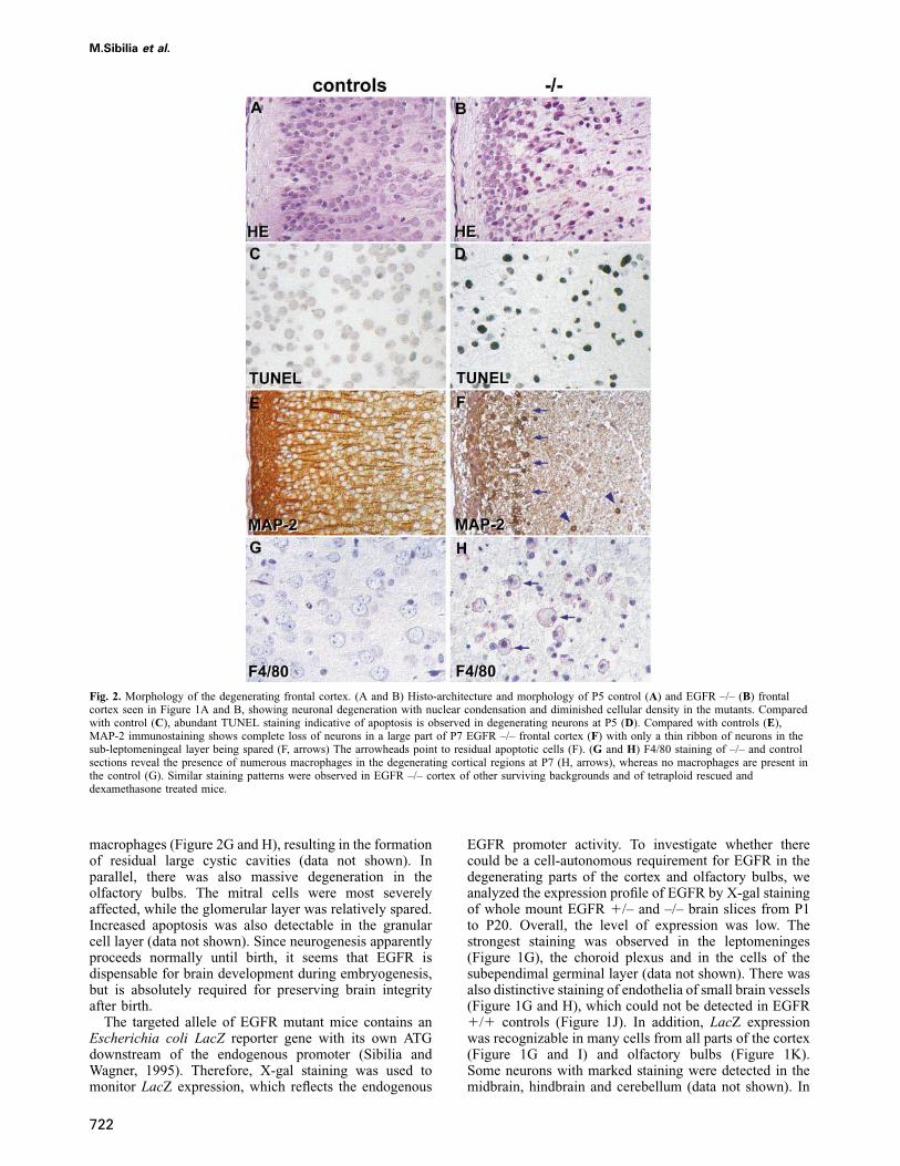
Fig. 2. Morphology of the degenerating frontal cortex. (A and B) Histo-architecture and morphology of P5 control (A) and EGFR –/– (B) frontalcortex seen in Figure 1A and B, showing neuronal degeneration with nuclear condensation and diminished cellular density in the mutants. Comparedwith control (C), abundant TUNEL staining indicative of apoptosis is observed in degenerating neurons at P5 (D). Compared with controls (E),MAP-2 immunostaining shows complete loss of neurons in a large part of P7 EGFR –/– frontal cortex (F) with only a thin ribbon of neurons in thesub-leptomeningeal layer being spared (F, arrows) The arrowheads point to residual apoptotic cells (F). (G and H) F4/80 staining of –/– and controlsections reveal the presence of numerous macrophages in the degenerating cortical regions at P7 (H, arrows), whereas no macrophages are present inthe control (G). Similar staining patterns were observed in EGFR –/– cortex of other surviving backgrounds and of tetraploid rescued anddexamethasone treated mice.
macrophages (Figure 2G and H), resulting in the formationof residual large cystic cavities (data not shown). Inparallel, there was also massive degeneration in theolfactory bulbs. The mitral cells were most severelyaffected, while the glomerular layer was relatively spared.Increased apoptosis was also detectable in the granularcell layer (data not shown). Since neurogenesis apparentlyproceeds normally until birth, it seems that EGFR isdispensable for brain development during embryogenesis,but is absolutely required for preserving brain integrityafter birth.
The targeted allele of EGFR mutant mice contains anEscherichia coli LacZ reporter gene with its own ATGdownstream of the endogenous promoter (Sibilia andWagner, 1995). Therefore, X-gal staining was used tomonitor LacZ expression, which reflects the endogenous
722
EGFR promoter activity. To investigate whether therecould be a cell-autonomous requirement for EGFR in thedegenerating parts of the cortex and olfactory bulbs, weanalyzed the expression profile of EGFR by X-gal stainingof whole mount EGFR �/– and –/– brain slices from P1to P20. Overall, the level of expression was low. Thestrongest staining was observed in the leptomeninges(Figure 1G), the choroid plexus and in the cells of thesubependimal germinal layer (data not shown). There wasalso distinctive staining of endothelia of small brain vessels(Figure 1G and H), which could not be detected in EGFR�/� controls (Figure 1J). In addition, LacZ expressionwas recognizable in many cells from all parts of the cortex(Figure 1G and I) and olfactory bulbs (Figure 1K).Some neurons with marked staining were detected in themidbrain, hindbrain and cerebellum (data not shown). In

Postnatal neurodegeneration in EGFR-deficient mice
Table I.
Crosses Genotypes
–/– �/– �/�
�/–��/– control 0 (0%) 110 (58%) 77 (41%)�/–��/– tetraploid (4N) 6 (12%) 46 (88%)
Frequency of genotypes from F2 progeny of tetraploid aggregationchimeras and 129/Sv EGFR heterozygote ( �/–) intercrosses at birth.Note that among the progeny of heterozygote intercrosses (control),no EGFR –/– fetuses were present at birth, whereas viable EGFR –/–pups were obtained from intercrosses rescued by tetraploidaggregation. One of the six tetraploid rescued pups was killed at P5for analysis whereas the rest died between P16 and P25.
the cortex, LacZ expression was essentially restricted toneurons, although some astrocytes were also positive,whereas oligodendrocytes consistently showed nostaining (data not shown). No clear increase in the intensityof staining with age and no detectable differences in thelevels of expression between EGFR �/– and –/– brainscould be observed (data not shown). Immunohisto-chemistry with anti-β-galactosidase antibody qualitativelyconfirmed the specificity of the lacZ staining (data notshown).
The neurodegeneration occurs in a strain-
independent manner and is not influenced by the
placental and lung defects
The development of the brain phenotype could be restrictedonly to EGFR mutant mice of certain genetic backgrounds,such as MF1 and C3H, or it could be a more generalphenotype occurring in all mouse strains, which wouldsuggest that EGFR is essential for neural cell survival.Alternatively, neurodegeneration could be a consequenceof impaired nutrition supply caused by other phenotypicalterations such as the placental defect or lung immaturity,since EGFR mutant mice of all backgrounds investigated(129/Sv, C57BL/6, CBA, MF1, C3H) showed placentaland lung phenotypes (data not shown). In order to investi-gate whether the brain phenotype would also arise in the129/Sv background, we rescued the placental defect bygenerating aggregation chimeras between tetraploid wild-type and diploid 129/Sv EGFR –/– embryos. Tetraploidcells, which can efficiently contribute to the developmentof all extra-embryonic tissues but not to the embryo itself(James et al., 1995), should complement the placentaldefect of EGFR –/– embryos and allow us to study EGFRfunction in 129/Sv embryos. While no 129/Sv EGFR –/–pups were ever obtained from heterozygote inter-crosses, six (12%) of the tetraploid aggregation chimeraswere EGFR –/– at birth (Table I), confirming that theplacental defects and no additional embryonic defects areresponsible for the midgestation lethality.
Newborn tetraploid rescued 129/Sv EGFR –/– aggrega-tion chimeras appeared similar to EGFR –/– pups ofC57BL/6, C3H and MF1 background. They had open eyesand were slightly smaller than their control littermates(data not shown). Some of them survived until day 25after birth (P25) and, similarly to EGFR mutant mice ofC3H and MF1 backgrounds, the 129/Sv –/– aggregationchimeras were severely growth-retarded and exhibited eye
723
Fig. 3. (A) Analysis of EGFR mutant mice rescued by tetraploidaggregation and transplacental dexamethasone administration. Externalappearance of 129/Sv tetraploid aggregation chimeras at postnatal day20 (P20). Like EGFR null mice of other backgrounds (Sibilia andWagner, 1995), the 129/Sv EGFR –/– (–/–) tetraploid aggregationchimeras are severely growth retarded and exhibit eye and hair growthdefects. (B) Viability of EGFR –/– mice after transplacentaldexamethasone administration. The percentage of live EGFR –/– miceat various stages after birth is shown for dexamethasone treated(square) and untreated (circle) mice. Transplacental dexamethasoneadministration significantly increases the survival of EGFR –/– mice inthe first 5 days after birth.
and hair defects (Figure 3A). Macroscopic and histologicalexaminations of brains isolated from 129/Sv EGFR –/–tetraploid aggregation chimeras at different postnatalstages revealed exactly the same onset and kinetics of theneurodegenerative process as observed in spontaneouslysurviving MF1 and C3H EGFR mutant mice (data notshown). PCR analysis of genomic DNA isolated fromplacentas, yolk sacs and tails of 129/Sv EGFR –/– tetra-ploid aggregation chimeras confirmed that the placentawas derived from wild-type tetraploid cells and the fetusfrom –/– cells (data not shown). These results demonstratethat in EGFR mutant mice the postnatal epithelial andbrain defects occur independently from the placentaldefect. Furthermore, they suggest that the genetic back-ground influences mainly the development of extra-embryonic tissues, but not of the postnatal epithelial andbrain defects.
At birth, EGFR mutant mice of all backgrounds haveimmature lungs and this defect is most probably

M.Sibilia et al.
Fig. 4. Massive apoptosis and increased c-fos expression in thedegenerating cortex of EGFR mutant brains at postnatal day 5 (P5). (Aand B) TUNEL staining of EGFR –/– brain sections reveal massiveapoptosis in the rostral part of the frontal cortex and olfactory bulb (B,inset) whereas no apoptotic cells can be detected in the control (A,inset). (C–H) In situ hybridization analysis showing the expression ofthe c-fos (C and D), VEGF (E and G) and flk-1 (F and H) genes in thecortex of EGFR –/– (D, G and H) and control (C, E and F) brains.Note that c-fos is upregulated in the degenerating cortical regions ofEGFR –/– brains (D, inset), but it is not expressed at detectable levelsin the controls (C, inset). There is no induction of VEGF mRNA inthe degenerating cortex of EGFR mutants (G) compared with thecontrols (E). Normal levels of expression of flk-1 in –/– cortical bloodvessels (H) and controls (F). Insets: magnifications of affected cortexin (A–D). In situ hybridization with sense control probes wereconsistently negative in degenerating areas (data not shown). Similardegenerative processes were observed in the cortex of survivingEGFR –/– of other backgrounds and of tetraploid rescued anddexamethasone treated mice.
responsible for the inability of the majority of mutantnewborns to initiate or sustain sufficient respiration (Miet-tinen et al., 1995; Sibilia and Wagner, 1995). AlthoughEGFR mutant mice of C3H and MF1 background cansurvive for 3 weeks after birth, a high number of mutants(60%) die during the first 5 postnatal days (Figure 3B).To investigate whether this early postnatal mortality wasattributable to lung immaturity and if persistent hypoxiaduring the first few days after birth could be responsiblefor the neurodegeneration, we attempted to amelioratethe lung phenotype by transplacental administration ofdexamethasone, a glucocorticoid analogue. Administrationof glucocorticoids to pregnant women is an establishedtherapy for promoting lung maturation in human fetuses(Ballard, 1989). As shown in Figure 3B, dexamethasonetreatment significantly increased the survival of EGFR –/–mice in the first days after birth, but there was noprolongation of the maximal lifespan. Corresponding tothe improved viability, histological analysis of newborndexamethasone-treated lungs revealed that the lungdysplasia of EGFR mutant offspring had been rescued(data not shown). However, in dexamethasone treatedEGFR mutant mice, the onset and severity of the neuro-degeneration were similar to spontaneously surviving
724
Fig. 5. Selective neuronal degeneration in thalamus and hippocampusof EGFR mutant brains. (A and B) Degenerating areas of the thalamusare revealed by the absence of immunohistochemical staining withMAP-2 antibodies in P7 –/– brains (arrows) (B). (C and D) GFAPstaining shows a pattern of ‘reactive astrogliosis’ in the EGFR –/–thalamus (D), which is absent in the control (C). (E–H) BrdU andTUNEL staining showing high proliferative activity of mutantastrocytes (G) and increased neuronal apoptosis (H, arrowheads) in thedegenerating thalamic regions compared with controls (E and F).Compared with controls (I), increased apoptosis in EGFR –/– granulecells of the dentate gyrus of the hippocampus at P19 (K, arrows). Allthese abnormalities were also observed in the brains of survivingEGFR –/– mice of other backgrounds and of tetraploid rescued anddexamethasone treated mice.
mutants (data not shown). Therefore, it appears unlikelythat the neurodegeneration is secondary to persistentperinatal hypoxia due to lung immaturity.
No evidence of hypoxia or pathological
angiogenesis
Since the placental defect and lung immaturity do notseem to alter the course of neurodegeneration, we nextinvestigated other possible mechanisms that could lead tothe massive apoptotic cell death. Corresponding to thedegenerating areas of the cortex and olfactory bulbs ofEGFR mutant brains which were affected by apoptosis(Figure 4A and B), there was a remarkable induction ofc-fos, which exactly mapped the extent of the degenerativeprocess (Figure 4C and D). In situ hybridization revealedno differences in c-fos expression between mutants andcontrols at birth and up to P3, with most pronouncedexpression in the hippocampus, internal granule cell layerof the cerebellum and subependymal germinal layer (datanot shown). The levels of c-fos gradually declined incontrols, but in EGFR mutants there was strong expressionof c-fos in neurons of the affected frontal cortex from P4
Postnatal neurodegeneration in EGFR-deficient mice
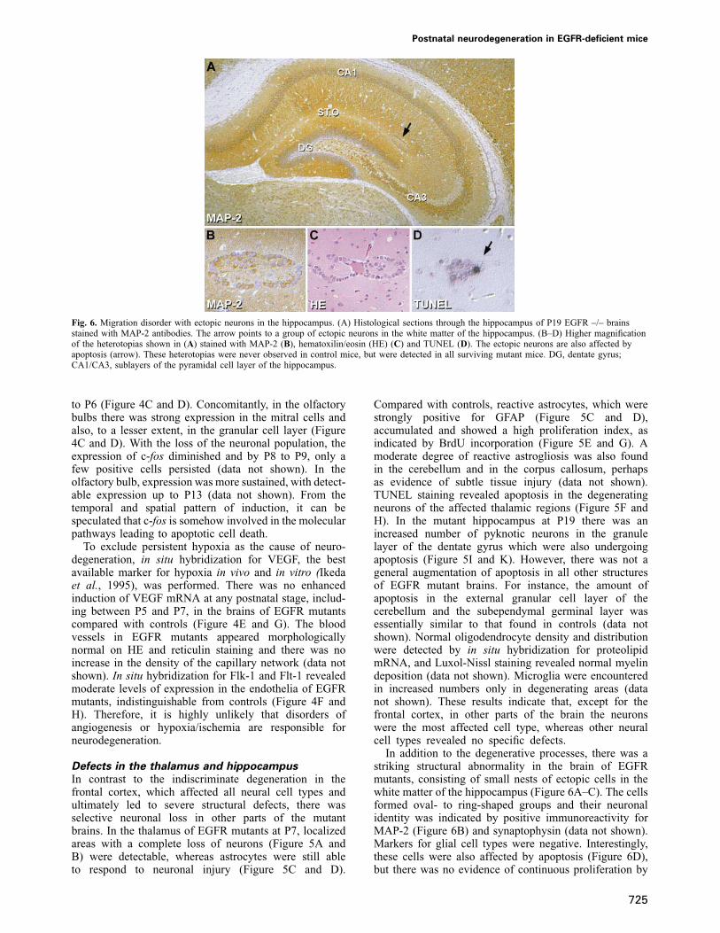
Fig. 6. Migration disorder with ectopic neurons in the hippocampus. (A) Histological sections through the hippocampus of P19 EGFR –/– brainsstained with MAP-2 antibodies. The arrow points to a group of ectopic neurons in the white matter of the hippocampus. (B–D) Higher magnificationof the heterotopias shown in (A) stained with MAP-2 (B), hematoxilin/eosin (HE) (C) and TUNEL (D). The ectopic neurons are also affected byapoptosis (arrow). These heterotopias were never observed in control mice, but were detected in all surviving mutant mice. DG, dentate gyrus;CA1/CA3, sublayers of the pyramidal cell layer of the hippocampus.
to P6 (Figure 4C and D). Concomitantly, in the olfactorybulbs there was strong expression in the mitral cells andalso, to a lesser extent, in the granular cell layer (Figure4C and D). With the loss of the neuronal population, theexpression of c-fos diminished and by P8 to P9, only afew positive cells persisted (data not shown). In theolfactory bulb, expression was more sustained, with detect-able expression up to P13 (data not shown). From thetemporal and spatial pattern of induction, it can bespeculated that c-fos is somehow involved in the molecularpathways leading to apoptotic cell death.
To exclude persistent hypoxia as the cause of neuro-degeneration, in situ hybridization for VEGF, the bestavailable marker for hypoxia in vivo and in vitro (Ikedaet al., 1995), was performed. There was no enhancedinduction of VEGF mRNA at any postnatal stage, includ-ing between P5 and P7, in the brains of EGFR mutantscompared with controls (Figure 4E and G). The bloodvessels in EGFR mutants appeared morphologicallynormal on HE and reticulin staining and there was noincrease in the density of the capillary network (data notshown). In situ hybridization for Flk-1 and Flt-1 revealedmoderate levels of expression in the endothelia of EGFRmutants, indistinguishable from controls (Figure 4F andH). Therefore, it is highly unlikely that disorders ofangiogenesis or hypoxia/ischemia are responsible forneurodegeneration.
Defects in the thalamus and hippocampus
In contrast to the indiscriminate degeneration in thefrontal cortex, which affected all neural cell types andultimately led to severe structural defects, there wasselective neuronal loss in other parts of the mutantbrains. In the thalamus of EGFR mutants at P7, localizedareas with a complete loss of neurons (Figure 5A andB) were detectable, whereas astrocytes were still ableto respond to neuronal injury (Figure 5C and D).
725
Compared with controls, reactive astrocytes, which werestrongly positive for GFAP (Figure 5C and D),accumulated and showed a high proliferation index, asindicated by BrdU incorporation (Figure 5E and G). Amoderate degree of reactive astrogliosis was also foundin the cerebellum and in the corpus callosum, perhapsas evidence of subtle tissue injury (data not shown).TUNEL staining revealed apoptosis in the degeneratingneurons of the affected thalamic regions (Figure 5F andH). In the mutant hippocampus at P19 there was anincreased number of pyknotic neurons in the granulelayer of the dentate gyrus which were also undergoingapoptosis (Figure 5I and K). However, there was not ageneral augmentation of apoptosis in all other structuresof EGFR mutant brains. For instance, the amount ofapoptosis in the external granular cell layer of thecerebellum and the subependymal germinal layer wasessentially similar to that found in controls (data notshown). Normal oligodendrocyte density and distributionwere detected by in situ hybridization for proteolipidmRNA, and Luxol-Nissl staining revealed normal myelindeposition (data not shown). Microglia were encounteredin increased numbers only in degenerating areas (datanot shown). These results indicate that, except for thefrontal cortex, in other parts of the brain the neuronswere the most affected cell type, whereas other neuralcell types revealed no specific defects.
In addition to the degenerative processes, there was astriking structural abnormality in the brain of EGFRmutants, consisting of small nests of ectopic cells in thewhite matter of the hippocampus (Figure 6A–C). The cellsformed oval- to ring-shaped groups and their neuronalidentity was indicated by positive immunoreactivity forMAP-2 (Figure 6B) and synaptophysin (data not shown).Markers for glial cell types were negative. Interestingly,these cells were also affected by apoptosis (Figure 6D),but there was no evidence of continuous proliferation by

M.Sibilia et al.
Fig. 7. Impaired proliferation of EGFR mutant astrocytes in vitro andreduced number of GFAP-positive astrocytes in mutant cortices afterbirth. (A) Cumulative cell number of control (circle) and EGFR –/–(square) astrocytes isolated from newborn (P1) cortices showing thatmutant astrocytes display a significantly reduced proliferation capacityin vitro. Primary cell suspensions (day 0), were initially plated at1�105 cells/cm2 and afterwards cells were passaged and counted whenapproaching confluence at the indicated timepoints and replated at 1:2or 1:3 split ratios. Isolates from ten controls and five EGFR –/–cortices were analysed and a representative growth curve from eachgroup is shown. Based on the result of the quantitative FACS analysis,the initial cell population was assumed to contain 1–1.3�104
astrocytes. (B) Flow-cytometric analysis of primary cell suspensionsprepared from P1, P2 and P3 EGFR –/– and littermate control corticesand stained for the astrocyte specific marker GFAP. The percentage ofGFAP-positive astrocytes is significantly reduced in EGFR mutant P2and P3 cortices. Data at each timepoint represent the mean � SD ofthe analysis of three independent cortices of each genotype (EGFR –/–and control). Background control represents the mean � SD of cellsincubated with the same concentration of purified rabbit IgG. Theseanalyses were performed in newborn mutant mice of C3H and MF1genetic background and the size and weight of the brain of the mutantand control mice were comparable.
BrdU staining. These ectopic neurons were found invariable amounts in all EGFR mutants older than P10,but never in controls, and most probably represent amigratory disorder. In contrast, other migrating cell typesappeared unaffected; for instance there was normalproliferation of the cells of the subependymal germinalcell layer, and the migration of these cells to the granulecell layer appeared uncompromised (data not shown). Insitu hybridization for c-fos revealed continuous expressionin the cells migrating into the olfactory bulbs, similar
726
in EGFR mutants and controls (data not shown). Thisphenotype may suggest a previously unrecognized functionof EGFR in the guidance of migrating hippocampalneurons.
Impaired proliferation of astrocytes in vitro and
in vivo
Until birth, the neurons in the different cortical layers donot appear to be affected in the absence of EGFR andthe cortical lamination is comparable to the controls.Neurodegeneration in the EGFR mutant cortex startsaround P4 and therefore, the defect leading to massiveapoptotic cell death is likely to occur during the firstpostnatal days. It is possible that neuronal cell death inthe cortex might be the consequence of a local lack oftrophic support by glial cells. Astrocytes are thought tobe critical for the nutritional and structural support ofneurons and most astrocyte differentiation or maturationseems to occur in the early postnatal period (Levison andGoldman, 1993). Since EGFR appears to play a role inthe proliferation and differentiation of astrocytes (Simpsonet al., 1982), we investigated whether this cell type wasaffected in EGFR mutant cortices. We first examinedwhether the absence of EGFR influences the proliferationrate of primary astrocytes isolated from postnatal day 1(P1) wild-type and mutant cortices. As shown in Figure7A, EGFR –/– astrocytes show a significantly reducedproliferation rate compared with controls and after 50days in culture, their cumulative cell number reached only25% of the value obtained with the controls. Morpholo-gically the cultured cells resembled type I astrocytesand immunofluorescence staining with antibodies againstGFAP, vimentin and GalC confirmed that after the firstpassage, more than 95% of the cells were astrocytes (datanot shown).
In vitro, astrocytes are known to proliferate in responseto EGF. Therefore, the reduced proliferation observed inastrocytes lacking EGFR could be due to the fact thatthey are unable to respond to EGF present in the serumof the culture medium. In order to investigate whether theabsence of EGFR also affected astrocyte proliferationin vivo, primary cell suspensions isolated from EGFRmutant and control cortices at P1, P2 and P3 were subjectedto flow-cytometric analysis after staining with anti-GFAPantibodies. In control cortices, the percentage of GFAP-positive cells increased from ~2% (P1) to 7% (P3) andthe major increment was observed between P1 and P2(Figure 7B). GFAP staining in the mutants was comparablewith the controls at P1, but at P2 and P3 the percentageof positive cells was significantly lower in the mutants,reaching only 30–50% of control levels. These resultsindicate that EGFR –/– astrocytes are impaired in theirproliferation capacity in vitro as well as in vivo. Alterna-tively, since a reduced number of GFAP-positive cells wasalready observed at birth, the absence of EGFR mightalso affect either the survival or the ability of neuralprecursor cells to efficiently differentiate into astrocytes.
Discussion
Depending on the genetic background, EGFR mutantmice die at midgestation (129/Sv), at birth (C57BL/6) orcan live up to postnatal day 20 (MF1 or C3H). The

Postnatal neurodegeneration in EGFR-deficient mice
multiple phenotypes are manifested in defects in theplacenta, the skin, the lung and also in the brain. In thisstudy, the brain lesions leading to a massive neurodegener-ation were characterized in different mouse strains. AroundP4, all mutant mice show a progressive neurodegenerationin the frontal cortex, olfactory bulb and thalamus leadingto the loss of extensive parts of the brain by P8. Thisdegeneration involves massive apoptosis and is accom-panied by upregulation of c-fos. In addition, a migrationdisorder consisting of ectopic neurons is observed in thehippocampus of mutant brains. A reduced number ofGFAP-positive astrocytes is present in mutant corticesand their proliferation capacity in vitro is also affected,suggesting that impaired proliferation and/or differenti-ation of astrocytes contribute to the brain phenotype.
When aggregation chimeras between 129/Sv EGFRmutant and tetraploid wild-type embryos were generated,viable EGFR mutant mice were obtained in a pure129/Sv genetic background. These mice survived up to3 weeks and developed similar defects as EGFR mutantsin other backgrounds. In particular, they displayed thesame pattern and degree of neurodegeneration, indicatingthat this degenerative process occurs in a strain-independ-ent manner. Furthermore, the rescue of the lung immaturityby dexamethasone administration did not alter the courseof neurodegeneration, excluding persistent hypoxia as thecause of the brain phenotype. There is no increase in themaximal lifespan in dexamethasone-treated mutant miceand it appears that the lung immaturity is not the limitingfactor for survival once the first few days of life havepassed. In contrast, the extensive neurodegeneration in thefrontal cortex, thalamus and olfactory bulbs most probablyadversely affects the viability of EGFR mutant mice.These defects do not influence the overall locomotoractivity of EGFR mutant mice, but might interfere withtheir feeding behavior.
Regional distribution of the neurodegenerative
lesions
It is unclear why only certain areas of the brain areaffected and why others are spared. The observed distribu-tion of lesions does not map to any vascular territory,nor is it compatible with the pattern of lesions foundin anoxic encephalopathy, where neuronal death occurspredominantly in the pyramidal cell layer of the hippo-campus (CA1) and in the cortex. In particular, there wasa conspicuous sparing of the retrosplenial cortex, withsharp demarcation to the degenerating tissue of the neigh-boring frontal cortex. Leptomeningeal hemorrhagesobserved on the surface of the degenerating cortex alwaysappeared concomitantly with the massive cell death andare likely to reflect a local inflammatory response whichattracts monocytes/macrophages to the lesioned areas.Alternatively, there could be a primary defect in EGFR –/–blood vessels, which render them more likely tospontaneous ruptures, especially since we detected EGFRexpression by X-gal staining in brain endothelia. EGFRexpression had indeed been described previously on humanbrain endothelia (Styren et al., 1993). However, neithermorphological analysis by HE and reticulin staining normolecular analysis by in situ hybridization for flk-1 andflt-1 revealed any vascular abnormalities such ashemangiomas or aneurysms in EGFR mutants and the
727
hemorrhages were always observed in conjunction withdegenerating areas in the brain.
Alternatively to a vascular defect, hypoxia or ischemiacould be responsible for the neurodegeneration. SinceVEGF expression was never enhanced in the brains ofEGFR mutant mice, hypoxia and ischemia are unlikely tobe involved in the neurodegeneration. In addition, VEGFcauses upregulation of its cognate receptors, flk-1 andflt-1 on endothelia (Wilting et al., 1996). However, normallevels of these receptors were detected in mutant brains.The late induction of c-fos just before the onset of thedegenerative process is also a strong argument against aperinatal cause, since c-fos is rapidly and transientlyinduced by hypoxia and ischemia (Kinouchi et al., 1994).In the nervous system, upregulation of c-fos has beenfound to precede apoptosis in many different models(Smeyne et al., 1993), and there is increasing evidencethat c-fos can act as a mediator of cell death in vivo(Preston et al., 1996; Hafezi et al., 1997). Therefore, ourfinding of high levels of c-fos expression concomitantwith progressive cell death, suggests that c-Fos might becausally involved in the molecular pathways leading toapoptosis. We were unable to test the hypothesis that inthe absence of c-Fos EGFR –/– brains would still beaffected by apoptosis, since from intercrosses betweenEGFR �/– c-fos �/– mice, we could not obtain survivingmice lacking both EGFR and c-Fos (M.Sibilia andE.F.Wagner, unpublished results).
Migratory defects
Ectopic neurons were observed in the white matter of thehippocampus. The position of these ectopic neurons is ina region of the hippocampus, where EGFR has beenshown to be expressed in rats (Tucker et al., 1993).Possibly, the presence of EGFR on resident or migratorycells offers cues for the correct migration into the appro-priate hippocampal structure. It appears likely that thisphenotype is the consequence of a migration disturbancerather than aberrant proliferation, since no proliferativeactivity was detected in the ectopic cells by BrdU staining.EGFR has been shown to control cell migration, sinceEGF injection can induce subependymal cells to migratefrom the lateral ventricle into the adjacent neural tissue(Craig et al., 1996). Further, in TGFα –/– mice there is adeficit in the rostral migration of neuronal progenitorcells in the adult subependyma (V.Tropepe, personalcommunication). EGF-like motifs are common in manymolecules with migratory function, such as astrotactin(Zheng et al., 1996) and reelin (Hirotsune et al., 1995),while EGFR has so far been mainly associated withproliferation and differentiation, but not neuronalmigration.
Neuronal versus astrocyte defects
It seems unlikely that exogenous mechanisms areresponsible for the degenerative process in the brain. Acell-autonomous defect appears to be the most likelyreason for the massive apoptosis observed in various brainregions, suggesting that EGFR might be essential for neuralcell survival. This is in agreement with our previouslypublished data where we showed that in chimeras,EGFR –/– ES cell derivatives can efficiently contribute tothe brain at E14.5. However, the EGFR –/– derivatives’

M.Sibilia et al.
contribution to the brain decreases after birth, suggestingthat neural cells lacking the EGFR might encounter aselective disadvantage compared with wild-type cells(Sibilia and Wagner, 1995). By X-gal staining, we detectEGFR expression in the subependymal layer, olfactorybulbs, cortex and in the basal forebrain. Our results arecompatible with other reports detecting EGFR on bothastrocytes and neurons (Gomez-Pinillla et al., 1988;Nieto-Sampedro et al., 1988; Topp et al., 1989;Kornblum et al., 1997). EGF and TGFα have beenshown to increase the survival of cortical, mesencephalicand cerebellar neurons in vitro (Kornblum et al., 1990;Casper et al., 1991; Alexi and Hefti, 1993) and EGFRimmunoreactivity has been demonstrated on neurons inthe cortex (Tucker et al., 1993). It is possible that EGFRstimulation is required for the survival of neurons in acell-autonomous manner and that the lack of EGFR leadsto apoptosis. Alternatively, in astrocytes, EGFR signalingmay lead to the secretion of neurotrophic factors that arecritical for the survival of neurons.
A reduced proliferation was indeed observed in EGFRmutant astrocytes, which is consistent with previous obser-vations showing that EGF promotes the proliferation ofprimary astrocytes in vitro (Simpson et al., 1982). Wealso detect reduced numbers of GFAP-positive astrocytesin mutant cortices at birth, and while the percentages ofpositive cells increase during the first postnatal days, theynever exceed 50% of control levels. Therefore, it seems thatalso in vivo the proliferation of EGFR mutant astrocytes isimpaired. In wa-1 mice, which are deficient in TGFα,reduced levels of GFAP mRNA have been observed butno obvious brain abnormalities were detected (Weickertand Blum, 1995). However, in this study the number ofGFAP-positive cells was not directly assessed (Weickertand Blum, 1995). Our data suggest that EGFR signalingis important in regulating the proliferation of astrocytesin vitro and in vivo. The observation that the EGFRand TGFα genes are amplified in several malignanthuman gliomas further supports the hypothesis that EGFRsignaling is important in glial cell proliferation (Yunget al., 1990; Wong et al., 1992).
It is very possible that the lack of EGFR could alsoaffect the differentiation efficiency of neural progenitorsinto the astrocyte lineage. Pluripotent progenitors in theventricular zone (VZ) of mouse embryonic forebrains andsubependymal layer (SEL) of adult brains have beenshown to proliferate in response to EGF and fibroblastgrowth factor 2 (FGF2) and differentiate into neurons,astrocytes or oligodendrocytes (for review see Weiss et al.,1996). There is increasing evidence that different typesof progenitor cells might exist in the VZ and SEL, whichexpress either FGF receptor 1 (FGFR1), EGFR or both(V.Tropepe, personal communication). Therefore, EGFRexpressing progenitors might be the ones that prevalentlydifferentiate into astrocytes. This is substantiated by recentfindings by Burrows and co-workers showing that increas-ing the level of EGFR signaling preferentially confers onprogenitor cells the ability to differentiate into astrocytes(Burrows et al., 1997). Alternatively, instead of directlyinducing astrocyte differentiation, EGF might be essentialfor the expansion and/or survival of progenitors along theastrocyte differentiation lineage (Johe et al., 1996) and,in the absence of EGFR, most of these progenitors would
728
die. The fact that FGF2 alone can induce astrocytedifferentiation of stem cells explains why in the absenceof EGFR there is only a reduction in, rather than acomplete block of, astrocyte differentiation in theforebrain.
It seems that in different degenerating parts of mutantbrains, different cell types are affected. A complete struc-tural disintegration was observed in the rostral part of thefrontal cortex and in the olfactory bulbs, implying thatneither neurons nor astrocytes may achieve sufficientsupport for survival in these areas. In contrast, in theretrosplenial cortex survival factors may diffuse from theneighboring midbrain and prevent both cell types fromundergoing apoptosis. In the thalamic lesions, there wasselective neuronal cell death with a vigorous ‘reactiveastrogliosis’ indicating that astrocytes were initially notaffected. These findings can possibly be explained by adifferent relative susceptibility of neurons and astrocytesto the loss of trophic support through EGFR signaling andby further quantitative differences in the vulnerability ofboth cell types in different regions of the mouse brain.Thus, in the thalamus, survival factors may suffice forastrocytes but not for neurons. Alternatively, neuronaldeath in the thalamus could be secondary to the corticallesions and affect only the neurons that project to frontaland not to retrosplenial cortical regions.
Mechanism of neurodegeneration
Although a reduced number of GFAP-positive cells isobserved in EGFR mutant cortices, we can not concludethat this defect is sufficient to cause massive death ofneurons. Conditional ablation of astrocytes in postnataltransgenic mice has been shown to induce several cere-bellar defects, including loss of granule neurons (Delaneyet al., 1996). Therefore, it is tempting to speculate that inEGFR mutant brains, a reduced number of astrocytesmight not give sufficient support to the neurons andcontribute in part to the neurodegeneration. This hypothesisis substantiated by studies where EGF- or TGFα-inducedsurvival of dopaminergic neurons was shown to be medi-ated, in part, by the presence of astrocytes in the culture,which divided in response to EGF or TGFα (Casper et al.,1991; Alexi and Hefti, 1993). Since EGFR is expressedin neurons as well as in astrocytes, it may be that in theabsence of EGFR, the massive neurodegeneration is causedby a combination of two mechanisms, one acting directlyon neurons, the other mediated by astrocytes. The pheno-type is therefore far more dramatic than the changesdescribed in other mouse models of neurodegeneration,such as in mice lacking various neurotrophic factors(Klein, 1994; Sendtner, 1995) or in mice with singleor combined deficiencies in the corresponding receptors(Klein, 1994; Minichiello and Klein, 1996). Specific rescueexperiments with conditional EGFR alleles or inactivatingthe EGFR in astrocytes or neurons will provide thedefinitive answer to which cell type causally contributesto the brain phenotype.
It would also be important to investigate whether relatedreceptor tyrosine kinases such as the other erbB familymembers could functionally compensate for the lack ofEGFR. These receptors are expressed in neuronal tissuesand it has recently been shown that mice lacking eithererbB2, erbB4 or neuregulin, the ligand for erbB3 and

Postnatal neurodegeneration in EGFR-deficient mice
erbB4, die at midgestation and show aberrant neuraldevelopment (Gassmann et al., 1995; Lee et al., 1995;Meyer and Birchmeier, 1995). Variations in the expressionpattern of the erbB receptors may even explain theregional distribution and the onset of the neurodegenerativeprocesses. The fact that the degenerative process affectsmature neurons suggests that EGFR signaling may beinvolved directly or indirectly in the maintenance ofpostmitotic neurons. If this can be substantiated, strategiesthat aim at enhancing EGFR signaling may be relevantfor the treatment of neurodegenerative conditions such asAlzheimer’s disease, where the balance between protectiveand noxious stimuli in neurons is disturbed.
Materials and methods
MiceThe generation of EGFR knock-out mice used in this study has beendescribed in detail previously (Sibilia and Wagner, 1995). Briefly, theEGFR gene was inactivated in ES cells by replacing parts of the firstexon with an E.coli lacZ reporter gene, which allows us to follow thestage- and tissue-specific expression pattern of EGFR during develop-ment. Correctly targeted ES cells were injected into C57BL/6 blastocystsand the resulting germline chimeras were mated to 129/Sv andC57BL/6 females to obtain EGFR �/– offspring of inbred 129/Sv andmixed 129/Sv�C57BL/6 genetic background, respectively. Starting frominbred 129/Sv EGFR �/– mice the mutant allele was subsequently bredinto C57BL/6, MF1 and C3H strains and backcrossed for severalgenerations. Heterozygous EGFR �/– mice of the respective geneticbackground were further intercrossed to generate homozygote EGFR –/–mice. Genotyping was performed by PCR as previously described (Sibiliaand Wagner, 1995).
Generation of tetraploid (4N) embryosB6CBAF1 (C57BL/6�CBA F1) female mice mated with males fromthe same F1 strain were used as donors of 2-cell stage embryos forthe production of tetraploid embryos. Tetraploidy was achieved byelectrofusion as described previously (Wang et al., 1997). Briefly, thetwo blastomeres were fused in 0.3 M mannitol following a short electricpulse at 95 V for 30 ms in an effective field (2 V) using the CF-100pulse generator (Biochemical Laboratory Service, Budapest). The fusedembryos (~95%) were selected and further cultivated for 24–40 h to the‘non-compacted’ morula stage (for tetraploid embryos: 4-cell-stage) ina microdrop of M16 medium under paraffin oil at 37°C in a 95% air/5% CO2 incubator.
Morula aggregationNon compacted 8-cell-stage morulae were isolated from oviducts of2.5 p.c. inbred 129/Sv EGFR �/– females intercrossed with 129/SvEGFR �/– males. Twenty-five per cent of these embryos are expectedto be EGFR –/–. After removal of the zona pellucida by acid Tyrode’ssolution, diploid morulae from EGFR �/– intercrosses were aggregatedat a 1:1 ratio with 4-cell-stage wild-type tetraploid morulae in M16medium. The aggregates were cultured for further 20–30 h in M16medium under paraffin oil at 37°C in a 95% air/5% CO2 incubator.Under these conditions �95% of the embryos spontaneously aggregatedovernight and formed blastocysts composed of diploid and tetraploidblastomeres. The chimeric blastocysts were transferred into the uterusof pseudopregnant recipients where the embryos developed to term.Pregnant recipients either delivered the fetuses spontaneously or weresubjected to Cesarean section on day 18.5 of gestation in order to collectyolk sacs and placentas. EGFR –/– fetuses were recognized by thepresence of open eyes at birth and their genotype was confirmed togetherwith the genotype of yolk sac and placenta by PCR, as describedpreviously (Sibilia and Wagner, 1995).
Transplacental dexamethasone administrationEGFR �/– females of C3H and MF1 genetic background were matedwith EGFR �/– males of the respective genetic background and removedafter they were plugged (day 0.5 of pregnancy). A stock concentrationof dexamethasone (Dex; Sigma Immunochemicals) at 10 mg/ml wasprepared in absolute ethanol and stored at –20°C. Before use, dexametha-sone was diluted 1:100 in sterile phosphate-buffered saline (PBS) solution
729
and injected intraperitoneally at day 15.5 of pregnancy (term of 19–20days) at a concentration of 2 mg/g body weight. Control animals wereinjected with PBS containing 1% absolute ethanol.
In vivo BrdU labelingA stock solution of BrdU was prepared in PBS at a concentration of5 mg/ml, stored at 4°C and further diluted in PBS before injection. Afinal concentration of 50 mg/g body weight of BrdU was injectedintraperitoneally into EGFR –/– mice and littermate controls 24 and12 h before sacrificing.
Histological analysis and β-galactosidase stainingMice were sacrificed and the isolated tissues were fixed immediately in4% paraformaldehyde (PFA) in PBS at 4°C for 16–24 h. After fixation,the tissues were dehydrated through RNase-free graded alcohols andtoluene and infiltrated with paraffin (Histowax, Reichert-Jung, Vienna)at 58°C overnight, under vacuum. Sections (2–4 mm) were cut, mountedon silanized slides and stained with hematoxylin and eosin (SigmaImmunochemicals). Brains used for X-gal staining were fixed for 10–20 min in 4% paraformaldehyde, cut into 2–3 mm slices and fixed foranother 10 min in 4% PFA. X-gal staining was performed as previouslydescribed (Sibilia and Wagner, 1995).
ImmunohistochemistryImmunohistochemistry was performed using polyclonal antibodies toGFAP (DAKO, 1:300), MAP-2 (Boehringer Mannheim, 1:1000)F4/80 (1:80), Ki67 (Novocastra Laboratories, 1:1000), and monoclonalantibodies to BrdU (Caltag Laboratories, 1:50) and β-galactosidase(Cappel, 1:400). An ultrasensitive Avidin-Biotinylated Enzyme Complex(ABC) staining kit (Pierce Chemical Co., Rockford, IL) was used onparaffin sections according to the protocol specified by the suppliers.Positive staining was visualized by incubation with DAB (VectorLaboratories, Burlingame, CA). Control sections were treated with PBSinstead of antibody. The APAAP-method, with neofuchsin as chromogen,was employed for F4/80 immunostaining (Dako). Microwave pretreat-ment in citric acid buffer was performed for MAP-2, β-galactosidase,BrdU and F4/80 immunostainings.
TUNELIn situ nick end-labeling (TUNEL) was performed using the in situ celldeath detection kit II (Boehringer Mannheim). Slides were deparaffinized,and sections were digested with proteinase K (20 mg/ml) for 15 min at37°C, followed by incubation with terminal transferase for 1 h at 37°Cin the presence of fluorescin-labeled dUTP. An alkaline phosphatase-coupled anti-fluorescin Fab fragment was used for detection, and5-bromo-4-chloro-3-indolyl phosphate and 4-nitro blue tetrazolium chlor-ide (Boehringer Mannheim) were employed as chromogens.
In situ hybridizationSense and antisense RNA probes were transcribed in vitro with T3and T7 RNA polymerase from linearized pBluescript vectors carryingc-fos (Hafezi et al., 1997), VEGF (Breier et al., 1995), flk-1 (Millaueret al., 1993), flt-1 (Breier et al., 1995) cDNAs in the presence ofdigoxygenin-11-UTP (Boehringer Mannheim). Fifty to 200 ng oflabeled transcripts (0.7–1.2 kb) were hybridized to tissue sections at65°C as described (Hafezi et al., 1997). Digoxygenin was detectedwith alkaline phosphatase-labeled anti-DIG Fab fragments and4-nitro blue tetrazolium chloride/5-bromo-4 chloro-3 inodyl phosphate(Boehringer Mannheim). Coverslips were mounted with glycerolgelatine.
Preparation of cortical astrocytesPrimary astrocyte cultures were prepared from brains of newbornEGFR –/– and littermate control pups using a modification of previouslydescribed procedures (Simpson et al., 1982; Bambrick et al., 1996).Cerebral cortices were dissected free of meninges and hippocampus andminced into small pieces. The cortical fragments were incubated at 37°Cin 2� Trypsin–EDTA (0.1% trypsin, 0.04% EDTA; GIBCO), 0.001%DNase I (Sigma) in DMEM/F12 (1:1; GIBCO) for 30 min. Enzymaticallysoftened tissues were titurated with a wide-bore pipette, resuspendedinto serum containing culture medium and passed through a 70 μmnylon cell strainer to obtain single cell suspensions. Cells were pelletedby centrifugation, resuspended in Dulbecco’s modified Eagle’s medium(DMEM) containing 10% fetal calf serum (PAA), Penicillin-Streptomycin(100 IU/ml to 100 μg/ml; GIBCO) and plated on poly-L-lysine (0.1 mg/ml; SIGMA) coated tissue culture dishes, initially at a concentration of1�105 cells/cm2. When approaching confluence, cells were trypsinized

M.Sibilia et al.
and viable cell numbers determined using a Neubauer-type hemocyto-meter with trypan blue staining. Cells were then replated at 1:2 or 1:3split ratio. Replated cultures were visually similar to long term primarycultures with respect to morphology and GFAP staining. Freshly preparedsuspensions of mouse cortical cells were galactocerebroside- (Gal-C;Sigma) negative and contained �5% neurons. After the first passage,cultures consisted of virtually �95% GFAP- and vimentin- (Sigma)positive cells as determined by immunofluoresence.
Cytometric analysis of GFAPCell suspensions of cortices were fixed in 4% PFA at 4°C, washed withPBS, permeabilized in 0.2% Triton X-100 in PBS for 5 min at roomtemperature, rewashed with PBS and then incubated for 1 h with rabbitpolyclonal antibodies to GFAP (DAKO; 1:100) in PBS � 1% bovineserum albumin (BSA; Sigma). As a negative control, the cells wereincubated with the same concentration of purified rabbit IgG. Cells werewashed twice with PBS � 0.1% Tween 20 (PBS-T; Fluka) and incubatedwith a 1:800 dilution of goat anti-rabbit IgG-Cy5 (Rockland). Cells werewashed twice, resuspended in 0.5 ml PBS-T and analysed with FACSVantage (Becton Dickinson).
Acknowledgements
We are grateful to Dr R.M.Zinkernagel for providing the F4/80 antibody,Dr P.Steinlein for help with FACS analysis, R.Kupinski and M.King formaintaining our mouse colony and V.Tropepe and A.Behrens for helpfuldiscussions. We thank V.Tropepe, M.Busslinger, W.Risau and D.van derKooy for critical reading of the manuscript. M.S. was a recipient of afellowship from the Associazione Italiana per la Ricerca sul Cancro(AIRC). This research was supported in part by the Austrian IndustrialResearch Promotion Fund.
References
Adamson,E.D. (1990) Developmental activities of the epidermalgrowth factor receptor. Curr. Top. Dev. Biol., 24, 1–29.
Alexi,T. and Hefti,F. (1993) Trophic actions of transforming growthfactor alpha on mesencephalic dopaminergic neurons developing inculture. Neuroscience, 55, 903–918.
Ballard,P.L. (1989) Hormonal regulation of pulmunary surfactant.Endocr. Rev., 10, 165–181.
Bambrick,L.L., de Grip,A., Seenivasan,V., Krueger,B.K. andYarowsky,P.J. (1996) Expression of glial antigens in mouseastrocytes: Species differences and regulation in vitro. J. Neurosci.Res., 46, 305–315.
Breier,G., Clauss,M. and Risau,W. (1995) Coordinate expression ofvascular endothelial growth factor receptor-1 (flt-1) and its ligandsuggests a paracrine regulation of murine vascular development.Dev. Dyn., 204, 228–239.
Burrows,R.C., Wancio,D., Levitt,P. and Lilien,L. (1997) Responsediversity and the timing of progenitor cell maturation are regulatedby developmental changes in EGFR expression in the cortex.Neuron, 19, 251–267.
Casper,D., Mytilineou,C. and Blum,M. (1991) EGF enhances thesurvival of dopamine neurons in rat embryonic mesencephalonprimary cell culture. J. Neurosci. Res., 30, 372–381.
Craig,C.G., Tropepe,V., Morshead,C.M., Reynolds,B.A., Weiss,S. andvan der Kooy,D. (1996) In vivo growth factor expansion ofendogenous subependymal neural precursor cell populations in theadult mouse brain. J. Neurosci., 16, 2649–2658.
Dardik,A., Smith,R.M. and Schultz,R.M. (1992) Colocalization oftransforming growth factor-alpha and a functional epidermal growthfactor receptor (EGFR) to the inner cell mass and preferentiallocalization of the EGFR on the basolateral surface of thetrophectoderm in the mouse blastocyst. Dev. Biol., 154, 396–409.
Delaney,C.L., Brenner,M. and Messing,A. (1996) Conditional ablationof cerebellar astrocytes in postnatal transgenic mice. J. Neurosci.,16, 6908–6918.
Derynck,R. (1992) The physiology of transforming growth factor-alpha. Adv. Cancer Res., 58, 27–52.
Derynck,R., Goeddel,D.V., Ullrich,A., Gutterman,J.U., Williams,R.D.,Bringman,T.S. and Berger,W. (1987) Synthesis of messenger RNAsfor transforming growth factors α and β and the epidermal growthfactor receptor by human tumors. Cancer Res., 47, 707–712.
Earp,H.S., Dawson,T.L., Li,X. and Yu,H. (1995) Heterodimerizationand functional interaction between EGF receptor family members:
730
a new signaling paradigm with implications for breast cancerresearch. Breast Cancer Res. Treat., 35, 115–132.
Fowler,K.J. et al. (1995) A mutation in the epidermal growth factorreceptor in waved-2 mice has a profound effect on receptorbiochemistry that results in impaired lactation. Proc. Natl Acad.Sci. USA, 92, 1465–1469.
Gassmann,M., Casagranda,F., Orioli,D., Simon,H., Lai,C., Klein,R.and Lemke,G. (1995) Aberrant neural and cardiac development inmice lacking the ErbB4 neuregulin recepto. Nature, 378, 390–394.
Gomez-Pinillla,F., Knauer,D.J. and Nieto-Sampedro,M. (1988)Epidermal growth factor receptor immunoreactivity in rat brain.Development and cellular localization. Brain Res., 438, 385–390.
Hafezi,F., Steinbach,J.P., Marti,A., Munz,K., Wang,Z.Q., Wagner,E.F.,Aguzzi,A. and Reme,C.E. (1997) The absence of c-fos preventslight-induced apoptotic cell death of photoreceptors in retinaldegeneration in vivo. Nature Med., 3, 346–349.
Hirotsune,S. et al. (1995) The reeler gene encodes a protein with anEGF-like motif expressed by pioneer neurons. Nat. Genet., 10, 77–83.
Ikeda,E., Achen,M.G., Breier,G. and Risau,W. (1995) Hypoxia-inducedtranscriptional activation and increased mRNA stability of vascularendothelial growth factor in C6 glioma cells. J. Biol. Chem., 270,19761–19766.
James,R.M., Klerkx,A.H., Keighren,M., Flockhart,J.H. and West,J.D.(1995) Restricted distribution of tetraploid cells in mousetetraploid↔diploid chimeras. Dev. Biol., 167, 213–226.
Johe,K.K., Hazel,T.G., Muller,T., Dugich-Djordjevic,M.M. andMcKay,R.D.G. (1996) Single factors direct the differentiation ofstem cells from the fetal and adult central nervous system. GenesDev., 10, 3129–3140.
Kinouchi,H., Sharp,F.R., Chan,P.H., Koistinaho,J., Sagar,S.M. andYoshimoto,T. (1994) Induction of c-fos, junB, c-jun, and hsp70mRNA in cortex, thalamus, basal ganglia, and hippocampusfollowing middle cerebral artery occlusion. J. Cereb. Blood FlowMetab., 14, 808–817.
Klein,R. (1994) Role of neurotrophins in mouse neuronal development.FASEB J., 8, 738–744.
Kornblum,H.I., Raymon,H.K., Morrison,R.S., Cavanaugh,K.P.,Bradshaw,R.A. and Leslie,F.M. (1990) Epidermal growth factor andbasic fibroblast growth factor: effects on an overlapping populationof neocortical neurons in vitro. Brain Res., 535, 255–263.
Kornblum,H.I., Hussain,R.J., Bronstein,J.M., Gall,C.M., Lee,D.C. andSeroogy,K.B. (1997) Prenatal ontogeny of the epidermal growthfactor receptor and its ligand, transforming growth factor alpha, inthe rat brain. J. Comp. Neurol., 380, 243–261.
Lazar,L.M. and Blum,M. (1992) Regional distribution anddevelopmental expression of epidermal growth factor andtransforming growth factor-alpha mRNA in mouse brain by aquantitative nuclease protection assay. J. Neurosci., 12, 1688–1697.
Lee,K.F., Simon,H., Chen,H., Bates,B., Hung,M.C. and Hauser,C.(1995) Requirement for neuregulin receptor erbB2 in neural andcardiac developmen. Nature, 378, 394–398.
Lemmon,M.A. and Schlessinger,J. (1994) Regulation of signaltransduction and signal diversity by receptor oligomerization. TrendsBiochem. Sci., 19, 459–463.
Levison,S.W. and Goldman,J.E. (1993) Both oligodendrocytes andastrocytes develop from progenitors in the subventricular zone ofpostnatal rat forebrain. Neuron, 10, 201–212.
Luetteke,N.C., Qiu,T.H., Peiffer,R.L., Oliver,P., Smithies,O. andLee,D.C. (1993) TGF alpha deficiency results in hair follicle andeye abnormalities in targeted and waved-1 mice. Cell, 73, 263–278.
Luetteke,N.C., Phillips,H.K., Qiu,T.H., Copeland,N.G., Earp,H.S.,Jenkins,N.A. and Lee,D.C. (1994) The mouse waved-2 phenotyperesults from a point mutation in the EGF receptor tyrosine kinase.Genes Dev., 8, 399–413.
Mann,G.B., Fowler,K.J., Gabriel,A., Nice,E.C., Williams,R.L. andDunn,A.R. (1993) Mice with a null mutation of the TGF alphagene have abnormal skin architecture, wavy hair, and curly whiskersand often develop corneal inflammation. Cell, 73, 249–261.
Meyer,D. and Birchmeier,C. (1995) Multiple essential functions ofneuregulin in development. Nature, 378, 386–390.
Miettinen,P.J., Berger,J.E., Meneses,J., Phung,Y., Pedersen,R.A.,Werb,Z. and Derynck,R. (1995) Epithelial immaturity and multiorganfailure in mice lacking epidermal growth factor receptor. Nature,376, 337–341.
Millauer,B., Wizigmann-Voos,S., Schnurch,H., Martinez,R., Moller,N.P.,Risau,W. and Ullrich,A. (1993) High affinity VEGF binding and

Postnatal neurodegeneration in EGFR-deficient mice
developmental expression suggest Flk-1 as a major regulator ofvasculogenesis and angiogenesis. Cell, 72, 835–846.
Minichiello,L. and Klein,R. (1996) TrkB and TrkC neurotrophinreceptors cooperate in promoting survival of hippocampal andcerebellar granule neurons. Genes Dev., 10, 2849–2858.
Nieto-Sampedro,M., Gomez-Pinilla,F., Knauer,D.J. and Broderick,J.T.(1988) Epidermal growth factor receptor immunoreactivity in ratbrain astrocytes. Response to injury. Neurosci. Lett., 91, 276–282.
Partanen,A.M. (1990) Epidermal growth factor and transforminggrowth factor-α in the development of epithelial-mesenchymalorgans of the mouse. Curr. Top. Dev. Biol., 24, 31–55.
Preston,G.A., Lyon,T.T., Yin,Y., Lang,J.E., Solomon,G., Annab,L.,Srinivasan,D.G., Alcorta,D.A. and Barrett,J.C. (1996) Induction ofapoptosis by c-Fos protein. Mol. Cell. Biol., 16, 211–218.
Prigent,S.A. and Lemoine,N.R. (1992) The type 1 (EGFR-related)family of growth factor receptors and their ligands. Prog. GrowthFactor Res., 4, 1–24.
Sendtner,M. (1995) Molecular biology of neurotrophic factors.Baillieres Clin. Neurol., 4, 575–591.
Seroogy,K.B., Lundgren,K.H., Lee,D.C., Guthrie,K.M. and Gall,C.M.(1993) Cellular localization of transforming growth factor-alphamRNA in rat forebrain. J. Neurochem., 60, 1777–1782.
Sibilia,M. and Wagner,E.F. (1995) Strain-dependent epithelial defectsin mice lacking the EGF receptor. Science, 269, 234–238.
Simpson,D.L., Morrison,R., de Vellis,J. and Herschman,H.R. (1982)Epidermal growth factor binding and mitogenic activity on purifiedpopulations of cells from the central nervous system. J. Neurosci.Res., 8, 453–462.
Smeyne,R.J., Vendrell,M., Hayward,M., Baker,S.J., Miao,G.G.,Schilling,K., Robertson,L.M., Curran,T. and Morgan,J.I. (1993)Continuous c-fos expression precedes programmed cell death in vivo.Nature, 363, 166–169.
Styren,S.D., DeKosky,S.T., Rogers,J. and Mufson,E.J. (1993) Epidermalgrowth factor receptor expression in demented elderly: localizationto vascular endothelial cells of brain, pituitary and skin. BrainRes., 615, 181–190.
Threadgill,D.W. et al. (1995) Targeted disruption of mouse EGFreceptor: effect of genetic background on mutant phenotype. Science,269, 230–234.
Topp,K.S., Faddis,B.T. and Vijayan,V.K. (1989) Trauma-inducedproliferation of astrocytes in the brains of young and aged rats.Glia, 2, 201–211.
Tucker,M.S., Khan,I., Fuchs Young,R., Price,S., Steininger,T.L.,Greene,G., Wainer,B.H. and Rosner,M.R. (1993) Localization ofimmunoreactive epidermal growth factor receptor in neonatal andadult rat hippocampus. Brain Res., 631, 65–71.
Wang,Z.Q., Kiefer,F., Urbanek,P. and Wagner,E.F. (1997) Generationof completely embryonic stem cell-derived mutant mice usingtetraploid blastocyst injection. Mech. Dev., 62, 137–145.
Weickert,C.S. and Blum,M. (1995) Striatal TGF-alpha: postnataldevelopmental expression and evidence for a role in the proliferationof subependymal cells. Brain Res. Dev. Brain Res., 86, 203–216.
Weiss,F.U., Daub,H. and Ullricj,A. (1997) Novel mechanisms of RTKsignal generation. Curr. Biol. Gen. Dev., 7, 80–86.
Weiss,S., Reynolds,B.A., Vescovi,A.L., Morshead,C., Craig,C.G. andvan der Kooy,D. (1996) Is there a neural stem cell in themammalian forebrain? Trends Neurosci., 19, 387–393.
Werner,M.H., Nanney,L.B., Stoscheck,C.M. and King,L.E. (1988)Localisation of immunoreactive epidermal growth factor receptorsin human nervous system. J. Histochem. Cytochem., 36, 81–86.
Wiley,L.M., Wu,J.X., Harari,I. and Adamson,E.D. (1992) Epidermalgrowth factor receptor mRNA and protein increase after the four-cell preimplantation stage in murine development. Dev. Biol., 149,247–260.
Wilting,J., Birkenhager,R., Eichmann,A., Kurz,H., Martiny-Baron,G.,Marme,D., McCarty,J.E., Christ,B. and Weich,H.A. (1996) VEGF121induces proliferation of vascular endothelial cells and expressionof flk-1 without affecting lymphatic vessels of chorioallantoicmembrane. Dev. Biol., 176, 76–85.
Wong,A.J., Ruppert,J.M., Bigner,S.H., Grzeschik,C.H., Humphrey,P.A.,Bigner,D.S. and Vogelstein,B. (1992) Structural alterations of theepidermal growth factor receptor gene in human gliomas. Proc.Natl Acad. Sci. USA, 89, 2965–2969.
Yung,W.K.A., Zhang,X., Steck,P.A. and Hung,M.C. (1990) Differentialamplification of the TGF-α gene in human gliomas. CancerCommun., 2, 201–205.
Zheng,C., Heintz,N. and Hatten,M.E. (1996) CNS gene encoding
731
astrotactin, which supports neuronal migration along glial fibers.Science, 272, 417–419.
Received October 2, 1997; revised November 18, 1997;accepted November 19, 1997