A Review on Microparticulate Drug Delivery System
15
BEPLS Vol 10 [3] February 2021 163 | P age ©2021 AELS, INDIA Bulletin of Environment, Pharmacology and Life Sciences Bull. Env. Pharmacol. Life Sci., Vol 10 [3] February 2021 : 163-177 ©2021 Academy for Environment and Life Sciences, India Online ISSN 2277-1808 Journal’s URL:http://www.bepls.com CODEN: BEPLAD REVIEW ARTICLE OPEN ACCESS A Review on Microparticulate Drug Delivery System Arpana S. Karnvar, Krishna P. Tank, Smita More, S. N. Dhole P. E. Society’s, Modern college of Pharmacy ( For Ladies ), Moshi, Pune 412105, Maharashtra, India. Savitribai Phule Pune University, Pune, Maharashtra, India. Corresponding Author's Email: [email protected], [email protected] ABSTRACT Microparticles are important part of drug delivery system because of their micron size and carrier properties. Microparticulate drug delivery system delivers the drug in the form of microparticles. The objective of this review is to study different features of microparticulate drug delivery system including its types, release mechanism, preparation methods. This review discusses the various aspects like types of microparticles, carriers used in microparticles preparation, release mechanism of drug advantages, disadvantages, and applications, techniques of preparation and evaluation of microparticles. Microparticulate drug delivery system have many advantages in comparison of conventional dosage form such as improved bioavailability and efficacy, controlled drug release, improved patient compliance and reduced toxicity. Microparticulate drug delivery system became an area of interest for many drugs to provide controlled and sustained drug release. Key words: Microparticles, polymers, entrapment efficiency, controlled release, sustained release. Received 01.11.2020 Revised 09.01.2021 Accepted 14.01.2021 INTRODUCTION Microparticulate drug delivery system provides the controlled and sustained delivery of drug for extended period of time. Microparticles can be outlined as “the particles having diameter in the 1- 1000μm size range.” In 1974, Kramer had suggested the microspheres of albumin as a new drug delivery system. Later, the role of microsphere as sustained drug release vehicle was proposed by Java Krishna and Catha in 1997 [1]. Microparticulate drug delivery system is suitable for solids, liquids as well as for gases. Microparticles formed can be administered directly at the site of action or by various routes such as intramuscular, pulmonary, ocular, intraperitoneal, intra-organ, nasal, etc. Microparticulate drug delivery system is useful for the delivery of vaccines, proteins andnucleic acid. Microparticles provide the protection tothe drug from the environment [2]. Over the last few years, biodegradable polymeric microparticles coated with hydrophilic polymer (E.g. PEG) are used potentially as it have long time circulating ability, target specific delivery and also able to deliver proteins, genes and peptides.[3] Microparticles are majorly of two types: 1) Matrix type - Microspheres are matrix type of microparticles. In microspheres, the drug is dispersed homogeneously. They may be either dissolved or suspended. [4] Microspheres follow first order of drug release.(3) 2) Reservoir type – Microcapsules are reservoir type of microparticles. They are heterogeneous system in which core material is surrounded by membrane shell to form a reservoir.(5) Microcapsule follows zero order of drug release. Microcapsules are further classified as Mono coated and Poly coated microcapsules [3].
Transcript of A Review on Microparticulate Drug Delivery System
BEPLS Vol 10 [3] February 2021 163 | P a g e ©2021 AELS, INDIA
Bulletin of Environment, Pharmacology and Life Sciences Bull. Env. Pharmacol. Life Sci., Vol 10 [3] February 2021 : 163-177 ©2021 Academy for Environment and Life Sciences, India Online ISSN 2277-1808 Journal’s URL:http://www.bepls.com CODEN: BEPLAD REVIEW ARTICLE OPEN ACCESS
A Review on Microparticulate Drug Delivery System
Arpana S. Karnvar, Krishna P. Tank, Smita More, S. N. Dhole P. E. Society’s, Modern college of Pharmacy ( For Ladies ), Moshi, Pune 412105, Maharashtra, India.
Savitribai Phule Pune University, Pune, Maharashtra, India. Corresponding Author's Email: [email protected], [email protected]
ABSTRACT
Microparticles are important part of drug delivery system because of their micron size and carrier properties. Microparticulate drug delivery system delivers the drug in the form of microparticles. The objective of this review is to study different features of microparticulate drug delivery system including its types, release mechanism, preparation methods. This review discusses the various aspects like types of microparticles, carriers used in microparticles preparation, release mechanism of drug advantages, disadvantages, and applications, techniques of preparation and evaluation of microparticles. Microparticulate drug delivery system have many advantages in comparison of conventional dosage form such as improved bioavailability and efficacy, controlled drug release, improved patient compliance and reduced toxicity. Microparticulate drug delivery system became an area of interest for many drugs to provide controlled and sustained drug release. Key words: Microparticles, polymers, entrapment efficiency, controlled release, sustained release. Received 01.11.2020 Revised 09.01.2021 Accepted 14.01.2021
INTRODUCTION Microparticulate drug delivery system provides the controlled and sustained delivery of drug for extended period of time. Microparticles can be outlined as “the particles having diameter in the 1-1000μm size range.” In 1974, Kramer had suggested the microspheres of albumin as a new drug delivery system. Later, the role of microsphere as sustained drug release vehicle was proposed by Java Krishna and Catha in 1997 [1]. Microparticulate drug delivery system is suitable for solids, liquids as well as for gases. Microparticles formed can be administered directly at the site of action or by various routes such as intramuscular, pulmonary, ocular, intraperitoneal, intra-organ, nasal, etc. Microparticulate drug delivery system is useful for the delivery of vaccines, proteins andnucleic acid. Microparticles provide the protection tothe drug from the environment [2]. Over the last few years, biodegradable polymeric microparticles coated with hydrophilic polymer (E.g. PEG) are used potentially as it have long time circulating ability, target specific delivery and also able to deliver proteins, genes and peptides.[3] Microparticles are majorly of two types:
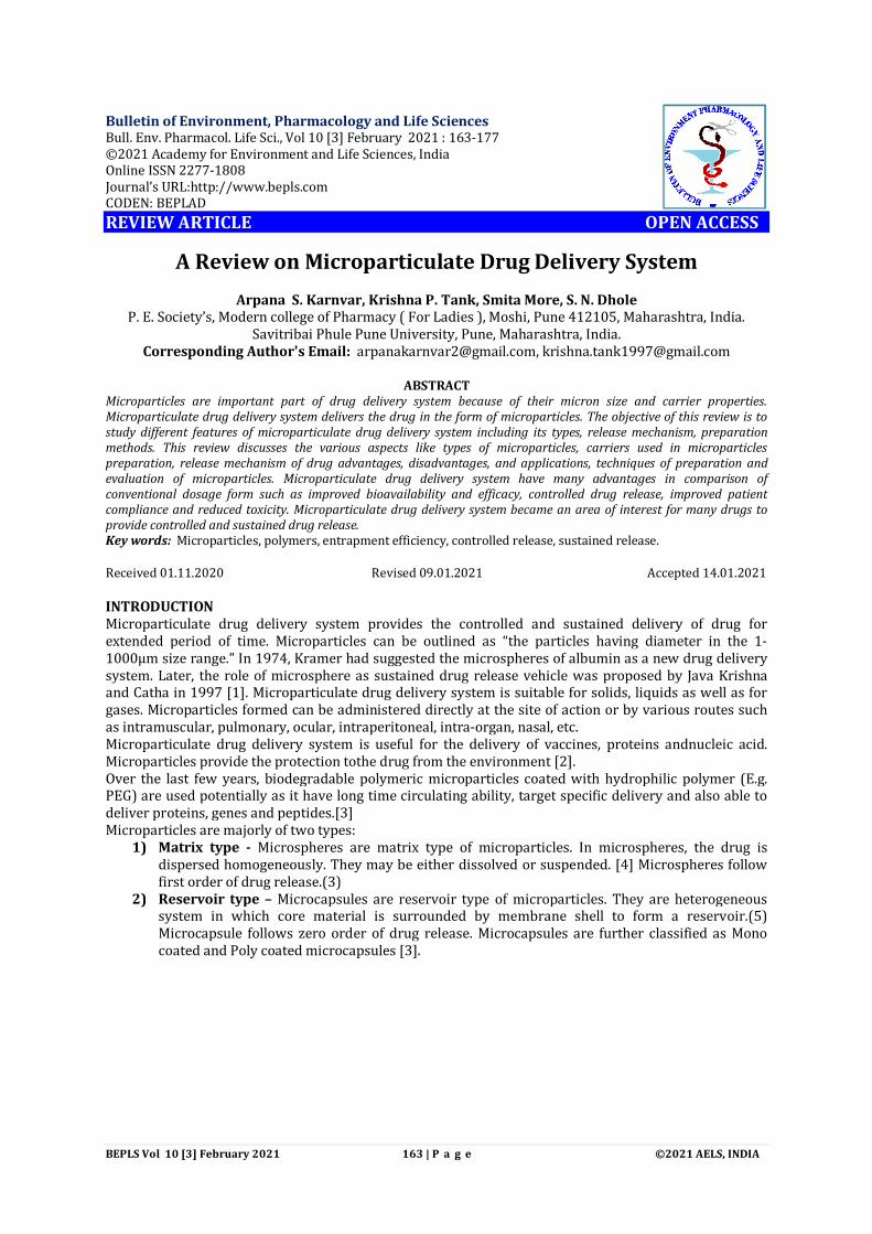
1) Matrix type - Microspheres are matrix type of microparticles. In microspheres, the drug is dispersed homogeneously. They may be either dissolved or suspended. [4] Microspheres follow first order of drug release.(3)
2) Reservoir type – Microcapsules are reservoir type of microparticles. They are heterogeneous system in which core material is surrounded by membrane shell to form a reservoir.(5) Microcapsule follows zero order of drug release. Microcapsules are further classified as Mono coated and Poly coated microcapsules [3].
BEPLS Vol 10 [3] February 2021 164 | P a g e ©2021 AELS, INDIA
Fig.1: Types of microparticles – Microcapsule and Microsphere
Advantages: Microparticulate drug delivery system offers many advantages including :
1) Taste masking 2) Odour masking 3) Protect the API or drugs from environment. 4) Increasing solubility by reducing particle size 5) Controlled drug delivery 6) Sustained release of drug 7) Targeted drug delivery system 8) Enhancing flow properties of powder 9) Increased bioavailability 10) Improved patient compliance 11) Reduction of volatility 12) Conversion of liquid into solid 13) Reduced dose dumping 14) Reduced drug toxicity 15) Enables safe handling of toxic substances (6)
Disadvantages: Despite of having many advantages, microparticulate drug delivery system has certain limitations:
1) More cost is involved due to material and techniques used for preparation 2) Less reproducibility 3) Temperature change, pH, evaporation, solvent addition, agitation may affects the stability of
core material 4) More time is required 5) Microparticles have large surface area as having small particle size and thus may have
limited drug loading (7) Composition of microparticles: Core material: Core material is the one on which the coating material is applied to form microparticles This core material may be the droplets of liquids or solid material and dispersions . The core material is any drug which has to be encapsulated for many reasons such as for protection from environment, for taste masking, for toxic substances, for sustained or controlled release of drug.(8) COATING MATERIAL / CARRIERS: There are various coating materials are used in microencapsulation process. This are:
Polymers : Water soluble: E.g. Gelatine, Methyl cellulose, etc. Water insoluble: E.g. Ethyl cellulose, Polymethacrylate, etc.
Waxes: E.g. Paraffin, Carnauba wax, Bees wax, etc. Enteric Resins: E.g. Shellac, Cellulose acetate phthalate, Zein. Polysaccharides: E.g. Chitin, Chitosan, Starch, Dextran, etc.
Polymers used: Non-biodegradable polymers:
Karnvar et al

BEPLS Vol 10 [3] February 2021 165 | P a g e ©2021 AELS, INDIA
E.g. Polyethylene vinyl acetate (PVA), Polyether urethane (PEU), Polydimethylsiloxam (PDS), Ethyl cellulose (EC), Polyvinyl chloride (PVC), Polyethylene, Cellulose acetate (CA). These are inert materials in environment of use. Hydrogels: E.g. Polyhydroxy ethyl methyl acrylate, cross linked Polyvinyl alcohol (PVA), Polyvinylpyrrolidone (PVP), Polyacrylamide, dextran. When come in contact with water they swell rather than dissolve. Biodegradable polymers: E.g. Polyglycolide(PGA). Polylactic acid (PLA), Polycaprolactone (PCL), Poly(lactic-co-glycolic acid) (PLGA). Soluble polymers: E.g. Hydroxypropylmethylcellulose (HPMC), Polyethylene glycol (PEG), Eudragit L. These polymers may be used individually or in combination to form controlled release dosage forms and to extend the release of drug for longer period of time. Properties of carriers:
Carriers should form cohesive film with core material. Carriers must be chemically compatible. They should be non-reactive. They should provide optimum strength, impermeability, flexibility and stability [8].
Factors Affecting Entrapment Efficiency Of Microparticles : The entrapment or encapsulation efficiency of microparticles can be influenced by various parameters. This parameters are as follows :
Table 1: Factor affecting the entrapment efficiency of microparticles [9] Low concentration of polymer High concentration of polymer High stirring speed Low stirring speed High drug : polymer ratio Low drug : polymer ratio High concentration of emulsifier Low concentration of emulsifier Low drug : polymer interaction High drug : polymer interaction Low concentration of cross linker High concentration of cross linker High solubility of polymer in organic solvent Low solubility of polymer in organic solvent High solubility of drug in continues phase Low solubility of drug in continues phase Low solubility of organic solvent in water High solubility of organic solvent in water
Release Mechanism of Microparticles: Depending on release mechanism, the encapsulated drug may release in sustained, controlled or targeted manner [10]. Drug release rate is depends upon :
Drug solubility Desorption of adsorbed drug Microparticle matrix degradation Diffusion of drug through microparticle matrix Combination of diffusion/ erosion process
Following are the major release mechanisms : Diffusion: In gastrointestinal tract, when microsphere comes in contact with aqueous media, the water diffuses inside the particle. Then the drug dissolves and drug solution diffuses through release coat to exterior. Erosion: The outer coating gets eroded with time and thus releases the drug entrapped within the particles. Polymer degrades by accumulation of monomer. When water penetrates within the particle it causes plasticization of matrix and thus change in microstructure which leads to polymer erosion.[10] Dissolution: When the coating material is soluble in dissolution media the drug release rate from microparticles is determined by rate of polymer coat dissolution [8]. Osmosis: Coating material acts as a semi-permeable membrane and thus creates osmotic pressure difference between drug solution and microparticle and allows drug solution to go out of the microparticles via small pores of the coating material.[8]
Low Drug Entrapment Efficiency High Drug Entrapment Efficiency
Karnvar et al

BEPLS Vol 10 [3] February 2021 166 | P a g e ©2021 AELS, INDIA
TECHNIQUES USED FOR MICROPARTICLE PREPARATION A) Physical methods :
a. Physico-chemical methods :- Co-acervation Solvent Evaporation Complex precipitation Layer-by-layer adsorption Supercritical fluid extraction Ionotropic gelation
b. Physico-mechanical methods:- Spray drying Spray congealing Vacuum encapsulation Pan coating Electrostatic encapsulation Air suspension (Fluidized bed) Extrusion Multiorifice centrifugal method
B) Chemical methods: Emulsion polymerization In-situ polymerization Interfacial polymerization (IFP)
Chemical methods: Chemical methods prepare microparticles by chemical reaction.
Emulsion Polymerization : In this technique, core material is added in the aqueous polymerization medium along with suitable emulsifier. Then, to this media the monomer is drop-by-drop added. Due to this process polymerizations starts. Primary nuclei is formed by precipitating the polymer molecule in the aqueous media, then these nuclei grows gradually as the polymerization proceeds. Simultaneously, the core material is entrapped to form microparticles.[11]
Interfacial Polymerization (IFP) : In this technique, the reactive monomers are used for polymerization. By polymerization the capsule shell is produced on the surface of particle or droplet. Multifunctional monomers are used . Generally, multifunctional acid chlorides and multifunctional isocyanates are used. These multifunctional monomers can be used individually or in combination. In liquid core material, the multifunctional monomers are dissolved and then dispersed in aqueous phase. This aqueous phase contains dispersing agent. To this mixture multifunctional amine is added as a co-reactant. This causes rapid polymerization at interface. As a result capsule shells forms. When acid chloride reacts with amine it forms polyamide or polynylon shell. When amine reacts with isocyanate it forms polyurea shell. Polyurethane shell will be formed by reaction of isocyanate with hydroxyl containing monomer.[11]
In-situ Polymerization : As same in IFP method, this method also forms the shell by polymerization monomers which are added into the encapsulation reactor. But, in this method the reactive agents are not added and polymerization occurs in continuous phase. Initially, prepolymer is formed and as the prepolymer grows, it gets adsorbed on the surface of core material to form shell.[11]
Karnvar et al

BEPLS Vol 10 [3] February 2021 167 | P a g e ©2021 AELS, INDIA
Fig. 2. Emulsion polymerization technique (Taken from EncyclopaediaBritannica)
Physical Methods: In these methods polymers are used as a starting material.
Physico-chemical Methods: Solvent Evaporation:
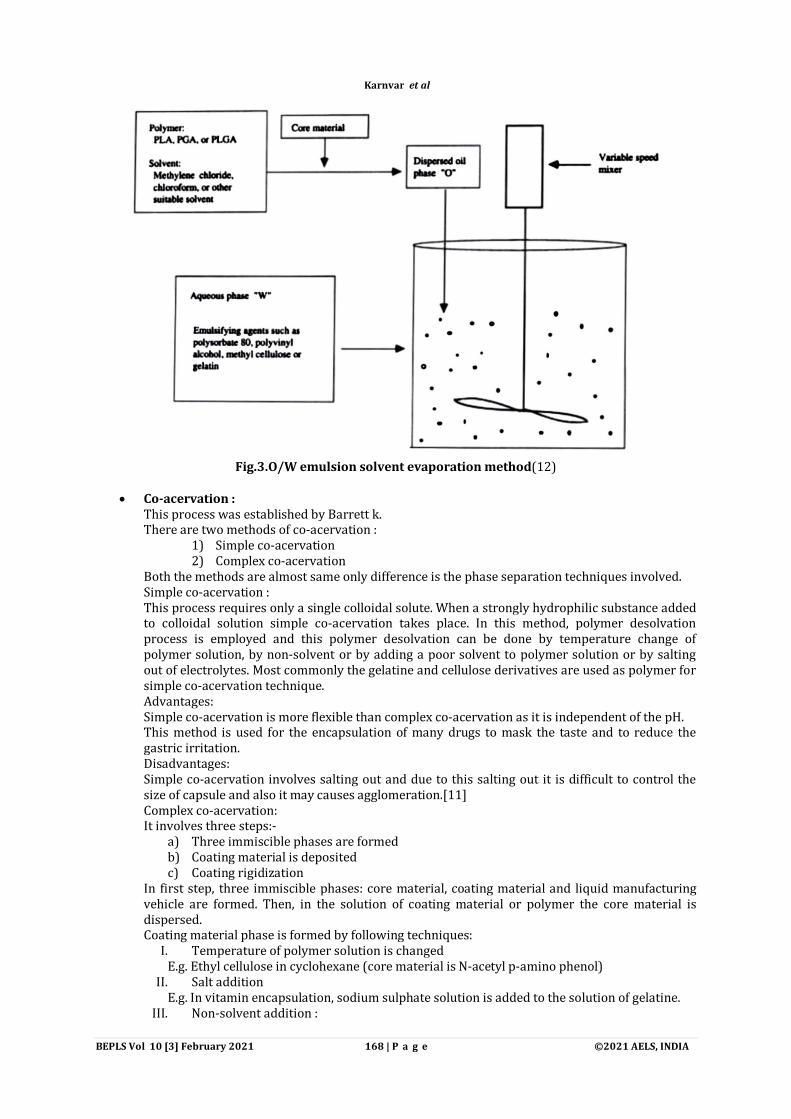
Most of the times the microparticles are get prepared by solvent evaporation method. It is an oil-in-water (o/w) type of procedure. Solvent evaporation technique involves following steps :
i. In volatile or organic solvent the coating material or polymer is get dissolved. This organic or volatile solvent is immiscible in liquid manufacturing vehicle.
ii. The drug which is to be encapsulated i.e., core material is then dispersed or dissolved in the above polymer solution.
iii. Then this mixture is added in the liquid manufacturing vehicle. It should be continuously agitated for obtaining desired size of microparticles.
iv. When optimum droplet size is formed’ the agitation speed is decreased and the solvent is evaporated under the reduced pressure and room temperature. Subsequent solvent evaporation yields the microparticles.(11)
Karnvar et al
BEPLS Vol 10 [3] February 2021 168 | P a g e ©2021 AELS, INDIA
Fig.3.O/W emulsion solvent evaporation method(12)
Co-acervation :
This process was established by Barrett k. There are two methods of co-acervation :
1) Simple co-acervation 2) Complex co-acervation
Both the methods are almost same only difference is the phase separation techniques involved. Simple co-acervation : This process requires only a single colloidal solute. When a strongly hydrophilic substance added to colloidal solution simple co-acervation takes place. In this method, polymer desolvation process is employed and this polymer desolvation can be done by temperature change of polymer solution, by non-solvent or by adding a poor solvent to polymer solution or by salting out of electrolytes. Most commonly the gelatine and cellulose derivatives are used as polymer for simple co-acervation technique. Advantages: Simple co-acervation is more flexible than complex co-acervation as it is independent of the pH. This method is used for the encapsulation of many drugs to mask the taste and to reduce the gastric irritation. Disadvantages: Simple co-acervation involves salting out and due to this salting out it is difficult to control the size of capsule and also it may causes agglomeration.[11] Complex co-acervation: It involves three steps:-
a) Three immiscible phases are formed b) Coating material is deposited c) Coating rigidization
In first step, three immiscible phases: core material, coating material and liquid manufacturing vehicle are formed. Then, in the solution of coating material or polymer the core material is dispersed. Coating material phase is formed by following techniques:
I. Temperature of polymer solution is changed E.g. Ethyl cellulose in cyclohexane (core material is N-acetyl p-amino phenol)
II. Salt addition E.g. In vitamin encapsulation, sodium sulphate solution is added to the solution of gelatine.
III. Non-solvent addition :
Karnvar et al

BEPLS Vol 10 [3] February 2021 169 | P a g e ©2021 AELS, INDIA
E.g. Isopropyl ether is added to methyl ethyl ketone solution. IV. Incompatible polymer addition to the polymer solution
E.g. Polybutadiene is added to the ethyl cellulose solution in toluene (core material is methylene blue)
V. Polymer-polymer interaction: E.g. At iso-electric point the gum arabic and gelatine interaction is induced. In second step, liquid polymer is deposited on the core material. In the last step, the microparticles produced are stabilized by desolvation, crosslinking or by thermal treatment.(6)
Fig.4. Schematic representation of co- acervation technique(13)
Ionotropic gelation:
In this method, the ionic polymers interact with oppositely charged ions and initiate the cross linking. Hydrogels are formed by this method. There are two types of ionotropic gelation method:
i. External ionotropic gelation : In this, cross linker ion is externally positioned.
ii. Internal gelation/Emulsification: In this, cross linker ion is inserted into polymer solution and it in an inactive form.
Sodium alginate and chitosan ate the mostly used polymers in ionotropic gelation method. (14) Physico-mechanical methods:
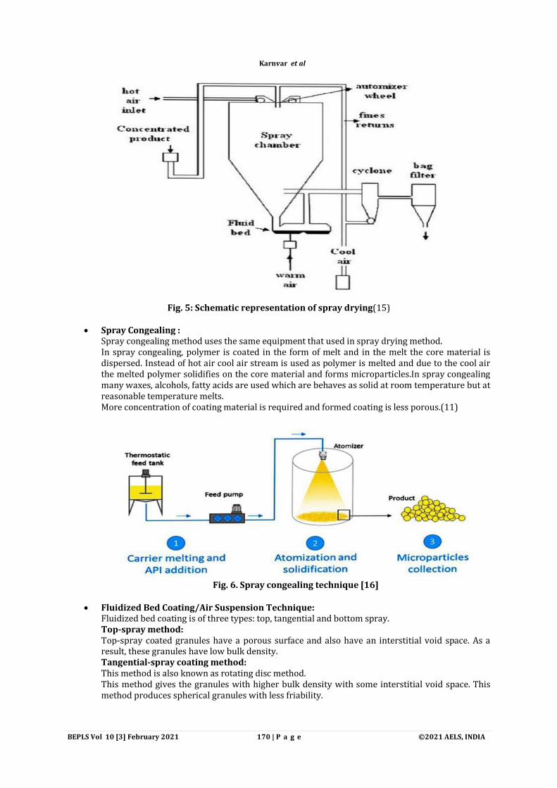
Spray Drying : It is a single step process. In spray drying method, polymer is dissolved in a solvent and then in this solvent the drug is dissolved or suspended. Then In the drying chamber the suspension or solution is atomized and microparticles are produced. These droplets can be dried by using heated carrier gas.These microparticles are 5-600 micron in size range.Mostly, water is used as a solvent for spray drying method as organic solvents maycauses toxicity.This method us rapid and gives reproducible results. This method is advantageous to be used for heat labile drugs as it requires less temperature.But, this method requires high cost as it involves the specialized equipmentand also there is difficulty in controlling the size of microparticles.(6)(11)
Karnvar et al
BEPLS Vol 10 [3] February 2021 170 | P a g e ©2021 AELS, INDIA
Fig. 5: Schematic representation of spray drying(15)
Spray Congealing :
Spray congealing method uses the same equipment that used in spray drying method. In spray congealing, polymer is coated in the form of melt and in the melt the core material is dispersed. Instead of hot air cool air stream is used as polymer is melted and due to the cool air the melted polymer solidifies on the core material and forms microparticles.In spray congealing many waxes, alcohols, fatty acids are used which are behaves as solid at room temperature but at reasonable temperature melts. More concentration of coating material is required and formed coating is less porous.(11)
Fig. 6. Spray congealing technique [16]
Fluidized Bed Coating/Air Suspension Technique:
Fluidized bed coating is of three types: top, tangential and bottom spray. Top-spray method: Top-spray coated granules have a porous surface and also have an interstitial void space. As a result, these granules have low bulk density. Tangential-spray coating method: This method is also known as rotating disc method. This method gives the granules with higher bulk density with some interstitial void space. This method produces spherical granules with less friability.
Karnvar et al

BEPLS Vol 10 [3] February 2021 171 | P a g e ©2021 AELS, INDIA
Bottom-spray method (Wurster process): This method was developed by E. Wurster. This apparatus consists of different parts like air distribution panel, coating chamber, control panel, nozzle for application of film coating. In the coating chamber the particles are suspended. These particles moves with upward moving air stream. On this moving core material the coating material is sprayed. During each pass the coating material is incremented on core material. This cyclic process continues until the desired thickness of coating is obtained. During the encapsulation process air stream dry out the product.(11) Following variables should be controlled for effective and efficient encapsulation process:
a. Particle size, shape, density, surface area, melting point, volatility, hardness, friability of core material.
b. Concentration, role of application and quantity of coating material. c. Air stream volume d. Operating temperature.
Fig.7: Fluidized bed coating technique.(17)
Multiorifice-centrifugal method:
This process has developed by ‘The Southwest Research Institute’ (SWRI). The apparatus is composed of a rotational cylinder having three peripheral grooves. The grooves are present at upper and lower periphery which carries the coating material. This coating material is present in solution or molten form. The middle groove have multiple orifice spaces closely and peripherally around the cylinder. The coating material is applied under centrifugal force by rotating the cylinder outward and forms a film through the orifice. Through the centrally placed inlet a counter rotating disc atomizes the core material. The counter rotating disc shoots the core material into the orifice. After that the core material encounters the coating material membrane and then centrifugal and impact force flings the core material across the coating membrane. Then the microparticles are congealed, hardened by various processes. This process encapsulates both solid as well as liquid materials.[11]
Karnvar et al

BEPLS Vol 10 [3] February 2021 172 | P a g e ©2021 AELS, INDIA
Fig.8:Multiorifice-centrifugal method. [18]
EVALUATION OF MICROPARTICLES
1) Particle Shape and Particle Size Determination: It can be determined by various techniques. Shape and surface morphology: It can be done by freezes etch electron microscopy, freeze fracture microscopy, etc. Crystallinity: It can be done by using differential scanning calorimetry and x-ray diffraction method.(1) Other methods for particle size determination include: a)Manual methods: 1. Sieving 2. Optical microscopy 3. Sedimentation 4. Electron microscopy:
Scanning ElectronMicroscopy (SEM), Transmission Electron Microscopy (TEM)
b) Automatic methods: 1. Flow cytometry 2. Light scattering:
Enhance Laser Diffraction Dynamic Light Scattering
3.Particle counters: The counter principle, Impaction and inertial techniques, Permeability, Optical particle counting.(6)
2) Bulk and Tap Density: Multivolume pychnometer is used to determine the bulk and tap density of microparticles.(3)
3) Flow properties: Angle of repose is determined by using fixed funnel method. Other method used is free-standing cone method.(3)
4) Microparticle yield/recovery: Microparticle yield can be determined by taking the ratio of amount of microparticles formed to the amount of drug and polymer used in its formulation.(6) It can be determined by following formula: Practical yield = Amount of encapsulated drug / Amount of added drug. Percent yield = (Practical yield / Theoretical yield )× 100
5) Entrapment Efficiency:
Karnvar et al

BEPLS Vol 10 [3] February 2021 173 | P a g e ©2021 AELS, INDIA
It can be calculated by the quantity of drug entrapped in microparticles and quantity of drug adsorbed on surface of polymer.(6) Percentage entrapped drug = (Amount of drug encapsulated / Amount of drug added for encapsulation) × 100
6) Differential Scanning Calorimetry (DSC): DSC is used to determine the physicochemical properties of the drug in microparticles. DSC gives the effect of excipients on thermal properties of the drug substances, similarities and differences of samples.(6)
7) Thermogravimetric Analysis (TGA): TGA determines the change in temperature. The temperature is gradually raised and then weight vs temperature is plotted. The curve smoothing and many other processes may done to find the definite points of inflection.(1)
8) Electrostatic Interaction: It is studied by FTIR assays and rheological assays by using potassium bromide pellets.(1)
9) In-vitro Release Studies: The drug release is obtained by determining the extent of polymer degradation of microparticles. The in-vitro drug release studies can be performed by various methods such as:
Dialysis method: In this method, microparticles are weighed accurately and placed in a dialysis bag. Then the bag is immersed in a continuous phase acceptor fluid. This stirred and the drug oozes out from the microparticle at different time is sampled and assayed.
Dissolution method: Drug release can be studied by using USP apparatus 2 or by using dissolution test method. It requires phosphate buffer of pH 6.8 and temperature of release medium should be 37±0.5. The sample is assayed using analytical methods like spectrophotometry.[1]
10) In-vivo Tissue Distribution Studies: It determines the efficacy of microparticles. For this studies various animal models are used such as adult Albino Mice, Rabbits, Wirstar Rats, etc. Drug is administered as dispersion in saline having 1% of tween 80. Dose of drug to be administer is calculated and administered to each animal. At certain time interval, the microparticles are injected through the vain of tail route and then animal is sacrificed by cervical dislocation. After, the organs like liver, lungs, spleen and heart are extracted and they are studied for the targeted action. This extracted tissues are stored at 20°c for 24 hours and then by using HPLC method the drug concentrated in each of the organ is calculated.[6]
APPLICATIONS Microparticulate drug delivery system have several applications :
1. Controlled drug delivery : Microparticulate drug delivery system releases the drug on controlled manner at predetermined rate for specified period of time. By using microparticle technology depot formulations can be developed.
2. Sustained drug delivery : By coating the drug particles in polymers, plasma concentration of drug can be maintained within therapeutic windows for longer period. It also reduces the dosing frequency and thus reduces toxic effects of drug.
3. Local drug delivery : Microparticulate drug delivery system can provide the local delivery of drug and maintain therapeutic concentration of the drug at the site of action. It reduces the systemic side effects.
4. Pulsatile drug delivery : Pulsatile delivery of antibiotics and vaccines is possible with microparticulate drug delivery system. Initial burst and then delayed release pulsed is effective in vaccine delivery.
5. Targeted drug delivery : Intracellular delivery: 1.Antisense therapy 2.Gene therapy 3.Ribozyme delivery 4.To cell organelles
Karnvar et al

BEPLS Vol 10 [3] February 2021 174 | P a g e ©2021 AELS, INDIA
5.Vaccine adjuvant 6.Cancer therapy Drug delivery to tissue: 1. To body cavity E.g. peritoneum 2. Antitumor microparticles to target an organ 3. Markers for detection/analysis E.g. Detect tumours. 4. Delivery of anti-cancer drugs. E.g.5-fluorouracil, doxorubicin
Table 2: List of drugs encapsulated in different microparticulate drug delivery system Drug Coating
Material Technique Result Ref.
5-fluorouracil PLAGA, PVA
Emulsion- extraction process
Microparticles can sustain the drug release over 18 days. (19)
Acetazolamide Eudragit RS, Eudragit RL
Solvent evaporation method
Controlled release is achieved with the microspheres without having initial peak levels. As a result these microsphere formulations may reduce the side effects, dosing frequency and may enhance patient adherence.
(20)
Acetaminophen Chitosan Spray drying method
This spray dried microspheres can be used as controlled drug delivery carriers. Particle size of microspheres was 3.8 to 4.2m and entrapment efficiency was 96.3-98.7%. Microspheres were found to be spherical,have smooth surface and found to follow Fick’s law of diffusion.
(21)
Albendazole HPMC, PVP, PVA
Spray drying method
The obtained microparticles showed enhanced dissolution rate of albendazole.
(22)
Betaxolol HCL
Eudragit, Montmorillonite
Oil-in-oil emulsion- solvent evaporation
Duration of in-vitro release was 12 hours much longer than conventional microspheres of BH (5hrs.) and BH solution (2.5hrs.). The microparticles showed less toxicity and also exhibited prolonged retention time and extended duration of action.
(23)
Curcumin Ethyl cellulose, Carbopol 934P
Emulsion-solvent evaporation
Free flowing microspheres were obtained. Drug release follows non-fickian model Due to controlled release of drug from microspheres these mucoadhesive microspheres may be used for both local as well as systemic action in effective H.pylori infection treatment.
(24)
Dexamethasone Albumin Spray drying This method produces the albumin microspheres with lipophilic drug loading. Size of microspheres was between 2 to 7.8m and showed 76.6 to 93.7% of entrapment efficiency.
(25)
Diclofenac sodium
Chitosan, Sod.alginate, Agar
Ionotropic gelation method
Spherical microspheres showed an increased drug release rate and controlled drug release for prolonged period of time
(26)
Furosemide Sodium alginate Ionic cross linking method
Microspheres showed 65% to 93% of entrapment efficiency. By varying different formulation parameters it was found that the entrapment efficiency and particle size also changed. It showed irregular transport mechanism.
(27)
Gliclazide Sod.alginate, Sod. CMC, HPMC, Carbopol 934P
Orifice –ionic gelation method
Microcapsules were spherical, discrete and free flowing. The drug release was found to be extended (16 hrs) and hence useful for long term delivery of drug. Its microencapsulation efficiency was 65-80% and showed good mucoadhesion.
(28)
Glimepiride Sod.alginate, HPMC
Ionic gelation and Emulsification gel method
Mucoadhesive microparticles were formed and showed good flow properties and encapsulation efficiencies. The drug release rate decreased with increased concentration of coating material. Microparticles formulated by emulsification gel technique showed slow release rate s compared to ionic gelation technique. So, former one is more preferable for mucoadhesive microparticles of glimepiride.
(29)
Ibuprofen Ethyl cellulose Solvent evaporation method
It showed that production variables affects the size distribution, amount of free drug and drug loading. It concluded that the parameters which affects the uncoated drug present on microparticle surface strongly influence the drug release from microparticles.
(30)
Karnvar et al

BEPLS Vol 10 [3] February 2021 175 | P a g e ©2021 AELS, INDIA
Ketoprofen Polysorbate 80, Carnauba wax
Emulsion congealing technique
This method produced spherical particles which showed sustained drug release. The formulated microspheres showed 200m average diameter. The obtained microsphere formulations were showed good flow properties, high drug load (about 50%w/w) and prolonged release of drug (for more than 24 hrs.).
(31)
Lovastatin Polylactic acid Emulsion –solvent evaporation technique
Microspheres obtained by this technique showed mean size of 2.650.69m and entrapment efficiency of 92.53.6%. It also showed increased drug loading (16.7% 2%). Microspheres followed sustained release pattern and showed significantly prolonged drug circulation time and increased bioavailability.
(32)
Metformin Homolipid obtained from C. hirus, Poloxamer 188, Phospholipon 90 H
Melt emulsification Obtained microparticles were spherical, stable and smooth surfaced. In-vivo studies for antidiabetic property showed higher reduction of glucose. It gave a new approach for delivery of metformin
(33)
Metronidazole Chitosan, Alginate
Tripolyphosphate cross- linking method
Microcapsule obtained had rough surface. Microcapsules showed entrapment efficiency in the range of 75.2-82%. Obtained microcapsules had improved mucoadhesive property at pH 1.2. Microcapsules showed slow drug release for extended period of time.
(34)
Naproxen Ethyl cellulose Spray drying method
Microparticles obtained had spherical shape and size range of 10-15m. Zero order release profile was followed by microparticles for skin permeation. It might prove as a potential delivery system with good efficacy.
(35)
Norfloxacin Sod. alginate Ionotropic gelation technique
Microbeads showed drug entrapment in range of 37.26% to 91.73%. Microbeads showed controlled release of drug. Release of norfloxacin from microbeads was observed to be affected by different concentration of sodium alginate.
(36)
Ondansetron Gellan gum Spray drying method
Microspheres were spherical and showed uniform particle size distribution. Microsphere formulation forms viscous gel when comes in contact with nasal mucosa. This gel reduces the rate of ciliary clearance and thus causes prolonged residence time.
(37)
Propranolol HCL
Bovine serum Albumin, Span 80, Tween 80
Emulsion –internal phase stabilization technique
Microspheres obtained were of 1-25m diameter. They showed 9% w/w of encapsulation and better bioadhesion (about 70%). Microspheres showed burst effect initially and then more gradual terminal release. Thus it showed biphasic release pattern. They showed sustained release form and controlled by diffusion. Microspheres provide steady plasma concentration of propranolol and increased bioavailability.
(38)
Repaglinide Chitosan, HPMC, Sod. alginate
Ionotropic gelation technique
Microparticles had 78.62% to 91.20% of entrapment efficiency and showed fickian and anomalous drug release profile. It also showed that with increased concentration of polymer there is decrease in amount of drug release.
(39)
Valsartan Polysorbate 20, Polysorbate 80
Spray drying method
Microparticles entrapped micelles showed enhanced dissolution of valsartan tablet. Obtained tablets showed enhanced dissolution rate irrespective of pH condition. They preserved the integrity and characteristics of encapsulated micelles.
(40)
CONCLUSION Microparticulate drug delivery system provides the controlled and sustained release of drug. Microparticles have size in micron range and have carriers system thus it is effective in providing the better drug delivery at controlled rate. This system has many advantages over the conventional dosage forms. It reduces the frequency of administration and have improved patient compliance and less side effects. Microparticles can be prepared by numerous techniques like solvent evaporation method, co-acervation method, spray drying and congealing, ionotropic gelation, etc. Microparticulate drug delivery system offers the better war for the preparation of drugs which cannot be formulated by conventional methods. Microparticulate drug delivery system is useful for targeted delivery of drug and applicable for the treatment of diseases including diabetes, arthritis, ocular diseases, cancer, etc. Microparticulate drug delivery system has become the area of interest for many drug formulations to provide the drug release for extended period of time.
Karnvar et al

BEPLS Vol 10 [3] February 2021 176 | P a g e ©2021 AELS, INDIA
REFERENCES 1. SathishMadhav NV, Kala S. (2015).Review on Microparticulate Drug Delivery System. Vol. 3, International
Journal of PharmTech Research Coden, 1242-1254 2. Lengyel M, Kállai-Szabó N, Antal V, Laki AJ, Antal I. (2019).Microparticles, microspheres, and microcapsules for
advanced drug delivery. Vol. 87, Scientia Pharmaceutica. MDPI AG. 3. Bale S, Khurana A, Singh M, Chandraiah Godugu (2016). Overview on Therapeutic Applications of
Microparticulate Drug Delivery Systems [Internet]. Vol. 33, Critical ReviewsTM in Therapeutic Drug Carrier Systems. Available from: www.begellhouse.com, 309-361.
4. Whelehan M, Marison IW. (2011). Microencapsulation using vibrating technology. Vol. 28, Journal of Microencapsulation. p. 669–88.
5. Peanparkdee M, Iwamoto S, Yamauchi R. (2016).Microencapsulation: AReviewof ApplicationIn The Food And Pharmaceutical Industries, Rev Agric Sci. ;4(0):56–65.
6. Pavan Kumar B, Chandiran IS, Bhavya B, Sindhuri M. (2011). Microparticulate drug delivery system: a review. 2011;1:19–37. Available from: www.ijpsrjournal.com
7. Shivani, SujithaH. (2009). ReviewArticleonMicroparticles [Internet]. Vol. 4, and Analytical Research. Available from: www.ijpar.com; p9-13
8. Singh MN, Hemant KSY, Ram M, Shivakumar HG. (2010). Microencapsulation: A promising technique for controlled drug delivery. Vol. 5. 20-30.
9. Dhakar R, Datta Maurya S, Bhagat S, Prajapati SK. (2008). Variables Influencing the Drug Entrapment Efficiency of Microspheres: A Pharmaceutical Review [Internet]. Available from: https://www.researchgate.net /publication/268436204
10. 10. Park K, Yeo Y. Microencapsulation Technology, Encyclopedia of Pharmaceutical Technology DOI: 10.1081/E-EPT-120028567, pp:2315–2327.
11. Das SK, David SR, Rajabalaya R, Halder T. (2011). Microencapsulation techniques and its practices Liquid Crystals View project Scurrula ferruginea View project. 6(2). Available from: https://www.researchgate.net/ publication/279498086
12. O’donnell PB, Mcginity JW. (1997). Preparation of microspheres by the solvent evaporation technique. Vol. 28, Advanced Drug Delivery Reviews. 29-37.
13. Iqbal K, Khan A, Sun D, Ashraf M, Rehman A, Safdar F, et al. (2019).Phase change materials, their synthesis and application in textiles—a review. Vol. 110, Journal of the Textile Institute. Taylor and Francis Ltd.; 625–38.
14. Ahirrao SP, Gide PS, Shrivastav B, Sharma P. (2009). Ionotropic Gelation: A Promising Cross Linking Technique for Hydrogels .ResearchAnd Reviews: Journal ofPharmaceutics And Nanotechnology.4[4]9-13
15. Saxena C, Vats S, Ts E, Vk S. Floating Microsphere: A Novel Approach Used to Develop Gastroretentive Drug Delivery System Floating Microsphere: A Novel Approach Used to Develop Gastroretentive Drug Delivery System [Internet]. Vol. 3, International Journal for Pharmaceutical Research Scholars (IJPRS) V. 2014. Available from: https://www.researchgate.net/publication/320187298
16. 16. Bertoni S, Albertini B, Passerini N. Spray congealing: An emerging technology to prepare solid dispersions with enhanced oral bioavailability of poorly water soluble drugs. Vol. 24, Molecules. MDPI AG; 2019.
17. Bakry AM, Abbas S, Ali B, Majeed H, Abouelwafa MY, Mousa A, et al. Microencapsulation of Oils: A Comprehensive Review of Benefits, Techniques, and Applications. Compr Rev Food Sci Food Saf. 2016 Jan 1;15(1):143–82.
18. Begum G, Madhusudhan Chettt C, Kota R, Susmitha K, Komali P, Jyothi Kiranmai S, et al. A Review on Microencapsulation. Rev Microencapsulation World J Pharm Sci [Internet]. 2018;6(4):25–36. Available from: http://www.wjpsonline.org/
19. Boisdron‐Celle M, Menei P, Benoit JP. Preparation and Characterization of 5‐Fluorouracil‐loaded Microparticles as Biodegradable Anticancer Drug Carriers. J Pharm Pharmacol. 1995;47(2):108–14.
20. Haznedar S, Dortunç B. Preparation and in vitro evaluation of Eudragit microspheres containing acetazolamide. In: International Journal of Pharmaceutics. Elsevier; 2004. p. 131–40.
21. Liu C, Desai KGH, Tang X, Chen X. Drug release kinetics of spray-dried chitosan microspheres. Dry Technol. 2006;24(6):769–76.
22. Ibrahim MA, Shazly GA, El-Badry M. Albendazole microparticles prepared by spray drying technique: Improvement of drug dissolution. Trop J Pharm Res. 2014 Dec 1;13(12):1963–70.
23. Tian S, Li J, Tao Q, Zhao Y, Lv Z, Yang F, et al. Controlled drug delivery for glaucoma therapy using montmorillonite/Eudragit microspheres as an ion-exchange carrier. Int J Nanomedicine. 2018 Jan 12;13:415–28.
24. Ali MS, Pandit V, Jain M, Dhar KL. Mucoadhesive microparticulate drug delivery system of curcumin against Helicobacter pylori infection: Design, development and optimization. J Adv Pharm Technol Res. 2014;5(1):48–56.
25. Pavanetto F, Genta I, Giunchedi P, Contit B, Conte U. (1994). Spray-dried albumin microspheres for the intra-articular delivery of dexamethasone. Vol. 11, J. Microencapsulation. 19-24.
26. Manjunatha K, Ramana M, Satyanarayana D. (2007). Design and evaluation of diclofenac sodium controlled drug delivery systems. Indian J Pharm Sci. 69(3):384–9.
27. Das M, Senapati P. (2008). Furosemide-loaded alginate microspheres prepared by ionic cross-linking technique: Morphology and release characteristics. Indian J Pharm Sci. ;70(1):77–84.
28. Prajapati SK, Tripathi P, Ubaidulla U, Anand V. (2008). Design and development of gliclazide mucoadhesive microcapsules: In vitro and In vivo evaluation. AAPS PharmSciTech. 9(1):224–30.
29. Satyanarayana J, Kumar AP, Kishore VS, Gopala TE, Murthy K. (2014).Formulation and Evaluation of
Karnvar et al

BEPLS Vol 10 [3] February 2021 177 | P a g e ©2021 AELS, INDIA
Mucoadhesive Microcapsules of Glimepiride. pp09. 30. Kristmundsdóttir T, Ingvarsdóttir K. (1994). Ibuprofen microcapsules: The effect of production variables on
microcapsule properties. Drug Dev Ind Pharm. 20(5):769–78. 31. Oliveira RB, Nascimento TL, Lima EM. (2012). Design and characterization of sustained release ketoprofen
entrapped carnauba wax microparticles. Drug Dev Ind Pharm. Jan;38(1):1–11. 32. Guan Q, Chen W, Hu X. (2015). Development of lovastatin-loaded poly(lactic acid) microspheres for sustained
oral delivery: In vitro and ex vivo evaluation. Drug Des Devel Ther.10;9:791–8. 33. Momoh MA, Kenechukwu FC, Attama AA. (2013). Formulation and evaluation of novel solid lipid microparticles
as a sustained release system for the delivery of metformin hydrochloride. Drug Deliv. 20(3–4):102–11. 34. Garud A, Garud N. (2013).Preparation and Evaluation of Chitosan Microcapsules of Metronidazole using
Tripolyphosphate Cross-linking Method.90-98 35. Arici M, Topbas O, Karavana SY, Ertan G, Sariisik M, Ozturk C. (2014).Preparation of naproxen-ethyl cellulose
microparticles by spray-drying technique and their application to textile materials. J Microencapsul. 1;31(7):654–66.
36. Kundu A, Datta S. (2012). Formulation and Characterization of Alginate Microbeads of Norfloxacin by Ionotropic Gelation Technique [Internet]. Available from: www.ijapbc.com 3[4].90-99
37. Mahajan HS, Gattani SG. (2009). Nasal administration of ondansetron using a novel microspheres delivery system. Pharm Dev Technol.14(2):226–32.
38. Abolghassem S, Tabassi S, Razavi N. (2003).Preparation and characterization of Albumin microspheres encapsulated with propranolol HCl. Daru. 11(4):137–41.
39. Bhadke S. (2006). Formulation And Development Of Repaglinide Microparticles By Ionotropic Gelation Technique [Internet]. Available from: http://hdl.handle.net/123456789/2138
40. Parvataneni DM, Devraj R, Mangamoori LN. (2013).Microparticles-entrapped micelles: A novel delivery system to improve solubility and dissolution rate of poorly water-soluble valsartan. J Microencapsul. 30(8):805–16.
CITATION OF THIS ARTICLE Arpana S. Karnvar, Krishna P. Tank, Smita More, S. N. Dhole. A Review On Microparticulate Drug Delivery System. Bull. Env. Pharmacol. Life Sci., Vol 10[3] February 2021 : 163-177.
Karnvar et al


![Intelligent drug delivery system - pgsitecdn.persiangig.com/dl/9MZwnq/student Intelligent drug delivery syste… · Table 2. Marketed technologies of pulsatile drug delivery [31]](https://static.fdocuments.in/doc/165x107/5f3dc762b8577c0d041fed9b/intelligent-drug-delivery-system-intelligent-drug-delivery-syste-table-2-marketed.jpg)