A Review of Mastitis Control Practices
52
A Review of Mastitis Control Practices A Senior Project presented to the Faculty of the Dairy Science Department California Polytechnic State University, San Luis Obispo In Partial Fulfillment of the Requirements for the Degree Bachelor of Science by Rebecca Gooder February, 2014 © 2014 Rebecca Gooder
Transcript of A Review of Mastitis Control Practices

A Review of Mastitis Control Practices
A Senior Project
presented to
the Faculty of the Dairy Science Department
California Polytechnic State University, San Luis Obispo
In Partial Fulfillment
of the Requirements for the Degree
Bachelor of Science
by
Rebecca Gooder
February, 2014
© 2014 Rebecca Gooder

1
TABLE OF CONTENTS
Abstract………………………………………………………………..…………………. 2
Introduction………………………………………………………………...…………….. 3
Discussion of Literature
Cow Hygiene…..………………………………..………………...…………...… 6
Milking Practices
Overview…………………………………………………..…………….. 9
Pre- and Postmilking Teat Dipping…………………………………….. 10
Types of Teat Disinfectants…………...………………………………... 12
Methods of Application……………………………...…………………. 17
Other Milking Techniques…………………………………...…………. 20
Dry Cow Therapy
Overview…………………………………………………………….….. 23
Antibiotic Infusion……………………………………………………… 23
Internal Teat Sealants…………………………………………………… 25
Combination Treatments………………………………………………... 28
Barrier Teat Dips………………………………………………………... 32
Vitamin Supplementation…………………………………..……….………….. 34
Records and Testing…………………………………………………………….. 36
Vaccination………………………………………………………………..……. 39
Conclusion………………………………………………………………..…………….. 42
References……………………………………………………….…………...…………. 44

2
ABSTRACT
The objective of this literature review was to investigate, compile and summarize
published research encompassing mastitis control practices, with an emphasis on dairy
cow hygiene, milking practices, dry cow therapy, the utilization of bulk tank analysis and
California Mastitis Tests, and vaccination programs. A significant amount of research has
been done in this field and the vast majority reports a positive correlation between these
management practices and the prevention and/ or control of mastitis in the dairy herd.
Many parallels have been made between the hygiene level of a cow and somatic cell
count since clean cows have less of a chance of acquiring an intramammary infection.
Several milking techniques assist in the prevention of mastitis, including pre- and
postmilking teat dipping. Dry cow therapy- antibiotics and internal and/or external teat
sealants- treats existing intramammary infections at drying off and protects cows from
acquiring new intramammary infections during the dry period. Somatic cell counts,
obtained through Bulk Tank Analysis testing, as well as use of the California Mastitis
Test, are good indicators of an intramammary infection. Vaccines have the ability to
lower microbial counts, provide protection against culling, and/ or reduce the severity of
clinical mastitis.
(Key words: mastitis, milking practices, dry cow therapy, vaccines)
Abbreviation key: CMT= California Mastitis Test, IMI= intramammary infection,
NMC= National Mastitis Council, SCC= somatic cell count

3
INTRODUCTION
Infection of the mammary gland has been deemed the most significant udder
health problem facing the dairy industry in the US (National Mastitis Council, 2007a).
This intramammary infection may be caused by contagious or environmental pathogens.
Contagious pathogens live and multiply on and in the cow’s mammary gland and are
transmitted from cow to cow primarily during the milking process. Major contagious
mastitis pathogens include Staphylococcus aureus and Streptococcus agalactiae.
Environmental pathogens reside in the environment where cows live. Major
environmental mastitis pathogens include streptococci other than S. agalactiae and
coliforms, or Gram-negative bacteria (Oliver et al., 2004). Mastitis can be a sub-clinical
or clinical infection (Barnouin, 2005). Clinical mastitis results in alterations in milk
composition and appearance, decreased milk production, elevated body temperature, and
swelling, redness or heat in infected mammary quarters. Subclinical mastitis, the most
common form of this disease, tends to persist, resulting in elevated milk somatic cell
count and decreased milk production. This may eventually lead to the development of
clinical mastitis. (Oliver et al., 2004).
The National Mastitis Council expects that dairies without an effective mastitis
control program will see about 40% of the herd infected in an average of two quarters.
Such infections can cost as much as $200 per cow per year (Current Concepts of Bovine
Mastitis, 1996). These economic losses are due to reduced milk production, treatment
costs, increased labor, milk withheld following treatment, death, and premature culling.
Reduced milk production is responsible for the majority of these losses- approximately
70%. (Sargeant et al., 2001). Estimating the loss due to mastitis is often done via the

4
negative relationship between milk yield and SCC. These estimates are crucial in
determining cost-effective strategies for mastitis control and prevention. (Gill et al.,
1990). Minimizing mastitis is not only necessary to maintain the economic viability of a
dairy, but essential in the quest to produce high quality milk. After all, studies have
shown that milk composition is altered by mastitis pathogens. Fat, lactose, and casein
contents are usually decreased, as are cheese yields (Pankey, 1989).
Best controlling instances of mastitis will require the minimization of
intramammary infections. Establishment of an intramammary infection requires
penetration of the aforementioned mastitis-causing pathogens through the teat canal. The
number and types of bacteria on the teat skin have a direct relationship to the incidence
and type of mastitis that occurs (Nickerson, 2001). There are several practices that dairy
producers can implement to successfully manage mastitis in their herd. The National
Mastitis Council published a ‘Recommended Mastitis Control Program’ that emphasizes
ten points: establishment of goals for udder health, maintenance of a clean, dry,
comfortable environment, proper milking procedures, proper maintenance and use of
milking equipment, good record keeping, appropriate management of clinical mastitis
during lactation, effective dry cow treatment, maintenance of biosecurity for contagious
pathogens and marketing of chronically infected cows, regular monitoring of udder health
status and period review of mastitis control program (National Mastitis Council
“Recommended Mastitis Control Program”). Comparing management practices of high
quality herds to medium or poor quality herds is one way to highlight management
practices associated with a high degree of control of clinical and subclinical udder

5
infections (Chassagne et al., 2005). Reviewing literature is another way to compare and
assess the current mastitis control program on a dairy.
This paper reviews mastitis control practices that have been recommended by the
National Mastitis Council. Practices highlighted include: cow hygiene, milking routines,
dry cow therapy, vitamin supplementation, milk testing, and vaccination strategies.

6
DISCUSSION OF LITERATURE
Cow Hygiene
Maintaining excellent cow hygiene is an essential practice for controlling mastitis
in the dairy herd as many studies have found a correlation between clean cows and lower
bulk tank SCC. Similarly, some studies have proved that poor hygiene results in udder
health problems (National Mastitis Council, 2007). Exposure to manure in cow housing
areas can influence the rate of clinical mastitis. Moreover, cleanliness of the udder is
thought to influence the quantity and type of bacteria that is present on teat surfaces.
Dirty teats and udders, as a result of moisture, mud and manure in the environment of the
cow, are considered to be sources of environmental bacteria in milk. Hygiene scores of
cows provide visible evidence of exposure to these potential sources (Schreiner and
Ruegg, 2003).
There are four ways in which manure can be transferred to the udder: direct, leg,
splash and tail. Direct transfer involves cows that may have lied down in a manure
contaminated free stall. Leg transfer occurs when cows have walked through manure that
has coat their feet and legs. Upon lying down, the udder rests on one of the hind feet and
manure is transferred. Splash transfer occurs when cows are walking through deep slurry
and manure splashes upwards toward the udder. Lastly, tail transfer occurs when the tail
becomes heavily soiled with manure which then comes into contact with the rear udder
and flank areas (National Mastitis Council, 2007).
Ruegg and Schreiner examined the relationship between udder and leg hygiene
scores of lactating dairy cattle and measures of subclinical mastitis. Udder and leg
hygiene scores were recorded by one person using a four-point scale ranging from one

7
(very clean) to four (very dirty). These scores were then compared to bacteriological
cultures of milk samples and monthly individual SCC values. Linear somatic cell scores
increased as udder hygiene scores increased. A significant association was reported
between the prevalence of intramammary contagious pathogens and udder hygiene score,
confirming the relationship between the measures of subclinical mastitis and
measurements of animal hygiene In addition, the prevalence of intramammary
environmental pathogens was significantly associated with udder hygiene score.
(Schreiner and Ruegg, 2003).
A 2003 National Mastic Council Annual Meeting Proceeding, entitled
‘Relationship of cow hygiene scores and SCC’, reported that a study conducted in the
Netherlands showed that herds with a bulk tank somatic cell count less than 150,000 had
four times fewer dry cows with greater than 30% of their udders covered with manure. In
their particular study, they utilized a similar scoring system to that of the Schreiner and
Ruegg study, ranking cows 1-5. A score of ‘1’ indicated an absolutely clean cow, while a
score of ‘5’ indicated a very dirty cow. The cow hygiene scorecard was broken down into
five general areas: tail head, flank, belly, udder, and rear legs and feet. Only the scores of
udder and lower rear legs had a significant effect on the somatic cell score. Herds with a
similar predominance of environmental mastitis infections and similar SCC levels may
expect to see a 40-50,000 change in herd SCC for each 1-unit change in cow hygiene
scores (National Mastitis Council, 2003).
A study performed by Sant’Anna and Paranhos da Costa in 2011 also followed a
similar evaluation to that of the Schreiner and Ruegg study. Four areas of each animal’s
body were evaluated: legs, flanks, abdomen, and udder. The scores were defined on a 1-4
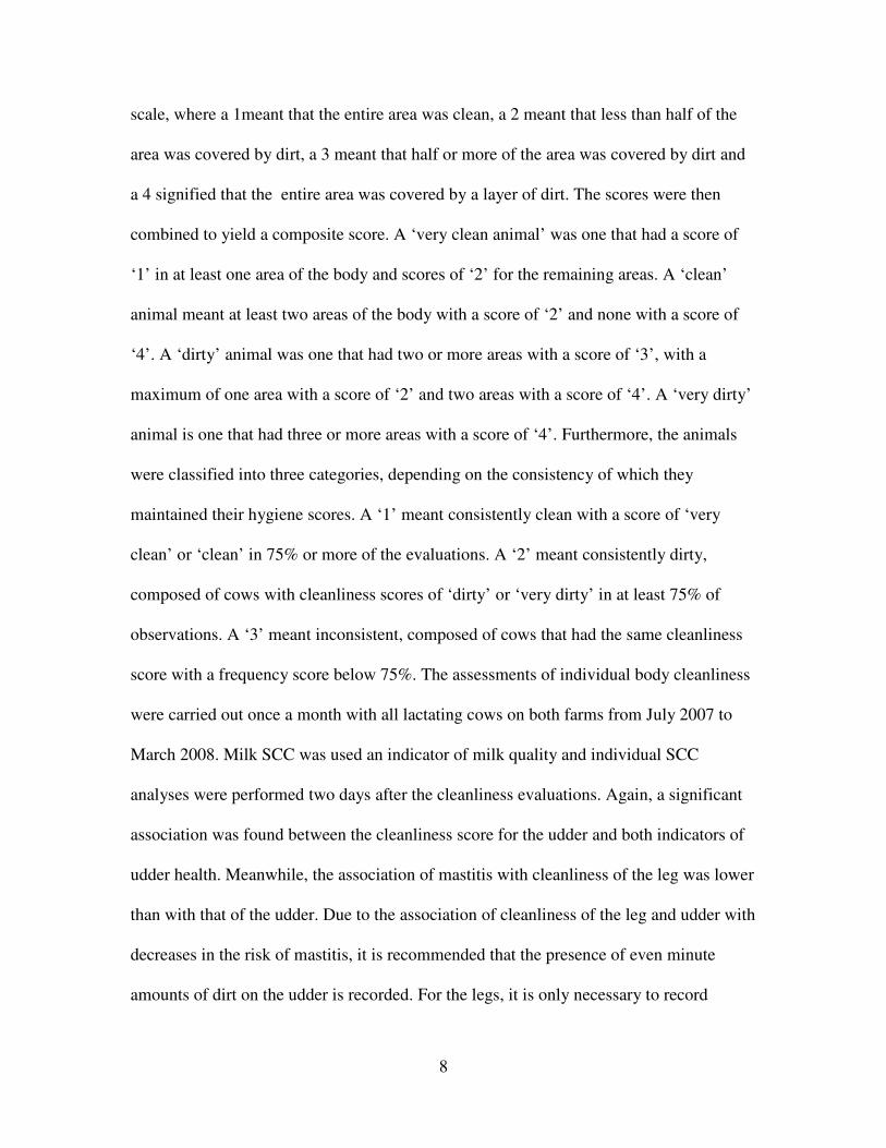
8
scale, where a 1meant that the entire area was clean, a 2 meant that less than half of the
area was covered by dirt, a 3 meant that half or more of the area was covered by dirt and
a 4 signified that the entire area was covered by a layer of dirt. The scores were then
combined to yield a composite score. A ‘very clean animal’ was one that had a score of
‘1’ in at least one area of the body and scores of ‘2’ for the remaining areas. A ‘clean’
animal meant at least two areas of the body with a score of ‘2’ and none with a score of
‘4’. A ‘dirty’ animal was one that had two or more areas with a score of ‘3’, with a
maximum of one area with a score of ‘2’ and two areas with a score of ‘4’. A ‘very dirty’
animal is one that had three or more areas with a score of ‘4’. Furthermore, the animals
were classified into three categories, depending on the consistency of which they
maintained their hygiene scores. A ‘1’ meant consistently clean with a score of ‘very
clean’ or ‘clean’ in 75% or more of the evaluations. A ‘2’ meant consistently dirty,
composed of cows with cleanliness scores of ‘dirty’ or ‘very dirty’ in at least 75% of
observations. A ‘3’ meant inconsistent, composed of cows that had the same cleanliness
score with a frequency score below 75%. The assessments of individual body cleanliness
were carried out once a month with all lactating cows on both farms from July 2007 to
March 2008. Milk SCC was used an indicator of milk quality and individual SCC
analyses were performed two days after the cleanliness evaluations. Again, a significant
association was found between the cleanliness score for the udder and both indicators of
udder health. Meanwhile, the association of mastitis with cleanliness of the leg was lower
than with that of the udder. Due to the association of cleanliness of the leg and udder with
decreases in the risk of mastitis, it is recommended that the presence of even minute
amounts of dirt on the udder is recorded. For the legs, it is only necessary to record

9
instances where there is a rather large accumulation of dirt. (Sant’Anna and Paranhos da
Costa, 2011).
Undoubtedly a good management tool, there is a strong factor of subjectivity that
must be accounted for. What appears ‘dirty’ to one observer might look fairly clean to
another, so it may be necessary to have a single employee in charge of scoring cows’
hygiene.
Milking Practices
An open teat sphincter during milking presents the perfect opportunity for mastitis
pathogens to invade. Proper milking routines, however, will make a cow less susceptible
to these pathogens.
Overview. An article on The Dairy Site, written by G.M. Jones, a professor of
Dairy Science at Virginia Tech, offers some key points for the milking routine. Pre-
dipping can serve as a substitute for washing teats with sanitizer; however, it is essential
that dirty teats are properly cleaned before a pre-dip solution is applied. The effectiveness
of pre-milking teat dipping is reduced by wet and dirty cows. The pre-dip shall remain on
the teat for approximately 30 seconds prior to being wiped off with a paper or cloth
towel. No one towel should be used on any two cows, as this is a prime method for
spreading infection. It is recommended to not even fold the cloth towel over and use the
backside of it for a second cow. Cloth towels are preferred by many milkers and do a
better job of getting teats dried. In addition, they may be cheaper; however, they must be
laundered in between uses. Regardless of towel type, drying is a crucial step in order to
avoid increased teat dip residues in the milk. It is permissible to use the same teat dip

10
prior to and after milking; however, it is advised that two different containers be used.
Dips should contain up to 10-14% skin conditioner, such as glycerol, lanolin, etc., to
prevent chaffing (Jones, 2006). Teat disinfectants are discussed in much more depth later
on.
Pre- and Postmilking Teat Dipping. Pre-milking teat disinfection was developed
as a simple, effective and economic method to control environmental pathogens by
reducing bacterial populations on teat skin before milking, thus minimizing their
penetration into the teat canal (Nickerson, 2001). This has proven to be necessary since
postmilking teat disinfectants have not proven to be effective in controlling mastitis due
to the environmental pathogens such as coliforms and streptococci other than S.
agalactiae (Oliver et al., 2001).
Nickerson, affirming that both pre- and postmilking teat dips are the most
effective procedures for preventing new intramammary infections, published an all-
encompassing article on choosing the best teat dip for mastitis control and milk quality.
There has been a great deal of evidence to support that this practice reduces the rate of
IMI among dairy cows. In addition, pre-dipping encourages milk letdown, thus speeding
up the milking process, and helps to ensure that the maximum amount of available milk is
harvested without causing damage to the sensitive teat tissues. Prior to this method,
which originated at the University of California, Davis, washing udders was common
practice. However, it was theorized that pre-dipping, as opposed to udder washing, may
lead to a reduced amount of water on teat ends remaining from the wash pen. As a result,
this procedure was deemed more effective than udder washing in killing bacteria. The
effectiveness was confirmed in later efficacy studies performed at Cornell University, the

11
University of Vermont, and the University of Tennessee (Nickerson, 2001). Pre-dipping
was found to reduce the incidence of new IMI, caused by environmental pathogens, by
more than 50% when compared with udder washing and drying with individual paper
towels. If applied on top of manure and dirt present on the teat skin, a pre-dip is not likely
to reduce the incidence of mastitis or lower the somatic cell count. In order to fully
appreciate the benefits of pre-dipping, manure and dirt must be removed (Nickerson,
2001).
Postmilking teat dipping has been deemed the most effective milking hygiene
practice for preventing new infections caused by the two most common contagious
mastitis pathogens- Staph. aureus and Strep. agalactiae. The vast majority of postmilking
teat dip products have the capability of reducing new intramammary infections by at least
50%, and, in some cases, as much as 95% (Nickerson, 2001).
Dipping all teats after each milking has a greater impact on reduction of milk
somatic cell counts and increased milk yields than any other milking practice. The teat
dip should leave a residue on the teat so that the antimicrobial action is still present when
the cow lies down in a free stall or any other place where sanitary conditions are less than
ideal. Recommended post-milking teat dipping practices include covering the entire teat
with the dip, making sure not to forget the backsides, especially if a sprayer is used. In
many cases, the back sides of teats are hardly touched when this method of teat dip
application is used in a parlor. At the very minimum, at least the bottom half of all teats
should be dipped after every milking, using an FDA approved teat dip (Jones, 2006).
Several teat disinfectants are available on the market and there is always debate
among researchers, extension specialists, veterinarians and producers on which one is

12
“best”. The answer, however, is often simple- one that has been registered by the FDA
and has been proven effective. As straightforward as that may sound, it is an important
point to make as producers do not want to run the risk of using a product that is either
providing absolutely no benefits or is inadvertently harmful to the teat skin, thus
potentially promoting a new intramammary infection to grow (Nickerson, 2001).
In August of 1995, the National Mastitis Council published a document that they
now update biannually, which summarizes the peer-reviewed scientific publications on
the efficacies of tested pre- and postmilking teat disinfectants, allowing producers,
veterinarians, etc. to make a more educated decision (Nickerson, 2001).
Types of Teat Disinfectants. Many different germicide classes are used in teat
dips and each is effective in their own manner. Iodine is a broad-spectrum germicide that
is fast acting and effective against essentially all mastitis-causing bacteria. Chlorine
destroys a wide range of microorganisms in a rapid fashion, yet these products must be
used within several hours of preparation because of short shelf life. Chlorhexidine
sanitizers adhere well to teat skin, provide antimicrobial activity over time, and do not
have deleterious effects on teat skin. Dodecyl Benzene Sulfonic Acid, or DDBSA, is
effective against gram-positive and gram-negative bacteria. Hydrogen peroxide provides
a wide spectrum of control against most mastitis-causing bacteria through its oxidizing
action. It may be combined with lactic acid, and it is through this combination that dead
teat skin is sloughed off, thus improving teat skin condition and minimizing bacterial
colonization on the teat skin surface. Fatty-acid based products, when water-based, are
generally recommended as both pre- and post-dips. On the other hand, organic solvent-
based products tend to be recommended for post-dipping. Nisin is extremely
13
bacteriocidal against Gram-positive and negative organisms through its lytic action on the
phospholipid components of the cytoplasmic membrane. Glycerol monolaurate is lipid
soluble and easily penetrates the bacterial cell membrane, which leads to the rupturing of
the cytoplasm. Quaternary ammonium products are microbicidal throughout denaturation
of cell proteins, inhibition of enzyme systems, and alteration membrane permeability,
which leads to disruption of the bacterial cell. Powdered teat sanitizers are recommended
during extremely cold and windy weather, when it is not advisable to use conventional
teat dip products due to the potential for frost bite (Nickerson, 2001). Regardless of the
active ingredient in the teat disinfectant, germicidal activity is a function of concentration
and contact time. Thus, higher concentrations require less contact time to be effective
(Pankey, 1989).
Phenols and phenolic compounds have been studied extensively as disinfectants
and have been shown to have a wide spectrum of antibacterial activity against both
Gram-positive and Gram-negative pathogens. A study by Oliver et al. examined the
effectiveness of a phenolic pre-milking teat disinfection compared with a negative control
using a split-udder experimental design (Oliver et al., 2001). The pre-milking teat
disinfectant was applied with a contact time of at least 30 seconds. The teats were then
dried thoroughly with single-use paper towels in preparation for attaching the milking
machine. The phenolic combination was applied as a postmilking teat disinfectant to teats
of all cows after milking machine removal. A quarter was considered newly infected
when the same bacterial species was isolated from two consecutive monthly samples, or
in samples from mammary glands of cows with clinical mastitis. The percentage of
quarters newly infected was 38% lower in quarters with teats pre-dipped and post-dipped

14
in the phenolic combination than in quarters with teats post-dipped only. New
intramammary infections by S. uberis and coagulase- negative staphylococci were
significantly lower in quarters with teats pre-dipped and post-dipped than in quarters with
teats post-dipped only. Overall, the teat disinfectant studied that contained this phenolic
combination was significantly effective against a wide variety of mastitis pathogens
including S. dysgalactiae, S. uberis, Gram-negative pathogens, and coagulase- negative
Staphylococcus species. This is a major difference between the results of this study and
previous reports on the efficacy evaluation of pre-milking teat disinfectants. This study
showed that applying a teat dip both prior to and after milking is significantly more
effective in preventing new intramammary infections than just postmilking teat
disinfection only (Oliver et al., 2001).
A gel, as opposed to a liquid pre-dip disinfectant, was developed and tested for its
ability to sanitize cows. The objective of this study by Ingawa and coworkers was to
compare the efficacy of using a gel with teat cleaners and sanitizers to prepare teats for
milking with the efficacy of washing and drying teats using plain water with or without a
.5% iodophor pre-dip. The first experiment used the gel (a water-based mixture
containing .5% iodophor as the active ingredient as well as a detergent, glycerin and an
aqueous gelling agent) with teat cleaning and sanitizing agents that were developed by
the authors of the study. The gel was allowed contact time with the teats of 30 seconds,
followed by the drying of each teat with a single-use paper towel. The second experiment
was simply cleaning the teats with water from a hose, using a spray nozzle, and then
drying the teats with a single-use paper towel. The third experiment was the same as the
second treatment, except that pre-dipping with a .5% iodophor solution was added.

15
Individual cow composite milk and teat end swab samples were collected once a week.
The gel and pre-dip treatments had similar raw milk bacterial counts. Both were lower
than in the wash treatment. The wash treatment had a significantly shorter preparation
time than the gel or pre-dip treatments, but as mentioned, the wash had the highest
bacterial count in milk and on teat ends. As a result, milk quality and udder heath would
likely suffer. Least square analysis of SCC data indicated lower SCC for the gel than for
either the pre-dip or wash treatments. Because using water before pre-dipping has been
shown to cause IMI and increase SCC, the gel treatment may prove itself to be an
effective method of udder preparation as it does not require water. No clinical cases of
mastitis were observed with the gel treatment during the study, and new IMI were
significantly reduced by the gel compared with the wash and pre-dip treatments. The
percentage of reduction in new clinical mastitis cases between the gel and the other two
treatments was 100%. Furthermore, the gel treatment was more efficient in the number of
cows milked per hour than the pre-dip treatment. And as has been noted in several
papers, the wash procedure did not provide adequate hygiene (Ingawa et al., 1992).
Winter in various parts of the country can present a large challenge to dairy
producers. Under conditions of severe cold or wind, postmilking teat dipping with
improperly formulated teat dips may facilitate teat chapping, resulting in increased
colonization by the Staphylococci that are normally associated with teat skin. In an effort
to minimize teat chapping, many dairy farms do not apply conventional, water-based teat
dips postmilking. The objective of a study by Goldberg et al. was to examine the
effectiveness of a powdered teat dip in the reduction of teat chapping. The starch in
powdered teat dips absorbs the surface water responsible for chapping. The most notable

16
positive result for the powdered dip was the consistent control of environmental mastitis
pathogens. The most important negative result was observed for the multiparous cows.
The powdered dip did not control mastitis caused by S. aureus. The presence of less than
average teat end conditions in the multiparous groups at initiation of the trial was
probably a predisposition to new intramammary infections with S. aureus (Goldberg et
al., 1994).
With a public that has in the past expressed concern over elevated concentrations
of iodine in milk, it has been necessary to study whether adequate mastitis control can be
implemented while limiting the amount of iodine that is present in the teat dips that are
being used. In a study by Bray and coworkers, six commercial dairy herds were used to
test the efficacy of three concentrations of iodine as a teat dip over a twelve month
period. Concentrations used were .1, .25 and 1% iodine with 3.5, 4.0 and 1.0ppm. Special
formulation procedures made it possible to increase free iodine concentrations in low
iodine dips. Quarter milk samples were collected for each bacteriological analysis from
each quarter of each cow at weeks 0 and 1 and at months 2, 4, 6, 9, and 12. Additional
samples were obtained by the dairy producer before administration of therapy for clinical
mastitis. A quarter was infected if two consecutive or preclinical samples contained the
same organism. The largest number of new infections occurred in cows whose teats were
dipped in the 1% iodine, which was the standard/ control treatment. The fewest number
of new infections occurred in the .1% iodine group. This was as expected, since the dip
contained a higher concentration of free iodine. Free iodine concentration is the most
important characteristic in the ability of the germicide to kill freely suspended vegetative
organisms. Regardless, the difference was not significant. This study determined that the

17
use of low iodine concentration teat dips with increased free iodine contents provided
protection against new infection equal to or greater than the standard 1% dip (Bray et al.,
1983).
Methods of Application. Teat disinfectants can be applied in an array of ways-
dipped, sprayed, automatic, manual, etc. The conventional method has been to dip teats
using some type of cup that contains the disinfectant. This can be done via recirculating
teat dip cups or non-circulating dip cups. Recirculating cups allow the product that has
come in contact with the teat skin to mix with the remainder of the dip cup contents. With
this type of applicator, if not kept extremely clean, there is the possibility of spreading
mastitis pathogens cow to cow. Non-circulating teat dip cups maintain the teat dip that
has contacted the teat skin separate from the rest of the dip cup contents, which is stored
in a separate reservoir. Because of this, the latter method is preferred (Nickerson, 2001).
In large commercial operations, spraying the teat disinfectant seems to be the
method of choice. Compared to dipping, this method tends to be faster, and as long as the
teat is sufficiently covered, this is a suitable method. However, many milkers who spray
teats do not cover an adequate portion. Teat spraying is as effective as teat dipping if it is
done properly, which means that the entire barrel of the teat in contact with the teat cup
liner must be covered with teat dip. This precision takes more time and more product than
teat dipping. Some producers have chosen to spray teats to reduce the possibility of
spreading bacteria from cow to cow with a dip cup, though contamination is highly
unlikely (Nickerson, 2001).
Pankey and Watts completed a study in which the application of postmilking teat
sanitizer by spraying, which evolved as an alternative to dipping, was evaluated in two

18
experimental challenge trials with Streptococcus agalactiae. Trial 1 lasted for nine weeks
and comprised 114 cows. Teats on the left side were dipped and sprayed on the right side.
Trial 2 lasted for seven weeks and comprised 105 cows. Left fore and right rear teats
were sprayed, while right fore and left rear teats served as untreated controls. In total,
eighteen S. Agalactiae intramammary infections were diagnosed in Trial 1. Ten of these
infections originated in the sprayed teats and the other eight came from the dipped
quarters. This difference, however, was not significant. In Trial 2, 28 intramammary
infections were confirmed- twenty in the control group and eight in the sprayed quarters.
In this case, the incidence of intramammary infections was reduced 58.6%. Teat spraying
proved to be as effective as dipping in preventing S. agalactiae intramammary infections.
Observations in the study and other reported studies indicate that teat spraying is only as
effective as its application. In order to recognize the full potential of a teat germicide,
spray must be applied to a teat from directly below to cover all sides, until a drop of
sanitizer collects on the distal end of the teat (Pankey and Watts, 1983).
Rather than manually applying a germicide to teats postmilking, a study
conducted by Galton evaluated the effects of an automatic postmilking teat dipping
system on new intramammary infections. Approximately 10% of the total worker routine
time during milking is spent dipping teats. In an effort to reduce this time, as well as
more consistently apply the teat disinfectant to all teats prior to machine removal, an
automatic postmilking teat dipping technology via the milking machine was developed
by Westfalia Surge Air Dip Technology®. 122 Holsteins were experimentally challenged
in a 22 week trial with S. agalactiae and S. aureus. Another trial challenged 148
Holsteins with S. uberis in a separate 22 week experiment. In both trials, cows were
19
divided into four groups: no postmilking teat dipping, manual postmilking teat dipping
with a proven efficacious iodophor teat dip, manual postmilking teat dipping with an
iodophor teat dip formulated for an automatic postmilking teat dipping system, and
automatically postmilking teat dipping via milking machines with an iodophor teat dip
formulated for the automated postmilking teat dipping system. The system has been
designed to apply approximately 3 to 4 mL of teat dip with a 1-2 second blast of air
towards the teat while the milking machine is attached and the liners are pulsating. The
bacterial suspensions were applied to teats of all the cows after pre-milking udder
preparation and immediately prior to milking machine attachment. This challenge was
used to simulate contamination of the teats and subsequent contamination of the milking
machines with mastitis pathogens to determine the efficacy of the automatic system
(Galton, 2004).
The pre-milking udder preparation was comprised of forestripping of 2-3 streams
of milk and application of a pre-milking teat dip with contact time of 2-30 seconds with
subsequent drying of the teats with cloth towels. The bacteriological status of each
mammary quarter was determined within one week before the experiment started. When
S. agalactiae or S. aureus were cultured in the weekly quarter milk samples, a new IMI
was confirmed. The postmilking teat dip treatments significantly reduced the number of
new intramammary infections caused by S. aureus compared with the negative control
treatment by 64.5, 76.5, and 88.2% and reduced the number of new intramammary
infections caused by S. agalactiae by 61.5, 77.8, and 94.4%, respectively. In addition, the
postmilking teat dip treatments reduced the numbers of new intramammary infections
caused by S. uberis by 63.5, 82.5, and 93.8% when compared against the negative control

20
treatment. The data indicated that automatically applying the postmilking teat dip via
milking machines prior to machine removal reduced the number of new intramammary
infections caused by the three mastitis pathogens used in the experimental challenge. This
is potentially due to more consistent and effective application and teat skin coverage of
the teat dip by the milking machine when compared to manual application of the teat dip.
Since the teat dip was applied by an air blast while milking machines were attached, there
may be greater penetration of the teat dip of the teat canals (Galton, 2004).
Interestingly, there is not a single governmental agency that requires data on the
ability of a teat dip to reduce the incidence of a new intramammary infection. The
National Mastitis Council has recommended a set of protocols in order to determine the
efficacy of a teat dip. These protocols have been designed to: determine efficacy of a teat
dip in preventing new intramammary infections following experimental exposure of teats
to mastitis pathogens, to determine efficacy of a teat dip in preventing naturally occurring
intramammary infections or to compare the efficacy of an experimental teat dip with
efficacy of a teat dip efficacious in reducing naturally occurring intramammary infections
(Hogan et al., 1990).
Other Milking Techniques. Galton et al. (1986) found that pre-milking
preparations resulting in the lowest bacterial counts were those that wetted and cleaned
teats only by use of water hose, wet towel, or pre-milking disinfectant dip followed by
manual drying with paper towel. It should be noted that manual drying of teats was
essential as part of any procedure in order to achieve the greatest reduction of bacterial
counts. This article also confirms an earlier stated idea regarding udder washes. Pre-
milking disinfectant dips may be better than udder wash sanitizers due to more effective

21
antibacterial properties. It was also determined that bacterial counts were not significantly
affected by the duration of drying. Apparently, the wetting and antibacterial properties of
the disinfectant dips were sufficient enough to reduce bacterial populations, regardless of
the drying time (Galton et al., 1986).
Another study by Galton and coworkers (1988) compared udder preparations of
wet towel plus drying and .1% iodophor pre-milking teat dipping plus drying to no
preparation. The number of new IMI was reduced by 41% for pre-milking teat dipping
plus drying when compared with wet towel plus drying. Furthermore, infections occurred
in 64.2% of the cows in the no preparation treatment compared with 53.5 and 25.0% for
wet towel plus drying and pre-milking teat dipping plus drying preparations. Manual
drying of teats is essential to reduce bacterial counts and teat dip residues in milk (Galton
et al., 1988).
Proper udder preparation includes forestripping, washing of teats with a minimal
amount of water, and manual drying of teats with a single-service paper towel. Ruegg and
Dohoo found similar results to those of the 1986 Galton et al. study, in that the lowest
bacterial counts have been shown to be produced with methods that wet and clean teats
only, by use of a water hose, wet towel, or pre-dip, followed by manual drying with a
paper towel (Ruegg and Dohoo, 1997).
Water plays a large role in fostering mastitis pathogens on the udder and teats.
Water laden with bacteria on udder and teat surfaces can enter teat cup liners and may
increase bacterial contamination of milk as well increase the incidence of an IMI. Wet
skin surfaces shed more bacteria than dry skin surfaces, and there is a positive

22
relationship between intramammary infections and bacteria present on teats (Galton et al.,
1982).
While wet cleaning has been shown to be better than dry cleaning, washing teats
with water directly from a hose must be followed by careful drying of the teats
(Magnusson et al., 2006).
A survey of 91 milking management practices and five measures of udder health
and production was conducted on 50 randomly selected dairies in Tulare County,
California by Goodger et al. It was determined that there was inconsistent application of
recommended milking practices such as careful teat dipping and use of paper towels. For
example, the National Mastitis Council rules for good milking technique recommend the
use of a single source paper towel to dry udders; yet, only 30% of the milkers in the
survey followed this practice. Of those that participated in this study, 17% teat dipped all
four teats. Furthermore, just a small percentage of managers appeared to recognize the
need to follow a total program of sanitation and proper milking procedures. Lack of
adherence may be due to a lack of data to support economic justification of these
practices; it could also be that there are no standard analytical programs that allow
managers to calculate the potential economic risks (Goodger et al., 1988). A study such
as this one is interesting as it suggests that despite the amount of available literature to
producers, these practices need to be managed in a way that ensures employee
compliance.

23
Dry Cow Therapy
Overview. The National Mastitis Council recommends several practices to ensure
effective dry cow management. These include drying cows off abruptly and treating each
quarter immediately following the last milking of lactation, disinfecting teats and
scrubbing the teat-end with an alcohol swab before infusing, treating all quarters of all
cows with a commercially available approved dry cow antibiotic and/ or an approved
internal teat sealant (National Mastitis Council “Recommended Mastitis Control
Program”).
Antibiotic Infusion. Antibiotic dry cow therapy was initially developed in the in
the 1940s to aid in the control of summer mastitis. Ever since the 1960s, this method has
been fundamental as a mastitis control strategy. Antibiotic dry cow therapy has two
functions: remove pre-existing intramammary infections that are present at drying off and
to prevent any new intramammary infections from forming during the dry period
(Bradley and Green, 2001).
Susceptibility of the bovine mammary gland to infection during the dry period is
considered to be greatest in the days after drying off and in the three weeks just prior to
calving (Green et al., 2007). Plenty of research has shown that many cases of clinical
mastitis in early lactation have stemmed from infections originating through the dry
period. This includes: 52% of clinical coliform mastitis through the first 100 days of
lactation, 61% of all new Gram-negative intramammary infections, 51% of all new
environmental streptococci intramammary infections, 56% of clinical mastitis cases due
to S. uberis, and 33% of clinical mastitis cases due to S. dysgalactiae (National Mastitis
Council, 2003b).

24
Dry cow therapy may resolve any teat canal or teat sinus infections, thus
increasing the rate of teat closure. This would aid in no pathogens getting in and starting
an intramammary infection (Berry and Hillerton, 2002).
Since antibiotic dry cow therapy has typically been aimed at the contagious
pathogens, the products typically contain antibiotics with a predominantly Gram-positive
spectrum. As a result of this, a study performed by Bradley and Green evaluated the
efficacy of an intramammary antibiotic dry cow preparation with significant Gram-
negative spectrum compared to a product with no Gram-negative efficacy. This was a
necessary study as the susceptibility of the non-lactating mammary gland to coliform
infection has long been recognized. Prior, it had been assumed that antibiotic dry cow
therapy could not play a role in the control of clinical coliform mastitis in early lactation,
due to inadequate spectrum and persistence of the antimicrobial agents employed. All
treatments were administered after the last milking and clinical mastitis was monitored
for the first 100 days of lactation. Data was collected from 881 cows, dried off between
March 8, 1999 and December 20, 1999, and calving between April 8, 1999 and April 29,
2000. This study demonstrated the clinical efficacy of a dry cow intramammary antibiotic
preparation, as measured by reduction in Gram-negative clinical mastitis in the
subsequent lactation. Cows in group B- those treated with a product with no Gram-
negative efficacy- were twice as likely to develop coliform mastitis (Bradley and Green,
2001).
Concerns have been raised about the “blanket” administration of dry cow therapy-
that practice of every quarter of every cow being infused at drying off. It has been
accused of being indiscriminate and resulting in an overuse of antibiotics, as a proportion
25
of the cows will be uninfected at dry off. As a result, we have seen the introduction of
selective dry cow therapy.
A Study by Berry and Hillerton evaluated the possible effects of selective dry cow
therapy applied to uninfected cows in two low SCC herds. No cases of clinical mastitis
were detected during the dry period in the treated quarters in any of the herds.
Significantly more quarters in the untreated groups were found to have clinical mastitis in
the dry period. There were significantly more new intramammary infections at calving in
the untreated group versus the treated groups (58 versus 19 quarters, respectively). All
cases of mastitis during the dry period were caused by S. uberis. This study showed that
dry cow therapy reduced the rate of new infection by approximately 80% and eliminated
more existing infections than by spontaneous cure, regardless of the primary pathogen
(Berry and Hillerton, 2002).
Teat Sealants. Rather than antibiotic infusion, internal teat sealants are another
suitable option. Internal teat sealants focus on the ‘prevention’ role of dry cow therapy.
All cows can benefit from this practice, whereas antibiotic treatment practices may only
be applicable to a very small percentage of a herd. Theoretically, cows that fail to form a
keratin plug will benefit the most from internal teat sealants (National Mastitis Council,
2003b).
An internal teat sealant fills the crevices of the teat canal and lower teat sinus
activity. A tight seal is formed and pathogens are prevented from entering the teat canal.
The teat sealers reside in the teat canal for the duration of the dry period and are then
removed at calving via manual stripping (Lim et al., 2007).

26
In early 2003, the US saw its very first internal teat sealant, Orbeseal®, become
available commercially. Prior to the US release, the product had been available for
several years abroad and was used mostly as an alternative to antibiotic dry cow therapy.
Orbeseal® is composed of bismuth subnitrate and is formulated into an inert viscous
malleable paste that does not contain any antibiotic or antimicrobial properties. It is
delivered in a manner very similar to the process of infusing a dry cow antibiotic
(National Mastitis Council, 2003b).
In order for this strategy to be successful without any prior antibiotic therapy,
candidates need to be carefully selected. A recent SCC of less than 200,000 and a
negative CMT test at dry off are reason enough to believe that solely an internal teat
sealant will suffice throughout the dry period. It is important to note that quarters will not
explode if an infection is accidentally sealed into the mammary gland and an antibiotic is
not co-administered. The natural keratin plug has been sealing intramammary infections
into mammary glands for many years prior to the development of internal sealants
(National Mastitis Council, 2003b).
A study conducted by Huxley et al. (2002) evaluated the efficacy of an internal
dry period teat sealer containing bismuth subnitrate. This product was compared against a
long-acting antibiotic preparation containing cephalonium. Product A was an internal teat
sealer with no antibacterial properties, containing 65%, wt/wt, bismuth subnitrate in an
oily base. It is a 4 gram product in mid-length-nozzled plastic tube for aseptic infusion
into the teat cistern. Product B was 250 mg of cephalonium in a mid-length-nozzled
plastic tube. Duplicate microbiological and single SCC “screening” samples were
collected aseptically from all quarters at two times points- drying off samples that were

27
taken after the last milking and immediately before dry cow product infusion and then
calving samples taken after calving and before the first machine milking. Product A, the
teat sealant, was deposited into the teat cistern, while product B, the antibiotic, was
infused into the teat cistern and then massaged up into the quarter. Cows that received
product A acquired significantly fewer intramammary infections caused by E. coli, all
Enterobacteriaceae, and all the major pathogens combined at the quarter level, and E.
coli and all Enterobacteriaceae at the cow level. Two cases of clinical mastitis occurred
in cows that received the antibiotic during the dry period, while there were no cases of
clinical mastitis in cows treated with the teat sealant. The difference, however, was not
significant (Huxley et al., 2002).
Despite this, the introduction of an internal teat sealer without antibacterial
properties into the mammary gland is a risk. Pathogens present around the teat sphincter
or in the environment could be inoculated into the quarter or the sealant material may
even act as a sanctuary for infection if the product is contaminated during the infusion
process. Whoever is responsible for infusing dry cows on the dairy needs to be aware of
this and properly trained in maintaining aseptic infusion techniques of the highest
standard. With that, the current drive to reduce the routine prophylactic antibiotic
treatments in animals- in an attempt to prevent the buildup of antimicrobial resistance in
humans- makes internal teat sealants a highly valuable option. As it was shown in this
study and in many others, an internal teat sealant based on bismuth subnitrate can
significantly reduce the number of new intramammary infections acquired during the dry
period. It is likely that the rationale for the use of high doses of long-acting antibiotics in

28
cows unaffected with major pathogens at drying off will become an increasingly
unacceptable practice (Huxley et al., 2002).
It is very critical to note that teat sealants are not designed to treat existing IMI. In
the cases of these cows, antibiotic therapy will need to be used. Failure to effectively treat
animals such as these will resort in a buildup of a reservoir of subclinical infections
within a herd. In turn, this will increase the bulk tank SCC and the incidence of clinical
mastitis (Huxley et al., 2002).
Combination Treatments. Several studies have examined the effects of
combining dry cow antibiotic therapy with internal teat sealants. The National Mastitis
Council has recommended using teat sealant together with dry cow antibiotic therapy, in
some cases, to provide higher protection against new intramammary infections (Halasa et
al., 2009).
One reason for combining these practices is that there is an inability to truly
differentiate infected versus non-infected quarters at drying off. Another reason is the
high rates of new IMI that occur at calving, despite intramammary antibiotic therapy at
drying off. In addition, the current commercial intramammary antibiotics have a limited
spectrum of activity against Gram-negative organisms, and do not persist until calving.
This leaves an opportunity for environmental pathogens to invade the teat canal,
particularly through the late dry period. It is important to remember that an internal teat
sealant is not a replacement for adequate management practices (National Mastitis
Council, 2003b).
Once the efficacy of internal teat sealants had been demonstrated in low SCC
uninfected cows, an obvious extension was to combine the use of an internal teat sealant

29
with an antibiotic in high SCC infected cows. This would combine the benefits of
enhanced cure with antibiotics with the enhanced ability of sealants to prevent new IMI.
It had been assumed that there would be a clear advantage in combining the two
treatments regardless of the infection status at drying off (Bradley et al., 2010).
A study conducted by Bradley and coworkers investigated the efficacy of
combination treatment in both cows that were infected and uninfected at drying off in
terms of reduction in intramammary infection and incidence of clinical mastitis in the
first 100 days of the subsequent lactation. At drying off, cows were categorized as high-
SCC infected or low-SCC uninfected using clinical mastitis and SCC history. Cows with
the last three monthly individual SCC <200,000 cells/ mL and no clinical mastitis within
that period were allocated into the ‘uninfected’ group. All other cows- with complete
records- were allocated into the ‘infected’ group. In the high-SCC infected group, within
each cow, ipsilateral quarters were randomly allocated to receive either antibiotic alone
(250mg of cephalonium) or a combination treatment (250 mg of cephalonium and 65%
bismuth subnitrate in a mineral oil paste). In the low-SCC uninfected group, within each
cow, quarters on the same side of the body were randomly allocated to either teat sealant
alone or a combination treatment. A total of 800 cows were enrolled between July 2007
and April 2008 from the six farms participating in the trial. 457 of those cows were high-
SCC infected and 433 were low-SCC uninfected. The results illustrate the positive
efficacy of the broad-spectrum cephalonium in the treatment and prevention of
intramammary infections in dry cows. Consistent with other research, the combination
treatment of both an antibiotic and the teat sealant was more effective than the antibiotic
by itself. Combination-treated quarters in the high-SCC infected cow category were less

30
likely to develop clinical mastitis. The combination treatment in the low-SCC uninfected
category did not prove to be significantly different from the teat sealant alone. While the
benefits of combining the two treatments are clear for cows that are high-SCC infected,
the benefits for the low-SCC uninfected cows are less clear. The incidence of clinical
mastitis caused by E. coli and Enterobacteriaceae was significantly increased when both
an antibiotic and teat sealant were used in combination. As other research states,
antibiotic dry cow therapy is actually a risk factor for coliform mastitis at calving and
suggests that the result of using antibiotics in combination with a teat sealant in low-SCC
uninfected cows would change the etiology of clinical mastitis in the next lactation
without affecting the overall incidence (Bradley et al., 2010).
Halasa et al. looked at multiple ways to control mastitis during the dry period,
including blanket dry cow therapy, selective dry cow therapy, cloxacillin compared with
other dry cow therapy products and teat sealant. Selective dry cow therapy originated as a
result of economic incentives as well as antibiotic bacterial resistance. SCC of an
individual cow approaching her dry off date in conjunction with her clinical
intramammary infection history is used in determining if she is a good candidate for
selective dry cow therapy. When compared with blanket dry cow therapy, selective dry
cow therapy showed less protection. Teat sealant injected quarters had 0.39 times less
risk of new intramammary infection than non – teat sealant injected quarters. Dry cow
therapy did not significantly protect cows against coliform induced intramammary
infections during the dry period. This is explained by the fact that new coliform
intramammary infections occur late in the dry period, when dry cow therapy might not
provide protection against new intramammary infections any longer because of a lower

31
concentration. The protection of dry cow therapy afforded to the cow lasted up to 21 days
post-calving (Halasa et al., 2009).
In addition to being used in dry cows, teat sealants may also be used in pre-
calving heifers. Parker et al. looked at the effect in heifers of infusion of a bismuth
subnitrate teat canal sealant and bacterial intramammary infection pre-calving on the
prevalence of post-calving intramammary infection and incidence of clinical mastitis in
the first two weeks post-calving. Peripartum clinical mastitis incidence is actually greater
in heifers than older herdmates. Because heifers have not been exposed to the milking
process and its associated mastitis risk factors, the epidemiology of mastitis peripartum
may differ between heifers and cows. Intramammary infections have been detected as
early as nine months pre-calving in heifers. Treatment with a teat sealant pre-calving
relative to no treatment with teat sealant tended to decrease the risk of post-calving
intramammary infection with any bacterial species. Furthermore, the use of teat sealant
tended to decrease the risk of all clinical mastitis cases. Insertion of a teat sealant
approximately thirty days before calving decreased the risk of post-calving
intramammary infection with Strep. uberis and clinical mastitis within the first 14 days of
lactation (Parker et al., 2007).
While a fair amount of mastitis research and control recommendations are
focused on the major pathogens, infections by the minor pathogens cause moderate
increases in SCC and losses in milk production. And today, the presence of minor
pathogens in quarter milk samples is a common observation in herds that have practiced
dry period therapy and teat dipping for a long time. The results of a study in 1986 by

32
Harmon et al. show that dry cow therapy has a marked impact on the prevalence of the
minor pathogens C. bovis and coagulase-negative staphylococci (Harmon et al., 1986).
The extensive research in this area prompted an appropriate question by Lam at
the 2014 Annual National Mastitis Council meeting- does dry cow therapy still deserve a
blanket recommendation? The USDA has estimated that blanket dry cow therapy-treating
every quarter of every cow with antibiotics at drying off, independent of their infectious
stage- is estimated at 72%, much lower than in Canada, which is estimated at 88%.
Interestingly, in the Netherlands, preventive use of dry cow antibiotics is no longer
permitted. The author of this report acknowledges that every time an animal is exposed to
antibiotics, there is some selection of resistant microorganisms, and the use of antibiotics
should therefore be minimized as much as possible, especially when considering that in
some cases, the same types of antibiotics are used in veterinary and in human medicine
(National Mastitis Council, 2014).
Barrier Teat Dips. Protecting the teats at the beginning of the dry period is
equally as important to protecting them throughout the dry period. Instead of using an
internal teat sealer, dairy producers can opt to use an external teat sealant, also called a
barrier teat dip. As we know, the teat canal is an important defense mechanism of
individual quarters and the formation of the teat canal keratin plug greatly decreases a
cow’s susceptibility to infections. Unfortunately, this keratin plug is not present right at
dry off (Lim et al., 2007).
In past studies, many researchers have failed to see any beneficial results of their
studies on barrier teat dips, with no sealant lasting more than 24 hours. Several factors
have been identified as influencing the duration of sealant adherence, including the

33
season of application, teat characteristics and formulation type. In this study, the median
average herd adherence was 4.4 days, with a range of 1.5 to 7.2 days. This value exceeds
what has been reported in previous studies using lactating cow barrier teat dips during the
dry period. In the process of identifying factors that affect the adherence of these external
teat sealers, only modifying the diet prior to drying off was statistically significant.
Because of this, it was assumed that management factors that decrease the probability of
milk leaking would likely enhance teat-end adherence (Lim et al., 2007).
In a study performed Nickerson and Boddie, two barrier teat dips were evaluated,
in natural exposure trials, for efficacy in preventing new intramammary infections by S.
aureus and S. agalactiae during experimental exposure trials. Results of this study
generally did not illustrate that the barrier products tested were effective. However,
favorable results with a product containing chlorous acid and chlorine dioxide and a
polymer barrier were demonstrated under both experimental and natural exposure trials.
The use of these two barrier teat dips did not reduce the incidence of new IMI. An
experimental .3% iodine barrier teat dip product used under natural exposure conditions
against a negative control was not effective in reducing new intramammary infections.
Yet, when the germicide concentration was increased to .5%, new intramammary
infections were reduced significantly. This is to perhaps say that the germicide
concentration in the barrier products tested was simply insufficient in preventing new
intramammary infections (Nickerson and Boddie, 1995).

34
Vitamin Supplementation
Another interesting consideration of mastitis management is the correlation
between vitamin A and β- carotene. A well-known function of vitamin A is the
maintenance of functional integrity of epithelial tissues lining the body cavities. This
functional state of the epithelium regulates the transfer of immunoglobulins and the
production of bactericidal agents such as keratin. Due to this, it is possible that when the
epithelium deviates from its healthy state that there would become an increased
susceptibility of the mammary gland to invasion by pathogens (Chew et al., 1982).
A study by Chew and coworkers evaluated 60 lactating Holsteins at the
Washington State University Dairy Center. Blood and milk samples and quarter CMT
scores were taken at two week intervals for a total of four periods. This was done to
investigate the relationship between vitamin A and β-carotene in blood and milk and the
severity of mammary gland infection in the lactating dairy cow. Although β-carotene has
no specific function in regulating the functional status of the bovine mammary gland
epithelium, its role as a vitamin A precursor should not be overlooked. Results of this
particular study supported the initial hypothesis that deficiencies or insufficiencies in
vitamin A and β-carotene may be associated with udder infections in cows. While
certainly not a cause-and-effect relationship, a deficiency in vitamin A and β-carotene
may have impaired functional integrity of the epithelium lining the mammary secretory
system. This would result in the successful invasion and establishment of mastitis
pathogens. The establishment of these organisms may be due to decreased secretion of
keratin, as mammary glands more susceptible to intramammary infections have a thin,
less dense and perhaps even detached keratin plug (Chew et al., 1982).
35
A study published several years later by Oldham et al. looked at the effects of
supplementing vitamin A or β-carotene during the dry period and early lactation on udder
health. Cows (n=82) were randomly assigned to one of three groups: 50,000 IU/d of
vitamin A per cow (about the equivalent to what the 1978 NRC recommended for daily
intake of dairy cows), 170,000 IU/d of vitamin A per cow, or 50,000 IU/d of vitamin A
plus 300 mg β-carotene per cow. Cows were supplemented during the two weeks before
drying off, throughout the dry period and for the first six weeks of lactation. Blood
samples for determination of serum concentrations of vitamin A and β –carotene were
collected from a coccygeal blood vessel into sterile vacutainers. (Oldham et al., 1991).
In this trial, the frequency of clinical mastitis and of new IMI during the dry
period, near parturition, and for the first six weeks of lactation did not differ among
treatment groups. These results contrast with those of various other studies. Chew and
Johnston found lower SCC in early lactation in cows receiving supplemental β-carotene
beginning before drying off and continuing through the first ten weeks of lactation. The
results of this study also differ with those of Dahlquist and Chew. They found fewer new
IMI in the early dry period in cows supplemented with high levels of β-carotene than in
cows receiving normal or high levels of vitamin A. However, the results of this study do
agree with those of Bindas et al., who found that β-carotene supplementation between 30
and 90 days of lactation had no effect on SCC. It is possible that these differences can be
attributed to the vitamin A and β-carotene status the experimental cows before
supplementation (Oldham et al., 1991). With the range of results, this is an area that
should be looked at more closely in the future to determine if this is a practical mastitis
control practice.

36
There have been scenarios in which herds have had a low bulk tank SCC,
maintained excellent hygiene across the dairy, yet still saw problems with clinical
mastitis. The low incidence of sub-clinical mastitis, indicated by the low herd SCC
demonstrated the producers have done an effective job of controlling against the major
contagious mastitis pathogens. Mastitis control programs that focus on these pathogens
have resulted in a change in the distribution of clinical mastitis (Gonzalez et al., 1990).
Gonzalez and coworkers performed a three year study where he found that the
total clinical mastitis incidence rates did not change. Yet, the cases changed to where the
clinical mastitis was now due to bacteria whose primary reservoir was the cow’s
environment. This now occupies the niche that was previously held by the contagious
pathogens (Gonzalez et al., 1990).
Records and Testing
Somatic Cell Count, commonly referred to as SCC, has long been an excellent
indicator of potential intramammary infection. Somatic cells are mostly cells of the
immune system. Somatic cells comprise approximately 80% of the cells in uninfected
quarters and about 99% of cells in an infected quarter. Somatic cells are part of the
natural defense mechanism and include lymphocytes, macrophages, polymorphonuclear
cells and some epithelial cells. Therefore, these somatic cells are a reflection of the
inflammatory response to an intramammary infection (National Mastitis Council, 2007b).
The dairy industry has placed a major emphasis on reducing SCC in an effort to
help producers remain profitable, keep dairy cattle in a healthy, clean and comfortable
environment, and improve milk quality for consumers. In order to achieve this, several
37
incentives have been implemented. Some federal orders offer pricing components for
reduced SCC and many co-ops are offering quality program incentives based on SCC.
And while the PMO standards remain stagnant, the dairy industry has strived to lower
SCC standards in the US. In 2004, the average SCC for US dairy herds was 295,000
cells/ ml. Three years later in 2007, that dropped to 276,000. Most recently in 2012, the
average SCC dropped down to 200,000 cells/ ml (National Mastitis Council). This
decline suggests that dairy producers are more closely monitoring those on-farm aspects
that have an effect on SCC (National Mastitis Council, 2009).
Uninfected cows should have a SCC around 165,000 cells/ ml. Reneau suggests
that 50% of uninfected cows should have a SCC less than 100,000, while 80% of
uninfected cows should be under 200,000. Counts higher than this typically hint at some
sort of udder damage, regardless of the cause. Nonetheless, the major mastitis pathogens
have more of an impact on SCC than minor pathogens. (Reneau, 1986).
Bulk tank somatic cell count is a useful measure of mastitis in the dairy herd, as
long the analysis is conducted accurately and frequently. A low bulk tank somatic cell
count has been shown to consistently correlate with a low level of mammary gland
inflammation. Most counts currently reported to producers are determined by milk plants
and regulatory agencies according to requirements of the Pasteurized Milk Ordinance, or
PMO (National Mastitis Council, 2001). A low bulk tank SCC suggests a successful
mastitis control program that includes postmilking teat disinfection, dry cow therapy,
proper milking management, treatment of clinical mastitis with antibiotics and the culling
of problem cows.
38
Barkema et al. found that farmers of herds with a low bulk tank SCC kept better
records and were more familiar with each cow in their herds. In addition, farmers of
herds with a low bulk tank SCC worked precisely, rather than fast (Barkema et al.,
1999b). As bulk tank SCC decreases, the prevalence of subclinical mastitis decreases.
However, the overall incidence rate of clinical mastitis and bulk tank SCC are not
correlated (Barkema et al., 1999a).
A typical bulk tank analysis test includes eight routine tests, including Laboratory
Pasteurized Count, Coliform Count and SCC. Yet it is not necessary to look at the
analysis in its entirety to monitor specific areas of concern, such as an increase in
mastitis. S. uberis, S. aureus and the Total Staph counts give a measure of these
individual bacteria and are useful in herds with a contagious mastitis problem. Attainable
targets for the S. uberis count and Total Staph count are less than 200 and less than 10 for
the S. aureus count. Because tests such as these only give a snapshot into milk quality on
a particular day, it is important that these tests are performed on a regular basis (National
Mastitis Council, 2005).
In a study by Barkema where a high bulk tank SCC was classified as one between
250,000 and 400,000, critical management practices responsible for lowering the bulk
tank SCC to “mid” (150,000-250,000) or “low” (<150,000) included post milk teat
dipping, dry cow therapy, milking technique, and antibiotic treatment of clinical cases.
This highlights the importance of using SCC as a way to assess effectiveness of mastitis
management practices (National Mastitis Council, 2001). While bulk tank SCC’s are
indeed a good indicator of a herd’s general udder health status, this test cannot be used to
identify problem cows or locate factors contributing to the high counts (5). Despite this,

39
bulk tank SCC’s can be used to alert producers to potential issues before they become
uncontrollable.
In addition to using SCC as an indicator of mastitis in the dairy herd, the use of
the California Mastitis Test, or CMT, is a useful way to detect IMI in early lactation dairy
cows. The CMT is a cow-side, qualitative measurement of the SCC in milk and is
therefore used to detect sub-clinical mastitis. Traditionally, the CMT has been used to
identify infected quarters in cows that were not in early lactation. The sensitivity of the
CMT is the ability to detect the presence of an IMI, while the specificity of the CMT is
the ability to detect quarters that did not have an IMI (Dingwell et al., 2003).
Dingwell et al. looked at a total of 1238 quarter CMT and culture result. The
prevalence of intramammary infections in early lactation was 10.0%. The major mastitis
pathogens isolated in early lactation in these herds were environmental Streptococci spp.
and E. coli. Since it is obviously beneficial to detect mastitis as soon as possible, the use
of the CMT may be a valuable tool in a fresh cow program (Dingwell et al., 2003).
A study performed by Green et al. in the United Kingdom found that the use of a
CMT after calving resulted in an increase of rates of clinical mastitis. This may be a
result of improved mastitis detection, or that an increased rate of clinical mastitis after
calving resulted in a greater probability of use of the test. Another possibility is that over-
diagnosis occurs with the use of this test (Green et al., 2007).
Vaccination Strategies
Mastitis vaccine research dates back at least three decades. Throughout this time,
several vaccines have become commercially available. In the United States, there are

40
vaccines that guard against S. aureus and E. coli, but none are currently available that
afford protection against any Streptococcus species (Ruegg, 2005).
The purpose of a vaccine is to enhance the immune response. However, an
improved immune response correlates to an increased SCC, so this can be a difficult
situation for dairy producers. Whenever vaccines are used as part of a mastitis control
program, it is imperative that they are handled properly, used before the expiration date,
etc. (Ruegg, 2005).
Tomita and coworkers looked at the efficacy of two different vaccines against E.
coli- J•Vac® and J5 bacterin®. All cows were vaccinated at drying off and at two weeks
before their anticipated calving date. This timing was based on the periods of greatest risk
for acquiring coliform mastitis, which has been shown to be during the early dry period,
late dry period, and at calving. Cows vaccinated with J5 bacterin® received a third dose at
calving, whereas cows vaccinated with J•Vac® did not. Immunization by either of these
vaccines did not affect the severity of clinical coliform mastitis; however, they did lower
the amount of E. coli isolated in challenged quarters. Furthermore, these vaccines
elevated antibody titers to E. coli J5 (Tomita et al., 2000).
A study by Wilson and coworkers evaluated Holsteins in three commercial herds
that were allocated to either J5 vaccination or untreated control groups. Vaccines were
administered at drying off and 21-28 days before expected calving date. In accordance
with the Tomita et al. study, J5 vaccination was not associated with reduced rates of
clinical coliform mastitis. There were significant benefits, however, and these included
protection against culling or death loss (Wilson, 2007).

41
Gurjar et al. assessed the efficacy of a J5 vaccination plan that differs from the
recommended use. Cows received their first dose of bacterin 15 days before drying off, a
second dose at the day of drying off, and a third dose two weeks after drying off. This
off-label use did reduce the severity of clinical mastitis, as evident by a decreased CMT
score, fewer flakes, etc. This treatment, however, did not prevent new IMI. In fact, an
IMI was established in 85% of the challenged quarters (Gurjar et al., 2013).
In a study looking at an experimental vaccine against S. aureus, heifers (n= 108)
were injected in the area of the supramammary lymph nodes twice before calving and
then sampled throughout the subsequent lactation (Nordhaug et al., 1994). None of the
vaccinated animals were diagnosed with clinical mastitis, and 8.6% were diagnosed with
cases of subclinical mastitis. A total of 16% of the control cows suffered from either
clinical or subclinical mastitis. Despite some adverse reactions (i.e. localized swelling),
the vaccine did have a protective effect (Nordhaug et al., 1994).
Calzolari and coworkers administered two doses of a vaccine, based on
inactivated and highly encapsulated S. aureus cells, to cows (n= 82) within a four week
span. The study determined that the vaccine resulted in significantly fewer S. aureus IMI
and lower microbial counts from infected quarters (Calzolari et al., 1997).
While the use of the S. aureus vaccine is not universally recommended, J5
vaccines are economically feasible for many operations (Ruegg, 2005).

42
CONCLUSION
Mastitis is a complex health issue that continues to affect dairy herds all across
the US and world. Fortunately, several management practices have a positive influence
on the prevention and incidence rate of mastitis caused by both the major and minor
contagious and environmental mastitis pathogens.
Poor cow hygiene is correlated to the acquisition of IMI. Hygiene scoring, formal
or informal, is a common practice on many dairies that allows producers to assess the risk
of cows acquiring an IMI caused by the environmental pathogens.
In the milk parlor, there are many procedures aimed at preventing mastitis
pathogens from entering the teat canal and generally keeping the udder free of bacteria.
Teat dipping has been deemed the most effective practice at controlling mastitis. Other
beneficial procedures include minimizing the use of water in the pre-milking routine.
Dry cow therapy, including antibiotics as well as teat sealants, is another aspect of
a successful mastitis management program. Dry cow therapy programs vary across
dairies; antibiotics or a teat sealant may be used alone, or in conjunction with one
another.
Vitamin A and β-carotene levels have been correlated to CMT scores. However,
more research needs to be completed in this area in order to deem this a valuable mastitis
control program practice.
SCC and the CMT, when used in the correct manner, are excellent indicators of
potential problems with mastitis.
And finally, vaccination strategies were discussed as a way to manage mastitis.
While they have the ability to provide protection against culling or death loss and lower

43
instances of clinical mastitis, they should still be used in conjunction with other control
practices.
This literature review has explored just a few of the many beneficial mastitis
control practices. It is important to utilize these studies as a way to stay informed on the
most effective practices. Proper management of clinical and subclinical mastitis will not
only promote the health status of a herd, but ensure a high quality of milk produced and
the economic viability of a dairy.

44
REFERENCES
Barkema, H. W., Y. H. Schukken, T. J. G. M. Lam, M. L. Beiboer, G. Benedictus, and A.
J. Brand. 1999a. Management practices associated with the incidence rate of
clinical mastitis. J. Dairy Sci. 82: 1643- 1654.
Barkema, H. W., J. D. Van Der Ploeg, Y. H. Shukken, T. J. G. M. Lam, G. Benedictus,
and A. Brand. 1999b. Management style and its association with bulk milk
somatic cell count and incidence rate of clinical mastitis. J. Dairy Sci. 82: 1655-
1663.
Barnouin, J., S. Bord, S. Bain, and M. Chassagne. 2005. Dairy management practices
associated with incidence rate of clinical mastitis in low somatic cell score herds
in France. J. Dairy Sci. 88: 3700-3709.
Berry, E. A. and J. E. Hillerton. 2002. The effect of selective dry cow treatment on new
intramammary infections. J. Dairy Sci. 85: 112- 121.
Bradley, A. J. and M. J. Green. 2001. An investigation of the impact of intramammary
antibiotic dry cow therapy on clinical coliform mastitis. J. Dairy Sci. 84: 1632-
1639.
Bradley, A. J., J. E. Breen, B. Payne, P. Williams, and M. J. Green. 2010. The use of a
cephalonium containing dry cow therapy and an internal teat sealant, both alone
and in combination. J. Dairy Sci. 93: 1566- 1577.
Bray, D. R., R. P. Natzke, R. W. Everett, and C. J. Wilcox. 1983. Comparison of teat dips
with differing iodine concentrations in prevention of mastitis infection. J. Dairy
Sci. 66: 2593- 2596.

45
Calzolari, A., J. A. Giraudo, H. Rampone, L. Odierno, A. T. Giraudo, C. Frigerio, S.
Bettera, C. Raspanti, J. Hernandez, M. Wehbe, M. Mattea, M. Ferrari, A.
Larriestra, and R. Nagel. 1997. Field trial of a vaccine against bovine mastitis. 2.
Evaluation in two commercial dairy herds. J. Dairy Sci. 80: 854-858.
Chassagne, M., J. Barnouin, and M. Le Guenic. 2005. Expert assessment study of
milking and hygiene practices characterizing very low somatic cell score herds in
France. J. Dairy Sci. 88: 1909- 1916.
Chew, B. P., L. L. Hollen, J. K. Hillers, and M. L. Herlugson. 1982. Relationship
between vitamin A and β-carotene in blood plasma and milk and mastitis in
Holsteins. J. Dairy Sci. 65: 2111- 2118.
Dingwell, R.T., R. E. Leslie, Y. H. Schukken, J. M. Sargeant, and L. L. Timms. 2003.
Evaluation of the California mastitis test to detect an intramammary infection
with a major pathogen in early lactation dairy cows. Can Vet J. 44: 413-416.
Galton, D. M., R. W. Adkinson, C. V. Thomas, T. W. Smith. 1982. Effects of pre-
milking udder preparation on environmental bacterial contamination of milk. J.
Dairy Sci. 65: 1540- 1543.
Galton, D. M., L. G. Peterson, and W. G. Merrill. 1986. Effects of pre-milking udder
preparation practices on bacterial counts in milk and on teats. J. Dairy Sci. 69:
260- 266.
Galton, D. M., L. G. Peterson, and W. G. Merrill. 1988. Evaluation of udder preparations
on intramammary infections. J. Dairy Sci. 71: 1417- 1421.
Galton, D. M. 2004. Effects of an automatic postmilking teat dipping system on new
intramammary infections and iodine in milk. J. Dairy Sci. 87: 225-231.

46
Gill, R., W. Howard, K. Leslie, and K. Lissemore. 1990. Economics of mastitis control. J.
Dairy Sci. 73: 3340- 3348.
Goldberg, J. J., P. A. Murdough, A. B. Howard, P. A. Drechsler, J. W. Pankey, G. A.
Ledbetter, L. L. Day, and J. D. Day. 1994. Winter evaluation of a postmilking
powdered teat dip. J. Dairy Sci. 77: 748- 758.
Gonzalez, R. N., D. E. Jasper, N. C. Kronlund, T. B. Farver, J. S. Cullor, R. B. Bushnell,
and J. D. Dellinger. 1990. Clinical mastitis in two California dairy herds
participating in contagious mastitis control programs. J. Dairy Sci. 73: 648-660.
Goodger, W., J. C. Galland, and V. E. Christiansen. 1988. Survey of milking
management practices on large dairies and their relationship to udder health and
production variables. J. Dairy Sci: 71: 2535- 2542.
Green, M. J., A. J. Bradley, G. F. Medley, and W. J. Browne. 2007. Cow, farm, and
management factors during the dry period that determine the rate of clinical
mastitis after calving. J. Dairy Sci. 90: 3764- 3776.
Gurjar, A. A., S. Klaessig, S. A. Salmon, R. J. Yancey, and Y. H. Schukken. 2013.
Evaluation of an alternative dosing regimen of a J-5 mastitis vaccine against
intramammary Escherichia coli challenge in nonlactating late-gestation dairy
cows. J. Dairy Sci. 96: 5053-5063.
Halasa, T., O. Osteras, H. Hogeveen, T. van Werven, and M. Nielen. 2009. Meta-
analysis of dry cow management for dairy cattle part 1 protection against new
intramammary infections. J. Dairy Sci. 92: 3134- 3149.

47
Harmon, R. J., W. L. Crist, R. W. Hemken, and B. E. Langlois. 1986. Prevalence of
minor udder pathogens after intramammary dry treatment. J. Dairy Sci. 69: 843-
849.
Hogan, J. S., D. M. Galton, R. J. Harmon, S. C. Nickerson, S. P. Oliver, and J. W.
Pankey. 1990. Protocols for evaluating efficacy of postmilking teat dips. J. Dairy
Sci. 73: 2580- 2585.
Huxley, J. N., M. J. Green, L. E. Green, and A. J. Bradley. 2002. Evaluation of the
efficacy of an internal teat sealer during the dry period. J. Dairy Sci. 85: 551- 561.
Ingawa, K. H., R. W. Adkinson, and R. H. Gough. 1992. Evaluation of a gel teat cleaning
and sanitizing compound for pre-milking hygiene. J. Dairy Sci. 75: 1224- 1232.
Jones, G. M. Milking practices recommended to assure milk quality and prevent mastitis.
The Dairy Site. Accessed 1/20/2014.
http://www.thedairysite.com/articles/714/milking-practices-recommended-to-
assure-milk-quality-and-prevent-mastitis
Lim, G. H., D. F. Kelton, K. E. Leslie, L. L. Timms, C. Church, and R. T. Dingwell.
2007. Herd management factors that affect duration and variation of adherence of
an external teat sealant. J. Dairy Sci. 90: 1301-1309.
Magnusson, M., A. Christiansson, B. Svensson, and C. Kolstrup. 2006. Effect of different
pre-milking manual teat-cleaning methods on bacterial spores in milk. J. Dairy
Sci. 89: 3866- 3875.
National Mastitis Council. The value and use of dairy herd improvement somatic cell
count. Accessed Nov. 21, 2013. http://nmconline.org/dhiscc.htm
48
National Mastitis Council. Recommended mastitis control program. Accessed Nov. 21,
2013. http://www.nmconline.org
National Mastitis Council. 2001. Somatic cell counts: Measures of farm management and
milk quality. Pages 29-35 in Natl. Mastitis Counc. Ann. Mtg. Proc., Reno, NV.
Natl. Mastitis Counc., Inc., Verona, WI.
National Mastitis Council. 2003a. Relationship of cow hygiene scores and SCC. Pages
362-363 in Natl. Mastitis Counc. Ann. Mtg. Proc., Fort Worth, TX. Natl. Mastitis
Counc., Inc., Verona, WI.
National Mastitis Council. 2003b. To seal or not to seal: Internal teat sealant strategies.
Pages 16-19 in Natl. Mastitis Counc. Reg. Mtg. Proc., Lansing, MI. Natl. Mastitis
Counc., Inc., Verona, WI.
National Mastitis Council. 2005. Practical use of bulk tank analysis for mastitis, milk
quality problems. Pages 94-100 in Natl. Mastitis Counc. Ann. Mtg. Proc.,
Orlando, FL. Natl. Mastitis Counc., Inc., Verona, WI.
National Mastitis Council. 2007a. A tool box for assessing cow, udder and teat hygiene.
Pages 31-43 in Natl. Mastitis Counc. Ann. Mtg. Proc., San Antonio, TX. Natl.
Mastitis Counc., Inc., Verona, WI.
National Mastitis Council. 2007b. Using SCC data to your best advantage. Pages 29-35 in
Natl. Mastitis Counc. Reg. Mtg. Proc., Visalia, CA. Natl. Mastitis Counc., Inc.,
Verona, WI.
National Mastitis Council. 2009. Reducing somatic cell counts: How are we getting there
on the farm. Pages 75-77 in Natl. Mastitis Counc. Ann. Mtg. Proc., Charlotte, NC.
Natl. Mastitis Counc., Inc., Verona, WI.

49
National Mastitis Council. 2014. Dry cow therapy: Does it still deserve a blanket
recommendation? Pages 64-72 in Natl. Mastitis Counc. Ann. Mtg. Proc., Fort
Worth, TX. Natl. Mastitis Counc., Inc., Verona, WI.
Nickerson, S.C. and R.L. Boddie. 1995. Efficacy of barrier-type postmilking teat
germicides against intramammary infection. J. Dairy Sci. 78: 2496- 2501.
Nickerson, S.C. 2001. Choosing the best teat dip for mastitis control and milk quality.
NMC-PDPW Milk Quality Conference. Proceedings. Pages 43-52.
Nordhaug, M. L., L L. Nesse, N. L. Norcross, and R. Gudding. 1994. A field trial with an
experimental vaccine against Staphylococcus aureus mastitis in cattle. 1. Clinical
parameters. J. Dairy Sci. 77: 1267- 1275.
Oldham, E. R., R. J. Eberhart, and L. D. Muller. 1991. Effects of supplemental vitamin A
or β-carotene during the dry period and early lactation on udder health. J. Dairy
Sci. 74: 3775- 3781.
Oliver, S.P., B. E. Gillespie, M. J. Lewis, S. J. Ivey, R. A. Almeida, D. A. Luther, D. L.
Johnson, K. C. Lamar, H. D. Moorehead, and H. H. Dowlen. 2001. Efficacy of
new pre-milking teat disinfectant containing a phenolic combination for the
prevention of mastitis. J. Dairy Sci. 84: 1545- 1549.
Oliver, S.P., B. E. Gillespie, S. J. Headrick, H. Moorehead, P. Lunn, H. H. Dowlen, D. L.
Johnson, K. C. Lamar, S. T. Chester, and W. M. Moseley. 2004. Efficacy of
extended ceftiofur intramammary therapy for treatment of subclinical mastitis in
lactating dairy cows. J. Dairy Sci. 87: 2393- 2400.
Pankey, J. W. and J. L. Watts. 1983. Evaluation of spray application of postmilking teat
sanitizer. J. Dairy Sci. 66: 355-358.
50
Pankey, J. W. 1989. Pre-milking udder hygiene. J. Dairy Sci. 72: 1308-1312.
Parker, K. I., C. Compton, F. M. Anniss, A. Weir, C. Heur, and S. McDougall. 2007.
Subclinical and clinical mastitis in heifers following the use of a teat sealant pre-
calving. J. Dairy Sci. 90: 207-218.
Reneau, J. 1986. Effective use of dairy herd improvement somatic cell counts in mastitis
control. J. Dairy Sci. 69: 1708- 1720.
Ruegg, P. L. and I. R. Dohoo. 1997. A benefit to cost analysis of the effect of pre-milking
teat hygiene on somatic cell count and intramammary infections in a commercial
dairy herd. Can Vet J. 38: 632-636.
Ruegg, P. Evaluating the effectiveness of mastitis vaccines. UW Milk Quality.
Sant’ Anna, A.C. and M. J. R. Paranhos da Costa. 2011. The relationship between dairy
cow hygiene and somatic cell count in milk. J. Dairy Sci. 94: 3835- 3844.
Sargeant, J. M., K. E. Leslie, J. E. Shirley, B. J. Pulkrabek, and G. H. Lim. 2001.
Sensitivity and specificity of somatic cell count and California mastitis test for
identifying intramammary infection in early lactation. J. Dairy Sci. 84: 2018-
2024.
Schreiner, D. A. and P.L. Ruegg. 2003. Relationship between udder and leg hygiene
scores and clinical mastitis. J. Dairy Sci. 86: 3460- 3465.
Tomita, G. M., C. H. Ray, S. C. Nickerson, W. E. Owens, G. F. Gallo. 2000. A
comparison of two commercially available Escherichia coli J5 vaccines against E.
coli intramammary challenge. J. Dairy Sci. 83: 2276- 2281.
Wilson, D.J., Y. T. Grohn, G. J. Bennett, R. N. Gonzalez, Y. H. Schukken, and J. Spatz.
2007. Comparision of J5 vaccinates and controls for incidence, etiologic agent,

51
clinical severity, and survival in in the herd following naturally occurring cases of
clinical mastitis. J. Dairy Sci. 90: 4282- 4288.