A Positive Correlation between Bacterial Autoaggregation...
10
A Positive Correlation between Bacterial Autoaggregation and Biofilm Formation in Native Sinorhizobium meliloti Isolates from Argentina Fernando G. Sorroche, a Mariana B. Spesia, a Ángeles Zorreguieta, b and Walter Giordano a Departamento de Biología Molecular, Universidad Nacional de Río Cuarto, Río Cuarto, Córdoba, Argentina, a and Fundación Instituto Leloir, IIBBA, CONICET and FCEyN, Universidad de Buenos Aires, Buenos Aires, Argentina b Sinorhizobium meliloti is a symbiotic nitrogen-fixing bacterium that elicits nodule formation on roots of alfalfa plants. S. meli- loti produces two exopolysaccharides (EPSs), termed EPS I and EPS II, that are both able to promote symbiosis. EPS I and EPS II are secreted in two major fractions that reflect differing degrees of subunit polymerization, designated high- and low-molecular- weight fractions. We reported previously that EPSs are crucial for autoaggregation and biofilm formation in S. meliloti reference strains and isogenic mutants. However, the previous observations were obtained by use of “domesticated” laboratory strains, with mutations resulting from successive passages under unnatural conditions, as has been documented for reference strain Rm1021. In the present study, we analyzed the autoaggregation and biofilm formation abilities of native S. meliloti strains iso- lated from root nodules of alfalfa plants grown in four regions of Argentina. 16S rRNA gene analysis of all the native isolates re- vealed a high degree of identity with reference S. meliloti strains. PCR analysis of the expR gene of all the isolates showed that, as in the case of reference strain Rm8530, this gene is not interrupted by an insertion sequence (IS) element. A positive correlation was found between autoaggregation and biofilm formation abilities in these rhizobia, indicating that both processes depend on the same physical adhesive forces. Extracellular complementation experiments using mutants of the native strains showed that autoaggregation was dependent on EPS II production. Our results indicate that a functional EPS II synthetic pathway and its proper regulation are essential for cell-cell interactions and surface attachment of S. meliloti. S inorhizobium meliloti is a Gram-negative alphaproteobacte- rium found in soil that, under nitrogen limitation conditions, is able to engage in a symbiotic association with the agriculturally important legume Medicago sativa (alfalfa). In nature, the bacte- rium plays an important role in the conversion of atmospheric nitrogen into forms that can be utilized by the plant. This process of nitrogen fixation is carried out in specialized structures called nodules that are formed in the legume roots. The interaction of the bacteria (termed rhizobia) and the plants shows a high degree of host specificity (8), and the successful infection of the roots is dependent upon a reciprocal molecular dialogue between the host plant and the rhizobia (11). Biofilms are defined as bacterial communities surrounded by a self-produced polymeric matrix and reversibly attached to an in- ert or a biotic surface (7). Bacteria may develop on plant roots as isolated cells, microcolonies, bacterial aggregates, or biofilms (31). Bacterial surface components, particularly exopolysaccharides (EPSs), flagella, and lipopolysaccharides (LPSs), in combination with bacterial functional signals, are crucial for the formation of rhizobial biofilms in all species studied so far (39). Rhizobial sur- face polysaccharides play important roles in symbiosis and the formation of active root nodules. Mutants defective in the pro- duction of EPSs, LPSs, and capsular polysaccharides usually show a reduced induction of effective nodules and are particularly af- fected in the process of infection through infection threads (18). S. meliloti produces two different EPSs, succinoglycan (also known as EPS I) and galactoglucan (EPS II) (22), which are both able to promote symbiosis. The perceptions of EPSs in the two basic types of nodule ontogeny (determinate versus indeterminate) appear to display differing rhizobial EPS requirements; e.g., EPS mutants of Rhizobium loti (in which LPSs are conserved) are fully effective with a determinate nodulating host but ineffective with an inde- terminate nodulating host (20). EPS I, the best-understood symbiotically important EPS, is re- quired for the invasion of alfalfa roots by S. meliloti strain Rm1021. EPS I is a polymer of repeating octasaccharide subunits (seven glucoses and one galactose), bearing succinyl, acetyl, and pyruvyl substituents (36). Mutations affecting EPS I biosynthesis result in a variety of developmental abnormalities during nodule formation, including delayed root hair curling, defective or aborted infection threads, and empty nodules with no bacteria or bacteroids. These findings suggest that EPS I has a signaling func- tion (12, 26). EPS II is composed of alternating glucose and galac- tose residues that are acetylated and pyruvylated, respectively (47). EPSs are produced in dual forms having high molecular weight (HMW) versus low molecular weight (LMW). The LMW fraction is an active biological form of EPS that is essential for the successful infection of leguminous plants that form indetermi- nate-type nodules (45). Under nonstarvation conditions in the laboratory, wild-type S. meliloti Rm1021 produces detectable quantities of succinoglycan but does not produce EPS II. The pro- duction of EPS II was observed under low-phosphate conditions (54) and in a mucR mutant (23). Strain Rm1021 carries an inser- tional mutation within the expR gene (35) that prevents EPS II production under standard culture conditions. The presence of a functional expR open reading frame (ORF) on a plasmid or in the genome is sufficient to promote the production of symbiotically active EPS II, e.g., in strain Rm8530, which has an intact expR and Received 10 December 2011 Accepted 21 March 2012 Published ahead of print 6 April 2012 Address correspondence to Walter Giordano, [email protected]. Copyright © 2012, American Society for Microbiology. All Rights Reserved. doi:10.1128/AEM.07826-11 4092 aem.asm.org Applied and Environmental Microbiology p. 4092– 4101 June 2012 Volume 78 Number 12 on August 30, 2018 by guest http://aem.asm.org/ Downloaded from
Transcript of A Positive Correlation between Bacterial Autoaggregation...
A Positive Correlation between Bacterial Autoaggregation and BiofilmFormation in Native Sinorhizobium meliloti Isolates from Argentina
Fernando G. Sorroche,a Mariana B. Spesia,a Ángeles Zorreguieta,b and Walter Giordanoa
Departamento de Biología Molecular, Universidad Nacional de Río Cuarto, Río Cuarto, Córdoba, Argentina,a and Fundación Instituto Leloir, IIBBA, CONICET and FCEyN,Universidad de Buenos Aires, Buenos Aires, Argentinab
Sinorhizobium meliloti is a symbiotic nitrogen-fixing bacterium that elicits nodule formation on roots of alfalfa plants. S. meli-loti produces two exopolysaccharides (EPSs), termed EPS I and EPS II, that are both able to promote symbiosis. EPS I and EPS IIare secreted in two major fractions that reflect differing degrees of subunit polymerization, designated high- and low-molecular-weight fractions. We reported previously that EPSs are crucial for autoaggregation and biofilm formation in S. meliloti referencestrains and isogenic mutants. However, the previous observations were obtained by use of “domesticated” laboratory strains,with mutations resulting from successive passages under unnatural conditions, as has been documented for reference strainRm1021. In the present study, we analyzed the autoaggregation and biofilm formation abilities of native S. meliloti strains iso-lated from root nodules of alfalfa plants grown in four regions of Argentina. 16S rRNA gene analysis of all the native isolates re-vealed a high degree of identity with reference S. meliloti strains. PCR analysis of the expR gene of all the isolates showed that, asin the case of reference strain Rm8530, this gene is not interrupted by an insertion sequence (IS) element. A positive correlationwas found between autoaggregation and biofilm formation abilities in these rhizobia, indicating that both processes depend onthe same physical adhesive forces. Extracellular complementation experiments using mutants of the native strains showed thatautoaggregation was dependent on EPS II production. Our results indicate that a functional EPS II synthetic pathway and itsproper regulation are essential for cell-cell interactions and surface attachment of S. meliloti.
Sinorhizobium meliloti is a Gram-negative alphaproteobacte-rium found in soil that, under nitrogen limitation conditions,
is able to engage in a symbiotic association with the agriculturallyimportant legume Medicago sativa (alfalfa). In nature, the bacte-rium plays an important role in the conversion of atmosphericnitrogen into forms that can be utilized by the plant. This processof nitrogen fixation is carried out in specialized structures callednodules that are formed in the legume roots. The interaction ofthe bacteria (termed rhizobia) and the plants shows a high degreeof host specificity (8), and the successful infection of the roots isdependent upon a reciprocal molecular dialogue between the hostplant and the rhizobia (11).
Biofilms are defined as bacterial communities surrounded by aself-produced polymeric matrix and reversibly attached to an in-ert or a biotic surface (7). Bacteria may develop on plant roots asisolated cells, microcolonies, bacterial aggregates, or biofilms (31).Bacterial surface components, particularly exopolysaccharides(EPSs), flagella, and lipopolysaccharides (LPSs), in combinationwith bacterial functional signals, are crucial for the formation ofrhizobial biofilms in all species studied so far (39). Rhizobial sur-face polysaccharides play important roles in symbiosis and theformation of active root nodules. Mutants defective in the pro-duction of EPSs, LPSs, and capsular polysaccharides usually showa reduced induction of effective nodules and are particularly af-fected in the process of infection through infection threads (18). S.meliloti produces two different EPSs, succinoglycan (also knownas EPS I) and galactoglucan (EPS II) (22), which are both able topromote symbiosis. The perceptions of EPSs in the two basic typesof nodule ontogeny (determinate versus indeterminate) appear todisplay differing rhizobial EPS requirements; e.g., EPS mutants ofRhizobium loti (in which LPSs are conserved) are fully effectivewith a determinate nodulating host but ineffective with an inde-terminate nodulating host (20).
EPS I, the best-understood symbiotically important EPS, is re-quired for the invasion of alfalfa roots by S. meliloti strainRm1021. EPS I is a polymer of repeating octasaccharide subunits(seven glucoses and one galactose), bearing succinyl, acetyl, andpyruvyl substituents (36). Mutations affecting EPS I biosynthesisresult in a variety of developmental abnormalities during noduleformation, including delayed root hair curling, defective oraborted infection threads, and empty nodules with no bacteria orbacteroids. These findings suggest that EPS I has a signaling func-tion (12, 26). EPS II is composed of alternating glucose and galac-tose residues that are acetylated and pyruvylated, respectively(47). EPSs are produced in dual forms having high molecularweight (HMW) versus low molecular weight (LMW). The LMWfraction is an active biological form of EPS that is essential for thesuccessful infection of leguminous plants that form indetermi-nate-type nodules (45). Under nonstarvation conditions in thelaboratory, wild-type S. meliloti Rm1021 produces detectablequantities of succinoglycan but does not produce EPS II. The pro-duction of EPS II was observed under low-phosphate conditions(54) and in a mucR mutant (23). Strain Rm1021 carries an inser-tional mutation within the expR gene (35) that prevents EPS IIproduction under standard culture conditions. The presence of afunctional expR open reading frame (ORF) on a plasmid or in thegenome is sufficient to promote the production of symbioticallyactive EPS II, e.g., in strain Rm8530, which has an intact expR and
Received 10 December 2011 Accepted 21 March 2012
Published ahead of print 6 April 2012
Address correspondence to Walter Giordano, [email protected].
Copyright © 2012, American Society for Microbiology. All Rights Reserved.
doi:10.1128/AEM.07826-11
4092 aem.asm.org Applied and Environmental Microbiology p. 4092–4101 June 2012 Volume 78 Number 12
on August 30, 2018 by guest
http://aem.asm
.org/D
ownloaded from
is termed expR� (17). EPS II-producing strain Rm8530, which hasa mucoid phenotype, displays a highly structured architecturalbiofilm, in contrast to the unstructured one formed by non-EPSII-producing strain Rm1021. In experiments with M. sativa (al-falfa), the Rm8530 expR� strain formed biofilms that covered theentire surface of the root, including root hairs, whereas strainRm1021 formed clusters of cells that adhered mostly to the mainroot (40). Wild-type S. meliloti reference strains carrying non-functional expR loci (and therefore unable to synthesize EPS II)fail to autoaggregate and develop a relatively small biomass at-tached to plastic surfaces.
Bacterial autoaggregation is a process whereby bacteria physi-cally interact with each other and settle to the bottom in a staticliquid suspension (33, 46). The adhesion of bacteria to varioussurfaces, and their self-aggregation, may be modulated by the reg-ulation of EPS synthesis (38). The presence of a functional copy ofthe expR regulator gene is necessary for autoaggregation. LMWEPS II, either alone or in combination with the HMW fraction,may function as a polymeric extracellular matrix that agglutinatesbacterial cells (46).
Laboratory strains of S. meliloti, such as Rm1021, apparentlyoften carry mutations resulting from successive passages underunnatural conditions. Two known examples in this strain are mu-tations in regulatory genes that control the expression of severalgenes, such as expR (16), and a mutation in the pstC gene thatcauses increases in the expression levels of eight genes related tophosphate deficiency stress (24).
For the purpose of characterizing indigenous, undomesticatedS. meliloti strains, we isolated bacteria from root nodules of alfalfaplants growing in fields that had not previously undergone inoc-ulation procedures. We then examined the correlation betweenbiofilm formation and autoaggregation in these native strains.The results of our analysis showed that EPS II plays a crucial role incell-cell interactions in both sessile and planktonic bacterial cells.
MATERIALS AND METHODSBacterial strains. Wild-type reference S. meliloti strains Rm1021 (30) andRm8530 (17) were grown as described previously (46). Native alfalfa mi-crosymbionts were obtained from plants growing in agricultural fieldswith no previous known inoculation procedures. Root nodules weretaken from 10 randomly chosen plants in each of four geographicallydistinct sites in Argentina (El Cerrito, San Rafael, Mendoza [SR]; UNRCfield, Río Cuarto, Córdoba [CU]; La Escondida field, Río Cuarto, Cór-doba [LE]; and Paso de los Indios, Chubut [PI]). The nodules were surfacesterilized and crushed, and their contents were plated onto petri disheswith tryptone yeast extract (TY) medium (50). Pure cultures were used infurther experiments and were grown in TY medium on a rotary shaker(200 rpm) at 30°C. The final concentrations of antibiotics used were asfollows: streptomycin at 500 �g/ml, neomycin at 200 �g/ml, and genta-micin at 40 �g/ml. The strains and phages used are listed in Table 1.
Plant nodulation tests. The nodulation phenotype was tested by in-oculation with native strains. Seeds of the alfalfa (M. sativa) “Pampeana”cultivar from INTA (Instituto Nacional de Tecnología Agropecuaria, Ar-gentina) were surface sterilized, germinated, and grown in a chamber at28°C under a 16-h light/8-h dark regimen, supplied with nitrogen-freeHoagland solution as needed (28). Thirty days after planting, inoculatedand uninoculated (control) plants were harvested. Nodules were sepa-rated from the roots, and the external morphology of the nodules wasexamined.
DNA extraction. Colonies were suspended in 500 �l sterile physiolog-ical saline solution and centrifuged at 10,000 rpm for 10 min. The super-natant was removed, and the pellet was suspended in 500 �l InstaGene
Matrix (Bio-Rad, Hercules, CA) (6). The suspension was incubated for 30min at 56°C and then heated for 10 min at 100°C. The supernatant wasused as a bacterial DNA template for PCR analysis.
Identification of isolated bacterial strains by partial 16S rRNA genesequencing. Direct PCR was performed by utilizing 1 �l DNA template ina 20-�l PCR mixture containing the universal primers 27F (5=-AGAGTTTGATCCTGGCTCAG-3=) and 1492R (5=-TACGGTTACCTTGTTACGACTT-3=) (25); numbering is based on the Escherichia coli 16S rRNA gene(3). Amplification was conducted for 35 cycles, at 94°C for 45 s, 55°C for60 s, and 72°C for 60 s. Purified PCR products of approximately 1,400 bpwere sequenced with an Applied Biosystems model 3730XL automatedDNA sequencing system (Applied Biosystems) by Macrogen Inc. Labora-tories (South Korea). The 16S rRNA gene sequences were subjected to aBLAST search program (National Center for Biotechnology Information)(1) to find identities between sequences.
Phylogenetic tree construction. Phylogenetic analyses were con-ducted by using MEGA, version 4, in order to produce a phylogenetic treereflecting the evolutionary relationship between alfalfa-nodulating strainsand reference strains by the neighbor-joining method (42), using theKimura two-parameter model.
Diagnostic PCR analysis of the expR gene. Diagnostic PCR analysis ofthe expR gene was conducted by using a procedure described previouslyby Pellock et al. (35), with minor modifications. The two primers used toamplify the expR region were RmndvA5=out (5=-GCGAGGAGATCCTGCCCGAG-3=) and Rmpyc5=out (5=-AGAGTGGCGTGAACATTCGG-3=). We used 1 �l DNA template in a 20-�l PCR mixture containing 2.5 UTaq polymerase (Invitrogen) under the manufacturer-recommendedbuffer conditions. Primers and deoxynucleoside triphosphates were usedat concentrations of 1 �M and 200 �M, respectively. The PCR programused was as follows: (i) 95°C for 5 min, (ii) 94°C for 30 s, (iii) 65°C for 30s, (iv) 72°C for 5 min, and (v) a hold at 4°C. Steps 2 to 4 were repeated 29times. The reaction was performed with a final volume of 25 �l. The PCRproduct was analyzed by electrophoresis in a 0.8% (wt/vol) agarose gel,with ethidium bromide (1 mg/ml), at 90 V for 45 min.
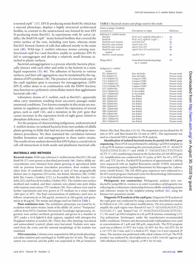
TABLE 1 Bacterial strains and phage used in this study
Strain or phage (GenBankaccession no.) Description or origin Reference
Reference S. meliloti strainsRm1021 SU47 str21 expR102::ISRm2011-1 30Rm8530 SU47 str21 expR101 (expR�) 17Rm8530 exoY mutant exoY210::Tn5 46Rm1021 expA mutant expA3::Tn5-233 17Rm8530 expA mutant expA3::Tn5-233 46Rm8530 expA exoY
mutantexpA3::Tn5-233 exoY210::Tn5 46
Rm1021 mucR mutant mucR31::Tn5 46
Phage �M12 Generalized transducing phagefor S. meliloti
10
Indigenous S. melilotistrains
PI1 (JQ666174) Paso del Indio Present studyPI2 (JQ666175) Paso del Indio Present studyCU4 (JQ666176) Campus UNRC Present studyCU5 (JQ666177) Campus UNRC Present studyCU9 (JQ666178) Campus UNRC Present studyCU10 (JQ666179) Campus UNRC Present studyLE7 (JQ666180) La Escondida Present studyLE16 (JQ666181) La Escondida Present studyLE17 (JQ666182) La Escondida Present studySR1 (JQ666183) San Rafael Present studySR2 (JQ666184) San Rafael Present studySR3 (JQ666185) San Rafael Present studySR4 (JQ666186) San Rafael Present studySR6 (JQ666187) San Rafael Present studySR7, (JQ666188) San Rafael Present studySR8 (JQ666189) San Rafael Present studySR9 (JQ666190) San Rafael Present studySR10 (JQ666191) San Rafael Present studySR11 (JQ666192) San Rafael Present studySR15 (JQ666193) San Rafael Present study
Cell-Cell Interactions in S. meliloti Native Strains
June 2012 Volume 78 Number 12 aem.asm.org 4093
on August 30, 2018 by guest
http://aem.asm
.org/D
ownloaded from
Phage transduction. The expA::Tn5 mutant alleles were transferredfrom the Rm1021 expA::Tn5 strain to recipient strains SR4, SR6, and SR9by using a generalized transduction method described previously by Fi-nan et al. (10), with some modifications. The cotransduction of the resis-tance markers (neomycin) and the dry-colony phenotype were verifiedfor each transductant strain. Donor and recipient strains were included ascontrols.
Autoaggregation assay. The bacteria were grown in 2 ml TY mediumsupplemented with appropriate antibiotics, incubated for 24 h at 30°C,diluted 1/100 in TY, and incubated for 48 h under the same conditions.The bacterial suspensions (5 ml) were then transferred into a glass tube(10 by 70 mm) and allowed to settle for 24 h at 4°C. A 0.2-ml aliquot of theupper portion of the suspension was transferred onto a microtiter plate,and the final optical density at 600 nm (OD600) (ODfinal) was measured. Acontrol tube was vortexed for 30 s, and the initial OD600 (ODinitial) wasdetermined. The autoaggregation percentage was calculated as follows:100 � [1 � (ODfinal/ODinitial)]. For both homologous and heterologousautoaggregation assays, cultures were centrifuged at 4,200 � g for 20 minprior to the settling period. For homologous assays, the pellet of a givenstrain was resuspended in the cell-free supernatant from an independentculture of the same strain. For heterologous assays, the pellet was resus-pended in the cell-free supernatant from a culture of a different strain.
Biofilm formation assay. Biofilm formation was determined macro-scopically by a quantitative assay with 96-well microtiter dishes, wherebybiofilms were stained with crystal violet (CV) based on a method de-scribed previously by O’Toole and Kolter (34), with modifications (14).The bacteria were grown in 2 ml TY medium supplemented with appro-priate antibiotics and incubated with agitation for 48 h at 30°C. The cul-tures were diluted with fresh medium to give an OD600 of 0.1. One hun-dred microliters of the suspension was added to each well and incubatedwith agitation for 24 h at 30°C. Bacterial growth was quantified by mea-suring the OD600. Planktonic cells were gently removed, 180 �l CV aque-ous solution (0.1%, wt/vol) was added, and staining proceeded for 15 min.Each CV-stained well was rinsed thoroughly and repeatedly with waterand then scored for biofilm formation by the addition of 150 �l 95%ethanol. The OD560 of solubilized CV was measured with a MicroELISAAuto Reader (series 700 microplate reader; Cambridge Technology). Par-allel, sterile control cultures were made with TY medium.
Quantification of rhizobial adsorption to roots. For the quantifica-tion of rhizobial adsorption to roots, we followed a protocol describedpreviously by Caetano-Anollés and Favelukes (4), except that our exper-imental unit consisted of a group of 15 alfalfa plants in which the totalnumber of adsorbed microcolonies was counted. Under each experimen-tal condition, at least 4 independent experiments were performed.
Statistical analysis. The autoaggregation assays were performed inquintuplicate. For the biofilm assays, each strain was plated onto at least 8wells of each microtiter dish. The data were subjected to a one-way anal-ysis of variance (ANOVA), followed by a comparison of multiple treat-ment levels with the control by using post hoc Fisher’s least-significant-difference (LSD) test. All statistical analyses were performed by usingInfostat, version 1.0.
Nucleotide sequence accession numbers. The nucleotide sequencesof the 16S rRNA gene from alfalfa-nodulating strains PI1, PI2, CU4, CU5,CU9, CU10, LE7, LE16, LE17, SR1, SR2, SR3, SR4, SR6, SR7, SR8, SR9,SR10, SR11, and SR15 determined in this study have been deposited in theGenBank nucleotide sequence database under accession numbersJQ666174, JQ666175, JQ666176, JQ666177, JQ666178, JQ666179,JQ666180, JQ666181, JQ666182, JQ666183, JQ666184, JQ666185,JQ666186, JQ666187, JQ666188, JQ666189, JQ666190, JQ666191,JQ666192, and JQ666193, respectively (Table 1).
RESULTSIsolation and phylogenetic analysis of alfalfa-nodulatingstrains. Native alfalfa microsymbionts were able to develop highlymucoid colonies after a 24-h incubation period in YEMA (yeast
extract mannitol) or TY medium. Acidification and a lack of ad-sorption of Congo red were observed when strains were grown inYEMA medium supplemented with bromothymol blue andCongo red, respectively. In order to confirm the symbiotic natureof the isolates, the nodulation phenotype was tested by inoculatingthe bacteria onto sterile alfalfa seeds. After 30 days, all isolateselicited characteristic root nodules in the host plant.
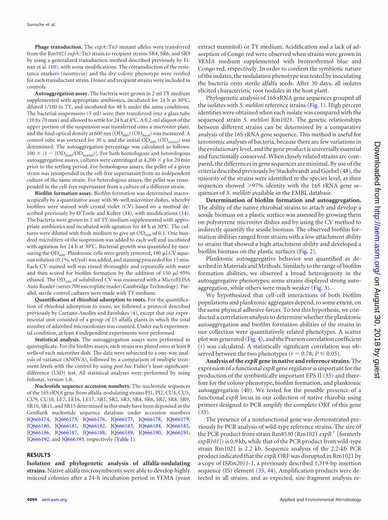
Phylogenetic analysis of 16S rRNA gene sequences grouped allthe isolates with S. meliloti reference strains (Fig. 1). High percentidentities were obtained when each isolate was compared with thesequenced strain S. meliloti Rm1021. The genetic relationshipsbetween different strains can be determined by a comparativeanalysis of the 16S rRNA gene sequence. This method is useful fortaxonomic analyses of bacteria, because there are few variations inthe evolutionary level, and the gene product is universally essentialand functionally conserved. When closely related strains are com-pared, the differences in gene sequences are minimal. By use of thecriteria described previously by Stackebrandt and Goebel (48), themajority of the strains were identified to the species level, as theirsequences showed �97% identity with the 16S rRNA gene se-quences of S. meliloti available in the EMBL database.
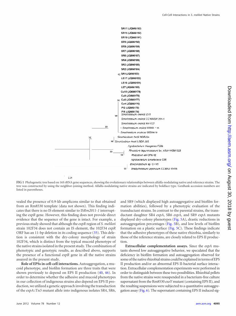
Determination of biofilm formation and autoaggregation.The ability of the native rhizobial strains to attach and develop asessile biomass on a plastic surface was assessed by growing themon polystyrene microtiter dishes and by using the CV method toindirectly quantify the sessile biomass. The observed biofilm for-mation abilities ranged from strains with a low attachment abilityto strains that showed a high attachment ability and developed abiofilm biomass on the plastic surfaces (Fig. 2).
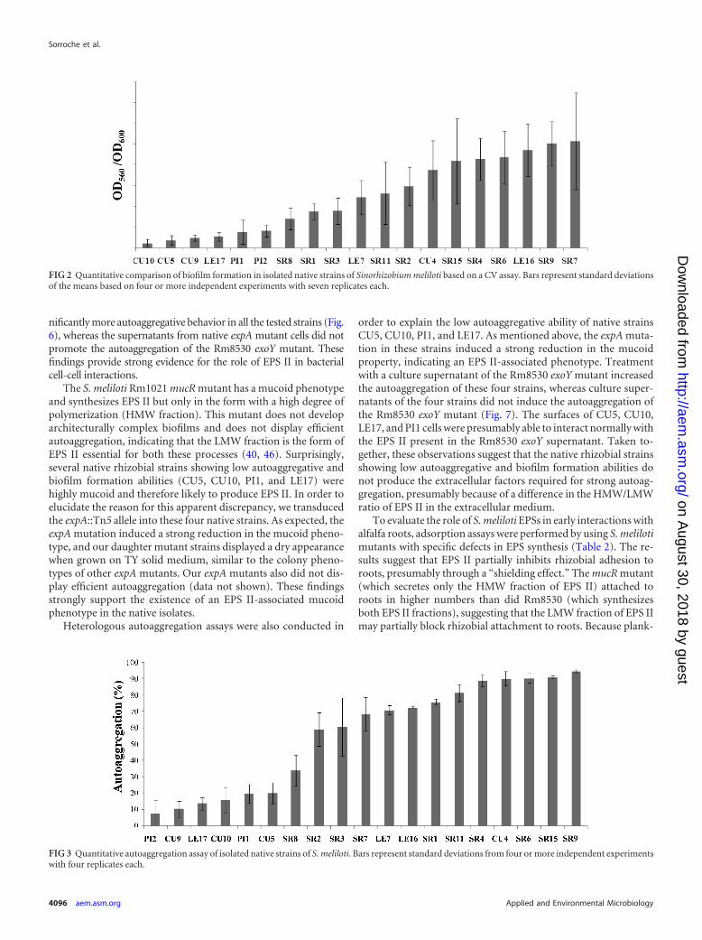
Planktonic autoaggregative behavior was quantified as de-scribed in Materials and Methods. Similarly to the range of biofilmformation abilities, we observed a broad heterogeneity in theautoaggregative phenotypes; some strains displayed strong auto-aggregation, while others were much weaker (Fig. 3).
We hypothesized that cell-cell interactions of both biofilmpopulations and planktonic aggregates depend, to some extent, onthe same physical adhesive forces. To test this hypothesis, we con-ducted a correlation analysis to determine whether the planktonicautoaggregation and biofilm formation abilities of the strains inour collection were quantitatively related phenotypes. A scatterplot was generated (Fig. 4), and the Pearson correlation coefficient(r) was calculated. A statistically significant correlation was ob-served between the two phenotypes (r � 0.78; P � 0.05).
Analysis of the expR gene in native and reference strains. Theexpression of a functional expR gene regulator is important for theproduction of the symbiotically important EPS II (35) and there-fore for the colony phenotype, biofilm formation, and planktonicautoaggregation (40). We tested for the possible presence of afunctional expR locus in our collection of native rhizobia usingprimers designed to PCR amplify the complete ORF of this gene(35).
The presence of a nonfunctional gene was demonstrated pre-viously by PCR analysis of wild-type reference strains. The size ofthe PCR product from strain Rm8530 (Rm1021 expR� [formerlyexpR101]) is 0.9 kb, while that of the PCR product from wild-typestrain Rm1021 is 2.2 kb. Sequence analysis of the 2.2-kb PCRproduct indicated that the expR ORF was disrupted in Rm1021 bya copy of ISRm2011-1, a previously described 1,319-bp insertionsequence (IS) element (35, 44). Amplification products were de-tected in all strains, and as expected, size-fragment analysis re-
Sorroche et al.
4094 aem.asm.org Applied and Environmental Microbiology
on August 30, 2018 by guest
http://aem.asm
.org/D
ownloaded from
vealed the presence of 0.9-kb amplicons similar to that obtainedfrom an Rm8530 template (data not shown). This finding indi-cates that there is no IS element similar to ISRm2011-1 interrupt-ing the expR gene. However, this finding does not provide directevidence that the sequence of the gene is intact. For example, aprevious study showed that although the expR region of S. melilotistrain 102F34 does not contain an IS element, the 102F34 expRORF has an 11-bp deletion in its coding sequence (35). This dele-tion is consistent with the dry-colony morphology of strain102F34, which is distinct from the typical mucoid phenotype ofthe native strains isolated in the present study. The combination ofphenotypic and genotypic results, as described above, supportsthe presence of a functional expR gene in all the native strainsassayed in the present study.
Role of EPSs in cell-cell interactions. Autoaggregation, a mu-coid phenotype, and biofilm formation are three traits that wereshown previously to depend on EPS II production (40, 46). Inorder to determine whether the adhesive and mucoid phenotypesin our collection of indigenous strains also depend on EPS II pro-duction, we utilized a genetic approach involving the transductionof the expA::Tn5 mutant allele into indigenous isolates SR4, SR6,
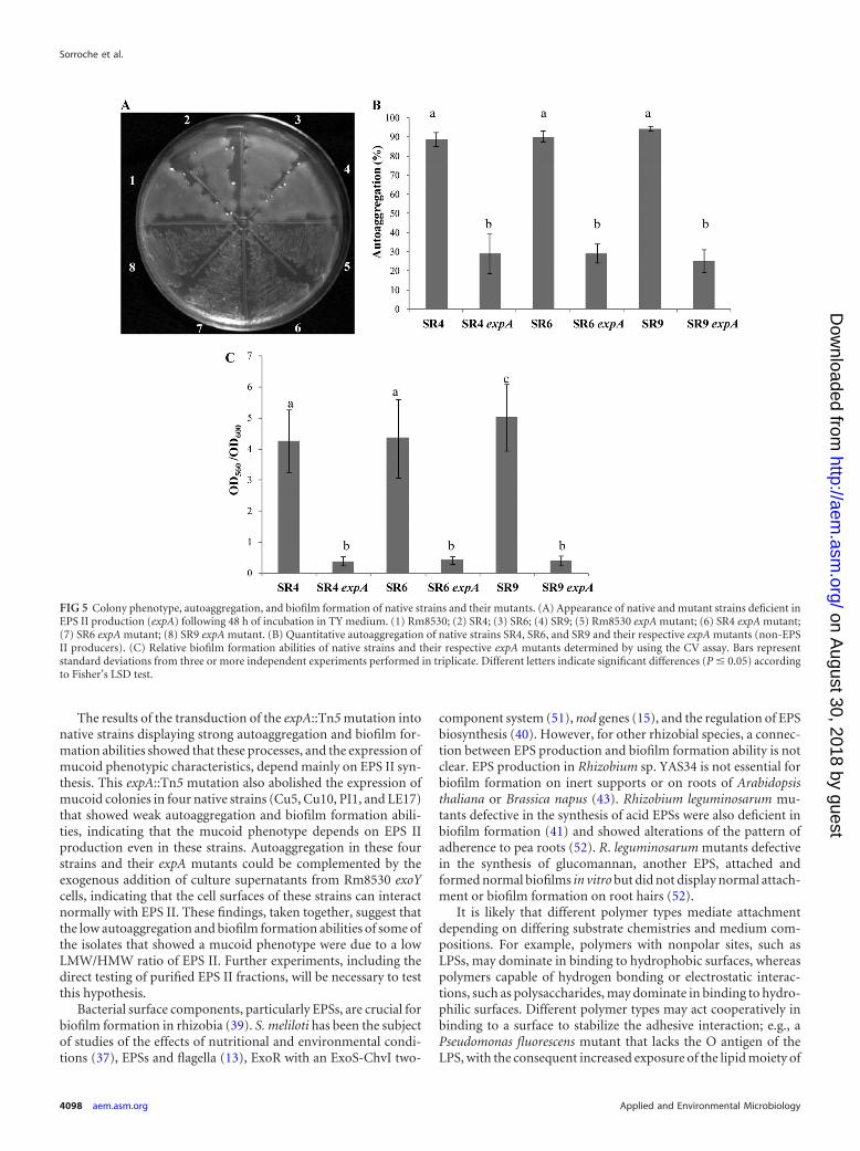
and SR9 (which displayed high autoaggregative and biofilm for-mation abilities), followed by a phenotypic evaluation of thetransductant strains. In contrast to the parental strains, the trans-ductant daughter SR4 expA, SR6 expA, and SR9 expA mutantsdisplayed dry-colony phenotypes (Fig. 5A), drastic reductions inautoaggregation percentages (Fig. 5B), and low levels of biofilmformation on a plastic surface (Fig. 5C). These findings indicatethat the adhesive phenotypes of these native rhizobia, similarly tothose of the reference strains, are closely related to EPS II produc-tion.
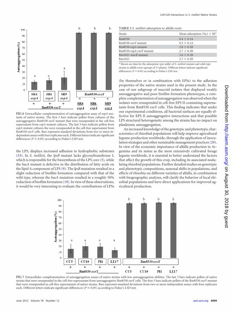
Extracellular complementation assays. Since the expA mu-tants showed low autoaggregative behavior, we speculated that thedeficiency in biofilm formation and autoaggregation observed forsome of the native rhizobial strains could be explained in terms of EPSII production and/or an abnormal EPS II-bacterial surface interac-tion. Extracellular complementation experiments were performed inorder to distinguish between these two possibilities. Rhizobial pelletsfrom the native strains were resuspended in a bacterium-free culturesupernatant from the Rm8530 exoY mutant (containing EPS II), andthe resulting suspensions were subjected to a quantitative autoaggre-gation assay (Fig. 6). The supernatant containing EPS II induced sig-
FIG 1 Phylogenetic tree based on 16S rRNA gene sequences, showing the evolutionary relationships between alfalfa-nodulating native and reference strains. Thetree was constructed by using the neighbor-joining method. Alfalfa-nodulating native strains are indicated by boldface type. GenBank accession numbers arelisted in parentheses.
Cell-Cell Interactions in S. meliloti Native Strains
June 2012 Volume 78 Number 12 aem.asm.org 4095
on August 30, 2018 by guest
http://aem.asm
.org/D
ownloaded from
nificantly more autoaggregative behavior in all the tested strains (Fig.6), whereas the supernatants from native expA mutant cells did notpromote the autoaggregation of the Rm8530 exoY mutant. Thesefindings provide strong evidence for the role of EPS II in bacterialcell-cell interactions.
The S. meliloti Rm1021 mucR mutant has a mucoid phenotypeand synthesizes EPS II but only in the form with a high degree ofpolymerization (HMW fraction). This mutant does not developarchitecturally complex biofilms and does not display efficientautoaggregation, indicating that the LMW fraction is the form ofEPS II essential for both these processes (40, 46). Surprisingly,several native rhizobial strains showing low autoaggregative andbiofilm formation abilities (CU5, CU10, PI1, and LE17) werehighly mucoid and therefore likely to produce EPS II. In order toelucidate the reason for this apparent discrepancy, we transducedthe expA::Tn5 allele into these four native strains. As expected, theexpA mutation induced a strong reduction in the mucoid pheno-type, and our daughter mutant strains displayed a dry appearancewhen grown on TY solid medium, similar to the colony pheno-types of other expA mutants. Our expA mutants also did not dis-play efficient autoaggregation (data not shown). These findingsstrongly support the existence of an EPS II-associated mucoidphenotype in the native isolates.
Heterologous autoaggregation assays were also conducted in
order to explain the low autoaggregative ability of native strainsCU5, CU10, PI1, and LE17. As mentioned above, the expA muta-tion in these strains induced a strong reduction in the mucoidproperty, indicating an EPS II-associated phenotype. Treatmentwith a culture supernatant of the Rm8530 exoY mutant increasedthe autoaggregation of these four strains, whereas culture super-natants of the four strains did not induce the autoaggregation ofthe Rm8530 exoY mutant (Fig. 7). The surfaces of CU5, CU10,LE17, and PI1 cells were presumably able to interact normally withthe EPS II present in the Rm8530 exoY supernatant. Taken to-gether, these observations suggest that the native rhizobial strainsshowing low autoaggregative and biofilm formation abilities donot produce the extracellular factors required for strong autoag-gregation, presumably because of a difference in the HMW/LMWratio of EPS II in the extracellular medium.
To evaluate the role of S. meliloti EPSs in early interactions withalfalfa roots, adsorption assays were performed by using S. melilotimutants with specific defects in EPS synthesis (Table 2). The re-sults suggest that EPS II partially inhibits rhizobial adhesion toroots, presumably through a “shielding effect.” The mucR mutant(which secretes only the HMW fraction of EPS II) attached toroots in higher numbers than did Rm8530 (which synthesizesboth EPS II fractions), suggesting that the LMW fraction of EPS IImay partially block rhizobial attachment to roots. Because plank-
FIG 2 Quantitative comparison of biofilm formation in isolated native strains of Sinorhizobium meliloti based on a CV assay. Bars represent standard deviationsof the means based on four or more independent experiments with seven replicates each.
FIG 3 Quantitative autoaggregation assay of isolated native strains of S. meliloti. Bars represent standard deviations from four or more independent experimentswith four replicates each.
Sorroche et al.
4096 aem.asm.org Applied and Environmental Microbiology
on August 30, 2018 by guest
http://aem.asm
.org/D
ownloaded from

tonic rhizobia were incubated with alfalfa plants for 4 h (4), thesefindings reflect the role that EPSs may play during the initial accessto the root; this test should therefore not be interpreted as a bio-film assay. Additional experiments are needed to better clarify theassociations between biofilm formation and other adhesion phe-notypes.
DISCUSSION
The inoculation of legume crop plants with selected, highly effi-cient rhizobia is an important method for the improvement ofsymbiotic nitrogen fixation in agricultural ecosystems and consti-tutes a major strategy for the sustainable input of nitrogen intoagricultural soils (27). However, the native rhizobial populationspresent in soils often display a superior competitive ability overinoculated strains on the basis of their large population size, po-sitional advantage, and/or superior adaptation to local environ-mental conditions (2, 49). The selection of efficient rhizobialstrains based on their adaptation to local ecological conditions cantherefore lead to the increased grain production of crops (27, 32).
We used several approaches to evaluate the rhizobial strainspresent in root nodules of alfalfa plants growing in fields in Ar-gentina that had no history of inoculation procedures. The iso-lated strains showed a mucoid phenotype when grown on petridishes. Such a phenotype was indicative of EPS II synthesis inpreviously characterized reference strains.
16S rRNA gene analyses of all the isolates revealed a high degreeof identity (approximately 98%) with reference S. meliloti strains,corresponding to a value of sequence divergence less than the3.0% required for differentiation between species (48). PCR anal-ysis of the chromosomal expR gene of these isolates revealed thatthis gene is not interrupted by an IS element, as is also the case forreference strain Rm8530 (35). ExpR is a LuxR homologue, whose
functions include the activation of EPS II production in the pres-ence of N-acyl-homoserine lactone (AHL), which is produced bythe sinR/sinI system in S. meliloti. Strain Rm1021 displays a dry (asopposed to mucoid) phenotype because its expR gene is inter-rupted by an IS element, and it therefore cannot produce EPS II.EPS II-producing strain Rm8530 displays a highly mucoid pheno-type. Rm8530 and the native strains isolated in this study harboran intact (not interrupted) copy of the expR gene, giving a 0.9-kbPCR product. Rm1021 yields a larger amplicon (2.2-kb PCR prod-uct) because the expR ORF is disrupted by a copy of ISRm2011-1,a 1,319-bp IS element.
We have shown previously that rhizobial cell surface compo-nents such as EPSs, in combination with bacterial functional sig-nals, are essential for the processes of autoaggregation (46) andbiofilm formation (39). Both processes play important ecologicalroles in the survival of rhizobia in their natural soil environmentand probably in the nitrogen-fixing symbiosis that occurs withinroot nodules, in which EPSs are essential for early stages of infec-tion (12). The findings of the present study illustrate a great vari-ability in both autoaggregation and biofilm formation abilitiesamong native soil isolates. This phenotypic diversity may resultfrom differential selective pressures in the soil microenvironmentor in the root nodules. Interestingly, a correlation analysis ofautoaggregation and biofilm formation abilities gave a Pearsoncorrelation coefficient of 0.78, indicating a positive correlationbetween these two variables. These findings suggest that the twoprocesses are related and that cell-cell interactions in the contextof both biofilm populations and planktonic aggregates depend, atleast under the conditions of our assays, on the same physicaladhesive forces. A similar positive correlation between autoaggre-gation and biofilm formation abilities was shown previously forMyroides odoratus, a Gram-negative bacillus (21).
FIG 4 Scatter plot of two variables: autoaggregation (percent) and relative biofilm formation ability (OD560/OD600). The circles are ordered pairs that representdifferent isolates.
Cell-Cell Interactions in S. meliloti Native Strains
June 2012 Volume 78 Number 12 aem.asm.org 4097
on August 30, 2018 by guest
http://aem.asm
.org/D
ownloaded from
The results of the transduction of the expA::Tn5 mutation intonative strains displaying strong autoaggregation and biofilm for-mation abilities showed that these processes, and the expression ofmucoid phenotypic characteristics, depend mainly on EPS II syn-thesis. This expA::Tn5 mutation also abolished the expression ofmucoid colonies in four native strains (Cu5, Cu10, PI1, and LE17)that showed weak autoaggregation and biofilm formation abili-ties, indicating that the mucoid phenotype depends on EPS IIproduction even in these strains. Autoaggregation in these fourstrains and their expA mutants could be complemented by theexogenous addition of culture supernatants from Rm8530 exoYcells, indicating that the cell surfaces of these strains can interactnormally with EPS II. These findings, taken together, suggest thatthe low autoaggregation and biofilm formation abilities of some ofthe isolates that showed a mucoid phenotype were due to a lowLMW/HMW ratio of EPS II. Further experiments, including thedirect testing of purified EPS II fractions, will be necessary to testthis hypothesis.
Bacterial surface components, particularly EPSs, are crucial forbiofilm formation in rhizobia (39). S. meliloti has been the subjectof studies of the effects of nutritional and environmental condi-tions (37), EPSs and flagella (13), ExoR with an ExoS-ChvI two-
component system (51), nod genes (15), and the regulation of EPSbiosynthesis (40). However, for other rhizobial species, a connec-tion between EPS production and biofilm formation ability is notclear. EPS production in Rhizobium sp. YAS34 is not essential forbiofilm formation on inert supports or on roots of Arabidopsisthaliana or Brassica napus (43). Rhizobium leguminosarum mu-tants defective in the synthesis of acid EPSs were also deficient inbiofilm formation (41) and showed alterations of the pattern ofadherence to pea roots (52). R. leguminosarum mutants defectivein the synthesis of glucomannan, another EPS, attached andformed normal biofilms in vitro but did not display normal attach-ment or biofilm formation on root hairs (52).
It is likely that different polymer types mediate attachmentdepending on differing substrate chemistries and medium com-positions. For example, polymers with nonpolar sites, such asLPSs, may dominate in binding to hydrophobic surfaces, whereaspolymers capable of hydrogen bonding or electrostatic interac-tions, such as polysaccharides, may dominate in binding to hydro-philic surfaces. Different polymer types may act cooperatively inbinding to a surface to stabilize the adhesive interaction; e.g., aPseudomonas fluorescens mutant that lacks the O antigen of theLPS, with the consequent increased exposure of the lipid moiety of
FIG 5 Colony phenotype, autoaggregation, and biofilm formation of native strains and their mutants. (A) Appearance of native and mutant strains deficient inEPS II production (expA) following 48 h of incubation in TY medium. (1) Rm8530; (2) SR4; (3) SR6; (4) SR9; (5) Rm8530 expA mutant; (6) SR4 expA mutant;(7) SR6 expA mutant; (8) SR9 expA mutant. (B) Quantitative autoaggregation of native strains SR4, SR6, and SR9 and their respective expA mutants (non-EPSII producers). (C) Relative biofilm formation abilities of native strains and their respective expA mutants determined by using the CV assay. Bars representstandard deviations from three or more independent experiments performed in triplicate. Different letters indicate significant differences (P � 0.05) accordingto Fisher’s LSD test.
Sorroche et al.
4098 aem.asm.org Applied and Environmental Microbiology
on August 30, 2018 by guest
http://aem.asm
.org/D
ownloaded from
the LPS, displays increased adhesion to hydrophobic substrates(53). In S. meliloti, the lpsB mutant lacks glycosyltransferase I,which is responsible for the biosynthesis of the LPS core (5), whilethe bacA mutant is defective in the distribution of fatty acids onthe lipid A component of LPS (9). The lpsB mutation resulted in aslight reduction of biofilm formation compared with that of thewild type, whereas the bacA mutation resulted in a roughly 50%reduction of biofilm formation (19). In view of these observations,it would be very interesting to evaluate the contributions of LPSs
(by themselves or in combination with EPSs) to the adhesionproperties of the native strains used in the present study. In thecase of our subgroup of mucoid isolates that displayed weaklyautoaggregative and poor biofilm formation phenotypes, a com-plete complementation of autoaggregation was observed when theisolates were resuspended in cell-free EPS II-containing superna-tants from Rm8530 exoY cells. This finding indicates that underour experimental conditions, all bacterial surfaces are equally ef-fective for EPS II autoaggregative interactions and that possibleLPS structural heterogeneity among the strains has no impact onplanktonic autoaggregation.
An increased knowledge of the genotypic and phenotypic char-acteristics of rhizobial populations will help improve agriculturallegume production worldwide, through the application of inocu-lation strategies and other sustainable management practices (29).In view of the economic importance of alfalfa production in Ar-gentina and its status as the most extensively cultivated foragelegume worldwide, it is essential to better understand the factorsthat affect the growth of this crop, including its associated nodu-lating rhizobial populations. Further detailed studies on genotypicand phenotypic compositions, seasonal shifts in populations, andeffects of rhizobia on different varieties of alfalfa, in combinationwith biogeographic analyses, will clarify the behavior of local rhi-zobial populations and have direct applications for improved ag-ricultural production.
FIG 6 Extracellular complementation of autoaggregation assay of expA mu-tants of native strains. The first 3 bars indicate pellets from cultures of theautoaggregative Rm8530 exoY mutant that were resuspended in the cell-freesupernatant from expA mutant cultures. The last 3 bars indicate pellets fromexpA mutant cultures the were resuspended in the cell-free supernatant fromRm8530 exoY cells. Bars represent standard deviations from two or more in-dependent assays with four replicates each. Different letters indicate significantdifferences (P � 0.05) according to Fisher’s LSD test.
FIG 7 Extracellular complementation of autoaggregation assays of native strains with low autoaggregation abilities. The last 3 bars indicate pellets of nativestrains that were resuspended in the cell-free supernatant from autoaggregative Rm8530 exoY cells. The first 3 bars indicate pellets of the Rm8530 exoY mutantthat were resuspended in cell-free supernatants of native strains. Bars represent standard deviations from two or more independent assays with four replicateseach. Different letters indicate significant differences (P � 0.05) according to Fisher’s LSD test.
TABLE 2 S. meliloti adsorption to alfalfa roots
Strain Mean adsorption (‰) � SEa
Rm8530 0.4 � 0.1ARm8530 exoY mutant 0.5 � 0.1ARm8530 expA mutant 2.8 � 0.1BRm8530 expA exoY mutant 2.7 � 0.1BRm1021 mucR mutant 2.6 � 0.1BRm1021 2.7 � 0.1Ba Shown are data for the adsorption (per mille) of S. meliloti mutant and wild-typestrains to alfalfa roots (groups of 15 plants). Different letters indicate significantdifferences (P � 0.05) according to Fisher’s LSD test.
Cell-Cell Interactions in S. meliloti Native Strains
June 2012 Volume 78 Number 12 aem.asm.org 4099
on August 30, 2018 by guest
http://aem.asm
.org/D
ownloaded from
ACKNOWLEDGMENTS
This work was supported by grants from the Secretaría de Ciencia y Téc-nica de la UNRC, Agencia Nacional de Promoción Científica y Tec-nológica (ANPCyT), and Consejo Nacional de Investigaciones Científicasy Técnicas (CONICET) of the República Argentina. F.G.S. and M.B.S.were supported by a fellowship from CONICET. W.G. and A.Z. are careermembers of CONICET.
We thank Juan Ignacio Ituarte for his assistance in alfalfa samplingprocedures and S. Anderson for editing the manuscript.
REFERENCES1. Altschul SF, et al. 1997. Gapped BLAST and PSI-BLAST: a new genera-
tion of protein database search programs. Nucleic Acids Res. 25:3389 –3402.
2. Bogino P, Banchio E, Bonfiglio C, Giordano W. 2008. Competitivenessof a Bradyrhizobium sp. strain in soils containing indigenous rhizobia.Curr. Microbiol. 56:66 –72.
3. Brosius J, Palmer JL, Kennedy HP, Noller HF. 1978. Complete nucle-otide sequence of a 16S ribosomal RNA gene from Escherichia coli. Proc.Natl. Acad. Sci. U. S. A. 75:4801– 4805.
4. Caetano-Anollés G, Favelukes G. 1986. Quantitation of adsorption ofrhizobia in low numbers to small legume roots. Appl. Environ. Microbiol.52:371–376.
5. Campbell GR, Reuhs BL, Walker GC. 2002. Chronic intracellular infec-tion of alfalfa nodules by Sinorhizobium meliloti requires correct lipopoly-saccharide core. Proc. Natl. Acad. Sci. U. S. A. 99:3938 –3943.
6. Cepeda C, Santos Y. 2000. Rapid and low-level toxic PCR-based methodfor routine identification of Flavobacterium psychrophilum. Int. Micro-biol. 3:235–238.
7. Costerton JW, Lewandowski Z, Caldwell DE, Korber DR, Lappin-ScottHM. 1995. Microbial biofilms. Annu. Rev. Microbiol. 49:711–745.
8. Denarié J, Debellé F, Promé JC. 1996. Rhizobium lipo-chitooligosaccharidenodulation factors: signaling molecules mediating recognition and morpho-genesis. Annu. Rev. Biochem. 65:503–535.
9. Ferguson GP, Roop RM, Walker GC. 2002. Deficiency of a Sinorhizo-bium meliloti bacA mutant in alfalfa symbiosis correlates with alteration ofthe cell envelope. J. Bacteriol. 184:5625–5632.
10. Finan TM, et al. 1984. General transduction in Rhizobium meliloti. J.Bacteriol. 159:120 –124.
11. Fisher RF, Long SR. 1992. Rhizobium-plant signal exchange. Nature357:655– 660.
12. Fraysse N, Couderc F, Poinsot V. 2003. Surface polysaccharide involve-ment in establishing the Rhizobium-legume symbiosis. Eur. J. Biochem.270:1365–1380.
13. Fujishige NA, Kapadia NN, De Hoff PL, Hirsch AM. 2006. Investiga-tions of Rhizobium biofilm formation. FEMS Microbiol. Ecol. 56:195–206.
14. Fujishige NA, Kapadia NN, Hirsch AM. 2006. A feeling for the micro-organism: structure on a small scale. Biofilms on plant roots. Bot. J. Linn.Soc. 150:79 – 88.
15. Fujishige NA, et al. 2008. Rhizobium common nod genes are required forbiofilm formation. Mol. Microbiol. 67:504 –515.
16. Galibert F, et al. 2001. The composite genome of the legume symbiontSinorhizobium meliloti. Science 293:668 – 672.
17. Glazebrook J, Walker GC. 1989. A novel exopolysaccharide can functionin place of the calcofluor-binding exopolysaccharide in nodulation of al-falfa by Rhizobium meliloti. Cell 56:661– 672.
18. Hirsch AM. 1999. Role of lectins (and rhizobial exopolysaccharides) inlegume nodulation. Curr. Opin. Plant Biol. 2:320 –326.
19. Hirsch AM, Lum MR, Fujishige NA. 2009. Microbial encounters of asymbiotic kind—attaching to roots and other surfaces, p 295–314. InEmons AMC, Ketelaar T (ed), Root hairs. Plant cell monographs, vol 12.Springer-Verlag, Berlin, Germany.
20. Hotter GS, Scott B. 1991. Exopolysaccharide mutants of Rhizobium lotiare fully effective on a determinate nodulating host but are ineffective onan indeterminate nodulating host. J. Bacteriol. 173:851– 859.
21. Jacobs A, Chenia HY. 2010. Biofilm-forming capacity, surface hydropho-bicity and aggregation characteristics of Myroides odoratus isolated fromSouth African Oreochromis mossambicus fish. J. Appl. Microbiol. 107:1957–1966.
22. Janczarek M. 2011. Environmental signals and regulatory pathways that
influence exopolysaccharide production in rhizobia. Int. J. Mol. Sci. 12:7898 –7933.
23. Keller M, et al. 1995. Molecular analysis of the Rhizobium meliloti mucRgene regulating the biosynthesis of the exopolysaccharides succinoglycanand galactoglucan. Mol. Plant Microbe Interact. 8:267–277.
24. Krol E, Becker A. 2004. Global transcriptional analysis of the phosphatestarvation response in Sinorhizobium meliloti strains 1021 and 2011. Mol.Genet. Genomics 272:1–17.
25. Lane DJ. 1991. 16S/23S rRNA sequencing, p 115–175. In Stackebrant E,Goodfellow M (ed), Nucleic acid techniques in bacterial systematics JohnWiley & Sons, New York, NY.
26. Leigh JA, Signer ER, Walker GC. 1985. Exopolysaccharide-deficientmutants of Rhizobium meliloti that form ineffective nodules. Proc. Natl.Acad. Sci. U. S. A. 82:6231– 6235.
27. Lindström K, Murwira M, Willems A, Altier N. 2010. The biodiversity ofbeneficial microbe-host mutualism: the case of rhizobia. Res. Microbiol.161:453– 463.
28. Löbler M, Hirsch AM. 1993. A gene that encodes a proline-rich nodu-lin with limited homology to PsENOD12 is expressed in the invasionzone of Rhizobium meliloti-induced alfalfa nodules. Plant Physiol. 103:21–30.
29. McInnes A, Thies JE, Abbott LK, Howieson JG. 2004. Structure anddiversity among rhizobial strains, populations and communities—a re-view. Soil Biol. Biochem. 36:1295–1308.
30. Meade H, Long S, Ruvkun G, Brown S, Ausubel F. 1982. Physical andgenetic characterization of symbiotic and auxotrophic mutants of Rhizo-bium meliloti induced by transposon Tn5 mutagenesis. J. Bacteriol. 149:114 –122.
31. Morris CE, Monier JM. 2003. The ecological significance of biofilmformation by plant-associated bacteria. Annu. Rev. Phytopathol. 41:429 –453.
32. Nievas F, Bogino P, Nocelli N, Giordano W. 2012. Genotypic analysis ofisolated peanut-nodulating rhizobial strains reveals differences amongpopulations obtained from soils with different cropping histories. Appl.Soil Ecol. 53:74 – 82.
33. Nikitina VE, Ponomareva EG, Alen’kina SA, Konnova SA. 2001. Therole of cell-surface lectins in the aggregation of Azospirilla. Microbiology70:471– 476.
34. O’Toole GA, Kolter R. 1998. Initiation of biofilm formation in Pseu-domonas fluorescens WCS365 proceeds via multiple, convergent signallingpathways: a genetic analysis. Mol. Microbiol. 28:449 – 461.
35. Pellock BJ, Teplitski M, Boinay RP, Bauer WD, Walker GC. 2002. ALuxR homolog controls production of symbiotically active extracellu-lar polysaccharide II by Sinorhizobium meliloti. J. Bacteriol. 184:5067–5076.
36. Reuber TL, Walker GC. 1993. Biosynthesis of succinoglycan, a symbiot-ically important exopolysaccharide of Rhizobium meliloti. Cell 74:269 –280.
37. Rinaudi L, et al. 2006. Effects of nutritional and environmental condi-tions on Sinorhizobium meliloti biofilm formation. Res. Microbiol. 157:867– 875.
38. Rinaudi L, Sorroche F, Zorreguieta A, Giordano W. 2010. Analysis ofmucR gene regulating biosynthesis of exopolysaccharides: implications forbiofilm formation in Sinorhizobium meliloti Rm1021. FEMS Microbiol.Lett. 302:15–21.
39. Rinaudi LV, Giordano W. 2010. An integrated view of biofilm formationin rhizobia. FEMS Microbiol. Lett. 304:1–11.
40. Rinaudi LV, Gonzalez JE. 2009. The low-molecular-weight fraction ofexopolysaccharide II from Sinorhizobium meliloti is a crucial determinantof biofilm formation. J. Bacteriol. 191:7216 –7224.
41. Russo DM, et al. 2006. Proteins exported via the PrsD-PrsE type I secre-tion system and the acidic exopolysaccharide are involved in biofilm for-mation by Rhizobium leguminosarum. J. Bacteriol. 188:4474 – 4486.
42. Saitou N, Nei M. 1987. The neighbor-joining method: a new method forreconstructing phylogenetic trees. Mol. Biol. Evol. 4:406 – 425.
43. Santaella C, Schue M, Berge O, Heulin T, Achouak W. 2008. Theexopolysaccharide of Rhizobium sp. YAS34 is not necessary for biofilmformation on Arabidopsis thaliana and Brassica napus roots but contrib-utes to root colonization. Environ. Microbiol. 10:2150 –2163.
44. Simon R, Hötte B, Klauke B, Kosier B. 1991. Isolation and character-ization of insertion sequence elements from gram-negative bacteria byusing new broad-host-range, positive selection vectors. J. Bacteriol. 173:1502–1508.
Sorroche et al.
4100 aem.asm.org Applied and Environmental Microbiology
on August 30, 2018 by guest
http://aem.asm
.org/D
ownloaded from
45. Skorupska A, Janczarek M, Marczak M, Mazur A, Król J. 2006. Rhizo-bial exopolysaccharides: genetic control and symbiotic functions. Microb.Cell Fact. 5:7.
46. Sorroche F, Rinaudi L, Zorreguieta A, Giordano W. 2010. EPS II-dependent autoaggregation of Sinorhizobium meliloti planktonic cells.Curr. Microbiol. 61:465– 470.
47. Spaink HP. 2000. Root nodulation and infection factors produced byrhizobial bacteria. Annu. Rev. Microbiol. 54:257–288.
48. Stackebrandt E, Goebel BM. 1994. Taxonomic note: a place for DNA-DNA reassociation and 16S rRNA sequence analysis in the present speciesdefinition in bacteriology. Int. J. Syst. Bacteriol. 44:846 – 849.
49. Streeter JG. 1994. Plant-environment interactions symbiotic nitrogenfixation, p 245–262. In Wilkinson RE (ed), Marcel Dekker Inc, NewYork, NY.
50. Vincent JM. 1970. A manual for the practical study of root-nodule bac-teria. Blackwell Scientific Publications, Oxford, England.
51. Wells DH, Chen EJ, Fisher RF, Long SR. 2007. ExoR is geneticallycoupled to the ExoS-ChvI two-component system and located in theperiplasm of Sinorhizobium meliloti. Mol. Microbiol. 64:647– 664.
52. Williams A, et al. 2008. Glucomannan-mediated attachment of Rhizo-bium leguminosarum to pea root hairs is required for competitive noduleinfection. J. Bacteriol. 190:4706 – 4715.
53. Williams V, Fletcher M. 1996. Pseudomonas fluorescens adhesion andtransport through porous media are affected by lipopolysaccharide com-position. Appl. Environ. Microbiol. 62:100 –104.
54. Zhan HJ, Lee CC, Leigh JA. 1991. Induction of the second exopolysac-charide (EPSb) in Rhizobium meliloti SU47 by low phosphate concentra-tions. J. Bacteriol. 173:7391–7394.
Cell-Cell Interactions in S. meliloti Native Strains
June 2012 Volume 78 Number 12 aem.asm.org 4101
on August 30, 2018 by guest
http://aem.asm
.org/D
ownloaded from