A Novel Technique of Uterine Manipulation in Laparoscopic Pelvic
6
Hindawi Publishing Corporation Minimally Invasive Surgery Volume 2010, Article ID 836027, 5 pages doi:10.1155/2010/836027 Research Article A Novel Technique of Uterine Manipulation in Laparoscopic Pelvic Oncosurgical Procedures: “The Uterine Hitch Technique” S. P. Puntambekar, A. M. Patil, N. V. Rayate, S. S. Puntambekar, R. M. Sathe, and M. A. Kulkarni Department of Minimally Invasive Oncology, Galaxy Laparoscopy Institute, Above Ayurved Rasashala, 25-A Karve Road, Pune, Maharashtra 411030, India Correspondence should be addressed to S. P. Puntambekar, [email protected] Received 23 July 2009; Accepted 26 November 2009 Academic Editor: Liselotte Mettler Copyright © 2010 S. P. Puntambekar et al. This is an open access article distributed under the Creative Commons Attribution License, which permits unrestricted use, distribution, and reproduction in any medium, provided the original work is properly cited. Aim. To describe a new technique of uterine manipulation in laparoscopic management of pelvic cancers. Material and Methods. We used a novel uterine hitch technique in 23 patients from May 2008 to October 2008. These patients underwent pelvic oncologic surgery including laparoscopic radical hysterectomy (n = 7), laparoscopic anterior resection (n = 4), laparoscopic abdominoperineal resection (n = 3), laparoscopic posterior exenteration (n = 4), or laparoscopic anterior exenteration (n = 5). The uterus was hitched to the anterior abdominal.wall by either a single suture in the fundus or by sutures through the round ligaments. Results. The uterine hitch technique was successfully accomplished in all procedures. It was performed in less than 5 minutes in all cases. It obviated the need for vaginal manipulation. An extra port for retraction could be avoided. There were no intraoperative complications. Conclusion. A practical, cheap and reproducible method for uterine manipulation, during pelvic oncologic surgery is described. It improves the stability of the uterus and also obviates the need for keeping an additional assistant for vaginal manipulation in any of the procedures. 1. Introduction Uterine manipulation is essential for laparoscopic proce- dures involving dissection of the female pelvis. Several uterine manipulators are available to help manoever the uterus facilitation proper dissection in the pelvis. At our institute, we routinely use the myoma screw for uterine manipulation during total laparoscopic hysterectomy or surgery for cervical [1], bladder, and rectal cancers. However its use is avoided in two situations. First, in endometrial cancer, where peritoneal spread of tumor cells is feared. Second, in patients with pyometra in cervical cancer, where insertion of the screw would lead to spillage of pus into the abdominal cavity. Various vaginal manipulators are used in laparoscopic surgery to manipulate the uterus. When these enter through the cancer tissue, like the cervical canal or the endometrial cavity, manipulation can sometimes result in tissue breakup and bleeding. This can theoretically lead to cancer cells enter- ing into the circulation and systemic spread. Some vaginal manipulators are comparatively expensive and require an additional assistant at the vaginal end. The uterus is often manipulated using a grasping forceps, but this entails use of one port for uterine manipulation or insertion of an additional port. Moreover, graspers often slip leading to tissue break up. This is especially common at the uterine cornua, which bleed during repeated manipulation with graspers. Hence, instead of a vaginal manipulator, a myoma screw, or a grasping forceps, we used a stitch to hitch up the uterus during laparoscopic pelvic oncosurgery. The uterus was hitched to the anterior abdominal wall by either a single suture in the fundus or by sutures through the round ligaments. This technique was very useful in patients requiring dissection in the puouch of Douglas as in cervical and rectal cancers.
Transcript of A Novel Technique of Uterine Manipulation in Laparoscopic Pelvic

Hindawi Publishing CorporationMinimally Invasive SurgeryVolume 2010, Article ID 836027, 5 pagesdoi:10.1155/2010/836027
Research Article
A Novel Technique of Uterine Manipulation in LaparoscopicPelvic Oncosurgical Procedures: “The Uterine Hitch Technique”
S. P. Puntambekar, A. M. Patil, N. V. Rayate, S. S. Puntambekar,R. M. Sathe, and M. A. Kulkarni
Department of Minimally Invasive Oncology, Galaxy Laparoscopy Institute, Above Ayurved Rasashala,25-A Karve Road, Pune, Maharashtra 411030, India
Correspondence should be addressed to S. P. Puntambekar, [email protected]
Received 23 July 2009; Accepted 26 November 2009
Academic Editor: Liselotte Mettler
Copyright © 2010 S. P. Puntambekar et al. This is an open access article distributed under the Creative Commons AttributionLicense, which permits unrestricted use, distribution, and reproduction in any medium, provided the original work is properlycited.
Aim. To describe a new technique of uterine manipulation in laparoscopic management of pelvic cancers. Material and Methods.We used a novel uterine hitch technique in 23 patients from May 2008 to October 2008. These patients underwent pelviconcologic surgery including laparoscopic radical hysterectomy (n = 7), laparoscopic anterior resection (n = 4), laparoscopicabdominoperineal resection (n = 3), laparoscopic posterior exenteration (n = 4), or laparoscopic anterior exenteration (n = 5).The uterus was hitched to the anterior abdominal.wall by either a single suture in the fundus or by sutures through the roundligaments. Results. The uterine hitch technique was successfully accomplished in all procedures. It was performed in less than 5minutes in all cases. It obviated the need for vaginal manipulation. An extra port for retraction could be avoided. There wereno intraoperative complications. Conclusion. A practical, cheap and reproducible method for uterine manipulation, during pelviconcologic surgery is described. It improves the stability of the uterus and also obviates the need for keeping an additional assistantfor vaginal manipulation in any of the procedures.
1. Introduction
Uterine manipulation is essential for laparoscopic proce-dures involving dissection of the female pelvis. Severaluterine manipulators are available to help manoever theuterus facilitation proper dissection in the pelvis.
At our institute, we routinely use the myoma screw foruterine manipulation during total laparoscopic hysterectomyor surgery for cervical [1], bladder, and rectal cancers.
However its use is avoided in two situations. First, inendometrial cancer, where peritoneal spread of tumor cells isfeared. Second, in patients with pyometra in cervical cancer,where insertion of the screw would lead to spillage of pus intothe abdominal cavity.
Various vaginal manipulators are used in laparoscopicsurgery to manipulate the uterus. When these enter throughthe cancer tissue, like the cervical canal or the endometrialcavity, manipulation can sometimes result in tissue breakup
and bleeding. This can theoretically lead to cancer cells enter-ing into the circulation and systemic spread. Some vaginalmanipulators are comparatively expensive and require anadditional assistant at the vaginal end.
The uterus is often manipulated using a grasping forceps,but this entails use of one port for uterine manipulation orinsertion of an additional port. Moreover, graspers often slipleading to tissue break up. This is especially common at theuterine cornua, which bleed during repeated manipulationwith graspers.
Hence, instead of a vaginal manipulator, a myoma screw,or a grasping forceps, we used a stitch to hitch up theuterus during laparoscopic pelvic oncosurgery. The uteruswas hitched to the anterior abdominal wall by either asingle suture in the fundus or by sutures through theround ligaments. This technique was very useful in patientsrequiring dissection in the puouch of Douglas as in cervicaland rectal cancers.

2 Minimally Invasive Surgery
When operating on endometrial cancer, the uterus washitched from the round ligaments, thus facilitating itseasy manipulation without spillage of cancer cells into theperitoneal cavity.
Here we describe the uterine hitch technique for uterinemanipulation, in patients who require radical pelvic dissec-tion or in whom the use of uterine manipulators is con-traindicated. We also describe the implications, advantages,and disadvantages of this technique.
2. Material and Methods
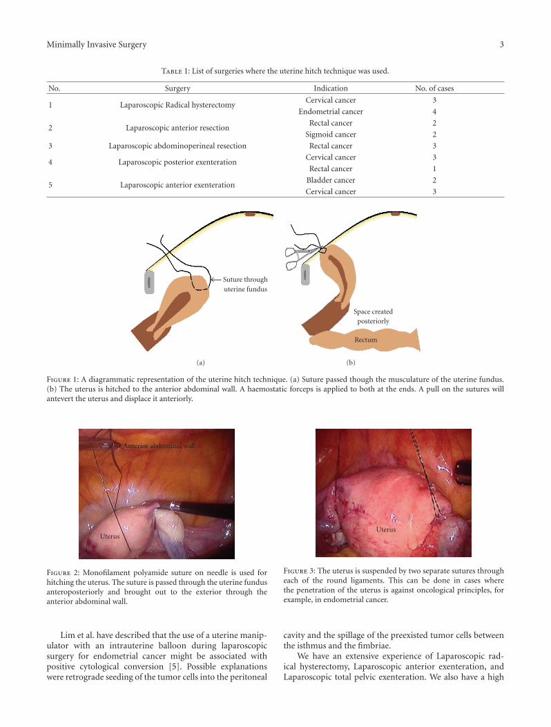
We performed the uterine hitch technique in 23 femalepatients who underwent laparoscopic pelvic oncosurgeriesat Galaxy Laparoscopy Institute from May 2008 to October2008. The procedures included laparoscopic radical hysterec-tomy for cervical cancer (n = 3) and for endometrial cancer(n = 4), laparoscopic anterior resection for rectal cancer (n =2) and sigmoid cancer (n = 2), laparoscopic abdominoper-ineal resection for rectal cancer (n = 3) and laparoscopicposterior exenteration for cervical cancer invading rectum(n = 3) and rectal cancer invading uterus (n = 1) andlaparoscopic anterior exenteration for bladder cancer (n = 2)and cervical cancer invading bladder (n = 3) (Table 1).
The uterine hitch technique was used in all the patientswho required a radical pelvic dissection or in whom the useof a uterine manipulator was contraindicated (endometrialcancer and pyometra).
The average age of the patients was 50 +/− 5 years.Previous surgeries included open tubal ligation (n = 20),caesarean section (n = 14), and open appendicectomy(n = 5).
All patients underwent a standard preoperative prepara-tion. Regional anesthesia, either spinal or epidural, was givenin combination with general anaesthesia. A Foley catheterwas placed into the bladder. The patient was placed in amodified Lloyd Davis position at approximately a 30 to a 45degrees angle.
A total of 4 or 5 ports were used (10 mm camera portthrough the umbilicus, a 10 mm port at the McBurney’spoint on right side, a 5 mm port pararectally at midclavicularline at level of umbilicus on the right side, and two 5 mmports on left side, which were the mirror image of the right-sided ports). The left pararectal ports were used for insertionof the myoma screw. This port couls now be omitted whenusing the uterine hitch.
2.1. The Uterine Hitch Technique. Figure 1 illustrates theuterine hitch technique.
The uterus was held using a nontraumatic grasper andbrought towards anterior abdominal wall. Monofilamentpolyamide suture material, number 2, 0 on a straight needle(or on a curved needle that was partially straightened out),was inserted through the layers of the anterior abdominalwall at the level of the uterine fundus. This needle was heldintraperitoneally with a laparoscopic needle holder and waspassed anteroposteriorly, through the fundus of the uterus.
The needle was then brought out of the abdomen just nearthe initial entry point (Figure 2).
Both threads were stretched and held with a haemostaticforceps close to the skin. In patients with endometrial cancer,the needle was not passed through the endometrial cavity.Instead, two separate sutures passing through each of theround ligament were used to suspend the uterus (Figure 3).
Traction on the threads resulted in uterine anteversionand anterior displacement of the uterus. This stretchedthe uterosacral ligaments and provided additional spaceposteriorly for dissection in the pouch of Douglas and thepararectal spaces (Figure 4).
During anterior dissection, the ends of the sutureswere released, brought intraperitoneally, and then pulledcranially with a grasper. Cranial traction on the suturesfacilitated dissection in the uterovesical or the prevesicalspace (Figure 4). Pulling on the sutures to the left or to rightfacilitated further lateral dissection.
3. Results
We have used the laparoscopic uterine hitch technique in23 patients. The procedure was easily accomplished in allpatients. The average time required for hitching up the uteruswas less than 5 minutes.
In one of the patients, the round ligament was torn dueto the suture cutting through the tissue. In one of the obesepatients, it was difficult to retrieve the needle through theabdomen, but it could be done by applying pressure on theabdomen when the needle was being removed. No othercomplication was noted.
We did not need to use an extra uterine manipulator inany of the cases.
4. Discussion
Various uterine manipulators are available for use inlaparoscopic pelvic surgeries. Though each one has its ownadvantages and disadvantages, none of the manipulatorshas all the attributes of an ideal manipulator and can beuniversally used.
Vaginal manipulators are being used successfully by somefor laparoscopic pelvic oncosurgery. Ramirez et al. describedthe use of a modified vaginal manipulator in laparoscopicradical hysterectomy for cervical and endometrial cancer [2].Similarly, Spirtos et al. used the uterine sound steri strippedto the tenaculum as a uterine manipulator for laparoscopicradical hysterectomy [3]. The vaginal manipulators requirean extra staff member to maintain the instrument in thecorrect position. Adjustments in the retraction are not indirect control of the operating surgeon, who has to instructthe assistant at the vaginal end as to what type of retractionis required. In patients with cervical stenoses, use of a uterinesound and cervical dilatation increases the risk of perforation[4]. In patients of cervical carcinoma, there is added risk oftearing of the cervix and bleeding or migration of the tissueinto the endometrial cavity and thus into the peritonealcavity.
Minimally Invasive Surgery 3
Table 1: List of surgeries where the uterine hitch technique was used.
No. Surgery Indication No. of cases
1 Laparoscopic Radical hysterectomyCervical cancer 3
Endometrial cancer 4
2 Laparoscopic anterior resectionRectal cancer 2
Sigmoid cancer 2
3 Laparoscopic abdominoperineal resection Rectal cancer 3
4 Laparoscopic posterior exenterationCervical cancer 3
Rectal cancer 1
5 Laparoscopic anterior exenterationBladder cancer 2
Cervical cancer 3
Suture throughuterine fundus
(a)
Space createdposteriorly
Rectum
(b)
Figure 1: A diagrammatic representation of the uterine hitch technique. (a) Suture passed though the musculature of the uterine fundus.(b) The uterus is hitched to the anterior abdominal wall. A haemostatic forceps is applied to both at the ends. A pull on the sutures willantevert the uterus and displace it anteriorly.
Anterior abdominal wall
Uterus
Figure 2: Monofilament polyamide suture on needle is used forhitching the uterus. The suture is passed through the uterine fundusanteroposteriorly and brought out to the exterior through theanterior abdominal wall.
Lim et al. have described that the use of a uterine manip-ulator with an intrauterine balloon during laparoscopicsurgery for endometrial cancer might be associated withpositive cytological conversion [5]. Possible explanationswere retrograde seeding of the tumor cells into the peritoneal
Uterus
Figure 3: The uterus is suspended by two separate sutures througheach of the round ligaments. This can be done in cases wherethe penetration of the uterus is against oncological principles, forexample, in endometrial cancer.
cavity and the spillage of the preexisted tumor cells betweenthe isthmus and the fimbriae.
We have an extensive experience of Laparoscopic rad-ical hysterectomy, Laparoscopic anterior exenteration, andLaparoscopic total pelvic exenteration. We also have a high

4 Minimally Invasive Surgery
Uterus
Rectum
Exaggerateduterosacral ligaments
Figure 4: Hitching up the uterus improves the stability of theuterus. The uterosacral ligaments are stretched and this eases theposterior dissection.
number of patients who undergo advanced laparoscopicpelvic colorectal and urological oncosurgeries.
In our published series of laparoscopic radical hysterec-tomy for cervical cancer, we used the myoma screw foruterine manipulation [1]. But a myoma screw could notbe used in cases of endometrial cancer or in uterus withpyometra, for risk of spillage of tumor cells or pus into theperitoneal cavity.
To obviate the disadvantages of a uterine manipulator, amyoma screw, or a grasping forceps, we adapted a techniquewhich is simple and easy. We have used laparoscopic uterinehitch technique for more than 6 months in 23 such patients.
The main advantage of this technique was that the stitchused for manipulation does not go through the tumortissue. Moreover the operating surgeon could directly controland adjust the retraction to his/her satisfaction (unlike thevaginal manipulation, in which the operating surgeon had toinstruct the assistant off and on). This reduced the operativetime and stress.
Ample space for dissection in the posterior compartmentof the pelvis was created when the uterus was hitched to theanterior abdominal wall.
This hitch resulted in adequate anteversion and anteriordisplacement. Cranial traction on the sutures facilitateddissection anterior to the uterus. In this position, there wasalso enough scope for lateral countertraction and dissectionon the lateral sides.
The left-upper pararectal port that was originally utilizedfor the myoma screw could now be used for assistance inretraction of the rectum and in suctioning. In some patientswe could even avoid the above port.
The problems anticipated were tearing of the uterus,inadvertent bleeding from the uterus, breaking of the suture,accidental perforation of the bladder, or abdominal wallvessel. But apart from some difficulty in passing the suturethrough the abdominal wall in an obese patient and tearingof the round ligament in one patient, we did not face anyother problem due to the uterine hitch.
Similar techniques have been used previously forlaparoscopy in benign conditions. Tsin and Colombero
described the laparoscopic leash technique to preventspecimen loss during laparoscopy [6]. Giesler furtherextended this concept with a puppet string variationto facilitate traction and manipulation in large broadligament fibroids [7]. The uterine hitch technique isan excellent alternative to routine manipulation tech-niques when operating laparoscopically on cancers in thepelvis.
5. Conclusion
The hitching of the uterus is an innovative method formanipulating the uterus in several laparoscopic pelviconcosurgical procedures. It is a simple, effective, easy, andeconomical technique which facilitates dissection poste-rior to the uterus and in cases where routine uterinemanipulators are relatively contraindicated (endometrialcarcinoma, pyometra). It can also be used in advancedoncosurgical procedures to reduce the number of portsused by one or make available one port (which is, oth-erwise, used for uterine manipulation, using some kindof instrumentation), for dissection or retraction of organsother than the uterus. It improves the stability of theuterus and also obviates the need for keeping an additionalassistant for vaginal manipulation in any of the proce-dures.
Article Precis
An easy and innovative method of uterine manipulation inlaparoscopic pelvic oncosurgery.
References
[1] S. P. Puntambekar, R. J. Palep, S. S. Puntambekar, et al.,“Laparoscopic total radical hysterectomy by the Pune tech-nique: our experience of 248 cases,” Journal of MinimallyInvasive Gynecology, vol. 14, no. 6, pp. 682–689, 2007.
[2] P. T. Ramirez, M. Frumovitz, R. Dos Reis, et al., “Modifieduterine manipulator and vaginal rings for total laparoscopicradical hysterectomy,” International Journal of GynecologicalCancer, vol. 18, no. 3, pp. 571–575, 2008.
[3] N. M. Spirtos, S. M. Eisenkop, J. B. Schlaerth, and S. C.Ballon, “Laparoscopic radical hysterectomy (type III) withaortic and pelvic lymphadenectomy in patients with stage Icervical cancer: surgical morbidity and intermediate follow-up,” American Journal of Obstetrics and Gynecology, vol. 187, no.2, pp. 340–348, 2002.
[4] H. T. Sharp, P. Williams, H. H. Hatasaka, and A. M. Poulson,“Comparison of the clearview uterine manipulator with theCohen cannula in laparoscopy,” Journal of the American Asso-ciation of Gynecologic Laparoscopists, vol. 2, no. 2, pp. 207–211,1995.
[5] S. Lim, H. S. Kim, K. B. Lee, C. W. Yoo, S. Y. Park, and S. S.Seo, “Does the use of a uterine manipulator with an intrauterineballoon in total laparoscopic hysterectomy facilitate tumor cellspillage into the peritoneal cavity in patients with endometrialcancer?” International Journal of Gynecological Cancer, vol. 18,no. 5, pp. 1145–1149, 2008.

Minimally Invasive Surgery 5
[6] D. A. Tsin and L. T. Colombero, “Laparoscopic leash: a sim-ple technique to prevent specimen loss during operativelaparoscopy,” Obstetrics and Gynecology, vol. 94, no. 4, pp. 628–629, 1999.
[7] C. F. Giesler and A. Vyas, “Laparoscopic challenges: the largeuterus,” OBG Management, vol. 20, no. 10, 2008.

Submit your manuscripts athttp://www.hindawi.com
Stem CellsInternational
Hindawi Publishing Corporationhttp://www.hindawi.com Volume 2014
Hindawi Publishing Corporationhttp://www.hindawi.com Volume 2014
MEDIATORSINFLAMMATION
of
Hindawi Publishing Corporationhttp://www.hindawi.com Volume 2014
Behavioural Neurology
EndocrinologyInternational Journal of
Hindawi Publishing Corporationhttp://www.hindawi.com Volume 2014
Hindawi Publishing Corporationhttp://www.hindawi.com Volume 2014
Disease Markers
Hindawi Publishing Corporationhttp://www.hindawi.com Volume 2014
BioMed Research International
OncologyJournal of
Hindawi Publishing Corporationhttp://www.hindawi.com Volume 2014
Hindawi Publishing Corporationhttp://www.hindawi.com Volume 2014
Oxidative Medicine and Cellular Longevity
Hindawi Publishing Corporationhttp://www.hindawi.com Volume 2014
PPAR Research
The Scientific World JournalHindawi Publishing Corporation http://www.hindawi.com Volume 2014
Immunology ResearchHindawi Publishing Corporationhttp://www.hindawi.com Volume 2014
Journal of
ObesityJournal of
Hindawi Publishing Corporationhttp://www.hindawi.com Volume 2014
Hindawi Publishing Corporationhttp://www.hindawi.com Volume 2014
Computational and Mathematical Methods in Medicine
OphthalmologyJournal of
Hindawi Publishing Corporationhttp://www.hindawi.com Volume 2014
Diabetes ResearchJournal of
Hindawi Publishing Corporationhttp://www.hindawi.com Volume 2014
Hindawi Publishing Corporationhttp://www.hindawi.com Volume 2014
Research and TreatmentAIDS
Hindawi Publishing Corporationhttp://www.hindawi.com Volume 2014
Gastroenterology Research and Practice
Hindawi Publishing Corporationhttp://www.hindawi.com Volume 2014
Parkinson’s Disease
Evidence-Based Complementary and Alternative Medicine
Volume 2014Hindawi Publishing Corporationhttp://www.hindawi.com