A new species of Varanus (Anguimorpha: Varanidae) from …madelaine/Ivanov_etal_2017.pdf ·...
32
Full Terms & Conditions of access and use can be found at http://www.tandfonline.com/action/journalInformation?journalCode=tjsp20 Download by: [Masarykova Univerzita v Brne] Date: 23 August 2017, At: 00:36 Journal of Systematic Palaeontology ISSN: 1477-2019 (Print) 1478-0941 (Online) Journal homepage: http://www.tandfonline.com/loi/tjsp20 A new species of Varanus (Anguimorpha: Varanidae) from the early Miocene of the Czech Republic, and its relationships and palaeoecology Martin Ivanov, Marcello Ruta, Jozef Klembara & Madelaine Böhme To cite this article: Martin Ivanov, Marcello Ruta, Jozef Klembara & Madelaine Böhme (2017): A new species of Varanus (Anguimorpha: Varanidae) from the early Miocene of the Czech Republic, and its relationships and palaeoecology, Journal of Systematic Palaeontology, DOI: 10.1080/14772019.2017.1355338 To link to this article: http://dx.doi.org/10.1080/14772019.2017.1355338 View supplementary material Published online: 22 Aug 2017. Submit your article to this journal View related articles View Crossmark data
Transcript of A new species of Varanus (Anguimorpha: Varanidae) from …madelaine/Ivanov_etal_2017.pdf ·...
Full Terms & Conditions of access and use can be found athttp://www.tandfonline.com/action/journalInformation?journalCode=tjsp20
Download by: [Masarykova Univerzita v Brne] Date: 23 August 2017, At: 00:36
Journal of Systematic Palaeontology
ISSN: 1477-2019 (Print) 1478-0941 (Online) Journal homepage: http://www.tandfonline.com/loi/tjsp20
A new species of Varanus (Anguimorpha:Varanidae) from the early Miocene of the CzechRepublic, and its relationships and palaeoecology
Martin Ivanov, Marcello Ruta, Jozef Klembara & Madelaine Böhme
To cite this article: Martin Ivanov, Marcello Ruta, Jozef Klembara & Madelaine Böhme (2017):A new species of Varanus (Anguimorpha: Varanidae) from the early Miocene of the CzechRepublic, and its relationships and palaeoecology, Journal of Systematic Palaeontology, DOI:10.1080/14772019.2017.1355338
To link to this article: http://dx.doi.org/10.1080/14772019.2017.1355338
View supplementary material
Published online: 22 Aug 2017.
Submit your article to this journal
View related articles
View Crossmark data
A new species of Varanus (Anguimorpha: Varanidae) from the early Miocene ofthe Czech Republic, and its relationships and palaeoecology
Martin Ivanova*, Marcello Rutab, Jozef Klembarac and Madelaine B€ohmed
aDepartment of Geological Sciences, Faculty of Science, Masaryk University, Kotl�a�rsk�a 2, 611 37 Brno, Czech Republic; bSchool of LifeSciences, Joseph Banks Laboratories, University of Lincoln, Lincoln LN6 7DL, UK; cDepartment of Ecology, Faculty of NaturalSciences, Comenius University in Bratislava, Mlynsk�a dolina, Ilkovi�cova 6, 842 15 Bratislava, Slovakia; dSenckenberg Center for
Human Evolution and Palaeoecology (HEP), University of T€ubingen, Institute for Geoscience Paleontological Collection and Museum,Sigwartstrasse 10, D-72076 T€ubingen, Germany
(Received 19 October 2016; accepted 26 June 2017; published online 22 August 2017)
Skeletal remains of a new early Miocene (Ottnangian, MN 4 mammal zone) monitor lizard, Varanus mokrensis sp.nov., are described from two karst fissures in the Mokr�a-Western Quarry (1/2001 Turtle Joint; 2/2003 Reptile Joint),Czech Republic, providing the first documented example of a European varanid for which osteological data permit awell-supported assignment to the genus Varanus. The new species is morphologically similar to the Recent Indo-Asiatic varanids of the Varanus bengalensis group. It differs from all other Varanus species on the basis of a singleautapomorphy and a combination of 11 characters. As a distinguishing feature of V. mokrensis, the parietal andsquamosal processes of the postorbitofrontal form a narrowly acute angle. The teeth show distinct, smooth cuttingedges along the mesial and distal margins of the apical portion of their crowns. This feature is not observed in mostextant Asiatic Varanus species and may represent a plesiomorphic condition. The results of parsimony phylogeneticanalyses, with and without character reweighting, reveal poor resolution within Varanus. A Bayesian analysis showsV. mokrensis to be closely related to extant representatives of the Indo-Asiatic Varanus clade, with close affinities tothe V. bengalensis species group. The topology of the Bayesian tree supports the hypothesis that Miocene monitorsfrom Mokr�a are representatives of a lineage that is ancestral to the well-defined clade of extant African varanids,including the early Miocene V. rusingensis. In addition, our results support a Eurasian origin for the varanid clade.The extant African Varanus species probably originated in the late Oligocene. The radiation of African varanidsprobably occurred during the late Oligocene to early Miocene time interval. The occurrence of Varanus in the earlyMiocene of Mokr�a-Western Quarry corresponds to the warm phase of the Miocene Climatic Optimum. Remains of adiverse aquatic and heliophobe amphibian fauna at the 2/2003 Reptile Joint site indicate more humid conditions thanthose at the 1/2001 Turtle Joint site.
http://zoobank.org/urn:lsid:zoobank.org:pub:B1553295-8AC7-42F0-91C4-51C4C13F1C9D
Keywords: morphology; palaeoecology; phylogeny; skull; Varanus
Introduction
The anguimorph genus Varanus includes today’s moni-
tor lizards (Varanidae Hardwicke & Gray, 1827), a clade
of mid-sized to large, mostly carnivorous (some frugivo-
rous) squamates widely distributed in tropical and sub-
tropical regions, and with a fossil record dating back to
the Late Cretaceous. Altogether, 79 valid species are cur-
rently assigned to Varanus (W. B€ohme 2003; Koch et al.
2010; Uetz et al. 2017). Most published checklists recog-
nise three major species groups formally supported by
mtDNA analyses (e.g. Ast 2001), namely an African
group, an Indo-Asiatic group (divided into two distinct
clades), and an Indo-Australian group. Unlike mtDNA
analyses, all morphological studies place the family
Varanidae (e.g. McDowell & Bogert 1954; Estes 1983;
Carroll 1993) in the clade Varanoidea M€unster, 1834
(D Platynota Baur, 1890 sensu Estes 1983; Carroll 1993)
together with two other extant families, the Helodermati-
dae Gray, 1837 and the Lanthanotidae Steindachner,
1878. However, several studies have recognized only
two extant family-rank clades within Varanoidea,
namely Helodermatidae and Varanidae. The close rela-
tionship between Varanus and Lanthanotus has long
been supported by morphological studies of extant taxa
(Rieppel 1980a, b), and further corroborated by mtDNA
analyses (Fuller et al. 1998; Ast 2001).
*Corresponding author. Email: [email protected]
� The Trustees of the Natural History Museum, London 2017. All rights reserved.
Journal of Systematic Palaeontology, 2017
https://doi.org/10.1080/14772019.2017.1355338
Dow
nloa
ded
by [
Mas
aryk
ova
Uni
verz
ita v
Brn
e] a
t 00:
36 2
3 A
ugus
t 201
7
This paper has three aims: (1) provide a detailed
description of the cranial and postcranial material of a
new species of Varanus from the early Miocene deposits
of the Mokr�a-Western Quarry locality in the Czech
Republic; (2) analyse the interrelationships of Varanus;
and (3) investigate the palaeoecology of the Mokr�a-Western Quarry. The anatomical terminology is based on
Fej�erv�ary-L�angh (1923), Fej�erv�ary (1935), Bahl (1937),
S€ave-S€oderbergh (1946, 1947), Bellairs (1949), Oelrich
(1956), Romer (1956), Shrivatsava (1963, 1964a, b),
Hoffstetter & Gasc (1969), Bellairs & Kamal (1981) and
Conrad (2004, 2006).
Brief review of the fossil record of varanoids and
VaranusThe interrelationships of varanoids remain in a state of
flux. Several key fossil taxa, including Saniwa Leidy,
1870, Saniwides Borsuk-Bia»ynicka, 1984 and Chermino-
tus Borsuk-Bia»ynicka, 1984 (Gilmore 1922, 1928;
Borsuk-Bia»ynicka 1984), have been largely neglected in
phylogenetic analyses, including many of the earliest
large-scale studies of squamate relationships (Pregill et al.
1986; Estes et al. 1988). Other extinct varanoid genera
described in the last few decades, including Estesia, Aio-
losaurus and Ovoo (Norell et al. 1992, 2007; Norell &
Gao 1997; Gao & Norell 2000), require proper phyloge-
netic placement. Based on comparative osteological
observations, Lee (1997) regarded the Late Cretaceous
Saniwides from Mongolia (Borsuk-Bia»ynicka 1984;
Pregill et al. 1986; Estes et al. 1988) as the sister taxon to
Varanus Merrem, 1820. However, Saniwa and Chermino-
tus, both assigned to Varanidae together with the Late
Cretaceous Telmasaurus and extant Varanus (Gao &
Norell 1998), are probably more closely related to Vara-
nus than Saniwides is (Norell & Gao 1997; Gao & Norell
1998; Norell et al. 2007; Rieppel & Grande 2007). The
Late Cretaceous (Campanian) Ovoo gurvel from Mongo-
lia has been described as a stem-Varanus (Norell et al.
2007). Aside from its possible impact on the interrelation-
ships of varanoids as a whole, our knowledge of Late Cre-
taceous varanoids has interesting palaeobiogeographical
implications. Specifically, data from these taxa may sup-
port an Asiatic rather than an African origin for the radia-
tion of European Varanus species.
The oldest fossils attributed to Varanus consist of verte-
brae from the late Eocene of the Birket Qarun Formation
in Egypt (Holmes et al. 2010). The earliest recorded Euro-
pean species in this genus, commonly referred to as Ibero-
varanus catalaunicus (Hoffstetter, 1969), comes from the
MN 3/MN 4 mammal zone transition of Spain (type mate-
rial from Can Mas, El Papiol, Barcelona), France and Ger-
many (M. B€ohme 2001). However, Iberovaranus has been
recently synonymized with Varanus and the species V.
catalaunicus is now regarded as a nomen dubium (Delfino
et al. 2013b). Other more or less contemporary localities
have yielded Varanus or Varanus-like remains, including
Spain (Varanus sp. from San Roque 4A; aff. Iberovaranus
sp. and Iberovaranus cf. I. catalaunicus from Agramon
and Ateca 1; M. B€ohme & Ilg 2003), France (Varanus sp.
from Artenay and B�eon 1; Rage & Bailon 2005), Ger-
many (Iberovaranus and Varanus from Petersbuch 2 and
28; M. B€ohme 2002; M. B€ohme & Ilg 2003) and the
Czech Republic (Mokr�a-Western Quarry site; Ivanov
et al. 2006).
Four valid extinct species of Varanus are reported from
Europe and Africa. The first species is V. rusingensis
Clos, 1995 from the early Miocene of Kenya, Rusinga
Island, Kiakanga Hill, the oldest known representatives of
which are from the Songhor site (»20 Ma; Clos 1995;
Rage & Bailon 2005). The second species is V. hofmanni
Roger, 1898 from the middle Miocene (MN 6 mammal
zone) of St€atzling, Germany (Roger 1898, 1900; Fej�erv�ary1918; Hoffstetter 1969), also known from the early to late
Miocene of France, Spain, Austria and Hungary (Hoffstet-
ter 1969; Alf�erez Delgado & Brea L�opez 1981; Tempfer
2005; Venczel 2006; Ivanov 2009). However, the type
material of V. hofmanni is represented by one cervical ver-
tebra, three trunk vertebrae and one caudal vertebra. This
taxon is regarded by some authors as a nomen dubium (M.
B€ohme 2002; Delfino et al. 2011). The third species is V.
(Varaneades) amnhophilis Conrad, Balcarcel & Mehling,
2012 from the late Miocene (Turolian) of Samos (Greece)
(Conrad et al. 2012). The fourth species is V. marathonen-
sis Weithofer, 1888 from the late Miocene and early Plio-
cene of Spain, Greece, Hungary and Turkey (Weithofer
1888; Bolkay 1913; Fej�erv�ary 1918; Rage & Sen 1976;
Sen & Rage 1979; Estes 1983; Delfino et al. 2013a;
P�erez-Ramos et al. 2016). Varanus sp. from the middle
Miocene of Gratkorn (Austria) is based on a well-pre-
served dentary, a fragmentary maxilla, and several verte-
brae and ribs (M. B€ohme & Vasilyan 2014).
Middle and late Miocene varanids from Kazakhstan (V.
pronini Zerova & Chkhikvadze, 1986), Ukraine (V. sem-
jonovi Zerova & Chkhikvadze 1986) and Moldova (V. tyr-
asiensis Zerova & Chkhikvadze, 1983; V. lungui Zerova
& Chkhikvadze, 1986) are known exclusively from sev-
eral isolated vertebrae (Lungu et al. 1983; Levshakova
1986; Zerova & Chkhikvadze 1984, 1986). Presumed
autapomorphies of these species may in fact be attributed
to incomplete preservation and/or intraspecific variation.
Therefore, the status of these four taxa must await a thor-
ough revision of available material.
An additional species, V. darevskii Levshakova, 1986,
known from incompletely preserved skull material from
the early Pliocene of Tadjikistan, has been attributed to the
subgenus Psammosaurus Fitzinger, 1826 and is hypothe-
sized to be closely related to the extant V. (Psammosaurus)
griseus (Daudin, 1803). However, Levshakova’s (1986)
assignment was questioned by Molnar (2004).
2 M. Ivanov et al.
Dow
nloa
ded
by [
Mas
aryk
ova
Uni
verz
ita v
Brn
e] a
t 00:
36 2
3 A
ugus
t 201
7
The large V. sivalensis Falconer, 1868 from the
Pliocene–early Pleistocene of the Siwalik Hills, India
(Falconer 1868) was described on the basis of a fragmen-
tary humerus and two trunk vertebrae. Although its
humerus is distinct from that of the very similar extant
V. komodoensis Ouwens, 1912, its trunk vertebrae
fall within the range of morphological variation docu-
mented in Recent V. salvator (Laurenti, 1768), thus cast-
ing doubt on its affinities to V. komodoensis (Hocknull
et al. 2009).
In summary, many nominal fossil varanid species are
based on insufficent material (mostly isolated verte-
brae). The European V. catalaunicus is a nomen dubium,
and V. pronini, V. semjonovi, V. tyrasiensis and V. lun-
gui are also regarded by some authors as nomina dubia
(Delfino et al. 2011) and need revision (Rage & Bailon
2005).
Material and methods
Abundant early Miocene (MN 4) vertebrate material,
including varanids, has been reported from two karst fis-
sures in Moravia (Czech Republic): 1/2001 Turtle Joint
and 2/2003 Reptile Joint (Fig. 1). The new early Miocene
Varanus species from Mokr�a-Western Quarry described
here represents one of the oldest known and most com-
plete European records of this genus. Its discovery is par-
ticularly important in light of its cranial anatomical
features, which are unique among early Miocene Euro-
pean varanids.
Four fossiliferous karst fissures were discovered in the
course of fieldwork undertaken in the years 2002–2005 on
karst floors at 410 m above sea level (asl), 395 m asl and
380 m asl. The two most fossiliferous fissures (altogether
7.5 m3, about 13 tons) are both from the floor at 380 m
asl, and are referred to as 1/2001 (from Turtle Joint) and
2/2003 (from Reptile Joint) hereafter. All material from
these two fissures was washed in sieves of 2 mm, 1 mm
and 0.5 mm mesh size. In order to separate fossil speci-
mens from the non-calcareous sand-clayey sediments, we
used H2O2 in aqueous solution with optimum concentra-
tion corresponding approximately to a 1 (H2O2 30%):100
(H2O) ratio. After dissolving the clayey component, we
used a regulated stream of running water for final wash-
ing. An environmental electron microscope LEO 1540 VP
and Leica MZ 16 microscopic system equipped with a
Leica DFC 480 digital camera (5 mpx) were used for
examination and photography of the fossil material.
Material has been deposited in the collections of the
Department of Geological Sciences, Masaryk University,
Brno, under registration numbers SMM/009-09-11/
372009, Pal. 1000–1238. The osteological specimens
used for comparative studies are listed in the Supplemen-
tal material.
Body size estimationWe follow Conrad et al. (2012, table 1) in using snout-
vent length (SVL) as a proxy for body size (tail length is
variable even within individual species). The length of the
posteriormost presacral vertebra (DVL) of fossil Varanus
from Mokr�a-Western Quarry was compared to vertebrae
of Varanus species in the ‘Indo-Asiatic A’ clade (sensu
Ast 2001), based on the assumption that the new taxon
scaled similarly to members of that clade. In particular,
the vertebra in question resembles closely those of taxa in
the V. bengalensis group (Conrad et al. 2012). Body size
estimations were based on a regression analysis performed
in STATISTICA, v. 10.
Phylogenetic analysis methodsIn order to assess the phylogenetic position of the new
Varanus species, we assembled a data matrix of 449 oste-
ological characters coded for 87 taxa. We included extant
and extinct Varanus species, as well as several outgroup
taxa selected according to the results of the most recent
squamate phylogenies (Supplemental Appendices 1, 2 and
3; Conrad et al. 2012). We performed two maximum par-
simony analyses and two Bayesian inference analyses.
The two parsimony analyses, carried out in TNT v. 1.1
(Goloboff et al. 2008), differed in the use of equal vs
implied weighting of characters (Goloboff 1993). In both
cases, a reduced version of the data matrix was used, after
applying safe taxonomic reduction (Wilkinson 1995) in
the Claddis package of R (Lloyd 2015). The parsimony
settings were as follows: heuristic searches; 50,000 ran-
dom stepwise addition sequences; tree bisection-recon-
nection branch-swapping algorithm, keeping a single tree
in memory at the end of each addition sequence; branch-
swapping subsequently applied to all trees in memory,
with the option of saving multiple trees (Quicke et al.
2001). The same settings were applied to the implied
weighting analysis. This technique assigns an implied
weight W to each character, measured as W D K/(K CMax ¡ Obs), where Max and Obs are, respectively, the
maximum possible and the observed number of changes
for a character. K is a constant of concavity, an integer
that ultimately determines the most parsimonious tree as
the tree which maximizes the W value across all charac-
ters. The Bayesian analyses (Supplemental Appendices 4
and 5) were carried out in MRBAYES v. 3.0b4 (Huelsen-
beck & Ronquist 2001) with the following default set-
tings: equal prior probability of all tree topologies;
unconstrained branch lengths; equal state frequencies.
Briefly, Bayesian analysis establishes the posterior proba-
bility of a tree given a data matrix, a model for character
evolution, and a prior model of character-state frequency
(e.g. Pollitt et al. 2005). The posterior probability of a tree
is proportional to its likelihood times the prior probability
of that tree (i.e. the probability without reference to the
New early Miocene species of Varanus from the Czech Republic 3
Dow
nloa
ded
by [
Mas
aryk
ova
Uni
verz
ita v
Brn
e] a
t 00:
36 2
3 A
ugus
t 201
7
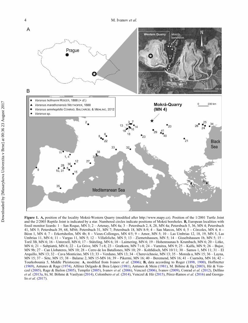
Figure 1. A, position of the locality Mokr�a-Western Quarry (modified after http://www.mapy.cz). Position of the 1/2001 Turtle Jointand the 2/2003 Reptile Joint is indicated by a star. Numbered circles indicate positions of Mokr�a boreholes. B, European localities withfossil monitor lizards: 1 – San Roque, MN 3; 2 – Artenay, MN 4a; 3 – Petersbuch 2, 8, 28, MN 4a; Petersbuch 5, 36, MN 4; Petersbuch41, MN 5; Petersbuch 39, 68, MN6; Petersbuch 31, MN 7; Petersbuch 18, MN 8-9; 4 – San Marcos, MN 4; 5 – C�orcoles, MN 4; 6 –B�eon 1, MN 4; 7 – Erkertshofen, MN 4b; 8 – Vieux-Collonges, MN 4/5; 9 ‒ Amor, MN 5; 10 – Las Umbrias 12, 18, 19, MN 5, LasUmbrias 11, MN 6; 11 ‒ Vargas 11, MN 5; 12 – Villafeliche, MN 5; 13 – Ziemetshausen, MN 5; 14 – Gisseltshausen 1b, MN 5; 15 –Toril 3B, MN 6; 16 – Unterzell, MN 6; 17 – St€atzling, MN 6; 18 – Laimering, MN 6; 19 – Hohenraunau b. Krumbach, MN 6; 20 ‒ Litke,MN 6; 21 ‒ Subpiatr�a, MN 6; 22 – La Grive, MN 7C8; 23 ‒ Gratkorn, MN 7C8; 24 – Varnitza, MN 9; 25 – Kalfa, MN 9; 26 – Bujor,MN 9b; 27 – Can Llobateres, MN 10; 28 ‒ Cerro de los Batallones, MN 10; 29 – Kohfidisch, MN 10/11; 30 – Samos 1, MN 11; 31 – ElArquillo, MN 13; 32 – Cava Monticino, MN 13; 33 ‒ Verduno, MN 13; 34 – Cherevichnoie, MN 13; 35 – Moreda a, MN 15; 36 – Layna,MN 15; 37 – S�ete, MN 15; 38 – Balaruc 2, MN 15-MN 16; 39 – Pikermi, MN 16; 40 – Beremend, MN 16; 41 – Csarn�ota, MN 16; 42 ‒Tourkobounia 5, Middle Pleistocene. A, modified from Ivanov et al. (2006); B, data according to Roger (1898, 1900), Hoffstetter(1969), Antunes & Rage (1974), Alf�erez Delgado & Brea L�opez (1981), Antunes & Mein (1981), M. B€ohme & Ilg (2003), H�ır & Ven-czel (2005), Rage & Bailon (2005), Tempfer (2005), Ivanov et al. (2006), Venczel (2006), Ivanov (2009), Conrad et al. (2012), Delfinoet al. (2013a, b), M. B€ohme & Vasilyan (2014), Colombero et al. (2014), Venczel & H�ır (2015), P�erez-Ramos et al. (2016) and Georga-lis et al. (2017).
4 M. Ivanov et al.
Dow
nloa
ded
by [
Mas
aryk
ova
Uni
verz
ita v
Brn
e] a
t 00:
36 2
3 A
ugus
t 201
7
original matrix). The tree landscape is explored using
Markov chain Monte Carlo (MCMC) algorithms to find
trees that have maximal posterior probability. The distri-
bution of the posterior probabilities is represented by a
50% majority-rule consensus in which the Bayesian sup-
port is given as a posterior probability on all internal
branches. The support value on a branch is interpreted as
the probability of retrieving the group subtended by that
branch, given the data matrix, the model of character evo-
lution, and the character-state frequency (e.g. Archibald
et al. 2003). We carried out 1,000,000 MCMC iterations,
sampling every 100th generation, and discarding the first
10,000 trees following the recommended burn-in. We
considered all posterior probability values above 90% to
be strong, and those below 50% to be weak (e.g. Tsuji
et al. 2010). In the Bayesian topologies, a value of 1 indi-
cates the probability of the partition indicated by the rele-
vant branch. For ease of figure presentation, we indicate
only partitions with a value of 1; all the others have a
value of 0.5. Using the Markov k (Mk) model for morpho-
logical data, we specified two types of rate distribution for
character changes across the tree, namely equal and
gamma, permitting respectively equal probability of
between-state changes and variable rates of change across
characters (e.g. Tsuji et al. 2010; Wright & Hillis 2014).
Stationarity was deemed to be satisfactory, with split fre-
quency values only slightly higher than 0.05.
Institutional abbreviations
MNHN: Mus�eum national d’Histoire naturelle, Paris,
France; NHMUK: Natural History Museum, London,
UK; NMA: Naturmuseum Augsburg, Augsburg, Ger-
many; NMP: National Museum, Prague, Czech Republic;
Pal.: Department of Geological Sciences, Masaryk Uni-
versity, Brno; SMF: Senckenberg Museum, Frankfurt,
Germany; UF, University of Florida, Gainesville, USA;
ZZSiD: Institute of Systematics and Evolution of Ani-
mals, Polish Academy of Sciences, Krak�ow, Poland.
Systematic palaeontology
Order Squamata Oppel, 1811
Superfamily VaranoideaM€unster, 1834Family Varanidae Hardwicke & Gray, 1827
Genus VaranusMerrem, 1820
yVaranus mokrensis sp. nov.
(Figs 2–7)
Diagnosis. Mid-sized Varanus with estimated snout-vent
length up to about 38 cm; differs from all other species in
the genus on the basis of a single autapomorphy – parietal
and squamosal processes of postorbitofrontal forming a
distinctly acute angle – and the following combination of
features: (1) short septomaxilla with flat dorsal surface;
(2) frontal and anterolateral postorbital processes of post-
orbitofrontal forming obtuse angle; (3) posterior margin
of nasal lobe of frontal situated in middle of dorsal width
of rostral process; (4) posterior margin of anteroposter-
iorly elongated frontal relatively narrow; (5) parietal with
wide subrectangular fronto-postfrontal processes, anteri-
orly arched fronto-parietal suture, and parietal foramen
situated significantly posterior to anterior margin of bone
(more than one-third of parietal plate length); (6) distance
between medially converging parietal crests of parietal
shortest in posterior quarter of parietal plate length; (7)
otoccipital with very distinct occipital crest at base of
paroccipital process; (8) pointed conical teeth with dis-
tally recurved apices and prominent smooth mesial and
distal cutting edges; (9) trunk vertebrae with moderately
developed depression between pars tectiformis and medial
margin of prezygapophyses and with posterolaterally
directed postzygapophyses forming a right angle; (10)
preacetabular process of ilium prominent and directed at
right angle to pubic process; and (11) bifurcated iliac crest
prominent.
Derivation of name. The species name mokrensis refers
to the Mokr�a-Western Quarry locality.
Holotype. 2/2003 Reptile Joint: parietal (Pal. 1097,
Fig. 3H, I).
Paratypes. 1/2001 Turtle Joint: left maxilla (Pal. 1023,
Fig. 2A, B; Pal. 1024, Fig. 2C–E), left septomaxilla (Pal.
1020, Fig. 2I, J). 2/2003 Reptile Joint: left postorbitofron-
tal (Pal. 1117, Fig. 3A, B), left frontal (Pal. 1090, Fig. 3E,
F), parietal (Pal. 1098, Fig. 3G), left palatine (Pal. 1134,
Fig. 4A, B), right pterygoid (Pal. 1131, Fig. 4D, E), right
quadrate (Pal. 1152, Fig. 4F), right prootic (Pal. 1104,
Fig. 4G, H), left otoccipital (Pal. 1109, Fig. 4J–M),
basioccipital (Pal. 1014, Fig. 4N), basisphenoid (Pal.
1114, Fig. 5A–C), right dentary (Pal. 1140, Fig. 5E–G),
left surangular (Pal. 1144, Fig. 5H, I), right articular (Pal.
1150, Fig. 5K, L), left ilium (Pal. 1154, Fig. 5M, N), cer-
vical vertebra (Pal. 1159, Fig. 6C, D), middle trunk verte-
bra (Pal. 1183, Fig. 6I–M). 1/2001 Turtle Joint: sacral
vertebra (Pal. 1066, Fig. 6Q).
Referred material. 1/2001 Turtle Joint: frontal (2 left C3 right), Pal. 1000–1004; parietal, Pal. 1005–1007; left
prootic, Pal. 1008; otoccipital (1 left C 3 right), Pal.
1009–1012; basisphenoid, Pal 1013; basioccipital, Pal.
1014–1015; postorbitofrontal (2 left C 2 right), Pal. 1016–
1019; septomaxilla (2 left C 1 right), Pal. 1020–1022;
maxilla (3 left C 2 right), Pal. 1023–1027; fragmentary
toothed bones, Pal. 1028–1032; left pterygoid, Pal. 1033;
right palatine, Pal. 1034; left dentary, Pal. 1035–1036; left
surangular, Pal. 1037; left articular, Pal. 1038; right quad-
rate, Pal. 1039; cervical vertebrae, Pal. 1040–1043; trunk
New early Miocene species of Varanus from the Czech Republic 5
Dow
nloa
ded
by [
Mas
aryk
ova
Uni
verz
ita v
Brn
e] a
t 00:
36 2
3 A
ugus
t 201
7
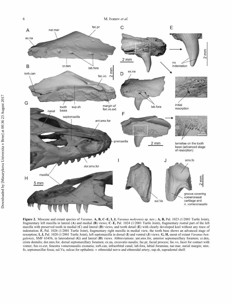
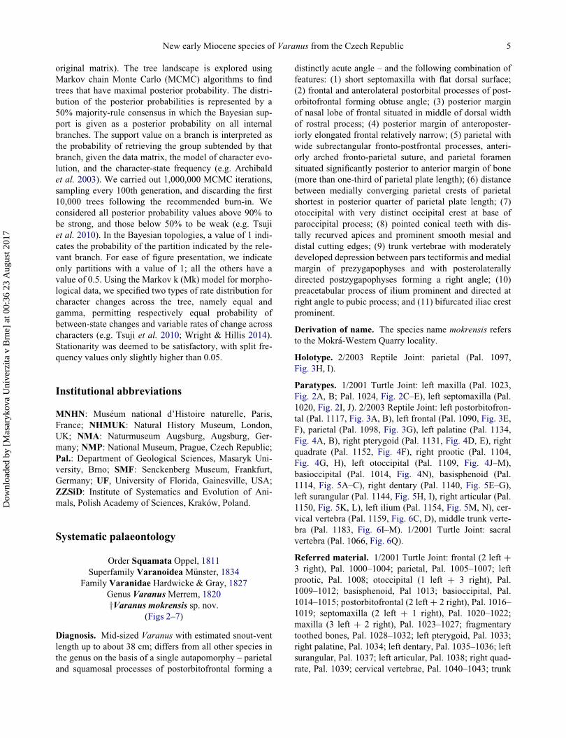
Figure 2. Miocene and extant species of Varanus. A, B, C–E, I, J, Varanus mokrensis sp. nov.; A, B, Pal. 1023 (1/2001 Turtle Joint),fragmentary left maxilla in lateral (A) and medial (B) views; C–E, Pal. 1024 (1/2001 Turtle Joint), fragmentary rostral part of the leftmaxilla with preserved tooth in medial (C) and lateral (D) views, and tooth detail (E) with clearly developed keel without any trace ofindentation; F, Pal. 1026 (1/2001 Turtle Joint), fragmentary right maxilla in medial view, the tooth base shows an advanced stage ofresorption; I, J, Pal. 1020 (1/2001 Turtle Joint), left septomaxilla in dorsal (I) and ventral (J) views. G, H, snout of extant Varanus ben-galensis, SMF 63456, in laterodorsal (G) and lateral (H) views. Abbreviations: ant.smx.for, anterior septomaxillary foramen; cr.den,crista dentalis; dor.smx.for, dorsal septomaxillary foramen; ex.na, excavatio nasalis; fac.pr, facial process; fac.vo, facet for contact withvomer; fen.vo.ext, fenestra vomeronasalis exonaria; iorb.can, infraorbital canal; lab.fora, labial foramina; nar.mar, narial margin; smx.fo, septomaxillar fossa; sul.Va, sulcus for opthalmic C ethmoidal nerve and ethmoidal artery; sup.sh, supradental shelf.
6 M. Ivanov et al.
Dow
nloa
ded
by [
Mas
aryk
ova
Uni
verz
ita v
Brn
e] a
t 00:
36 2
3 A
ugus
t 201
7

vertebrae, Pal. 1044–1065; sacral vertebrae, Pal. 1066–
1068; caudal vertebrae, Pal. 1069–1087.
2/2003 Reptile Joint: frontal (5 left C 4 right), Pal.
1088–1096; parietal, Pal. 1097–1103, prootic (2 left C 3
right), Pal. 1104–1108; otoccipital (2 left C 3 right), Pal.
1109–1113; basisphenoid, Pal. 1114–1116; postorbito-
frontal (3 left C 2 right), Pal. 1117–1121; septomaxilla (2
left C 1 right), Pal. 1122–1124; maxilla (2 left C 4 left or
right), Pal. 1125–1130; pterygoid (2 left C 1 right), Pal.
1131–1133; palatine (2 left C 1 right), Pal. 1134–1136;
dentary (3 left C 4 right), Pal. 1137-1143; surangular (3
left C 2 right), Pal. 1144–1148; articular (left C right),
Pal. 1149–1150; quadrate (2 left C 1 right), Pal 1151–
1153; ilium (3 left C 2 right), Pal. 1154–1158; cervical
vertebrae, Pal. 1159–1171; trunk vertebrae, Pal. 1172–
1206; sacral vertebrae, Pal. 1207–1210; caudal vertebrae,
Pal. 1211–1238.
Type locality and horizon. Mokr�a-Western Quarry,
Czech Republic: 1/2001 Turtle Joint and 2/2003 Reptile
Joint. Both joints date back to the early Miocene, Eggen-
burgian or Ottnangian, Orleanian, MN 4 (Ivanov et al.
2006; Sabol et al. 2007).
Description
Skull
Maxilla. The subtriangular maxilla contacts the premax-
illa anteriorly, the septomaxilla, vomer and premaxilla
medially, and the prefrontal, lacrimal, jugal, ectopterygoid
and maxillary process of the palatine posteriorly (Fig. 2A–
F). Its anteromedial extension forms the ventral rim of the
exonarial fenestra (excavatio nasalis sensuMertens 1942).
All preserved maxillae are fragmentary with broken ante-
rior and posterior extremities.
The sharp supradental shelf is expanded medially in its
anterior part where it meets the vomer forming the pos-
terolateral emargination of the external vomeronasal pit.
The orifice of the infraorbital canal (for the nervus alveo-
laris superior and maxillary artery; Bellairs 1949), situated
immediately dorsal to the supradental shelf, is delimited
anterodorsally by a short, distinct crest. Anteriorly, the
supradental shelf continues medially into a distinct facet
for the contact with the vomer, and also forms the poste-
rior margin of the exonarial fossa. The most complete left
maxilla shows six tooth positions. Seven labial foramina
occur in the most completely preserved maxilla. Foramina
are more closely spaced anteriorly at the level of the pos-
terior termination of the exonarial fenestra. A distinct
depression on the dorsal surface of the anterior part of the
maxillary fragments indicates that the excavatio nasalis
was relatively short anteroposteriorly.
Remarks. The prefrontal process is not preserved,
but the facial process was presumably very tall in lateral
view, as suggested by the inclination of the narial margin,
and enlarged posteriorly near the contact with the prefron-
tal and lacrimal. In its general shape (Fig. 2G, H), the
maxilla resembles that of V. bengalensis (Daudin, 1802),
although in adults of the latter species the laterally
depressed maxillary teeth do not have distinctly broad
basal portions (Mertens 1942, pl. 25, figs 195, 196) and
their crowns may be rounded in lateral view (Mertens
1942, pl. 25, fig. 196).
Septomaxilla. The approximately trapezoidal, paired
septomaxilla is situated between the nasal process of the
premaxilla and the maxillae. Laterally, it borders the flat
anteromedial margin of the maxilla (Fig. 2I, J). Its anterior
part forms a short, stout process.
A distinct, ventrally positioned septomaxillary fossa is
present. In life, this fossa housed the cartilaginous capsule
of the Jacobson’s organ. The anteromedial margin of the
fossa is broken. A triangular process with a distally nar-
rowing longitudinal groove projects posteriorly from the
fossa. This process extended dorsally over the paraseptal
cartilage (Bellairs 1949; Jollie 1960). The sharp postero-
lateral process of the septomaxilla formed the roof of the
free posterior termination of the chondrocranial lamina
transversalis anterior. In dorsal view, a distinct sulcus is
visible along the medial margin of the septomaxilla (sul-
cus pro ramus frontalis ophthalmici nerve [V] sensu Bahl
1937). In life, this sulcus probably accommodated the nar-
rowing medial branch of the ethmoidal nerve (Shrivatsava
1963; Va, nervus ophthalmicus C nervus ethmoideus
sensu Bellairs 1949) and the collateral ethmoidal artery. A
short crest is visible anterolaterally proximal to the suture
with the widened rostral portion of the maxilla. The crest
originally bordered a distinct groove that presumably
extended anteriorly almost to the level of the foramen sep-
tomaxillaris anterior (for its position in extant V. benga-
lensis, see Fig. 2G). The lacrimal duct (ld 2 – lower
lacrimal duct sensu Shrivatsava 1963) was situated within
the groove in life, and presumably ran ventrally and medi-
ally and opened below the Jacobson’s organ (Bellairs
1949).
Postorbitofrontal. The paired postorbitofrontal is
enlarged posteriorly and forms three processes (Fig. 3A,
B) situated along the medial and anterolateral margins of
the bone. It articulates with the parietal and frontal medi-
ally and with the squamosal posteriorly.
In dorsal view, the postorbitofrontal contacts the pos-
terolateral process of the frontal anteromedially via a
long, robust, proximally wide frontal process (processus
frontalis). The gracile distal termination of the process is
not preserved, but it is assumed to have been very elon-
gate, based on the strongly enlarged triangular facet for
the postorbitofrontal attachment observed in the frontal
New early Miocene species of Varanus from the Czech Republic 7
Dow
nloa
ded
by [
Mas
aryk
ova
Uni
verz
ita v
Brn
e] a
t 00:
36 2
3 A
ugus
t 201
7
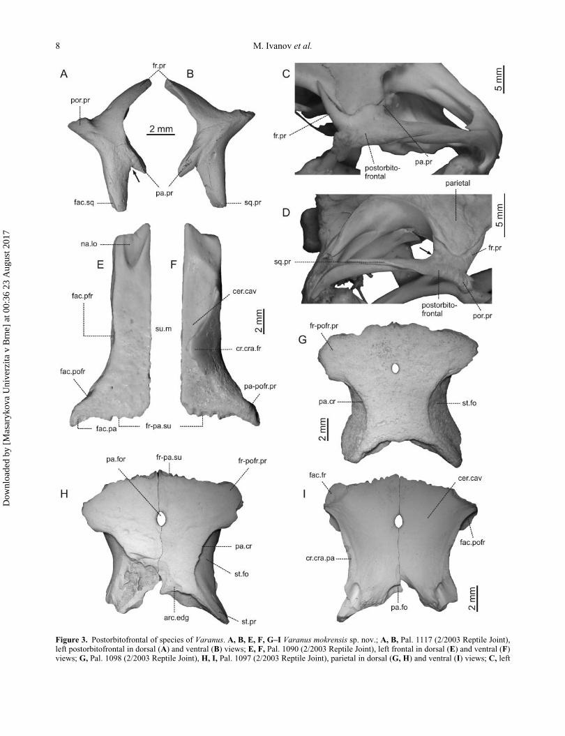
Figure 3. Postorbitofrontal of species of Varanus. A, B, E, F, G–I Varanus mokrensis sp. nov.; A, B, Pal. 1117 (2/2003 Reptile Joint),left postorbitofrontal in dorsal (A) and ventral (B) views; E, F, Pal. 1090 (2/2003 Reptile Joint), left frontal in dorsal (E) and ventral (F)views; G, Pal. 1098 (2/2003 Reptile Joint), H, I, Pal. 1097 (2/2003 Reptile Joint), parietal in dorsal (G, H) and ventral (I) views; C, left
8 M. Ivanov et al.
Dow
nloa
ded
by [
Mas
aryk
ova
Uni
verz
ita v
Brn
e] a
t 00:
36 2
3 A
ugus
t 201
7

bone. The postorbital and frontal processes are orientated
slightly ventrally, and form an obtuse angle between
them. The short posteromedially directed parietal process
is attached to the anterolateral border of the parietal, and
forms a conspicuously acute angle with the posteriorly
directed squamosal process (Fig. 3A – see arrow). The
robust squamosal process was probably short and,
together with the squamosum, participated in the forma-
tion of the anterior supratemporal arch.
Remarks. The angle between the frontal and parietal
processes may increase with age (Mertens 1942, pl. 23,
p. 167) (Fig. 3C, SMF 54157), as documented for Varanus
flavescens (Hardwicke & Gray, 1827); ZZSiD 456 and V.
benagalensis (Fig. 3D, NHMUK 1974.2479). Such an
increase results from the increasing anteroposterior width
of the processus fronto-postorbitofrontalis of the parietal.
Frontal. The paired frontal forms most of the dorsal part
of the orbital margin. The frontal cranial crests (cristae
cranii sensu Oelrich 1956; ventro-lateral processes sensu
Scanlon 2005) are the most prominent structures on the
ventral surface of the bone. They extend ventromedially
and almost completely enclose the olfactory canal
(Fig. 3E, F). The canal is subtriangular in cross section.
In dorsal view, the medial and lateral margins of the
frontal are parallel. The bone carries a short anteromedial
process and a large posterolateral process. The bone is
widest at the level of the parietal suture. Its medial margin
is straight and smooth. Its posterolateral margin forms a
strong process directed slightly posteriorly. This process
would be wedged between the postorbitofrontal and the
parietal in life. The triangular parietal tab is well devel-
oped. The posterior margin of the distinct lobus nasalis
(margo nasalis posterior), where the frontal meets the pos-
terolateral process of the nasal, is situated at the level of
the mid-width of the rostral process of the frontal. The
medial descending process has a broad base. The lateral
descending process is broken off close to its base. The flat
dorsal surface of the frontal is smooth anterior to the
orbital margin. The broad orbital portion of the bone
shows traces of the dorsal sculpture in the form of polygo-
nal fields separated by low weak ridges. In ventral view,
the medioventrally directed ventrolateral process of the
frontal cranial crest is distinctly developed. The frontopar-
ietal suture is interdigitated. In lateral view, the triangular
depression for the articulation with the prefrontal is con-
spicuously developed, and its posterior margin reaches
slightly posterior to the mid-length of the bone. The artic-
ular facet for the postorbitofrontal is indistinct. In medial
view, the anterior portion of the dorsal margin of the fron-
tal narrows, and the medial descending process of the
bone is slightly inclined ventrally. In anterior view, the
anterior margin of the crista cranii frontalis is directed dis-
tinctly ventrally, and its posterior margin is arched in a
medial direction.
Parietal. The unpaired parietal covers dorsally the poste-
rior portion of the braincase (Fig. 3G–I). It articulates
with the frontal anteriorly and the parietal posteriorly.
The frontoparietal suture is interdigitating. A flexible
movable joint occurs between the parietal and the prootic.
In dorsal view, the flat parietal table is covered by a
weakly developed ornamentation consisting of irregular
shallow grooves. The anterolateral processes are antero-
posteriorly broadened and project slightly laterally. The
distance between the medially converging parietal crests
is shortest at the level of the posterior quarter of the parie-
tal table length. Farther posteriorly, the parietal crest
extends onto the dorsal surface of the incompletely pre-
served supratemporal processes. The basal portions of the
posterolaterally diverging supratemporal processes form
an almost right angle. The parietal crest contributes to the
dorsal margin of a large and anteroposteriorly short supra-
temporal fossa, which extends from the postorbitofrontal
articulation, through the parietal process, to the unpre-
served posterior tip of the supratemporal process. The
fossa is mediolaterally broadest immediately posterior to
the level of the maximal constriction of the parietal crests.
The anteromedial part of the fossa is covered by the
slightly extending parietal crest. The parietal foramen lies
at a level slightly anterior to the mid-length of the parietal
table.
In ventral view, the facet for the articulation with the
posterolateral process of the frontal is anterolaterally
broad. The crista cranii parietalis is deepest halfway
between the parietal foramen and the posterior border of
the parietal table. Distinct marginal grooves are preserved
on either side of the crista cranii in one specimen (Pal.
1097). The posterior section of the crista cranii is short
and forms the anterolateral constriction of a deep depres-
sion visible at the base of the supratemporal process. The
parietal fossa forms a wide and shallow depression on the
posterior wall of the parietal table. In lateral view, the
bases of the supratemporal processes are orientated ven-
trally. A ledge-like lateral margin of the parietal table
postorbitofrontal of extant Varanus flavescens, SMF 54157, in laterodorsal view; D, right postorbitofrontal of extant Varanus bengalen-sis, NHMUK 1974.2479, in laterodorsal view. Abbreviations: arc.edg, arcuate edge; cer.cav, cerebral cavity; cr.cra.fr, crista cranii fron-talis; cr.cra.pa, crista cranii parietalis; fac.fr, facet for contact with frontal; fac.pa, facet for contact with parietal; fac.pfr, facet forcontact with prefrontal; fac.pofr, facet for contact with postorbitofrontal; fac.sq, facet for contact with squamosum; fr-pa.su, fronto-pari-etal suture; fr-pofr.pr, frontalo-postorbitofrontal process; fr.pr, frontal process; na.lo, nasal lobe; pa.cr, parietal crest; pa.fo, parietal fossa;pa.for, parietal foramen; pa-pofr.pr, parietal-postorbitofrontal process; pa.pr, parietal process; por.pr, postorbital process; sq.pr, squamo-sal process; st.fo, supratemporal fossa; st.pr, supratemporal process.
J
New early Miocene species of Varanus from the Czech Republic 9
Dow
nloa
ded
by [
Mas
aryk
ova
Uni
verz
ita v
Brn
e] a
t 00:
36 2
3 A
ugus
t 201
7

Figure 4. Miocene and extant species of Varanus. A, B, D–H, J–N, Varanus mokrensis sp. nov. A, B, Pal. 1134 (2/2003 Reptile Joint),left palatine in dorsal (A) and ventral (B) views; D, E, Pal. 1131 (2/2003 Reptile Joint), right pterygoid in dorsal (D) and ventral (E)views; F, Pal. 1152 (2/2003 Reptile Joint), right quadrate in caudal view; G, H, Pal. 1104 (2/2003 Reptile Joint), right prootic in lateral(G) and medial (H) views; J–M, Pal. 1109 (2/2003 Reptile Joint), left otoccipital in dorsal (J), caudal (K), cranial (L) and medial (M)views; N, Pal. 1014 (1/2001 Turtle Joint), basioccipital in ventral view. C, I. extant Varanus bengalensis, SMF 63456; C, left palatine inventral view; I, left prootic in lateral view. Abbreviations: ant.sci.can, anterior semicircular canal; ar.con, articular condyle; bsph.pr,
10 M. Ivanov et al.
Dow
nloa
ded
by [
Mas
aryk
ova
Uni
verz
ita v
Brn
e] a
t 00:
36 2
3 A
ugus
t 201
7

covers the articulation of the parietal with the parietal pro-
cess of the postorbitofrontal.
Remarks. The course of the parietal crests indicates a
well-developed attachment for the musculus pseudotem-
poralis superficialis, such as is found in extant duropha-
gous species of Varanus, including V. niloticus (Linnaeus,
1766), V. exanthematicus (Bosc, 1792) and V. olivaceus
Hallowell, 1857 (Mertens 1942, pl. 23, figs 166, 168). In
these species and in the extinct V. rusingensis from the
early Miocene of Kenya (Clos 1995), the parietal crests
approach one another closely in a posterior direction
(Clos 1995). Such crests often occur in adult specimens
and may form a distinct ridge in the midline of the poste-
rior margin of the bone (e.g. V. olivaceus; Mertens 1942,
pl. 24, fig. 172; SMF 72156). Specimens of V. mokrensis
represent adults of average size (about 374.9 mm SVL).
In this taxon, the converging parietal crests fail to come in
contact with one another and the features of the dentition
indicate that it had a weaker pseudotemporalis superficia-
lis muscle than the above-mentioned species. We infer,
therefore, that V. mokrensis was not a durophagous taxon.
Palatine. The irregularly shaped, toothless palatine bears
three processes (Fig. 4A, B). The wide and posteriorly
directed pterygoid process (broken off in all specimens) is
connected to the pterygoid. The thin, anteromedially
directed vomerine process connects to the vomer. The
maxillary process is directed anteroposteriorly, but its
anterior extremity is not preserved. The posterior end of
this process contacts the jugal and ectopterygoid. The
anterior border of the palatine forms the posterior margin
of the internal nostril.
In anterior view, the infraorbital foramen (maxillo-pala-
tine foramen sensu Mertens 1942 and Jollie 1960) forms
the orifice of the superior alveolar canal running through
the base of the maxillary process.
Remarks. The elliptical to almost circular infraorbi-
tal foramen of the palatine and the proximally slender
processus vomerinus are also observed in extant V. benga-
lensis (MNHN 1884-4, 1886-649). The elongate maxillary
process forms an obtuse angle with the vomerine process,
and this suggests that the posterior nares were probably
wide and short (as indicated also by the short septomax-
illa) unlike in V. bengalensis (Fig. 4B, C – see arrows).
The same development of posterior nares occurs in Afri-
can monitors, which are considered to form the sister
group to all other Varanus species.
Pterygoid. In ventral view, the triradiate pterygoid is
slightly curved medially and shows no trace of dentition
(Fig. 4D, E). Its anterior portion is flat, whereas its poste-
rior portion forms a long and gracile quadrate process ori-
entated posterolaterally. The anteromedial portion of the
pterygoid projects into a flat and wide palatine process.
The latter process articulates with the posterior border of
the palatine. Anterolaterally, the pterygoid articulates
with the ectopterygoid. The posterior terminations of all
three processes are broken in all specimens.
In dorsal view, the crista columellaris anterior is wide
and weakly developed. The crista columellaris posterior,
forming the posteromedial border of the elongated fossa
columellae, is sharp and decreases in height towards the
posterior termination of the columellar fossa. The anterior
margin of the columellar fossa reaches as far as the poste-
rior termination of the markedly deep sulcus which
widens and diminishes anteriorly. The basisphenoid
process projects medially. The articular surface for the
basipterygoid process of the basisphenoid is slightly
rugose. In ventral view, the basal portion of the palatal
process narrows posteriorly. The more gracile ectoptery-
goid process is broken near its base. The low but distinct
crista transversa dorsalis courses close to the lateral
margin of the ectopterygoid process. The suborbital
incisure is shallow and gradually merges into the wide
base of the pterygoid process.
Quadrate. Only the trochlear part of the quadrate is
present (Fig. 4F). It has a wide articular condyle for the
junction with the articular. A small triangular facet for the
articulation with the pterygoid is present at the medial
border of the articular condyle. The tympanic fossa is
deep and wide, and bears a wide outer ridge for the attach-
ment of the dorsal and anterior borders of the tympanic
membrane. The lateromedially wide and strongly devel-
oped posterior crest extends as far as the tympanic part of
the articular condyle.
Prootic. The prootic forms the anterior and antero-
ventral portion of the auditory capsule (Fig. 4G, H). The
prootic articulates with the supratemporal process of the
basisphenoid process; co.fo, columellar fossa; coch.cav, cochlear cavity; cr.al.pot, crista alaris prootica; cr.co.ant, crista columellarisanterior; cr.co.post, crista columellaris posterior; cr.pot, crista prootica; cr.tra.dor, crista transversa dorsalis; ect.pr, ectopterygoid pro-cess; fac.pr, facial process; fac.st, facet for contact with supratemporal; for.ro, foramen rotundum; hor.sci.can, horizontal semicircularcanal; inf.pr.pot, inferior process of prootic; IX, foramen for glossopharyngeal nerve; mx.pr, maxillary process; occo.tub, occipitocondy-lar tubercle; oc.cr, occipital crest; pal.pr, palatal process; paoc.pr, paroccipital process; post.cr, posterior crest of quadratum; post.pr.pot,posterior process of prootic; post.sci.can, posterior semicircular canal; pt.pr, pterygoid process; qu.pr, quadrate process; sph-oc.tub,spheno-occipital tubercle; sul.ve.jug, sulcus venae jugularis; suor.inc, suborbital incisure; sup.alv.can, superior alveolar canal; sup.pr.pot, superior process of prootic; tri.no, trigeminal notch; ty.fo, tympanic fossa; vest.cav, vestibular cavity; VII, foramen for the facialnerve; VII.pal, foramen for palatine branch of the facial nerve; vo.pr, vomerine process; X, foramen for vagus nerve; XI, foramen foraccessory nerve; XII, foramen for hypoglossal nerve.
J
New early Miocene species of Varanus from the Czech Republic 11
Dow
nloa
ded
by [
Mas
aryk
ova
Uni
verz
ita v
Brn
e] a
t 00:
36 2
3 A
ugus
t 201
7
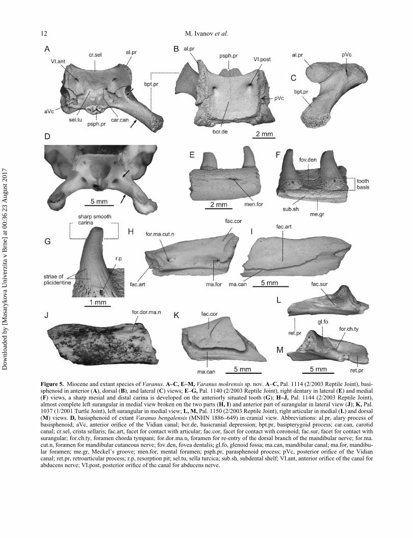
Figure 5. Miocene and extant species of Varanus. A–C, E–M, Varanus mokrensis sp. nov. A–C, Pal. 1114 (2/2003 Reptile Joint), basi-sphenoid in anterior (A), dorsal (B), and lateral (C) views; E–G, Pal. 1140 (2/2003 Reptile Joint), right dentary in lateral (E) and medial(F) views, a sharp mesial and distal carina is developed on the anteriorly situated tooth (G); H–J, Pal. 1144 (2/2003 Reptile Joint),almost complete left surangular in medial view broken on the two parts (H, I) and anterior part of surangular in lateral view (J); K, Pal.1037 (1/2001 Turtle Joint), left surangular in medial view; L, M, Pal. 1150 (2/2003 Reptile Joint), right articular in medial (L) and dorsal(M) views. D, basisphenoid of extant Varanus bengalensis (MNHN 1886–649) in cranial view. Abbreviations: al.pr, alary process ofbasisphenoid; aVc, anterior orifice of the Vidian canal; bcr.de, basicranial depression; bpt.pr, basipterygoid process; car.can, carotidcanal; cr.sel, crista sellaris; fac.art, facet for contact with articular; fac.cor, facet for contact with coronoid; fac.sur, facet for contact withsurangular; for.ch.ty, foramen chorda tympani; for.dor.ma.n, foramen for re-entry of the dorsal branch of the mandibular nerve; for.ma.cut.n, foramen for mandibular cutaneous nerve; fov.den, fovea dentalis; gl.fo, glenoid fossa; ma.can, mandibular canal; ma.for, mandibu-lar foramen; me.gr, Meckel’s groove; men.for, mental foramen; psph.pr, parasphenoid process; pVc, posterior orifice of the Vidiancanal; ret.pr, retroarticular process; r.p, resorption pit; sel.tu, sella turcica; sub.sh, subdental shelf; VI.ant, anterior orifice of the canal forabducens nerve; VI.post, posterior orifice of the canal for abducens nerve.
12 M. Ivanov et al.
Dow
nloa
ded
by [
Mas
aryk
ova
Uni
verz
ita v
Brn
e] a
t 00:
36 2
3 A
ugus
t 201
7

Figure 6. Varanus mokrensis sp. nov. A, B, Pal. 1040 (1/2001 Turtle Joint), cervical vertebra in lateral (A) and caudal (B) views; C, D,Pal. 1159 (2/2003 Reptile Joint), cervical vertebra in lateral (C) and dorsal (D) views; E–H, Pal. 1172 (2/2003 Reptile Joint), anteriortrunk vertebra in lateral (E), dorsal (F), ventral (G) and cranial (H) views; I–M, Pal. 1183 (2/2003 Reptile Joint), middle trunk vertebrain lateral (I), dorsal (J), ventral (K), cranial (L) and caudal (M) views; N–P, Pal. 1207 (2/2003 Reptile Joint), sacral vertebra in dorsal(N), ventral (O) and cranial (P) views;Q, Pal. 1066 (1/2001 Turtle Joint), sacral vertebra in ventral view; R, S, Pal. 1215 (2/2003 ReptileJoint), anterior caudal vertebra in dorsal (R) and ventral (S) views. Abbreviations: cd, condyle; ct, cotyle; hy, hypapophysis; na, neuralarch; nc, neural canal; ns, neural spine; po, postzygapophysis; pof, postzygapophyseal articular facet; pr, prezygapophysis; prf, prezyga-pophyseal articular facet; syn, synapophysis; t.pr, transverse process.
New early Miocene species of Varanus from the Czech Republic 13
Dow
nloa
ded
by [
Mas
aryk
ova
Uni
verz
ita v
Brn
e] a
t 00:
36 2
3 A
ugus
t 201
7

parietal and the epipterygoid anterodorsally, the supraoc-
cipital posterodorsally, the exoccipital posteriorly, the
basioccipital posteroventrally and the basisphenoid ante-
roventrally. It is irregularly triaxial (Bellairs & Kamal
1981; Rieppel & Zaher 2000), and shows an alary process
(processus prooticus anterioris superioris sensu Bellairs &
Kamal 1981), an anterior inferior process (processus proo-
ticus anterioris inferioris – sensu Bellairs & Kamal 1981),
and a posteriorly directed posterior process.
In lateral view, the anterior inferior process is dorso-
ventrally flattened. It is blunt and its anterior surface bears
a shallow groove for the articulation with the alary pro-
cess of the basisphenoid. A distinct rounded trigeminal
notch occurs between the anterior inferior and the anterior
superior processes. In extant Varanus, the shallow groove
is adjacent to the posterior narrower part of a membrane
perforated by the exit orifice for the trigeminal nerve. The
anterior superior process forms a movable articulation
with the underside of the lateral bevelled edge of the pari-
etal and with the external dorsal extremity of the epiptery-
goid. The lateral crest (crista alaris prootici) is wide and
blunt. The posterior process of the prootic contacts the
external surface of the paroccipital process of the otocci-
pital. The inferior process of the prootic forms a crest
(crista prootica; crista otosphenoidea – sensu Bahl 1937)
situated dorsal to a deep groove for the jugular vein (sul-
cus pro vena capitalis lateralis – sensu Bellairs & Kamal
1981). A small foramen for the exit of the palatal branch
of the facial nerve (VII. pal.) lies in the anterior section of
the groove. A much larger foramen for the exit of the
hyomandibular branch (VII. hm.) is situated just posterior
to the former foramen. In medial view, the cavum vestibu-
lare housing the membraneous labyrinth (utriculus Csaculus) has a wide orifice. A somewhat smaller deep
cochlear cavity, surrounded by strongly damaged cochlear
ridge, occurs on its anteroventral portion.
A large orifice for the vertically orientated anterior
semicircular canal is situated at the anterior margin of the
vestibular cavity. The orifice for the horizontal semicircu-
lar canal occurs inside the vestibular cavity and close to
the dorsal margin of the latter, and is situated posteroven-
tral to the orifice of the anterior semicircular canal. The
horizontal semicircular canal passes along the external
wall of the prootic and continues to the opisthotic portion
of the otoccipital, close to the base of the inner margin of
the processus prooticus posterioris. One to two minute
foramina are situated at the base of the alary process,
immediately anteroventral to the foramen for the anterior
semicircular canal. A large foramen for the facial nerve
(VII) is situated close to the base of the anterior inferior
process.
Remarks. The posterior orifice for the Vidian canal
of the basisphenoid is situated close to the junction of this
bone with the prootic. Such an orifice is also documented
in the extinct V. amnhophilis and V. priscus (Owen, 1859)
(Conrad et al. 2012, p. 5, fig. 2G, J; Head et al. 2009, p.
449). The prootic of V. mokrensis generally resembles
that of V. bengalensis (MNHN 1886-649; MNHN 1884-4;
SMF 63456), but differs from the latter (Fig. 4I – see
arrow) in showing a more robust lateral edge of the alary
process (crista alaris prootici, Fig. 4G – see arrow).
Otoccipital. The otoccipital forms the posterolateral
wall of the cerebral cavity and participates in the forma-
tion of the posterior wall of the auditory capsule (Fig. 4J–
M). A substantial portion of the laterally projecting paroc-
cipital process is formed by the opisthotic (Bellairs &
Kamal 1981). The exoccipital portion articulates with the
basioccipital ventrolaterally. The opisthotic portion is sit-
uated almost at a right angle to the exoccipital, and articu-
lates with the prootic anteriorly and laterally, and with the
posterior portion of the supraoccipital dorsally (Bahl
1937; Mertens 1942).
The opisthotic forms a posterolaterally enlarged and
ventrally directed process. The dorsally located rounded
facet for the junction with the supratemporal occurs at the
distal termination of the paroccipital process. The orifice
of the horizontal semicircular canal is situated at the base
of the paroccipital process. The inner exit of the semicir-
cular canal is situated within the recessus utriculi, in close
proximity to the medioventrally situated ampulla anterior,
leading to the small orifice for the posterior semicircular
canal. The orifice for the posterior semicircular canal is
situated at the dorsal surface of the opisthotic portion. A
depression on the base of the paroccipital process marks
Figure 7. Varanus mokrensis sp. nov. A, B, Pal. 1154 (2/2003Reptile Joint), left ilium in lateral (A) and dorsal (B) views.Abbreviations: il.cr, iliac crest; is.pr, ischial process; pac.pr, pre-acetabular process; poac.pr, postacetabular process; pub.pr,pubic process.
14 M. Ivanov et al.
Dow
nloa
ded
by [
Mas
aryk
ova
Uni
verz
ita v
Brn
e] a
t 00:
36 2
3 A
ugus
t 201
7
the posterior boundary of the fenestra vestibuli. There is a
well-preserved composite foramen for the exit of the cra-
nial nerves XCXI, the entry of the occipital branch of the
inner carotid artery, and the exit of the jugular vein. The
small foramen for the hypoglossal nerve (XII) is separated
from the foramina for cranial nerves XCXI by a bony sep-
tum. Small foramina for the nerves XI (dorsally) and XII
(ventrally) are situated close to the medial margin of the
otoccipital. The crista tuberalis is dorsally arched. The
dorsal surface of the paroccipital process bears a deep and
laterally enlarged depression posteriorly limited by the
distinct lateral occipital crest. The slit-like jugular fora-
men for the common exit of the jugular vein and cranial
nerve X occurs at the posteroventral margin of the
otoccipital.
Basioccipital. The basioccipital forms the floor of the
posterior portion of the braincase (Fig. 4N). It articulates
with the basisphenoid anteriorly and with the exoccipitals
laterally.
In ventral view, the anteriorly widened margin of the
basioccipital is either slightly convex medially (Pal. 1014)
or straight (Pal. 1015). The basipterygoid processes are
situated laterally; their anterolateral margins protrude ven-
trally into the falciform spheno-occipital tubercles (Pal.
1014). The lateral margin of the basioccipital is slightly
concave between the basipterygoid process and the
strongly damaged occipitocondylar tubercle forming the
medial portion of the occipital condyle. The ventral sur-
face of the basioccipital is more or less flat. Distinct
depressions are present at the base of the basipterygoid
processes of larger specimens (Pal. 1014). The medial
crest is indistinct (Pal. 1014). In dorsal view, a shallow
oval depression marks the point where the medulla oblon-
gata terminates.
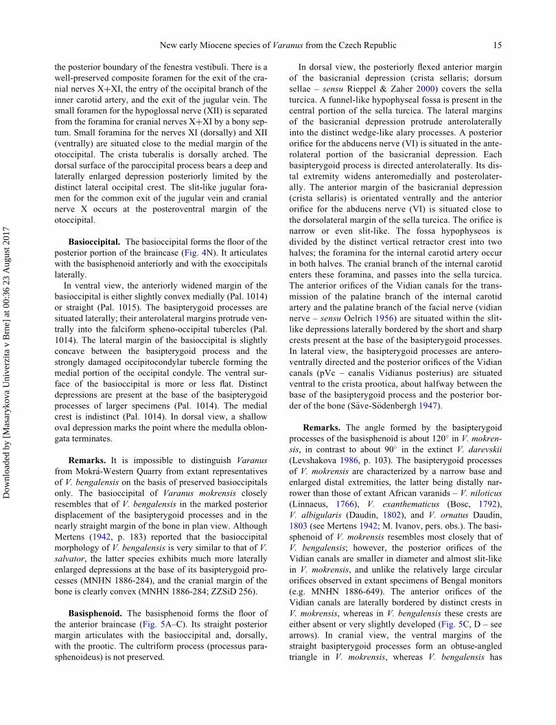
Remarks. It is impossible to distinguish Varanus
from Mokr�a-Western Quarry from extant representatives
of V. bengalensis on the basis of preserved basioccipitals
only. The basioccipital of Varanus mokrensis closely
resembles that of V. bengalensis in the marked posterior
displacement of the basipterygoid processes and in the
nearly straight margin of the bone in plan view. Although
Mertens (1942, p. 183) reported that the basioccipital
morphology of V. bengalensis is very similar to that of V.
salvator, the latter species exhibits much more laterally
enlarged depressions at the base of its basipterygoid pro-
cesses (MNHN 1886-284), and the cranial margin of the
bone is clearly convex (MNHN 1886-284; ZZSiD 256).
Basisphenoid. The basisphenoid forms the floor of
the anterior braincase (Fig. 5A–C). Its straight posterior
margin articulates with the basioccipital and, dorsally,
with the prootic. The cultriform process (processus para-
sphenoideus) is not preserved.
In dorsal view, the posteriorly flexed anterior margin
of the basicranial depression (crista sellaris; dorsum
sellae – sensu Rieppel & Zaher 2000) covers the sella
turcica. A funnel-like hypophyseal fossa is present in the
central portion of the sella turcica. The lateral margins
of the basicranial depression protrude anterolaterally
into the distinct wedge-like alary processes. A posterior
orifice for the abducens nerve (VI) is situated in the ante-
rolateral portion of the basicranial depression. Each
basipterygoid process is directed anterolaterally. Its dis-
tal extremity widens anteromedially and posterolater-
ally. The anterior margin of the basicranial depression
(crista sellaris) is orientated ventrally and the anterior
orifice for the abducens nerve (VI) is situated close to
the dorsolateral margin of the sella turcica. The orifice is
narrow or even slit-like. The fossa hypophyseos is
divided by the distinct vertical retractor crest into two
halves; the foramina for the internal carotid artery occur
in both halves. The cranial branch of the internal carotid
enters these foramina, and passes into the sella turcica.
The anterior orifices of the Vidian canals for the trans-
mission of the palatine branch of the internal carotid
artery and the palatine branch of the facial nerve (vidian
nerve – sensu Oelrich 1956) are situated within the slit-
like depressions laterally bordered by the short and sharp
crests present at the base of the basipterygoid processes.
In lateral view, the basipterygoid processes are antero-
ventrally directed and the posterior orifices of the Vidian
canals (pVc – canalis Vidianus posterius) are situated
ventral to the crista prootica, about halfway between the
base of the basipterygoid process and the posterior bor-
der of the bone (S€ave-S€odenbergh 1947).
Remarks. The angle formed by the basipterygoid
processes of the basisphenoid is about 120� in V. mokren-
sis, in contrast to about 90� in the extinct V. darevskii
(Levshakova 1986, p. 103). The basipterygoid processes
of V. mokrensis are characterized by a narrow base and
enlarged distal extremities, the latter being distally nar-
rower than those of extant African varanids – V. niloticus
(Linnaeus, 1766), V. exanthematicus (Bosc, 1792),
V. albigularis (Daudin, 1802), and V. ornatus Daudin,
1803 (see Mertens 1942; M. Ivanov, pers. obs.). The basi-
sphenoid of V. mokrensis resembles most closely that of
V. bengalensis; however, the posterior orifices of the
Vidian canals are smaller in diameter and almost slit-like
in V. mokrensis, and unlike the relatively large circular
orifices observed in extant specimens of Bengal monitors
(e.g. MNHN 1886-649). The anterior orifices of the
Vidian canals are laterally bordered by distinct crests in
V. mokrensis, whereas in V. bengalensis these crests are
either absent or very slightly developed (Fig. 5C, D – see
arrows). In cranial view, the ventral margins of the
straight basipterygoid processes form an obtuse-angled
triangle in V. mokrensis, whereas V. bengalensis has
New early Miocene species of Varanus from the Czech Republic 15
Dow
nloa
ded
by [
Mas
aryk
ova
Uni
verz
ita v
Brn
e] a
t 00:
36 2
3 A
ugus
t 201
7

relatively shorter basipterygoid processes (Fig. 5C, D –
see arrows).
Lower jaw. Dentary. Several highly fragmentary denta-
ries are preserved (Fig. 5E–G). Both the rostral and poste-
rior portions of all fragments are broken. In medial view,
the narrow sulcus Meckeli remains open throughout the
entire length of the dentary. The best-preserved fragment
(Pal. 1140) possesses three tooth positions with two dam-
aged teeth (Fig. 5E, F).
Surangular. One of several preserved surangulars is
almost complete; only its posterior margin is missing
(Fig. 5H–J). It is a massive and laterally compressed bone
(Fig. 5H–K), anteriorly sutured to the dentary, dorsally to
the coronoid, ventrally and medially to the splenial and
articular, and ventrolaterally to the angular.
In lateral view, a distinct orifice for the mandibular
cutaneous nerve (posterior supra-angular foramen – sensu
Oelrich 1956), being a part of the mandibular division of
cranial nerve V, is situated in close proximity to the bro-
ken posterior tip of the bone. The canal passes through the
posterior portion of the dentary, which is triangular in
cross section, and leads into the bone. Another foramen
for re-entry of the dorsal branch of the mandibular nerve
is situated close to the base of the coronoid process. The
slit-like foramen which is visible in anterolabial view is
surrounded by the bony tissue of the surangular and situ-
ated in a shallow depression. In medial view, there is a
distinct groove situated at the anterior termination of the
fossa Meckeli. The groove was originally enclosed by the
articular from both ventral and medial sides and also by
the cuneate angular in its anterior portion. This wide man-
dibular canal, which is circular in cross section, served for
the entry of the inferior main branch of the mandibular
nerve (nervus alveolaris inferior) and mandibular artery.
At the same time, it served for the re-entry of the mandib-
ular vein (Bahl 1937). The deep groove situated at the
anterodorsal portion of the surangular represents the re-
entry of the canal for another branch of the mandibular
nerve that passes through the entire length of the bone.
Remarks. The distinct foramen for re-entry of the
dorsal branch of the mandibular nerve (Fig. 5J) is situated
far from the anterior border of the bone. In African vara-
nids, particularly in V. niloticus (e.g. MNHN 1964-50),
this foramen is situated distinctly anteriorly. It is difficult
to observe this foramen in numerous articulated speci-
mens, e.g. V. albigularis albigularis (Daudin, 1802)
(Mertens 1942, pl. 26, fig. 201) or V. griseus (e.g. MNHN
1895-366). The surangular of V. mokrensis closely resem-
bles that of V. salvator (Laurenti, 1768) (MNHN 1888-
198) particularly by the length/height ratio and the convex
body of the bone; the surangular of V. bengalensis
(MNHN 1886-649) and V. flavescens (UF 64743) is rela-
tively flat in contrast to that of V. mokrensis. However,
the foramen for the re-entry of the dorsal branch of the
mandibular nerve is situated even more posteriorly in Var-
anus from Mokr�a-Western Quarry, approximately half-
way between the anterior border of the bone and the
orifice of the distinct cutaneous nerve.
Articular. The articular is an elongated bone whose
massive retroarticular process extends far behind the
quadrate/articular joint (Fig. 5L, M). Only the posterior
portion of the bone is preserved; it has a distinct trans-
versely concave glenoid fossa. The retroarticular process
is long and stout and its distal extremity widens posteri-
orly. A well-developed retroarticular fossa is situated pos-
terior to the articular facet. An aperture for the entrance of
the chorda tympani branch of cranial nerve VII occurs
within the fossa.
Dentition (Figs 2C–F, 5E–G). The teeth are conical
and laterally flattened. They have mesiodistally broadened
bases with distinctly developed longitudinal striations (as
a result of furrowed plicidentine). The tooth bases of the
fully developed teeth are either massive with small resorp-
tion pits or are formed by a system of laminae indicating
an advanced stage of resorption preceding the tooth
replacement (Rieppel 1978). Distinct and sharp cutting
edges are present at the mesial and distal margins of the
tooth crown; the surface of the cutting edge is smooth.
Remarks. The most immediately recognizable trait
of the Varanus mokrensis dentition is the distinctly devel-
oped smooth cutting edge along the mesial and distal mar-
gins of the apical part of the tooth crown. Most of the
extant Asiatic varanids have developed trenchant and pos-
teriorly directed teeth with serrated cutting edges. Vara-
nids with a molariform dentition show adaptations to
durophagy (e.g. extant V. niloticus, V. exanthematicus, V.
olivaceus and extinct V. rusingensis; Mertens 1942; Riep-
pel & Labhardt 1979; Clos 1995) and may have developed
a short cutting edge without indentation, but this character
was observed only in subadult specimens (Ivanov 2009).
In members of the closest outgroup to Varanus (i.e. platy-
notan representatives of Lanthanotus, Helodermatidae
and Anguidae) the cutting edge is either absent or devel-
oped (but lacks indentation). Therefore, it is possible to
presuppose that the presence of the smooth edge in adult
specimens of Varanus represents plesiomorphic
condition.
Postcranial skeleton
Cervical vertebrae. Most of the preserved cervical ver-
tebrae are fragmentary, usually with broken neural spines
and hypapophyses (Fig. 6A–D). The height of the neural
spine depends on the position within the cervical region
of the vertebral column. In lateral view, the posterior cer-
vical vertebrae with distally broken neural spines were
16 M. Ivanov et al.
Dow
nloa
ded
by [
Mas
aryk
ova
Uni
verz
ita v
Brn
e] a
t 00:
36 2
3 A
ugus
t 201
7

characterized by neural spines probably as anteroposter-
iorly long as dorsoventrally high, or somewhat lower in
the cranio-caudal direction. Anterior cervical vertebrae
possess distinctly anteroposteriorly enlarged and conspic-
uously low neural spines. Anteriorly, the neural spine
reaches the anterior margin of the neural arch. Posteriorly,
the neural spine extends as far as the posterior termination
of the neural arch. The bases of the transverse processes
are directed laterally. The anterior margin of the hypa-
pophysis extends as a low ridge as far as the vicinity of
the cotylar rim. The hypapophysis distinctly rises at about
centrum midlength. In dorsal view, the oval prezygapo-
physeal articular facets extend laterally to form the wide
prezygapophyseal area. The interzygapophyseal constric-
tion occurs at about vertebral mid-length. In ventral view,
the anterior margin of the vertebral centrum base is wid-
ened laterally and gradually passes into the transverse pro-
cess. The posterior margin of the centrum is narrow
anterior to the laterally wide condylar base. In anterior
view, the neural arch is moderately vaulted. Its anterior
margin is triangular to slightly round in cross section and
is situated between the markedly dorsally tilted prezyga-
pophyses. The cotyle is prominently depressed dorsoven-
trally. The distal termination of the hypapophysis is pear
shaped and is distinctly developed particularly in the pos-
terior cervical vertebrae.
Remarks. Fragmentary cervical vertebrae of Varanus
mokrensis have developed hypapophyses with laterally
widened distal terminations. This is typical for all repre-
sentatives of the genus Varanus. Several fragmentary ver-
tebrae from the cervical region indicate that the neural
spine of Varanus mokrensis was low and antero-posteri-
orly distinctly enlarged in contrast to all observed extant
representatives of Indo-Asiatic group (Ast 2001). In this
respect, cervical vertebrae of V. mokrensis resemble
extant V. griseus recently inhabiting Africa and south-
western Asia.
Trunk vertebrae. Numerous well-preserved trunk
vertebrae enable studies of the intracolumnar variability,
particularly as regards the shape of the neural spine
(Fig. 6E–M). In lateral view, the neural spine of the mid-
dle trunk vertebrae is about two to three times anteropos-
teriorly longer than tall. Its anterior margin is
posterodorsally inclined; this inclination is more distinct
in the posterior trunk vertebrae. Therefore, the distal ter-
mination of the neural spine is relatively short in the pos-
terior trunk vertebrae. The synapophyses are massively
built and markedly large in several vertebrae. The lateral
foramina, which occur close behind the posterodorsal
margin of synapophyses, are very small and indistinct. In
dorsal view, the interzygapopyseal constriction occurs
about at two-thirds of the vertebra length close before the
posterolaterally directed and laterally relatively narrow
postzygapophyses (compared to prezygapophyses). The
prezygapophyses are strongly laterally widened, and the
prezygapophyseal articular facets are of anterolaterally
enlarged oval shape. The dorsal margin of the neural spine
is posteriorly slightly laterally thickened. In ventral view,
the vertebral centrum is elongated and wide anteriorly
whereas the basal portion of the markedly laterally wid-
ened condyle is strongly constricted. Synapophyses are
directed laterally. In cranial view, the prezygapophyses
are conspicuously tilted dorsally, the synapophyses are
short and directed laterally. The neural arch is moderately
vaulted and its roof-like posterior margin rises dorsally to
the base of the neural spine. The neural canal is irregularly
circular to subtriangular in cross section. The cotyle is
depressed dorsoventrally. Small paracotylar foramina are
developed in depressions at both sides between the cotyle
and upraising base of the prezygapophyses in many verte-
brae. In some cases, the foramina are not paired and they
are often absent.
Sacral vertebrae. All sacral vertebrae are fragmen-
tary (Fig. 6N–Q). In ventral view, they differ from the ver-
tebrae of the trunk region by the distinctly shorter centrum
length/neural arch width ratio. The dorsoventrally
depressed transverse processes are not distally narrowed.
Typical is the presence of a groove at the posterior margin
of the transverse processes. This groove is strongly wid-
ened medially. In posterior view, the condyle is distinctly
laterally widened with a wide and shallow medial groove
extending dorsoventrally.
Caudal vertebrae. Several preserved caudal verte-
brae are fragmentary and with a broken neural spine
(Fig. 6R, S). Rarely preserved isolated neural spines are
posterodorsally strongly elongated. In dorsal view, the
prezygapophyseal articular facets are oval and the dorso-
ventrally flattened transverse processes are directed
laterally.
Ilium. One almost complete left ilium is character-
ized by the distinct bifurcated iliac crest situated dorsal to
the acetabulum (Fig. 7A, B). In lateral view, the postace-
tabular process is massively built with a dorsoventrally
wide base. The preacetabular process is prominent with a
blunt termination; it is directed at a right angle to the
pubic process of the ilium. The posterior margin of the
ischial process is perpendicular to the direction of the pos-
teriorly projected postacetabular process.
Comparisons and discussionFive extinct species of the genus Varanus – V. hofmanni,
V. amnhophilis, V. marathonensis, V. darevskii and V.
sivalensis (eastern European taxa with poor description or
illustration are excluded) – are known from Eurasia, and
two of these have been recorded from adjacent territories
in eastern Europe and Asia: Varanus darevskii from
New early Miocene species of Varanus from the Czech Republic 17
Dow
nloa
ded
by [
Mas
aryk
ova
Uni
verz
ita v
Brn
e] a
t 00:
36 2
3 A
ugus
t 201
7
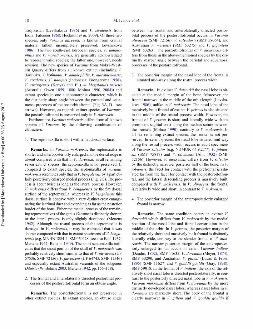
Tadjikistan (Levshakova 1986) and V. sivalensis from
India (Falconer 1868; Hocknull et al. 2009). Of these two
species, only Varanus darevskii is known from cranial
material (albeit incompletely preserved; Levshakova
1986). The two south-east European species, V. amnho-
philis and V. marathonensis, are generally acknowledged
to represent valid species; the latter one, however, needs
revision. The new species of Varanus from Mokr�a-West-
ern Quarry differs from all known extinct (including V.
darevskii, V. hofmanni, V. amnhophilis, V. marathonensis,
V. sivalensis, V. hooijeri (Indonesia; Brongersma 1958),
V. rusingensis (Kenya) and V. ( D Megalania) priscus
(Australia; Owen 1859, 1880; Molnar 1990, 2004)) and
extant species in one autapomorphic character, which is
the distinctly sharp angle between the parietal and squa-
mosal processes of the postorbitofrontal (Fig. 3A, D – see
arrows). However, as regards extinct species of Varanus,
the postorbitofrontal is preserved only in V. darevskii.
Furthermore, Varanus mokrensis differs from all known
species of Varanus by the following combination of
characters:
1. The septomaxilla is short with a flat dorsal surface.
Remarks. In Varanus mokrensis, the septomaxilla is
shorter and anteroposteriorly enlarged and the dorsal ridge is
absent compared with that in V. darevskii; in all remaining
seven extinct species, the septomaxilla is not preserved. If
compared to extant species, the septomaxilla of Varanus
mokrensis resembles only that in V. bengalensis by a particu-
larly posteriorly enlarged medial process (Fig. 2G). The pro-
cess is about twice as long as the lateral process. However,
V. mokrensis differs from V. bengalensis by the flat dorsal
surface of the septomaxilla, whereas in V. bengalensis this
dorsal surface is concave with a very distinct crest emargi-
nating the lacrimal duct and extending as far as the posterior
border of the bone. Either the medial process of the remain-
ing representatives of the genus Varanus is distinctly shorter,
or the lateral process is only slightly developed (Mertens
1942). Although the rostral process of the septomaxilla is
damaged in V. mokrensis, it may be estimated that it was
shorter compared with that in extant specimens of V. benga-
lensis (e.g. MNHN 1884-4; SMF 60428; see also Bahl 1937;
Mertens 1942; Bellairs 1949). The short septomaxilla indi-
cates that the snout portion of the skull of V. mokrensis was
probably relatively short, similar to that of V. olivaceus (UF
57536; SMF 72156), V. flavescens (UF 64743; SMF 11546)
and especially extant Australian varanids of the subgenus
Odatria (W. B€ohme 2003;Mertens 1942, pp. 156–158).
2. The frontal and anterolaterally directed postorbital pro-
cesses of the postorbitofrontal form an obtuse angle.
Remarks. The postorbitofrontal is not preserved in
other extinct species. In extant species, an obtuse angle
between the frontal and anterolaterally directed postor-
bital process of the postorbitofrontal occurs in Varanus
olivaceus (SMF 72156), V. salvadorii (SMF 58064), and
Australian V. mertensi (SMF 53275) and V. giganteus
(SMF 53263). The postorbitofrontal of V. mokrensis dif-
fers from those in the above-mentioned species by the dis-
tinctly sharper angle between the parietal and squamosal
processes of the postorbitofrontal.
3. The posterior margin of the nasal lobe of the frontal is
situated mid-way along the rostral process width.
Remarks. In extinct V. darevskii the nasal lobe is sit-
uated at the medial margin of the bone. Moreover, the
frontal narrows in the middle of the orbit length (Levsha-
kova 1986), unlike in V. mokrensis. The nasal lobe of the
massively built frontal of extinct V. priscus is also situated
in the middle of the rostral process width. However, the
frontal of V. priscus is short and laterally wide with the
prominent sagittal crest along the median suture between
the frontals (Molnar 1990), contrary to V. mokrensis. In
all six remaining extinct species, the frontal is not pre-
served. In extant species, the nasal lobe situated mid-way
along the rostral process width occurs in adult specimens
of Varanus salvator (e.g. NHMUK 64.9.2.77), V. jobien-
sis (SMF 75817) and V. olivaceus (Ahl, 1932) (SMF
72156). However, V. mokrensis differs from V. salvator
by the distinctly narrower posterior half of the bone. In V.
jobiensis, the facet for contact with the prefrontal is situ-
ated far from the facet for contact with the postorbitofron-
tal, and the lateral descending process is massively built,
compared with V. mokrensis. In V. olivaceus, the frontal
is relatively wide and short, in contrast to V. mokrensis.
4. The posterior margin of the anteroposteriorly enlarged
frontal is narrow.
Remarks. The same condition occurs in extinct V.
darevskii which differs from V. mokrensis by the medial
position of the nasal lobe and frontal constriction in the
middle of the orbit. In V. priscus, the posterior margin of
the relatively short and massively built frontal is distinctly
laterally wide, contrary to the slender frontal of V. mok-
rensis. The narrow posterior margin of the anteroposter-
iorly enlarged frontal occurs in extant Varanus indicus
(Daudin, 1802); SMF 11635, V. doreanus (Meyer, 1874);
SMF 33290, and Australian V. gilleni (Lucas & Frost,
1895) (SMF 11627) and V. gouldii gouldii (Gray, 1838);
SMF 59018. In the frontal of V. indicus, the axis of the rel-
atively short nasal lobe is directed posterolaterally, in con-
trast to the posteriorly directed nasal lobe in V. mokrensis.
Varanus mokrensis differs from V. doreanus by the more
distinctly developed nasal lobes, whereas nasal lobes in V.
doreanus are markedly short. The body of the frontal is
clearly narrower in V. gilleni and V. gouldii gouldii if
18 M. Ivanov et al.
Dow
nloa
ded
by [
Mas
aryk
ova
Uni
verz
ita v
Brn
e] a
t 00:
36 2
3 A
ugus
t 201
7

compared to V. mokrensis. The frontal of Varanus mok-
rensis is particularly similar to that of V. bengalensis
(Mertens 1942, pl. 22, pp. 160, 161; SMF 60428; MNHN
1886-649), although the body of the bone is more slender
in V. mokrensis. In V. mokrensis, the posterior margin of
the nasal lobe is situated mid-way along the rostral pro-
cess width. In adult specimens of V. bengalensis, the nasal
lobe occurs close to the lateral margin of the rostral pro-
cess (MNHN 1886-649). The posterior margin of the fron-
tal of V. mokrensis is relatively narrow, if compared with
that of V. bengalensis (MNHN 1886-649; Mertens 1942,
pl. 22, pp. 160, 161).
5. Parietal with wide, roughly rectangular fronto-post-
frontal processes, and anteriorly arched fronto-parietal
suture, and with the parietal foramen situated posteri-
orly far from the anterior margin of bone (more than
one-third of the parietal plate length).
Remarks. Regarding extinct species, the parietal is
known in V. darevskii, V. rusingensis, V. hooijeri and V.
priscus. Varanus darevskii lacks wide fronto-postfrontal
processes. Varanus rusingensis (and possibly V. hooijeri)
has developed wide fronto-postfrontal processes; how-
ever, both species differ from V. mokrensis by the much
more developed constriction of converging parietal crests.
Varanus priscus differs from V. mokrensis by its much
larger dimensions, more slender fronto-postfrontal pro-
cesses, the presence of a straight frontoparietal suture, and
the parietal foramen situated close to the anterior margin
of the bone (Molnar 1990). Regarding extant species, the
wide, roughly rectangular fronto-postorbitofrontal process
of the parietal occurs only in Varanus bengalensis (e.g.
SMF 63456), V. flavescens (e.g. SMF 11546) and V. rudi-
collis (Gray, 1845); e.g. SMF 59216. All of the above-
mentioned species possess a straight frontoparietal suture,
whereas in V. mokrensis this suture is arched anteriorly.
Varanus mokrensis further differs from V. bengalensis by
the greater distance between medially converging parietal
crests. The shortest distance between the medially con-
verging parietal crests is situated very close to the poste-
rior border of the parietal plate in V. flavescens, contrary
to V. mokrensis in which this constriction is shifted to the
posterior quarter of the parietal plate length. Moreover,
the parietal foramen is situated more anteriorly in V. fla-
vescens if compared to V. mokrensis.
6. Distance between the medially converging parietal
crests of the parietal is the shortest in the posterior
fourth of the parietal plate.
Remarks. The distance between the supratemporal
fossae (measured at the level of the narrowest portion of
the parietal), in the posterior quarter of the parietal plate,
is distinctly shorter in Varanus mokrensis in contrast to
extinct V. darevskii (Levshakova 1986, p. 104, fig. 2), but
not as short as in extinct V. rusingensis (Clos 1995,
p. 256, fig. 1A), V. hooijeri (Brongersma 1958, pl. IV/1)
and V. priscus (Molnar 1990, p. 440, fig. 2C, D). The
same distance between the medially converging parietal
crests as observed in V. mokrensis occurs in extant V. gri-
seus (Mertens 1942, pl. 22, p. 162; NMP 22180; NMP
22948; NMP 22181; MNHN 1895-366) and V. rudicollis
(SMF 59216; SMF 67586). Varanus mokrensis differs
substantially from V. griseus by the presence of a wide,
roughly rectangular fronto-postorbitofrontal process,
which is narrow in V. griseus, and by the narrower and
anteriorly arched margin of the frontoparietal suture. The
frontoparietal suture is straight in V. rudicollis.
7. Otoccipital with very distinct occipital crest at base of
the paroccipital process.
Remarks. Regarding extinct species, the otoccipital
is known only in V. amnhophilis and V. priscus. In V.
mokrensis, the paroccipital process of the otoccipital is
relatively long and massively built, with a distinctly ven-
trally enlarged distal termination which is similar to that
of extinct V. amnhophilis (Conrad et al. 2012, p. 5,
fig. 2G–I). In V. priscus, the otoccipital is rather fragmen-
tary with a broken distal termination (Head et al. 2009, p.
448, fig. 1). Both extinct species lack the distinct depres-
sion on the dorsal surface of the paroccipital process
which is conversely present in V. mokrensis. The mas-
sively built and distally enlarged paroccipital process of
the otoccipital occurs in extant Asiatic varanids of the V.
bengalensis group, in particular V. bengalensis (e.g.
MNHN 1886-649; MNHN 1884-4) and V. flavescens (e.g.
UF 64743), as well as V. salvator (Laurenti, 1768);
NHMUK 1961.1761, V. olivaceus (e.g. UF 53917) and V.
salvadorii (Peters & Doria, 1878); SMF 67670. However,
the depression at the dorsal surface of the paroccipital pro-
cess is never as distinct as in Varanus mokrensis. More-
over, the elongate and prominent posterior occipital crest
in V. mokrensis is directed distinctly lateroventrally and is
straight along most of its length. These features are not
present in extant representatives of the genus Varanus.
8. Pointed conical teeth with distally recurved apices and
prominent smooth mesial and distal cutting edges.
Remarks. The distinct and smooth mesial and distal
cutting edges in pointed teeth do not occur in extinct V.
rusingensis, V. hooijeri and V. priscus (Clos 1995; Bron-
gersma 1958; Anderson 1930; Hecht 1975) or in Varanus
sp. from Gratkorn (M. B€ohme & Vasilyan 2014). In the
remaining extinct species the teeth are unknown. As
regards extant species, the smooth cutting edges occur in
several species of subgenera Odatria Gray, 1838, Empa-
gusia Gray, 1838 and Varanus: V. (Odatria) timorensis
New early Miocene species of Varanus from the Czech Republic 19
Dow
nloa
ded
by [
Mas
aryk
ova
Uni
verz
ita v
Brn
e] a
t 00:
36 2
3 A
ugus
t 201
7

(Gray, 1831), V. (O.) tristis (Schlegel, 1839), V. (O.) ere-
mius Lucas & Frost, 1895, V. (O.) caudolineatus Bou-
lenger, 1885, V. (Empagusia) nebulosus (Gray, 1831), V.
(E.) rudicollis and V. (V.) giganteus (Gray, 1845). Vara-
nus mokrensis differs from the above-mentioned Varanus
(Odatria) species by the anteroposteriorly wider base of
the subrectangular frontal-postorbitofrontal process of the
parietal and a distinctly higher ratio of the maximum
parietal roof width to the narrowest portion of the parietal
roof. Varanus mokrensis differs from Varanus (Empagu-
sia) species in its distinctly shorter septomaxilla; more-
over, the parietal roof of V. (E.) rudicollis is laterally wide
and anteroposteriorly short relative to that of V. mokren-
sis. The new taxon differs from V. (V.) giganteus in the
shortest distance between the medially converging parie-
tal crests in the posterior quarter of the parietal plate,
whereas in V. (V.) giganteus the same constriction is situ-
ated much more anteriorly (SMF 53263).
9. Trunk vertebrae with moderately developed depression
between pars tectiformis and medial margin of prezy-
gapophyses and posterolaterally directed postzyga-
pophyses which form a right angle.
Remarks. The trunk vertebrae have distinctly smaller
dimensions compared with those of extinct Varanus amnho-
philis, V. marathonensis, V. sivalensis and V. priscus. The
precondylar constriction (81% of the condylar width, n D9) is less distinct compared with V. amnhophilis (75% in
the first and 77% in the second preserved dorsal vertebra;
Conrad et al. 2012), V. hofmanni (78%, type specimen
NMA 86-471) and V. rusingensis (78%, n D 7; Clos 1995).
Moreover, V. mokrensis exhibits postzygapophyses that are
narrow compared with the prezygapophyses, in contrast to
the conditions in V. sivalensis and V. marathonensis (Hock-
null et al. 2009; Fej�erv�ary 1918; Rage & Sen 1976). Vara-
nus mokrensis differs from extinct V. hofmanni and
Varanus sp. from Gratkorn (M. B€ohme & Vasilyan 2014)
by the less markedly developed depression between the pars
tectiformis and the medial margin of the prezygapophyses,
as well as by the posterolaterally directed postzygapophyses
which delimit a right angle. Regarding extant species, the
trunk vertebrae of V. mokrensis closely resemble those of V.
griseus (NMP 22180, 22181 NMP) in the width of pre- and
postzygapophyses and angle, which together form left and
right postzygapophysis. However, the posterior part of the
neural arch of the extant V. griseus rises less conspicuously
than that of V. mokrensis.
10. Preacetabular process of the ilium prominent and
directed at a right angle to the pubic process.
Remarks. Regarding extinct taxa, the ilium is known
only in V. rusingensis (Clos 1995, p. 256, fig. 1E) and V.
priscus (Hecht 1975, pp. 243–244). Varanus rusingensis
differs from V. mokrensis by the less massively built and
much more anteriorly inclined preacetabular process. In
V. priscus this process is very long, knob-like and oriented
horizontally, unlike in V. mokrensis. The prominent prea-
cetabular process of the ilium, which is directed antero-
dorsally rather than anteriorly and forms a right angle to
the long pubic process, is known in extant Asiatic V. ben-
galensis (MNHN 1886-649) and Australian V. varius
(White, 1790); MNHN 1851-853. Varanus bengalensis
differs from V. mokrensis in the absence of a distinct and
very long bifurcated iliac crest. In V. varius the acetabular
part of the ilium is much smaller than that of V. mokrensis.
11. Bifurcated iliac crest prominent.
Remarks. In V. rusingensis and V. priscus, the bifur-
cated iliac crest is absent. Moreover, the excavation situ-
ated on the inner surface below the iliac crest is
prominent in V. rusingensis, whereas in V. mokrensis this
excavation is shallow. Regarding extant representatives,
the distinctly developed bifurcated iliac crest is absent in
Varanus bengalensis (MNHN 1886-649). In V. varius, the
acetabular part of the ilium is much smaller compared
with V. mokrensis.
Results
Body size estimationThe average SVL/DVL for the extant Indo-Asiatic A
clade of Varanus (Conrad et al. 2012, table 1) is 36.22
(standard deviation [SD] of 2.562; y intercept [yi] D42.21; x intercept [xi] D ¡64.96; R2 D 0.994). If V. mok-
rensis is scaled similarly to other ‘Indo-Asiatic A’-clade
(Ast 2001) Varanus, we estimate (Fig. 8) its length as
374.9 mm (§ 19.94 mm with a 95% confidence interval)
based on the length of the posterior dorsal vertebra
(DVL). Based on the data concerning extant representa-
tives of Varanus (Pianka et al. 2004; Conrad et al. 2012,
table 2), V. mokrensis was a medium-sized monitor.
Phylogenetic analysisComplete lists of characters and character states are
reported in Supplemental Appendices 1, 2 and 3. We ana-
lysed 449 morphological characters coded for 87 taxa.
The coding comes from Conrad et al. (2012). Safe taxo-
nomic reduction identified six ‘unstable’ taxa, including
five species of Varanus present in the analysis of Conrad
et al. (2012), namely: B. Qarun, V. bivittatus, V. cf. mara-
thonensis (topotypic fragmentary skull where a possible
association with the holotypic vertebra needs a revision –
Rage & Sen 1976; Estes 1983; Conrad et al. 2012),
20 M. Ivanov et al.
Dow
nloa
ded
by [
Mas
aryk
ova
Uni
verz
ita v
Brn
e] a
t 00:
36 2
3 A
ugus
t 201
7

V. keithhornei (Wells & Wellington, 1985), V. maratho-
nensis (holotypic trunk vertebra – Rage & Sen 1976; Estes
1983) and V. panoptes horni (B€ohme, 1988). These taxa
were excluded from the maximum parsimony analyses.
Maximum parsimony analyses were terminated when a
maximum of 50,000 trees were obtained. The unweighted
analysis yielded hardly any resolution. The reweighted
strict consensus of 3136 trees shows very poor resolution
within the Varanus clade (Fig. 9), with the only exception
being a relatively well-supported Indo-Australian Odatria
clade. Although the topology of the majority consensus
tree derived from the reweighted analysis also showed
poor resolution (Fig. 10), it showed that most extant Afri-
can species, together with the extinct V. rusingensis from
the early Miocene (MN 3 Zone) of Kenya (Clos 1995),
belong to a well-supported (but rather unresolved) clade.
This clade forms the sister group to the extant V. rose-
nbergi (Mertens, 1957). Varanus mokrensis is a member
of a lineage that is ancestral to most African species,
including V. exanthematicus, V. ornatus, V. niloticus and
V. albigularis. As regards the results of the Bayesian anal-
yses (Figs 11, 12), the analysis that employed gamma
rates (Fig. 12) showed generally better resolution, with a
relatively well-supported Indo-Australian clade as well as
an Indo-Asiatic A clade (see Ast 2001), including a well-
supported Soterosaurus clade.
Bayesian analyses run with equal and gamma rates
show V. mokrensis to be a member of the extant represen-
tatives of the Indo-Asiatic A clade of Varanus with close
affinities to the V. bengalensis species group (Empagusia
clade; see W. B€ohme 2003 and Koch et al. 2013). The
topology of the Bayesian tree using both rate types sup-
ports the hypothesis that Miocene monitors from Mokr�aare representatives of a lineage ancestral to the extant
African varanids plus the early Miocene V. rusingensis
(Figs 11, 12). There are six well-documented trans-
Tethyan mammal dispersal phases between Eurasia and
Africa throughout the Palaeogene, with the last important
dispersal phase (from Laurasia to Africa) at the Priabo-
nian/Rupelian boundary (Gheerbrant 1987, 1990, 2001;
Gheerbrant & Rage 2006). It is possible that the first rep-
resentatives of varanids might have dispersed into Africa
from Eurasia as early as the late Eocene, i.e. long before
the initial rifting and formation of the Eritrean Red Sea in
the late Oligocene (27.5‒23.8 Ma; Bosworth et al. 2005).
This assumption is supported by the fact that most out-
group members of the genus Varanus come from Eurasia.
The oldest known varanids with possible affinity to
4 6 8 10 12 14 16 18 20
DVL
100
200
300
400
500
600
700
800DVL:SVL: y = -64.9563 + 42.2126*x; 2
(mm
)
(mm)
Varanus (Varaneades) amnhophilis
“Indo-Asian A” - clade VaranusVaranus mokrensis sp. nov.
SV
L
Figure 8. Body size estimation (snout-vent length, SVL) of Varanus mokrensis sp. nov. and Varanus amnhophilis (AMNH FR 30630)based on comparison of posterior dorsal vertebra length (DVL). The solid black trend line was calculated using extant species (see Con-rad et al. 2012, table 1) from the ‘Indo-Asiatic A’ clade of Varanus (SVL/DVL, y intercept [yi] D 42.21, x intercept [xi] D -64.96, R2 D0.996). Grey bars indicate predictive size range within 95% confidence interval; the vertical bars indicate predictive size range within95% prediction interval.
New early Miocene species of Varanus from the Czech Republic 21
Dow
nloa
ded
by [
Mas
aryk
ova
Uni
verz
ita v
Brn
e] a
t 00:
36 2
3 A
ugus
t 201
7

ScrocodilurusParasaniwaEosaniwaParvidermaHhorridumHtexanaHsuspectumParavaranusConiasaurusDolichosaurusAigialosaurusMhoffmanniPontosaurusAdriosaurusOvooYquarryLanthanotusCherminotusVtogianusVgiganteusVpanoptespanoptesVbeccariVprasinusVyuwonoiVrosenbergiVornatusVniloticusVolivaceusVhooijeriJqatraniValbigularisVcfbengalensisVrusingensisVexanthematicusVmokrensisVmytiliniVgriseusVspenceriVvariusSensidensTelmasaurusSaniwidesAiolosaurusProplatynotiaSfeistiNeucarinatus
VpriscusNcayluxi
VkomodoensisVnebulosusVmertensiVflavescensVbengalensisVkingorumVjobiensisVindicusVdoreanusVrudicollisVcumingiVglebopalmaVstorriVbaritjiVdumeriliiVacanthurusVscalarisVeremiusVpilbarensisVgouldiiVsalvadoriiVmelinusVsemiremexVtristisVtimorensisVkordensisVbrevicaudaVprimordiusVmitchelliVgilleniVsalvatorVmarmoratusVamnhophilis
Figure 9. Reweighted strict consensus tree.
22 M. Ivanov et al.
Dow
nloa
ded
by [
Mas
aryk
ova
Uni
verz
ita v
Brn
e] a
t 00:
36 2
3 A
ugus
t 201
7

ScrocodilurusParasaniwaEosaniwaParvidermaHhorridumHtexanaHsuspectumParavaranusConiasaurusDolichosaurusAigialosaurusMhoffmanniPontosaurusAdriosaurus
OvooYquarry
LanthanotusCherminotus
VtogianusVgiganteusVpanoptespanoptesVbeccariVprasinus
VyuwonoiVrosenbergi
VornatusVniloticusVolivaceusVhooijeri
Jqatrani
ValbigularisVcfbengalensis
VrusingensisVexanthematicus
VmokrensisVmytilini
VgriseusVspenceri
TelmasaurusSaniwidesAiolosaurusProplatynotia
Neucarinatus
Vpriscus
Ncayluxi
Vkomodoensis
VnebulosusVmertensiVflavescensVbengalensisVkingorumVjobiensisVindicusVdoreanus
VrudicollisVcumingi
VglebopalmaVstorriVbaritjiVdumeriliiVacanthurusVscalarisVeremiusVpilbarensisVgouldiiVsalvadoriiVmelinusVsemiremexVtristisVtimorensisVkordensisVbrevicaudaVprimordiusVmitchelliVgilleniVsalvatorVmarmoratusVamnhophilis
VvariusSensidens
Sfeisti
Figure 10. Reweighted majority consensus tree.
New early Miocene species of Varanus from the Czech Republic 23
Dow
nloa
ded
by [
Mas
aryk
ova
Uni
verz
ita v
Brn
e] a
t 00:
36 2
3 A
ugus
t 201
7

African monitors are known from isolated vertebrae from
the late Eocene Birket Qarun Formation (Holmes et al.
2010) and the early Oligocene Jebel Qatrani Formation,
both in the Fayum Province, Egypt (Varanus stem-clade,
probably closely related to Saniwa; Smith et al. 2008).
Although findings of the late Eocene varanids coincide
with nuclear DNA estimates of the origination of Varanus
(40.1 Ma; Portik & Papenfuss 2012), the phylogenetic
position of these taxa is uncertain (Figs 11, 12; Conrad
et al. 2012). It is plausible that the radiation of Eurasian
varanids into Africa continued throughout the Oligocene,
and multiple colonization events of Africa (probably via
the Arabian Peninsula) cannot be excluded. The position
of the early Miocene (»17‒18 Ma; Sabol et. al. 2007)
Shinisaurus crocodilurusParasaniwa wyomingensis
‘Varanus’ - Birket QarunHeloderma horridumHeloderma suspectum
Heloderma texanaVaranid - Yale Quarry
Lanthanotus borneensisNecrosaurus eucarinatus
Cherminotus longifronsOvoo gurvel
Varanus baritjiNecrosaurus cayluxi
Saniwa feistiTelmasaurus grangeri
Varanus melinusVaranus storri
Eosaniwa koehniVaranus timorensis
Proplatynotia longirostrataParavaranus angustifrons
Saniwides mongoliensisVaranus primordius
Parviderma inexacta
Varanus amnhophilis
Aiolosaurus oriensVaranus mitchelliVaranus glebopalma
Varanus scalarisSaniwa ensidensVaranus tristis
Varanus semiremexVaranus cumingiVaranus marmoratus
Dolichosaurus longicollis
Varanus salvatorVaranus prasinus
Varanus kordensisVaranus beccari
Varanus kingorumVaranus doreanus
Varanus jobiensisVaranus mertensi
Varanus brevicaudaVaranus marathonensiscf.
Coniasaurus crassidens
Aigialosaurus dalmaticusVaranus panoptes panoptes
Varanus togianus
Varanus giganteus
Mosasaurus hoffmanni
Adriosaurus suessiPontosaurus lesinensis
1.00
1.00
1.001.00
1.00
1.00
1.00
Varanus griseus
Varanus gouldiiVaranus pilbarensis
Varanus salvadoriiVaranus komodoensis
Varanus priscusVaranus varius
Varanus eremiusVaranus gilleni
Varanus mytiliniVaranus bivittatus
Varanus acanthurusVaranus dumerilii
Varanus marathonensisVaranus nebulosus
Varanus flavescensVaranus indicus
Varanus rudicollisVaranus bengalensis
Varanus hooijeri
Varanus exanthematicusVaranus rusingensis
Varanus niloticus
Varanus panoptes horni
Varanus ornatusVaranus albigularis‘Saniwa’ - Jebel Qatrani
Varanus olivaceusVaranus spenceri
Varanus rosenbergi
Varanus bengalensiscf.Varanus yuwonoi
Varanus keithhornei
1.001.00
Varanus mokrensis
Figure 11. The majority rule of consensus topology used in the Bayesian analysis (without constraints). Bold – fossil varanids;y – extinct Varanus species; black dots – posterior probability (PP) 100%, remaining nodes 50%.
24 M. Ivanov et al.
Dow
nloa
ded
by [
Mas
aryk
ova
Uni
verz
ita v
Brn
e] a
t 00:
36 2
3 A
ugus
t 201
7
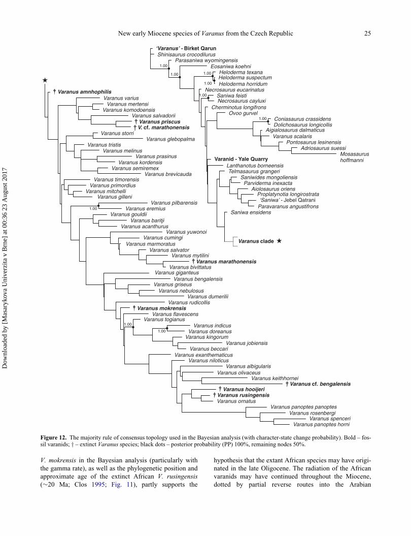
V. mokrensis in the Bayesian analysis (particularly with
the gamma rate), as well as the phylogenetic position and
approximate age of the extinct African V. rusingensis
(»20 Ma; Clos 1995; Fig. 11), partly supports the
hypothesis that the extant African species may have origi-
nated in the late Oligocene. The radiation of the African
varanids may have continued throughout the Miocene,
dotted by partial reverse routes into the Arabian
Varanus amnhophilis
Varanus mitchelli
Varanus tristis
Varanus gilleni
Varanus primordiusVaranus timorensis
Varanus melinus
Varanus storri
Varanus komodoensis
Varanus varius
Varanus acanthurus
Varanus gouldii
Varanus semiremexVaranus kordensis
Varanus mertensi
Varanus baritji
Varanus mokrensis
Varanus marmoratus
Varanus eremius
Varanus prasinus
V. marathonensiscf.Varanus priscus
Varanus salvadorii
Varanus togianusVaranus flavescens
Varanus griseus
Varanus giganteus
Varanus salvator
Varanus cumingi
Varanus pilbarensis
Varanus brevicauda
Varanus glebopalma
Varanus rudicollis
Varanus nebulosus
Varanus bivittatus
Varanus mytilini
Varanus yuwonoi
Varanus niloticusVaranus exanthematicus
Varanus beccari
Varanus kingorumVaranus doreanus
Varanus bengalensis
Varanus dumerilii
Varanus indicus
Varanus marathonensis
Varanus olivaceus
Varanus hooijeri
Varanus ornatusVaranus rusingensis
Varanus albigularis
Varanus jobiensis
Varanus keithhornei
Varanus panoptes panoptes
Varanus bengalensiscf.
Varanus rosenbergi
Varanus panoptes horniVaranus spenceri
1.00
1.00
1.00
‘Varanus’ - Birket QarunShinisaurus crocodilurus
Parasaniwa wyomingensis
Necrosaurus eucarinatus
Eosaniwa koehni
Cherminotus longifrons
Varanid - Yale Quarry
Necrosaurus cayluxiSaniwa feisti
Heloderma horridumHeloderma suspectumHeloderma texana
Telmasaurus grangeriLanthanotus borneensis
Ovoo gurvel
Saniwa ensidens
Saniwides mongoliensisParviderma inexacta
Aiolosaurus oriensProplatynotia longirostrata
Paravaranus angustifrons‘Saniwa’ - Jebel Qatrani
Aigialosaurus dalmaticusVaranus scalaris
Dolichosaurus longicollisConiasaurus crassidens
Pontosaurus lesinensisAdriosaurus suessi
Mosasaurus
1.00
1.00 1.00
1.00
1.00
1.00
hoffmanni
Varanus clade
Figure 12. The majority rule of consensus topology used in the Bayesian analysis (with character-state change probability). Bold – fos-sil varanids; y – extinct Varanus species; black dots – posterior probability (PP) 100%, remaining nodes 50%.
New early Miocene species of Varanus from the Czech Republic 25
Dow
nloa
ded
by [
Mas
aryk
ova
Uni
verz
ita v
Brn
e] a
t 00:
36 2
3 A
ugus
t 201
7

Peninsula, as suggested by the position of V. yemenensis
(B€ohme, Joger & Sch€atti, 1989; Portik & Papenfuss
2012). However, the Bayesian analysis with gamma rates,
where the extinct V. rusingensis occurs in a terminal posi-
tion (Fig. 12), reveals that the diversification of the Afri-
can varanid clade may have been completed as early as
the early Miocene. In addition, the dispersal of North
African varanids into Iberia is unlikely, given that the
accurately dated North African-Iberian dispersal of mam-
mals (6.2 Ma) via ephemeral land bridges in the Strait of
Gibraltar occurred long before the onset of the Messinian
Salinity Crisis (5.59 Ma) resulting from the latest Miocene
glaciation at 6.26 Ma (Gibert et al. 2013). The only known
late middle Miocene varanid, an as yet unpublished Vara-
nus from the Vall�es-Pened�es Basin in Catalonia (Spain)
possibly referable to Varanus marathonensis (Delfino
et al. 2011), is most probably of Eurasiatic origin, as is V.
amnhophilis reported from the late Miocene (7.6‒6.9 Ma)
of Samos, Greece (Fig. 10; Conrad et al. 2012).
Discussion
PalaeoclimateThe amphibian and reptile assemblage from the 1/2001
Turtle Joint and 2/2003 Reptile Joint is rather diverse
(Ivanov et al. 2006; Ivanov 2008, 2009). Based on M.
B€ohme et al. (2006), different ecophysiological groups
occur in the early Miocene of Mokr�a-Western Quarry
(Supplemental Table 1).
The herpetofauna of the Mokr�a-Western Quarry corre-
sponds to the warm humid phase of the MCO (Miocene
Climatic Optimum sensu M. B€ohme 2003) during the
early Miocene when mean annual temperatures (MAT) in
Central Europe reached about 20�C. In the North Alpine
Foreland Basin (NAFB), the occurrence of cordylid liz-
ards indicates MAT > 17.4�C (M. B€ohme 2003). Simi-
larly, the presence of boid snakes with Bavarioboa cf.
hermi Szyndlar & Schleich, 1993 in the southern part of
the Moravian Karst (1/2001 Turtle Joint; 2/2003 Reptile
Joint) also indicates a rather high MAT. The large boid
snake from Mokr�a-Western Quarry (Boidae gen. et sp.
indet., large form), probably belonging to Pythonidae, is
important because Central European representatives of
Pythonidae Fitzinger, 1826 are known so far only from
Griesbeckerzell, Germany (late MN 5 and base of MN 6).
Pythons represent the most thermophilic members of the
Squamata in the Miocene of Europe, and document
MAT > 18.6�C (Ivanov & B€ohme 2011).
Regarding humidity, there is a difference in the mean
annual precipitation (MAP) between the two karst fis-
sures. While the assemblage composition in the 1/2001
Turtle Joint indicates MAP D 461 § 253 mm (Supple-
mental Table 1), the assemblage in the 2/2003 Reptile
Joint indicates MAP D 864 § 254 mm. The amphibian
and reptile assemblage of the 1/2001 Turtle Joint, where
heliophobic taxa are absent altogether, developed in a rel-
atively drier phase of the MCO when MAP values were
comparable with today’s long-term average value for this
area (MAP D 490.1 mm – www.chmi.cz).
PalaeoenvironmentRemains of fully aquatic amphibians in the Karst fissures
suggest the possible presence of nearby lacustrine envi-
ronments. The absence of ostracods and the scarcity of
gastropods indicate oligotrophic to weakly mesotrophic
conditions (Ivanov et al. 2006). The diverse amphibian
fauna (Supplemental Table 1; Ivanov et al. 2006; Ivanov
2008) includesMioproteus sp., Triturus aff. roehrsi Herre,
1955, Triturus cf. marmoratus (Latreille, 1800), Triturus
sp., Chioglossa meini Estes & Hoffstetter, 1976, Merten-
siella mera Hodrov�a, 1984,Mertensiella sp., and Rana sp.
(synklepton R. esculenta Linnaeus, 1758). Among
amniotes are turtles of the families Testudinidae Batsch,
1788 and Geoemydidae Theobald, 1868 (Testudo sp., Pty-
chogaster sp.; �A. H. Luj�an, pers. comm.), and squamate
reptiles (Natrix sp.). This assemblage indicates the pres-
ence of damp and marshy habitats with stagnant or slow-
flowing waters in both localities of the Mokr�a-Western
Quarry. However, species adapted to more humid terres-
trial environments are also recorded, such as Chelotriton
sp. whose adults probably inhabited damp habitats with
dense vegetation (Estes 1981). This is inferred from the
known habitat preferences of the closely related extant
Tylototriton Anderson, 1871 which usually inhabits
humid, high mountain areas with dense vegetation,
although adults of this species are known to cover distan-
ces of several tens of metres away from water reservoirs
(Kuzmin et al. 1994). Although the development of
amphibians is partly associated with aquatic habitats, the
extinct pelobatid frog Pelobates sanchizi Venczel, 2004
was probably adapted to the dry sandy soil and well-
drained substrates within inundation areas (Venczel 2004;
M. B€ohme & Vasilyan 2014). Bufo sp. is also adapted to
drier conditions. Squamates belong to semiaquatic/helio-
philic forms (Natrix sp.) and to heliophilic, fossorial or
terrestrial forms, such as cf. Blanus sp., Varanus sp.,
Pseudopus sp., Lacerta sp., type I, Lacerta sp., Bavario-
boa cf. hermi, and ‘Coluber’ sp., type I, and Vipera sp.
(‘Oriental vipers’ and ‘European vipers’). Although the
fillings of both karst fissures are approximately coeval
(Ivanov et al. 2006), the scarcity or absence of certain
aquatic or heliophobic forms of amphibians at the site 1/
2001 Turtle Joint (rare occurrence of the genus Triturus
Rafinesque, 1815; the absence of the genera Chioglossa
Bocage, 1864 and Mertensiella Wolterstorff, 1925) indi-
cates drier conditions (Ivanov 2008). This assumption is
confirmed by the relatively higher proportion of ‘Oriental
vipers’ in the faunal assemblage from this fissure.
26 M. Ivanov et al.
Dow
nloa
ded
by [
Mas
aryk
ova
Uni
verz
ita v
Brn
e] a
t 00:
36 2
3 A
ugus
t 201
7
The conclusions of our study are fully consistent with
results of analyses of medium- and small-sized mammals
(Ivanov et al. 2006; Sabol et al. 2007), the occurrence of
which indicates a warm climate with a diverse mosaic of
vegetation cover, possibly indicating open steppe and
open forest environments with a system of rivers and
swamps (Sabol et al. 2007).
Acknowledgements
We thank Drs D. K€ohler (Senckenberg Forschungsinstitut
und Naturmuseum Frankfurt), D. Gower (Natural History
Museum, London), A. Martin (Mus�eum national d’Histoire
naturelle, Paris) and J. Moravec (N�arodn�ı museum, Prague)
and Professors F. W. King (Florida Museum of Natural
History, Gainesville) and Z. Szyndlar (Instytut Systematyki
i Ewolucji Zwierzat Polskiej Akademii Nauk, Krak�ow) foraccess to specimens in their care. We also thank two anon-
ymous reviewers and the Associate Editor for their con-
structive criticism. This project was supported by the
SYNTHESYS GB-TAF-4392 (MI), the Specific research
project in the Faculty of Science at Masaryk University,
Brno (MI) and the Slovak Research and Development
Agency Grant No. 16-0180-1 (JK).
Supplemental data
Supplemental material for this article can be accessed at:
https://doi.org/10.1080/14772019.2017.1355338
References
Ahl, E. 1932. Eine neue Eidechse und zwei neue Fr€osche vonderInsel Jobi. Mitteilungen aus dem Zoologischen Museum inBerlin, 17, 892–899.
Alf�erez Delgado, F. & Brea L�opez, P. 1981. Estudio preliminarde los restos de peces, anfibos y reptiles del yacimento deCorcoles (Guadalajara). Bolet�ın de la Real Sociedad Espa-~nola de Historia Natural, Secci�on Geol�ogica, 79, 5–20.
Anderson, C. 1930. Meiolania platyceps Owen and Varanus(Megalania) priscus (Owen). Records of the AustralianMuseum, 17, 309–316.
Anderson, J. 1871. Description of a new genus of newts fromwestern Yunan. Proceedings of the Zoological Society ofLondon, 1871, 423–425.
Antunes, M. T. & Mein, P. 1981. Vert�ebr�es du mioc�ene moyende Amor (Leiria) – importance stratigraphique. Ciencias daTerra, 6, 169–188.
Antunes, M. T. & Rage, J.-C. 1974. Notes sur la g�eologie et lapal�eontologie du Mioc�ene de Lisbonne. XIV: quelquesSquamata (Reptilia). Boletim da Sociedade Geol�ogica dePortugal, 19, 47–60.
Archibald, J. K., Mort, M. E. & Crawford, D. J. 2003. Bayes-ian inference of phylogeny: a non-technical primer. Taxon,52, 187‒191.
Ast, J. 2001. Mitochondrial DNA evidence and evolution inVaranoidea (Squamata). Cladistics, 17, 211–226.
Bahl, K. N. 1937. Skull of Varanus monitor (Linn.). Records ofthe Indian Museum, 39, 133‒174.
Batsch, A. J. C. G. 1788. Versucheiner Anleitung, zur Kenntnißund Geschichte der Thiere und Mineralien, f€ur akademischeVorlesungen entworfen, und mit den no€thigsten Abbildungenversehen. Erster Theil. Allgemeine Geschichte der Natur;besondre der Sa€ugthiere, Vo€gel, Amphibien und Fische.Akademische Buchhandlung, Jena.
Baur, G. 1890. On the characters and systematic position of thelarge sea lizards, Mosasauridae. Science, 16, 1–262.
Bellairs, A. d’A. 1949. Observations of the snout of Varanus,and a comparison with that of other lizards and snakes. Jour-nal of Anatomy, 83, 116–146.
Bellairs, A. d’A. & Kamal, A. M. 1981. The chondrocraniumand the development of the skull in recent reptiles. Pp. 1–263 in C. Gans & T. S. Parsons (eds) Biology of the Reptilia11. Academic Press, London.
Bocage, J. V. B. du. 1864. Note sur la D�ecouverte d’un Zoo-phyte de la familie Hyalochaetides sur la cote du Portugal.Proceedings of the Zoological Society of London, 1864,265–269.
B€ohme, M. 2001. The oldest representative of a brown frog(Ranidae) from the early Miocene of Germany. Acta Palae-ontologica Polonica, 46, 119–124.
B€ohme, M. 2002. Palaeoclimate and aquatic ecosystems in theNeogene of Europe – new research approaches based onectothermic vertebrates. Unpublished habilitation thesis,Department of Geo- and Environmental Science, Ludwig-Maximillians University, Munich. [In German.]
B€ohme, M. 2003. The Miocene Climatic Optimum: evidence fromthe ectothermic vertebrates of Central Europe. Palaeogeogra-phy, Palaeoclimatology, Palaeoecology, 195, 389–401.
B€ohme, M. & Ilg, A. 2003. FosFARbase. Updated at: www.wahre-staerke.com/, accessed 25 August 2016.
B€ohme, M. & Vasilyan, D. 2014. Ectothermic vertebrates fromthe late Middle Miocene of Gratkorn (Austria, Styria).Palaeobiodiversity and Palaeoenvironments, 94, 21–40.
B€ohme, M., Ilg, A, Ossig, A. & K€uchenhoff, H. 2006. Newmethod to estimate paleoprecipitation using fossil amphib-ians and reptiles and the middle and late Miocene precipita-tion gradients in Europe. Geology, 34, 425–428.
B€ohme, W. 1988. Der Arguswaran (Varanus panoptes Storr,1980) auf Neuguinea: Varanus panoptes horni spp. n. Sala-mandra, 24(2/3), 87–101.
B€ohme, W. 2003. Checklist of the living monitor lizards of theworld (family Varanidae). Zoologische Verhandlungen, 341(25), 4–43.
B€ohme, W., Joger, U. & Sch€atti, B. 1989. A new monitor lizard(Reptilia: Varanidae) from Yemen, with notes on ecology,phylogeny, and zoology. Fauna of Saudi Arabia, 10, 433–448.
Bolkay, St. J. 1913. Additions to the fossil herpetology of Hun-gary from the Pannonian and Praeglacial periode. Mitteilun-gen aus dem Jahrbuch der ko€niglichen ungarischenGeologischen Reichsanstalt, 21, 217–230.
Borsuk-Bia»ynicka, M. 1984: Anguiomorphans and related liz-ards from the Late Cretaceous of the Gobi Desert, Mongolia.Palaeontologica Polonica, 46, 5–105.
Bosc, L. 1792. Lacerta exanthematica. Actes de la Soci�et�ed’Histoire naturelle de la Paris, 1, 25.
Bosworth, W., Huchon, P. & McClay, K. 2005. The Red Seaand Gulf of Aden Basins. Journal of African Earth Sciences,43, 334–378.
New early Miocene species of Varanus from the Czech Republic 27
Dow
nloa
ded
by [
Mas
aryk
ova
Uni
verz
ita v
Brn
e] a
t 00:
36 2
3 A
ugus
t 201
7

Boulenger, G. A. 1885. Catalogue of the lizards in the BritishMuseum (Natural History). Vol. 2, 2nd edition, Printed byorder of the Trustees, London, xiii C 497 pp.
Brongersma, L. D. 1958. On an extinct species of the genusVaranus (Reptilia, Sauria) from the island of Flores. Zoo-logische Mededelingen, 36(7), 113–125.
Carroll, R. L. 1993. Pala€ontologie und Evolution der Wirbel-tiere. Georg Thieme Verlag, Stuttgart, 684 pp.
Clos, L. M. 1995. A new species of Varanus (Reptilia: Sauria)from the Miocene of Kenya. Journal of Vertebrate Paleon-tology, 15, 254–267.
Colombero, S., Angelone, C., Bonelli, E., Carnevale, G., Cav-allo, O., Delfino, M., Giuntelli, P., Mazza, P., Pavia, G.,Pavia, M. & Repetto, G. 2014. The Messinian vertebrateassemblages of Verduno (NW Italy): another brick for a lat-est Miocene bridge across the Mediterranean. Neues Jahr-buch f€ur Geologie und Pala€ontologie, Monatschefte, 272,287–234.
Conrad, J. L. 2004. Skull, mandible, and hyoid of Shinisauruscrocodilurus AHL (Squamata, Anguiomorpha). ZoologicalJournal of the Linnean Society, 141, 399–434.
Conrad, J. L. 2006. Postcranial skeleton of Shinisaurus crocodi-lurus (Squamata: Anguiomorpha). Journal of Morphology,267, 759–775.
Conrad, J. L, Balcarcel, A. M. & Mehling, C. M. 2012. Earli-est example of a giant monitor lizard (Varanus, Varanidae,Squamata). Plos One, 7(8), e41767.
Daudin, F. M. 1802. Histoire naturelle, g�en�erale et particuliCredes Reptiles, ouvrage faisant suite, a l’histoire naturelle,g�en�erale et particuli�ere compos�ee par Leclerc de Buffon, etredi�ee par C. S. Sonnini, vol. 3., F. Dufart, Paris, 426 pp.
Daudin, F. M. 1803. Histoire Naturelle Generale et Particulieredes Reptiles. Vol. 6. F. Dufart, Paris, 108 pp.
Delfino, M., Abella J., S�anchez, I. & Alba, D. M. 2013a. Anearly complete Varanus skeleton from the late Miocene ofCerro de los Batallones (Madrid Basin). P. 33 in Abstracts of6th Annual Meeting of the European Association of Verte-brate Palaeontologists. Villers-sur-Mer, France, 10–15 June2013.
Delfino, M., Rage, J-C., Bolet, A. & Alba, D. M. 2013b. EarlyMiocene dispersal of the lizard Varanus into Europe: reas-sessment of vertebral material from Spain. Acta Palaeonto-logica Polonica, 58, 731–735.
Delfino, M., Alba, D. M., Carmona, R., Luj�an, �A. H. &Robles, J. M. 2011. European monitor lizards (Anguimor-pha, Varanidae, Varanus): new materials and new perspec-tives. Abstracts of the 71st Annual Meeting Society ofVertebrate Paleontology, Paris Las Vegas, Las Vegas,Nevada, USA, November 2–5, 2011. Journal of VertebratePaleontology, 31(Suppl. 2), 97–98.
Estes, R. 1981. Gymnophiona, Caudata. Part 2. Pp. 1–113 in P.Wellnhofer (ed.) Handbuch der Pala€oherpetologie 2 (Ency-clopedia of paleoherpetology 2). Gustav Fischer Verlag,Stuttgart.
Estes, R. 1983. Sauria Terrestria. Part 10A. Pp. 1–249 in P.Wellnhofer (ed.) Handbuch der Pala€oherpetologie 10A(Encyclopedia of paleoherpetology 10A). Gustav FischerVerlag, Stuttgart.
Estes, R., de Queiroz, K. & Gauthier, J. 1988. Phylogeneticrelationships within Squamata. Pp. 119–281 in R. Estes &G. Pregill (eds) Phylogenetic relationships of the lizard fam-ilies. Stanford University Press, Stanford.
Estes, R. & Hoffstetter, R. 1976. Les Urod�eles du Mioc�ene deLa Grive-Saint-Alban (Is�ere, France). Bulletin du Mus�eumnational d’histoire naturelle, s�er. 3, 398, 297–343.
Falconer, H. 1868. On fossil species of Emys from the SewalikHills, referable to the existing Emys tecta (Bell). Pp. 382–388 in C. Murchison (ed.) Paleontological memoirs andnotes of the late Hugh Falconer, AM, MD. Volume 1: FaunaAntiqua Sivalensis. Robert Hardwicke Publishers, London.
Fej�erv�ary, G. J. de. 1918. Contributions to a monography onfossil Varanidae and on Megalanidae. Annales Musei Natio-nalis Hungarici, 15, 341–467.
Fej�erv�ary, G. J. de. 1935. Further contributions to a monographof the Megalanidae and fossil Varanidae – with notes onRecent Varanians. Annales Musei Nationalis Hungarici, 29,1–130.
Fej�erv�ary-L�angh, A. M. 1923. Beitr€age zu einer monographieder fossilen Ophisaurier. Palaeontologia Hungarica, 1(7),123–220.
Fitzinger, L. 1826. Neue Classification der Reptilien nach ihrennat€urlichen Verwandtschaften nebst einer Verwandschafts-Tafel und einem Verzeichnisse der Reptilien-Sammlung desK. K. Zoologischen Museums zu Wien. J. G. Heubner, Wien,66 pp.
Fuller, S., Baverstock, P. & King, D. 1998. Biogeographic ori-gins of goannas (Varanidae): a molecular perspective.Molecular Phylogenetics and Evolution, 9, 294–307.
Gao, K. & Norell, M. A. 1998. Taxonomic revision of Carusia(Reptilia: Squamata) from the Late Cretaceous of the GobiDesert and phylogenetic relationships of anguiomorphan liz-ards. American Museum Novitates, 3230, 1–51.
Gao, K. & Norell, M. A. 2000. Taxonomic composition andsystematics of Late Cretaceous lizard assemblages fromUkhaa Tolgod and adjacent localities, Mongolian Gobi Des-ert. Bulletin of the American Museum of Natural History,249, 1–118.
Georgalis, G., Villa, A. & Delfino, M. 2017. The last Europeanvaranid: demise and extinction of monitor lizards (Squa-mata, Varanidae) from Europe. Journal of Vertebrate Pale-ontology, doi: 10.1080/02724634.2017.1301946
Gheerbrant, E. 1987. Les Vert�ebr�es continentaux de l’AdrarMgorn (Maroc, Pal�eoc�ene); une dispersion de mammif�erestranst�ethysienne aux environs de la limite Mesozo€ıque/C�enozo€ıque? Geodinamica Acta, 1, 233–245.
Gheerbrant, E. 1990. On the early biogeographical history ofthe African placentals. Historical Biology, 4, 107–116.
Gheerbrant, E. 2001. Histoire pal�eobiog�eographique desmammif�eres pal�eog�enes du domaine arabo-africain. HDRthesis, University of Paris VI.
Gheerbrant, E. & Rage, J.-C. 2006. Paleobiogeography ofAfrica: how distinct from Gondwana and Laurasia? Palaeo-geography, Palaeoclimatology, Palaeoecology, 241, 224–246.
Gibert, L., Scott, G. R., Montoya, P., Ruiz-S�anchez, F. J.,Morales, J., Luque, L., Abella, J. & Leria, M. 2013. Evi-dence for an African-Iberian mammal dispersal during thepre-evaporitic Messinian. Geology, 41, 691–694.
Gilmore, C. W. 1922. A new description of Saniwa ensidensLeidy, an extinct varanid lizard from Wyoming. Proceed-ings of the United States National Museum, 60, 1–28.
Gilmore, C. W. 1928. Fossil lizards of North America.Memoirsof the National Academy of Sciences, 22, 1–201.
Goloboff, P. A. 1993. Estimating character weights during treesearch. Cladistics, 9, 83–91.
Goloboff, P. A., Farris, J. S. & Nixon, K. C. 2008. TNT, a freeprogram for phylogenetic analysis. Cladistics, 24, 1–13.
Gray, J. E. 1831. A Synopsis of the species of the class Reptilia.Pp. 1–29 in E. Griffith (ed.), The Animal Kingdom, Vol. 9,Whittaker, Treacher & Comp., London.
28 M. Ivanov et al.
Dow
nloa
ded
by [
Mas
aryk
ova
Uni
verz
ita v
Brn
e] a
t 00:
36 2
3 A
ugus
t 201
7

Gray, J. E. 1837. General arrangement of the Reptilia. Proceed-ings of the Zoological Society of London, 1837(5), 131–132.
Gray, J. E. 1838. Catalogue of the slender-tongued saurians,with description of many new genera and species. Annals ofNatural History, 1, 274–283, 388–394.
Gray, J. E. 1845. Catalogue of the specimens of lizards in thecollection of the British Museum. Trustees of the BritishMuseum/Edward Newman, London, xxvii C 289 pp.
Hallowell, E. 1857. Notes on the reptiles in the collection of themuseum of the Academy of Natural Sciences. Proceedingsof the Academy of Natural Sciences of Philadelphia, 8(4),146–153.
Hardwicke, F. R. & Gray, J. E. 1827. A synopsis of the speciesof saurian reptiles, collected in India by Major-GeneralHardwicke. Zoological Journal, London, 3, 214–229.
Head, J. J., Barrett Fls, P. M. & Rayfield, E. J. 2009.Neurocranial osteology and systematic relationships of Var-anus (Megalania) prisca Owen, 1859 (Squamata: Varani-dae). Zoological Journal of the Linnean Society, 155, 445–457.
Hecht, M. K. 1975. The morphology and relationships of thelargest known terrestrial lizard, Megalania prisca Owen,from the Pleistocene of Australia. Proceedings of the RoyalSociety of Victoria, 87, 239–250.
H�ır, J. & Venczel, M. 2005. New middle Miocene vertebratelocalities from Subpiatr�a (Bihor District, Romania). ActaPalaeontologica Romaniae, 5, 211–221.
Hocknull, S. A., Piper, P. J., van den Bergh, G. D., Due, R. A.,Morwood, M. J. & Kurniawan, I. 2009. Dragon’s paradiselost: palaeobiogeography, evolution and extinction of the larg-est-ever terrestrial lizards (Varanidae).PLoSONE, 4(9), e7241.
Hodrov�a, M. 1984. Salamandridae of the Upper Pliocene Iva-novce locality (Czechoslovakia). Acta Universitatis Caroli-nae, Geologica, 4, 331–352.
Hoffstetter, R. 1969. Pr�esence de Varanidae (Reptilia, Sauria)dans le Mioc�ene de Catalogne. Considerations sur l’histoirede la familie. Bulletin du Mus�eum National d’HistoireNaturelle, 40, 1051–1064.
Hoffstetter, R. & Gasc, J.-P. 1969. Vertebrae and ribs of mod-ern reptiles. Pp. 200–310 in C. Gans, A. d’A. Bellairs & T.S. Parsons (eds) Biology of the Reptilia. Volume 1, Morphol-ogy A. Academic Press, New York.
Holmes, R. B., Murray, A. M., Attia, Y. S., Simons, E. L. &Chatrath, P. 2010. Oldest known Varanus (Squamata: Var-anidae) from the Upper Eocene and Lower Oligocene ofEgypt: support for an African origin of the genus. Palaeon-tology, 53, 1099–1110.
Huelsenbeck, J. P. & Ronquist, F. 2001. MRBAYES. Bayesianinference of phylogenetic trees. Bioinformatics, 17, 754–755.
Ivanov, M. 2008. Early Miocene amphibians (Caudata, Salien-tia) from the Mokr�a-Western Quarry (Czech Republic) withcomments on the evolution of Early Miocene amphibianassemblages in Central Europe. Geobios, 41, 465–492.
Ivanov, M. 2009. Evolution of Neogene and Quaternary squa-mate assemblages (Diapsida: Squamata) in Central Europe.Unpublished Associate Professor thesis, Masaryk Univer-sity, 319 pp. [In Czech.]
Ivanov, M. & B€ohme, M. 2011. Snakes from Griesbeckerzell(Langhian, Early Badenian), North Alpine Foreland Basin(Germany), with comments on the evolution of snake faunain Central Europe during the Miocene Climatic Optimum.Geodiversitas, 33, 411–449.
Ivanov, M., Musil, R. & Brzobohat�y, R. 2006. Terrestrial andmarine faunas from the Miocene deposits of the Mokr�a
Plateau (Drahany Upland, Czech Republic) – impact onpalaeogeography. Beitra€ge zur Pala€ontologie, 30, 223–239.
Jollie, M. J. 1960. The head skeleton of the lizard. Acta Zoolog-ica, Stockholm, 41, 1–64.
Koch, A., Aulyia, M. & Ziegler, T. 2010. Updated checklist ofthe living monitor lizards of the world (Squamata: Varani-dae). Bonn Zoological Bulletin, 57(2), 127–136.
Koch, A., Ziegler, T., B€ohme, W., Arida, E. & Auliya, M.2013. Pressing Problems: Distribution, threats, and conser-vation status of the monitor lizards (Varanidae: Varanusspp.) of Southeast Asia and the Indo-Australian Archipelago.Herpetological Conservation and Biology, 8(Monograph 3),1–62.
Kuzmin, S. L., Dasgupta, R. & Smirina, �E. M. 1994. Ecology ofthe Himalayan newt (Tylototriton verrucosus) in DarjeelingHimalayas, India.Russian Journal of Herpetology, 1(1), 69–76.
Latreille, P. A. 1800. Histoire naturelle des salamandres deFrance: pr�ec�ed�ee d’un tableau m�ethodique des autres rep-tiles. Villier, Paris, 61 pp.
Laurenti, J. N. 1768. Austriaci Viennensis specimen medicum,exhibens synopsin reptilium emendatam cum experimentiscirca Venena et antidota reptilium Austriacorum quodauthoritate et consensu. J. Thomae, Caes. Reg. Aulae Typo-gographi, Wien, 217 pp.
Lee, M. S. Y. 1997. The phylogeny of varanoid lizards and theaffinities of snakes. Philosophical Transactions of the RoyalSociety of London, Series B, 352, 53–91.
Leidy, J. 1870. Descriptions of Emys jeansesi, E. haydeni,Ba€ena arenosa, and Saniwa ensidens. Proceedings of theAcademy of Natural Sciences of Philadelphia, 1870, 123–124.
Levshakova, I. J. 1986. A new Varanus from lower Pliocene ofTajikistan. Trudy Zoologicheskogo Instituta Akademii NaukSSSR, 157, 101–116. [In Russian.]
Linnaeus, C. 1758. Systema naturæ per regna tria naturæ,secundum classes, ordines, genera, species, cum characteri-bus, differentiis, synonymis, locis. Tomus I. Editio decima,reformata, Laurentii Salvii, Holmiæ, 824 pp.
Linnaeus, C. 1766. Systema Naturae per Regna Tria Naturae,Secundum Classes, Ordines, Genera, Species, cum Charac-teribus, Differentiis, Synonymis, Locis. 12th edition, Salvius,Holmiæ, Sweden, 122 pp.
Lloyd, G. T. 2015. Claddis: measuring morphological diversity andevolutionary tempo. R package version 0.1. Updated at: https://github.com/graemetlloyd/Claddis, accessed 12 October 2015.
Lucas, A. H. S. & Frost, C. 1895. Preliminary notice of certainnew species of Lizards from central Australia. Proceedingsof the Royal Society of Victoria, 7, 264–269.
Lungu, A. N., Zerova, G. A. & Chkhikvadze, V. M. 1983. Pri-mary evidence on the Miocene Varanus of the North BlackSea littoral. Sovshcheniia Akademii Nauk Gruzinskoi SSR,110, 417–420. [In Russian.]
McDowell, S. B. Jr. & Bogert, C. M. 1954. The systematicposition of Lanthanotus and the affinities of the anguinomor-phan lizards. Bulletin of the American Museum of NaturalHistory, 105, 1–142.
Merrem, B. 1820. Versuch eines Systems der Amphibien/Tenta-men systematis amphibiorum. J. C. Krieger, Marburg, 191 pp.
Mertens, R. 1942. Die Familie der Warane (Varanidae). ZweiterTeil: Der Sch€adel. Abhandlungen der SenckenbergischenNaturforschenden Gesellschaft, 465, 117–234.
Mertens, R. 1957. Ein neuer melanitischer Waran aus dems€udlichen Australien (V. gouldii rosenbergi, subsp. nov.).Zoologischer Anzeiger, 159, 17–20.
New early Miocene species of Varanus from the Czech Republic 29
Dow
nloa
ded
by [
Mas
aryk
ova
Uni
verz
ita v
Brn
e] a
t 00:
36 2
3 A
ugus
t 201
7

Meyer, A. B. 1874. Eine Mittheilung von Hrn. Dr. Adolf Meyer€uber die von ihm auf Neu-Guinea un den Inseln Jobi,Mysore und Mafoor im Jahre 1873 gesammelten Amphib-ien. Monatsberichte der Ko€niglichen Preussische Akademiedes Wissenschaften zu Berlin, 1874, 128–140.
Molnar, R. E. 1990. New cranial elements of a giant varanidfrom Queensland. Memoirs of the Queensland Museum, 29,437–444.
Molnar, R. E. 2004. The long and honourable history of moni-tors and their kin. Pp. 10–67 in E. R. Pianka, D. R. King &R. A. King (eds) Varanoid lizards of the world. Indiana Uni-versity Press, Bloomington.
M€unster, G. L. F. W. 1834. Petrefacta Germaniae tam ea: quaein Museo Universitatis Regiae Borussicae Fridericiae Wil-helmiae Rhenanae servantur quam alia quaecunque inMuseis Hoeninghusiano Muensteriano aliisque extant, icon-ibus et descriptionibus illustrata (Abbildungen und Bes-chreibungen der Petrefacten Deutschlands und derangra€nzenden La€nder, Theil 2 (1834-1840). Lithographi-schen Anstalt, Arnz & Comp., D€usseldorf.
Norell, M. A. & Gao, K. 1997. Braincase and phylogenetic rela-tionships of Estesia mongoliensis from the Late Cretaceousof the Gobi Desert and the recognition of a new clade of liz-ards. American Museum Novitates, 3211, 1–25.
Norell, M. A., Gao, K. & Conrad, J. 2007. A new platynotanlizard (Diapsida: Squamata) from the Late Cretaceous GobiDesert (€Omn€ogov), Mongolia. American Museum Novitates,3605, 1–22.
Norell, M. A., McKenna, M. C. & Novacek, M. J. 1992. Este-sia mongoliensis, a new fossil varanoid from the Late Creta-ceous Barun Goyot Formation of Mongolia. AmericanMuseum Novitates, 3045, 1–24.
Oelrich, T. M. 1956. The anatomy of the head of Ctenosaurapectinata (Iguanidae). Miscellaneous Publications of theUniversity of Michigan Museum of Zoology, 94, 1–122.
Oppel, M. 1811. Die Ordnungen, Familien und Gattungen derReptilien als Prodrom einer Naturgeschichte derselben.Joseph Lindauer, Munich, 86 pp.
Ouwens, P. A. 1912. On a large Varanus species from the islandof Komodo. Bulletin of the Botanical Gardens Buitenzorg,2, 1–3.
Owen, F. R. S. 1859. Description of some remains of a giganticland-lizard (Megalania prisca, Owen) from Australia. Philo-sophical Transactions of the Royal Society of London, 149,43–48, 2 pls.
Owen, F. R. S. 1880. Description of some remains of the gigan-tic land-lizard (Megalania prisca, Owen), from Australia.Part 2. Philosophical Transactions of the Royal Society ofLondon, 171, 1037–1050, 5 pls.
P�erez-Ramos A., Abella J., Luj�an, �A. H., Alba, D. M., Del-fino, M. &Morales, J. 2016. Three-dimensional reconstruc-tion of the skull of Varanus marathonensis from the LateMiocene of Batallones-3 (Madrid, Spain). P. 113 in E. Man-zanares et al. (eds) 1st International Meeting of Early-stageResearchers in Palaeontology/1XIV Encuentro de J�ovenesInvestigadores en Paleontolog�ıa, new perspectives on theevolution of phanerozoic biotas and ecosystems. 13–16 April2016, Alpuente, Valencia, Spain, 204 pp.
Peters, W. C. H. & Doria, G. 1878. Catalogo del retilli e delbatraci raccolti da O. Beccari, L. M. D’Alberts e A. A.Bruijn nella sotto-regione Austro-Malese. Annali del MuseoCivico de Storia Naturale di Genova, 1(13), 323–450.
Pianka, E. R, King, D. R. & King, R. A. eds. 2004. Varanoidlizards of the world. Indiana University Press, Bloomington,608 pp.
Pollitt, J. R., Fortey, R. A.&Wills, M. A. 2005. Systematics ofthe trilobite families Lichidae Hawle & Corda, 1847 andLichakephalidae Tripp, 1957: the application of Bayesianinference to morphological data. Journal of SystematicPalaeontology, 3, 225–241.
Portik, D. M. & Papenfuss, T. J. 2012. Monitors cross the RedSea: the biogeographic history of Varanus yemenensis.Molecular Phylogenetics and Evolution, 62, 561–565.
Pregill, G. K., Gauthier, J. A. & Greene, H. W. 1986. Theevolution of helodermatid squamates, with description of anew taxon and an overview of Varanoidea. Transactions ofSan Diego Society of Natural History, 21, 167–202.
Quicke, D. L. J., Taylor, J. & Purvis, A. 2001. Changing thelandscape: a new strategy for estimating large phylogenies.Systematic Biology, 50, 60–66.
Rafinesque, C. S. 1815. Analyse de la nature ou Tableau del’univers et des corps organis�es. Jean Barravecchia,Palermo, 242 pp.
Rage, J.-C. & Bailon, S. 2005. Amphibians and squamate rep-tiles from the late early Miocene (MN 4) of B�eon 1 (Mon-tr�eal-du-Gers, southwestern France). Geodiversitas, 27,413–441.
Rage, J.-C. & Sen, S. 1976. Les amphibiens et les reptiles duPlioc�ene sup�erieur de Calta (Turquie). G�eologiem�editerran�eenne, 3(2), 127–134.
Rieppel, O. 1978. The phylogeny of cranial kinesis in lower ver-tebrates, with special reference to the Lacertilia. Neues Jahr-buch f€ur Geologie und Pala€ontologie, Abhandlungen, 156,353–370.
Rieppel, O. 1980a. The phylogeny of anguinomorph lizards.Denkschriften der Schweizerischen NaturforschendenGesellschaft, 94, 1–86.
Rieppel, O. 1980b. The postcranial skeleton of Lanthanotus bor-neensis (Reptilia, Lacertilia). Amphibia-Reptilia, 1, 95–112.
Rieppel, O.&Grande, L. 2007. The anatomy of the fossil varanidlizard Saniwa ensidens Leidy, 1870, based on a newly discov-ered complete skeleton. Journal of Paleontology, 81, 643–665.
Rieppel, O. & Labhardt, L. 1979. Mandibular mechanics inVaranus niloticus (Reptilia: Lacertilia). Herpetologica, 35(2), 158–163.
Rieppel, O. & Zaher, H. 2000. The braincase of mosasaurs andVaranus, and the relationships of snakes. Zoological Journalof the Linnean Society, 129, 489–514.
Roger, O. 1898. Wirbelthierreste aus dem Dinotheriensande, II.Theil. Bericht des Naturwissenschaftlichen Vereins f€urSchwaben und Neuburg (a.V.) in Augsburg, 33, 385–396.
Roger, O. 1900. Wirbelthierreste aus dem Dinotheriensande, III.Theil. Bericht des Naturwissenschaftlichen Vereins f€urSchwaben und Neuburg (a.V.) in Augsburg, 34, 53–70.
Romer, A. S. 1956. Osteology of the reptiles. University of Chi-cago Press, Chicago, 772 pp.
Sabol, M., Fejfar, O., Hor�a�cek, I. & �Cerm�ak, S. 2007. TheEarly Miocene micromammalian assemblage from Mokr�a –1/2001 Turtle Joint site (Morava, Czech Republic) – prelimi-nary results. Scripta Facultatis Scientiarum Naturalium Uni-versitatis Masarykiane Brunensis, Geology, 36, 57–64.
S€ave-S€oderbergh, G. 1946. On the fossa hypophyseos and theattachment of the retractor bulbi group in Sphenodon, Vara-nus and Lacerta. Arkiv fo€r zoology, 38A, 1–24.
S€ave-S€oderbergh, G. 1947. Notes on the brain-case in Sphen-odon and certain Lacertilia. Zoologiska Bidrag fra
�n
Uppsala, 25, 489–516.Scanlon, J. D. 2005. Cranial morphology of the Plio–
Pleistocene giant madtsoiid snake Wonambi naracoortensis.Acta Palaeontologica Polonica, 50, 139–180.
30 M. Ivanov et al.
Dow
nloa
ded
by [
Mas
aryk
ova
Uni
verz
ita v
Brn
e] a
t 00:
36 2
3 A
ugus
t 201
7

Schlegel, H. 1839. Abbildungen neuer oder unvollsta€ndigbekannter Amphibien, nach der Natur oder dem Lebenentworfen und mit einem erla€uternden Texte begleitet. Arnz& Comp., D€usseldorf, xiv C 141 pp.
Sen, S. & Rage, J.-C. 1979. Pliocene vertebrate fauna of Calta.Bulletin of the Geological Society of Turkey, 22, 155–160.[In Turkish.]
Shrivatsava, R. K. 1963. The structure and the development ofthe chondrocranium of Varanus. Part I. The development ofthe ethmoidal region. Folia anatomica Japonica, 39, 53–83.
Shrivatsava, R.K. 1964a. The structure and the development of thechondrocranium of Varanus. Part II. The development of theorbito-temporal region. Journal of Morphology, 115, 97–108.
Shrivatsava, R. K. 1964b. The structure and the development ofthe chondrocranium of Varanus. Part III. The otic and occip-ital regions, basal plate, viscerocranium and certain featuresof the osteocranium of a juvenile. Morphologisches Jahr-buch, 106, 147–187.
Smith, K. T., Bhullar, B.-A.S. & Holroyd, P. A. 2008. EarliestAfrican record of Varanus stem-clade from OligoceneEgypt. Journal of Vertebrate Paleontology, 28, 909–913.
Steindachner, F. 1878. €Uber zwei Eidechsen-Arten aus S€ud-Amerika und Borneo. Denkschriften der Kaiserlichen Aka-demie der Wissenschaften, 38, 93–96.
Szyndlar, Z. & Schleich, H. H. 1993. Description of MioceneSnakes from Petersbuch 2 with comments on the Lower andMiddle Miocene Ophidian faunas of Southern Germany.Stuttgarter Beitra€ge zur Naturkunde, Ser. B., 192, 1–47.
Tempfer, P. M. 2005. The Herpetofauna (Amphibia: Caudata,Anura; Reptilia: Scleroglossa) of the Upper Miocene localityKohfidisch (Burgerland, Austria). Beitra€ge zurPala€ontologie, 29, 145–253.
Theobald, W. 1868. Catalogue of reptiles in the Museum of theAsiatic Society of Bengal. Journal of the Asiatic Society ofBengal, 37, i–vi C 7–88, i–iii.
Tsuji, L. A., M€uller, J. & Reisz, R. R. 2010. Microleter mckin-zieorum gen. et sp. nov. from the Lower Permian of Okla-homa: the basalmost parareptile from Laurasia. Journal ofSystematic Palaeontology, 8, 245–255.
Uetz, P., Freed, P. & Ho�sek, J. eds. 2017. The reptile database.Updated at: http://www.reptile-database.org, accessed 26March 2017.
Venczel, M. 2004. Middle Miocene anurans from the CarpathianBasin. Palaeontographica Abteilung A, 271, 151–174.
Venczel, M. 2006. Lizards from the Late Miocene of Polg�ardi(W-Hungary). Nymphaea, Folia naturae Bihariae, 33, 25–38.
Venczel, M. & H�ır, J. 2015. Lissamphibians and squamate rep-tiles from the early middle Miocene of Litke, Northern Hun-gary. Geobios, 48, 491–504.
Weithofer, A. 1888. Beitr€age zur Kenntnis der Fauna von Pike-rmi bei Athen. Beitra€ge zur Pala€ontologie und GeologieOesterreich-Ungarns und des Orients, 6(3), 225–292.
Wells, R. W. & Wellington, C. R. 1985. A classification of theAmphibia and Reptilia of Australia. Australian Journal ofHerpetology, Supplementary Series, 1, 1–61.
White, J. 1790. Journal of a voyage to new South Wales, withsixty-five plates of non descript animals, birds, lizards, ser-pents, curious cones of trees and other natural productions.Debrett, London, 229 pp.
Wilkinson, M. 1995. Coping with abundant missing entries inphylogenetic inference using parsimony. Systematic Biol-ogy, 44, 501–514.
Wolterstorff, W. 1925. Katalog der Amphibien-Sammlung imMuseum f€ur Natur- und Heimatkunde zu Magdeburg: ErsterTeil, Apoda, Caudata. Abhandlungen und Berichte aus demMuseum f€ur Natur- und Heimatkunde, 4, 231–310.
Wright, A. M & Hillis, D. M. 2014. Bayesian analysis using asimple likelihood model outperforms parsimony for estima-tion of phylogeny from discrete morphological data. PLoSONE, 9, e109210.
Zerova, G. A. & Chkhikvadze, V. M. 1983. Primary evidenceon the Miocene Varanus of the north Black Sea littoral.Soobshcheniya Akademii Nauk Gruzinskoi SSR, 110(2),417–420. [In Russian.]
Zerova, G. A. & Chkhikvadze, V. M. 1984. Review of Ceno-zoic lizards and snakes of the USSR. Izvestia AkademiiNauk, Georgian Academy of Science, 10(5), 319–326. [InRussian with English abstract.]
Zerova, G. A. & Chkhikvadze, V. M. 1986. Neogene varanidsof the USSR. Pp. 689–694 in Z. Ro�cek (ed.) Studies in her-petology. Charles University, Prague.
New early Miocene species of Varanus from the Czech Republic 31
Dow
nloa
ded
by [
Mas
aryk
ova
Uni
verz
ita v
Brn
e] a
t 00:
36 2
3 A
ugus
t 201
7