A liver fibrosis cocktail? Psoriasis, methotrexate and genetic hemochromatosis
6
BioMed Central Page 1 of 6 (page number not for citation purposes) BMC Dermatology Open Access Case report A liver fibrosis cocktail? Psoriasis, methotrexate and genetic hemochromatosis Joseph Mathew* 1 , May Y Leong 1 , Nick Morley 2 and Alastair D Burt 3 Address: 1 Department of Histopathology, Royal Cornwall Hospital, Truro, TR1 3 LJ, UK, 2 Research and Development Unit, Royal Cornwall Hospital, Truro, TR1 3 LJ, UK and 3 School of Clinical and Laboratory Sciences, University of Newcastle upon Tyne, NE2 4HH, UK Email: Joseph Mathew* - [email protected]; May Y Leong - [email protected]; Nick Morley - [email protected]; Alastair D Burt - [email protected] * Corresponding author Abstract Background: Pathologists are often faced with the dilemma of whether to recommend continuation of methotrexate therapy for psoriasis within the context of an existing pro-fibrogenic risk factor, in this instance, patients with genetic hemochromatosis. Case presentations: We describe our experience with two male psoriatic patients (A and B) on long term methotrexate therapy (cumulative dose A = 1.56 gms and B = 7.88 gms) with hetero- (A) and homozygous (B) genetic hemochromatosis. These patients liver function were monitored with routine biochemical profiling; apart from mild perivenular fibrosis in one patient (B), significant liver fibrosis was not identified in either patient with multiple interval percutaneous liver biopsies; in the latter instance this patient (B) had an additional risk factor of partiality to alcohol. Conclusion: We conclude that methotrexate therapy is relatively safe in patients with genetic hemochromatosis, with no other risk factor, but caution that the risk of fibrosis be monitored, preferably by non-invasive techniques, or by liver biopsy. Background Liver fibrosis, increased liver collagen content, [1-3] dam- age to the canals of Hering, [4] and cirrhosis, have been reported in patients being treated with methotrexate for psoriasis [5] or rheumatoid arthritis [6] but not in the con- text of inflammatory bowel disease;[7,8] some studies suggest that this risk of liver fibrosis is overstated [9-11]. The duration of methotrexate therapy,[12] patient age, weekly oral dosage schedule,[13] cumulative methotrex- ate dose,[14] risk factors for non-alcoholic steatohepatitis (obesity, diabetes mellitus, glucose intolerance)[6,15-17] and concurrent alcohol ingestion [14,18] have been iden- tified as significant risk factors in the evolution of liver fibrosis in these patients. By contrast long-term low-dose methotrexate therapy, in the absence of excess alcohol ingestion or other profibrogenic agents or diseases, have been reported as being relatively free of the risk of devel- oping fibrosis [19,20]. It has been recommended that baseline pre-treatment liver biopsies are necessary to define the initial histologic status of the liver [21,22]. Although annual, or other suitable interval, liver biopsies are recommended with this treatment, to monitor devel- opment of liver fibrosis,[12,13,17,21] its necessity in the first five years of treatment[12] or the need at all for this invasive procedure [11] has been questioned; recent guidelines from the British Society of Gastroenterology do not include methotrexate as an indication for liver biopsy [23]. Ultrasound liver examination [24] and measure- Published: 29 November 2005 BMC Dermatology 2005, 5:12 doi:10.1186/1471-5945-5-12 Received: 11 June 2005 Accepted: 29 November 2005 This article is available from: http://www.biomedcentral.com/1471-5945/5/12 © 2005 Mathew et al; licensee BioMed Central Ltd. This is an Open Access article distributed under the terms of the Creative Commons Attribution License (http://creativecommons.org/licenses/by/2.0 ), which permits unrestricted use, distribution, and reproduction in any medium, provided the original work is properly cited.
-
Upload
joseph-mathew -
Category
Documents
-
view
216 -
download
4
Transcript of A liver fibrosis cocktail? Psoriasis, methotrexate and genetic hemochromatosis
BioMed CentralBMC Dermatology
ss
Open AcceCase reportA liver fibrosis cocktail? Psoriasis, methotrexate and genetic hemochromatosisJoseph Mathew*1, May Y Leong1, Nick Morley2 and Alastair D Burt3Address: 1Department of Histopathology, Royal Cornwall Hospital, Truro, TR1 3 LJ, UK, 2Research and Development Unit, Royal Cornwall Hospital, Truro, TR1 3 LJ, UK and 3School of Clinical and Laboratory Sciences, University of Newcastle upon Tyne, NE2 4HH, UK
Email: Joseph Mathew* - [email protected]; May Y Leong - [email protected]; Nick Morley - [email protected]; Alastair D Burt - [email protected]
* Corresponding author
AbstractBackground: Pathologists are often faced with the dilemma of whether to recommendcontinuation of methotrexate therapy for psoriasis within the context of an existing pro-fibrogenicrisk factor, in this instance, patients with genetic hemochromatosis.
Case presentations: We describe our experience with two male psoriatic patients (A and B) onlong term methotrexate therapy (cumulative dose A = 1.56 gms and B = 7.88 gms) with hetero-(A) and homozygous (B) genetic hemochromatosis. These patients liver function were monitoredwith routine biochemical profiling; apart from mild perivenular fibrosis in one patient (B), significantliver fibrosis was not identified in either patient with multiple interval percutaneous liver biopsies;in the latter instance this patient (B) had an additional risk factor of partiality to alcohol.
Conclusion: We conclude that methotrexate therapy is relatively safe in patients with genetichemochromatosis, with no other risk factor, but caution that the risk of fibrosis be monitored,preferably by non-invasive techniques, or by liver biopsy.
BackgroundLiver fibrosis, increased liver collagen content, [1-3] dam-age to the canals of Hering, [4] and cirrhosis, have beenreported in patients being treated with methotrexate forpsoriasis [5] or rheumatoid arthritis [6] but not in the con-text of inflammatory bowel disease;[7,8] some studiessuggest that this risk of liver fibrosis is overstated [9-11].The duration of methotrexate therapy,[12] patient age,weekly oral dosage schedule,[13] cumulative methotrex-ate dose,[14] risk factors for non-alcoholic steatohepatitis(obesity, diabetes mellitus, glucose intolerance)[6,15-17]and concurrent alcohol ingestion [14,18] have been iden-tified as significant risk factors in the evolution of liverfibrosis in these patients. By contrast long-term low-dose
methotrexate therapy, in the absence of excess alcoholingestion or other profibrogenic agents or diseases, havebeen reported as being relatively free of the risk of devel-oping fibrosis [19,20]. It has been recommended thatbaseline pre-treatment liver biopsies are necessary todefine the initial histologic status of the liver [21,22].Although annual, or other suitable interval, liver biopsiesare recommended with this treatment, to monitor devel-opment of liver fibrosis,[12,13,17,21] its necessity in thefirst five years of treatment[12] or the need at all for thisinvasive procedure [11] has been questioned; recentguidelines from the British Society of Gastroenterology donot include methotrexate as an indication for liver biopsy[23]. Ultrasound liver examination [24] and measure-
Published: 29 November 2005
BMC Dermatology 2005, 5:12 doi:10.1186/1471-5945-5-12
Received: 11 June 2005Accepted: 29 November 2005
This article is available from: http://www.biomedcentral.com/1471-5945/5/12
© 2005 Mathew et al; licensee BioMed Central Ltd. This is an Open Access article distributed under the terms of the Creative Commons Attribution License (http://creativecommons.org/licenses/by/2.0), which permits unrestricted use, distribution, and reproduction in any medium, provided the original work is properly cited.
Page 1 of 6(page number not for citation purposes)

BMC Dermatology 2005, 5:12 http://www.biomedcentral.com/1471-5945/5/12
ment of serum type III procollagen aminopeptide (PII-INP) [25-27] have been suggested as alternatives to liverbiopsy; the former is useful only if the US appearances arenormal as it is not discriminatory in identifying fibrosis[24]. We reflect on our experience with two patientsreceiving methotrexate for psoriasis, with genetic hemo-chromatosis (GH) as an "additional" risk factor for thedevelopment of liver fibrosis.
Case presentationsThe clinical histories of two patients (patient A andpatient B) with psoriasis, being treated with methotrexate,are summarised. During the course of their treatment,liver biopsies were performed as baseline to rule out andsubsequently monitor the development of liver fibrosisas, at the present time PIIINP monitoring of liver fibrois is
not available in our institution. The possibility of GH wasqueried on histological grounds and subsequently con-firmed with genotyping.
Patient AA 47 year-old male patient presented to the dermatologistOctober 1998 following an episode of severe flare up ofpsoriasis, which he had had for 15 years. His conditionimproved with coal tar, hydrocortisone ointment andUVB. However, after stopping UVB, his flare up recurred.Methotrexate was introduced to his treatment regime.After a satisfactory baseline blood test and a trial dose of2.5 mg, he started methotrexate (10 mg/week) in April1999. Two weeks later the dose was increased to 15 mg/week. In June 1999, the dose was increased to 17.5 mg/week and then back to 15 mg/week in July 1999 with
Perl's Prussian Blue stain for iron shows a typical pattern of iron accumulation seen in heterozygous GH (Patient A) (×20)Figure 1Perl's Prussian Blue stain for iron shows a typical pattern of iron accumulation seen in heterozygous GH (Patient A) (x20)
Page 2 of 6(page number not for citation purposes)

BMC Dermatology 2005, 5:12 http://www.biomedcentral.com/1471-5945/5/12
improvement of his psoriasis. There was no history ofexcess alcohol ingestion.
He had his first liver biopsy, six months later (October1999) which showed moderate acinar zone 3 and 2 mac-rovesicular steatosis, with lesser amounts of microvesicu-lar steatosis. Acinar zone 1, grade 1 parenchymal ironaccumulation, with an acinar gradient, suggestive of het-erozygous form of GH was seen with Perl's Prussian Blue(figure 1); iron was not detected in sinusoidal Kupffercells or macrophages. There was no evidence of fibrosis or,accumulation of copper or alpha 1 anti-trypsin. Geno-typic studies confirmed heterozygosity for the HFE genemutation H63D (in the absence of C282Y).
He had his second liver biopsy in November 2001; totalcumulative methotrexate dose of 1.56 gm from his firstliver biopsy. The biopsy showed moderate macrovesicularsteatosis, mild steatohepatitis, grade 1 iron accumulationsimilar to his first biopsy but no fibrosis or contra-indica-tion to the continued use of methotrexate.
Patient BThis 58 year-old patient was first seen by the dermatolo-gist in October 1992 with psoriasis. He responded wellwith UVL and Alphosyl HC. In February 1993, his psoria-sis got worse and methotrexate was started with a test doseof 5mg. Following satisfactory baseline blood tests, thatwas gradually increased to 30 mg/week. His skin condi-tion improved significantly with treatment.
Grade 3 hepatocyte iron accumulation with an acinar distribution pattern consistent with homozygous GH (Patient B) (Perl's Prussian Blue: ×10)Figure 2Grade 3 hepatocyte iron accumulation with an acinar distribution pattern consistent with homozygous GH (Patient B) (Perl's Prussian Blue: ×10).
Page 3 of 6(page number not for citation purposes)
BMC Dermatology 2005, 5:12 http://www.biomedcentral.com/1471-5945/5/12
He stopped methotrexate before Christmas 1994 but sub-sequently he restarted it in March 1995. He had a liverbiopsy in August 1995 (cumulative methotrexate dose of2115 mg) which showed moderate steatosis, moderatedeposition of hemosiderin within hepatocytes and no evi-dence of fibrosis. His liver function tests and full bloodcounts were normal at that time.
He was lost for follow up for 2 years between May 1996 toMay 1998. When he was seen again in May 1998 he hadslowly rising ALT and was still on methotrexate. Anotherliver biopsy (cumulative methotrexate dose of 5000 mg)was performed which showed moderate to severe mac-rovesicular steatosis with smaller amounts of microvesic-ular steatosis, predominantly in acinar zones 3 and 2.Grade 3 parenchymal iron accumulation, with an acinar
gradient, was seen in acinar zone 1 (Figure 2) suggestiveof homozygous GH. There was no evidence of fibrosis andthus no contra-indication for continued use of methotrex-ate. Genotypic studies confirmed homozygosity for HFEgene mutation C282Y, following which regular venesec-tion was arranged. He continued on methotrexate untilMarch 1999 when his ALT was raised at 96. However, asstopping methotrexate did not improve his liver functiontest result it was restarted together with UVL therapy inMay 1999 for flare up.
He continued with methotrexate despite failed attempt toreduce the dose. He had another liver biopsy in October2001 (total cumulative methotrexate dose of 7880 mg)which showed moderate steatosis, moderate to severesteatohepatitis, marginal perivenular fibrosis but no resid-
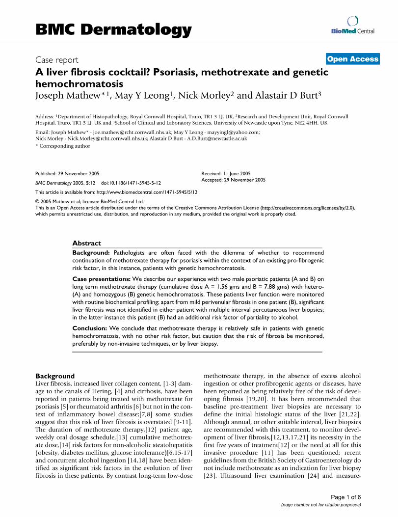
Following venesection (Patient B), there is no histological evidence of residual iron (×20)Figure 3Following venesection (Patient B), there is no histological evidence of residual iron (×20).
Page 4 of 6(page number not for citation purposes)
BMC Dermatology 2005, 5:12 http://www.biomedcentral.com/1471-5945/5/12
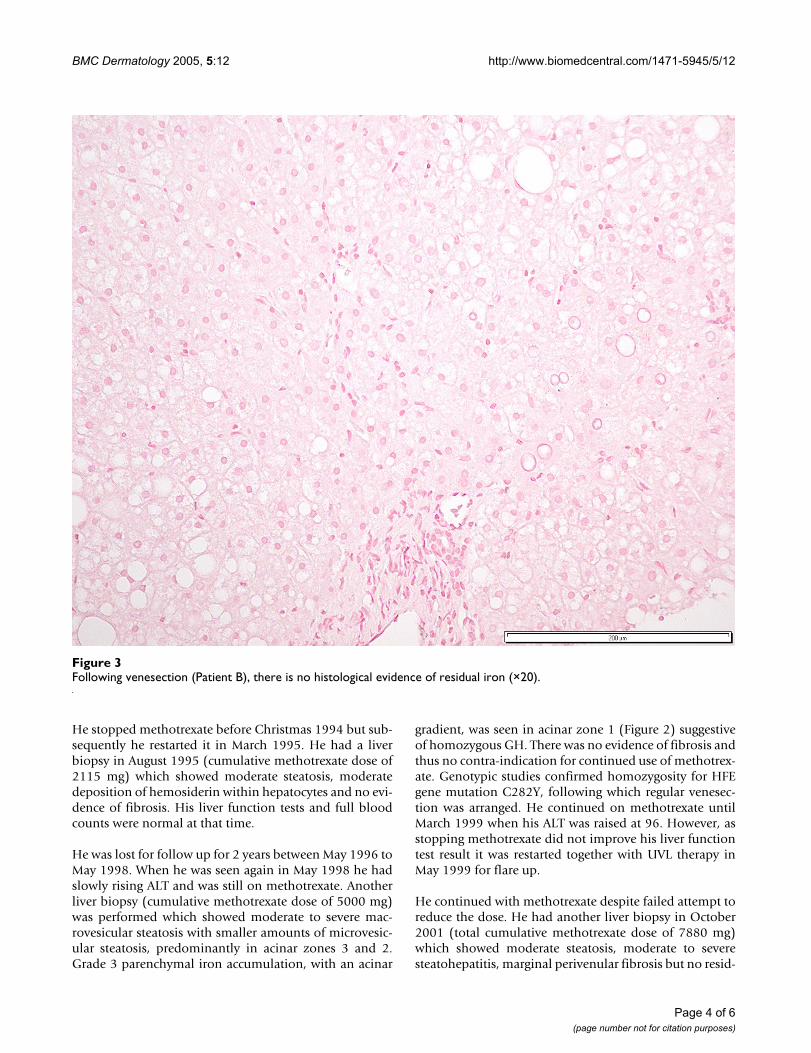
ual iron (Figure 3); the former features were reminiscentof alcohol-induced liver damage (Figure 4). As there wasno significant fibrosis, continued methotrexate adminis-tration was not contra-indicated although he was advisedto curtail his alcohol ingestion.
DiscussionPathologists are occasionally faced with a decision ofwhether to recommend continuation of methotrexatetherapy when a patient has an "additional" risk factor forthe development of liver fibrosis and/or cirrhosis; in thisinstance with two patients on long-term methotrexatetherapy who had GH. As far as we are aware, there havebeen no reports in the english literature that addresses thisspecific issue.
Patients with GH are at risk of developing liver fibrosisand, in the long term, liver cirrhosis; [28-30] there is aalso, a risk of developing fibrosis with methotrexate ther-apy alone [31,32]. Cessation of methotrexate therapy isadvised in the presence of significant liver fibrosis [13].
We have described our experience of two hemochroma-totic patients on low dose on methotrexate therapy for rel-atively short periods of three and five years and relativelylow cumulative doses. Although one of these patientswith homozygous GH was also partial to alcohol, he didnot show evidence of significant fibrosis apart from equiv-ocal alcohol-associated perivenular fibrosis. In suchinstances, adequate counselling of the patient of the riskof excess alcohol in this setting is probably appropriate.
There was no evidence of liver damage or fibrosis in ourpatient with heterozygous GH.
Although measurement of PIIINP can be useful in defin-ing the development of liver fibrosis,[25] elevated or ris-ing levels are not specific or sensitive for liver fibrosis; [25-27] however, liver biopsy has been recommended inpatients whose PIINP levels increase from normal whilstthey are being monitored for liver fibrosis [27]. ClearlyPIIINP measurements will not distinguish fibrosis due tomethotrexate or GH, in combination or alone or, fibrosisdeveloping as a result of any other profibrogenic agent.We are unable to offer PIIINP measurements for evaluat-ing liver fibrosis in our institution.
ConclusionWe conclude that methotrexate therapy is relatively safe inpatients with GH but caution that the risk of fibrosis bemonitored, preferably by non-invasive techniques, or byliver biopsy. We recommend that patients in this settingbe advised of the potential for liver damage especiallywith "additonal" risk factors.
List of abbreviationsserum type III procollagen aminopeptide (PIIINP)
genetic hemochromatosis (GH)
ultraviolet B (UVB)
HFE: gene symbol
This figure shows a reticulin (a) and a Haematoxylin Van Gieson stain (b) of two terminal hepatic venules in which there is equivocal perivenular fibrosis (×40)Figure 4This figure shows a reticulin (a) and a Haematoxylin Van Gieson stain (b) of two terminal hepatic venules in which there is equivocal perivenular fibrosis (x40).
Page 5 of 6(page number not for citation purposes)

BMC Dermatology 2005, 5:12 http://www.biomedcentral.com/1471-5945/5/12
ultraviolet light (UVL)
alanine aminotransferase (ALT)
Competing interestsThe author(s) declare that they have no competing inter-ests.
Authors' contributionsJM is the local liver pathologist who made the diagnosisof GH and together with MYL interrogated and preparedthe manuscript. ADB provided expert review, advice andcontributed to the preparation of the manuscript, as didNM who also initiated contact with these patients andsecured consent. All authors have read and approved themanuscript.
References1. Bjorkman DJ, Hammond EH, Lee RG, Clegg DO, Tolman KG:
Hepatic ultrastructure after methotrexate therapy for rheu-matoid arthritis. Arthritis Rheum 1988, 31:1465-1472.
2. Nohlgard C, Rubio CA, Kock Y, Hammar H: Liver fibrosis quanti-fied by image analysis in methotrexate-treated patients withpsoriasis. J Am Acad Dermatol 1993, 28:40-45.
3. Jaskiewicz K, Voigt H, Blakolmer K: Increased matrix proteins,collagen and transforming growth factor are early markersof hepatotoxicity in patients on long-term methotrexatetherapy. J Toxicol Clin Toxicol 1996, 34:301-305.
4. Hytiroglou P, Tobias H, Saxena R, Abramidou M, Papadimitriou CS,Theise ND: The canals of hering might represent a target ofmethotrexate hepatic toxicity. Am J Clin Pathol 2004,121:324-329.
5. Nyfors A: Liver biopsies from psoriatics related to meth-otrexate therapy. 3. Findings in post-methotrexate liverbiopsies from 160 psoriatics. Acta Pathol Microbiol Scand [A] 1977,85:511-518.
6. Shergy WJ, Polisson RP, Caldwell DS, Rice JR, Pisetsky DS, Allen NB:Methotrexate-associated hepatotoxicity: retrospective anal-ysis of 210 patients with rheumatoid arthritis. Am J Med 1988,85:771-774.
7. Lemann M, Zenjari T, Bouhnik Y, Cosnes J, Mesnard B, Rambaud JC,Modigliani R, Cortot A, Colombel JF: Methotrexate in Crohn'sdisease: long-term efficacy and toxicity. Am J Gastroenterol2000, 95:1730-1734.
8. Te HS, Schiano TD, Kuan SF, Hanauer SB, Conjeevaram HS, BakerAL: Hepatic effects of long-term methotrexate use in thetreatment of inflammatory bowel disease. Am J Gastroenterol2000, 95:3150-3156.
9. Boffa MJ, Chalmers RJ, Haboubi NY, Shomaf M, Mitchell DM:Sequential liver biopsies during long-term methotrexatetreatment for psoriasis: a reappraisal. Br J Dermatol 1995,133:774-778.
10. Boffa MJ, Chalmers RJ: Methotrexate for psoriasis. Clin Exp Der-matol 1996, 21:399-408.
11. Aithal GP, Haugk B, Das S, Card T, Burt AD, Record CO: Monitor-ing methotrexate-induced hepatic fibrosis in patients withpsoriasis: are serial liver biopsies justified? Aliment PharmacolTher 2004, 19:391-399.
12. Reynolds FS, Lee WM: Hepatotoxicity after long-term meth-otrexate therapy. South Med J 1986, 79:536-539.
13. Robinson JK, Baughman RD, Auerbach R, Cimis RJ: Methotrexatehepatotoxicity in psoriasis. Consideration of liver biopsies atregular intervals. Arch Dermatol 1980, 116:413-415.
14. Nyfors A, Poulsen H: Morphogenesis of fibrosis and cirrhosis inmethotrexate-treated patients with psoriasis. Am J Surg Pathol1977, 1:235-243.
15. Langman G, Hall PM, Todd G: Role of non-alcoholic steatohepa-titis in methotrexate-induced liver injury. J Gastroenterol Hepa-tol 2001, 16:1395-1401.
16. Kremer JM, Koff R: A debate: should patients with rheumatoidarthritis on methotrexate undergo liver biopsies? Semin Arthri-tis Rheum 1992, 21:376-386.
17. Malatjalian DA, Ross JB, Williams CN, Colwell SJ, Eastwood BJ:Methotrexate hepatotoxicity in psoriatics: report of 104patients from Nova Scotia, with analysis of risks from obes-ity, diabetes and alcohol consumption during long term fol-low-up. Can J Gastroenterol 1996, 10:369-375.
18. Ashton RE, Millward-Sadler GH, White JE: Complications inmethotrexate treatment of psoriasis with particular refer-ence to liver fibrosis. J Invest Dermatol 1982, 79:229-232.
19. Van Dooren-Greebe RJ, Kuijpers AL, Mulder J, De Boo T, Van deKerkhof PC: Methotrexate revisited: effects of long-termtreatment in psoriasis. Br J Dermatol 1994, 130:204-210.
20. Lanse SB, Arnold GL, Gowans JD, Kaplan MM: Low incidence ofhepatotoxicity associated with long-term, low-dose oralmethotrexate in treatment of refractory psoriasis, psoriaticarthritis, and rheumatoid arthritis. An acceptable risk/bene-fit ratio. Dig Dis Sci 1985, 30:104-109.
21. Kevat S, Ahern M, Hall P: Hepatotoxicity of methotrexate inrheumatic diseases. Med Toxicol Adverse Drug Exp 1988, 3:197-208.
22. Dolan OM, Burrows D, Irvine A, Walsh M: The value of a baselineliver biopsy prior to methotrexate treatment. Br J Dermatol1994, 131:891-894.
23. British Society of Gastroenterology: Guidelines on the use of liverbiopsy in clinical practice. 2004.
24. Coulson IH, McKenzie J, Neild VS, Joseph AE, Marsden RA: A com-parison of liver ultrasound with liver biopsy histology in pso-riatics receiving long-term methotrexate therapy. Br JDermatol 1987, 116:491-495.
25. Zachariae H, Aslam HM, Bjerring P, Sogaard H, Zachariae E, Heicken-dorff L: Serum aminoterminal propeptide of type III procolla-gen in psoriasis and psoriatic arthritis: relation to liverfibrosis and arthritis. J Am Acad Dermatol 1991, 25:50-53.
26. Boffa MJ, Smith A, Chalmers RJ, Mitchell DM, Rowan B, Warnes TW,Shomaf M, Haboubi NY: Serum type III procollagen aminopep-tide for assessing liver damage in methotrexate-treated pso-riatic patients. Br J Dermatol 1996, 135:538-544.
27. Zachariae H, Heickendorff L, Sogaard H: The value of amino-ter-minal propeptide of type III procollagen in routine screeningfor methotrexate-induced liver fibrosis: a 10-year follow-up.Br J Dermatol 2001, 144:100-103.
28. Mathew J, Burt AD: Cellular mechanisms of fibrogenesis ingenetic haemochromatosis. In Cells of the hepatic sinusoid Volume4. Edited by: Knook DL and Wisse E. , The Kupffer Cell Foundation,Netherlands; 1993:274-276.
29. Wojcik JP, Speechley MR, Kertesz AE, Chakrabarti S, PC A: Naturalhistory of C282Y homozygotes for hemochromatosis. Can JGastroenterol 2002, 16:297-302.
30. Hubscher SG: Iron overload, inflammation and fibrosis ingenetic haemochromatosis. J Hepatol 2003, 38:521-525.
31. Whiting-O'Keefe QE, Fye KH, Sack KD: Methotrexate and histo-logic hepatic abnormalities: a meta-analysis. Am J Med 1991,90:711-716.
32. Zachariae H, Sogaard H, Heickendorff L: Methotrexate-inducedliver cirrhosis. Clinical, histological and serological studies--afurther 10-year follow-up. Dermatology 1996, 192:343-346.
Pre-publication historyThe pre-publication history for this paper can be accessedhere:
http://www.biomedcentral.com/1471-5945/5/12/prepub
Page 6 of 6(page number not for citation purposes)
http://www.ncbi.nlm.nih.gov/entrez/query.fcgi?cmd=Retrieve&db=PubMed&dopt=Abstract&list_uids=3196365
http://www.ncbi.nlm.nih.gov/entrez/query.fcgi?cmd=Retrieve&db=PubMed&dopt=Abstract&list_uids=3196365
http://www.ncbi.nlm.nih.gov/entrez/query.fcgi?cmd=Retrieve&db=PubMed&dopt=Abstract&list_uids=3196365
http://www.ncbi.nlm.nih.gov/entrez/query.fcgi?cmd=Retrieve&db=PubMed&dopt=Abstract&list_uids=7678843
http://www.ncbi.nlm.nih.gov/entrez/query.fcgi?cmd=Retrieve&db=PubMed&dopt=Abstract&list_uids=7678843
http://www.ncbi.nlm.nih.gov/entrez/query.fcgi?cmd=Retrieve&db=PubMed&dopt=Abstract&list_uids=7678843
http://www.ncbi.nlm.nih.gov/entrez/query.fcgi?cmd=Retrieve&db=PubMed&dopt=Abstract&list_uids=8667468
http://www.ncbi.nlm.nih.gov/entrez/query.fcgi?cmd=Retrieve&db=PubMed&dopt=Abstract&list_uids=8667468
http://www.ncbi.nlm.nih.gov/entrez/query.fcgi?cmd=Retrieve&db=PubMed&dopt=Abstract&list_uids=8667468
http://www.ncbi.nlm.nih.gov/entrez/query.fcgi?cmd=Retrieve&db=PubMed&dopt=Abstract&list_uids=3195601
http://www.ncbi.nlm.nih.gov/entrez/query.fcgi?cmd=Retrieve&db=PubMed&dopt=Abstract&list_uids=3195601
http://www.ncbi.nlm.nih.gov/entrez/query.fcgi?cmd=Retrieve&db=PubMed&dopt=Abstract&list_uids=3195601
http://www.ncbi.nlm.nih.gov/entrez/query.fcgi?cmd=Retrieve&db=PubMed&dopt=Abstract&list_uids=8555032
http://www.ncbi.nlm.nih.gov/entrez/query.fcgi?cmd=Retrieve&db=PubMed&dopt=Abstract&list_uids=8555032
http://www.ncbi.nlm.nih.gov/entrez/query.fcgi?cmd=Retrieve&db=PubMed&dopt=Abstract&list_uids=8555032
http://www.ncbi.nlm.nih.gov/entrez/query.fcgi?cmd=Retrieve&db=PubMed&dopt=Abstract&list_uids=9167333
http://www.ncbi.nlm.nih.gov/entrez/query.fcgi?cmd=Retrieve&db=PubMed&dopt=Abstract&list_uids=3704718
http://www.ncbi.nlm.nih.gov/entrez/query.fcgi?cmd=Retrieve&db=PubMed&dopt=Abstract&list_uids=3704718
http://www.ncbi.nlm.nih.gov/entrez/query.fcgi?cmd=Retrieve&db=PubMed&dopt=Abstract&list_uids=7369769
http://www.ncbi.nlm.nih.gov/entrez/query.fcgi?cmd=Retrieve&db=PubMed&dopt=Abstract&list_uids=7369769
http://www.ncbi.nlm.nih.gov/entrez/query.fcgi?cmd=Retrieve&db=PubMed&dopt=Abstract&list_uids=7369769
http://www.ncbi.nlm.nih.gov/entrez/query.fcgi?cmd=Retrieve&db=PubMed&dopt=Abstract&list_uids=1626283
http://www.ncbi.nlm.nih.gov/entrez/query.fcgi?cmd=Retrieve&db=PubMed&dopt=Abstract&list_uids=1626283
http://www.ncbi.nlm.nih.gov/entrez/query.fcgi?cmd=Retrieve&db=PubMed&dopt=Abstract&list_uids=9193771
http://www.ncbi.nlm.nih.gov/entrez/query.fcgi?cmd=Retrieve&db=PubMed&dopt=Abstract&list_uids=9193771
http://www.ncbi.nlm.nih.gov/entrez/query.fcgi?cmd=Retrieve&db=PubMed&dopt=Abstract&list_uids=9193771
http://www.ncbi.nlm.nih.gov/entrez/query.fcgi?cmd=Retrieve&db=PubMed&dopt=Abstract&list_uids=7130740
http://www.ncbi.nlm.nih.gov/entrez/query.fcgi?cmd=Retrieve&db=PubMed&dopt=Abstract&list_uids=7130740
http://www.ncbi.nlm.nih.gov/entrez/query.fcgi?cmd=Retrieve&db=PubMed&dopt=Abstract&list_uids=7130740
http://www.ncbi.nlm.nih.gov/entrez/query.fcgi?cmd=Retrieve&db=PubMed&dopt=Abstract&list_uids=8123573
http://www.ncbi.nlm.nih.gov/entrez/query.fcgi?cmd=Retrieve&db=PubMed&dopt=Abstract&list_uids=8123573
http://www.ncbi.nlm.nih.gov/entrez/query.fcgi?cmd=Retrieve&db=PubMed&dopt=Abstract&list_uids=3967557
http://www.ncbi.nlm.nih.gov/entrez/query.fcgi?cmd=Retrieve&db=PubMed&dopt=Abstract&list_uids=3967557
http://www.ncbi.nlm.nih.gov/entrez/query.fcgi?cmd=Retrieve&db=PubMed&dopt=Abstract&list_uids=3967557
http://www.ncbi.nlm.nih.gov/entrez/query.fcgi?cmd=Retrieve&db=PubMed&dopt=Abstract&list_uids=3041245
http://www.ncbi.nlm.nih.gov/entrez/query.fcgi?cmd=Retrieve&db=PubMed&dopt=Abstract&list_uids=3041245
http://www.ncbi.nlm.nih.gov/entrez/query.fcgi?cmd=Retrieve&db=PubMed&dopt=Abstract&list_uids=7857846
http://www.ncbi.nlm.nih.gov/entrez/query.fcgi?cmd=Retrieve&db=PubMed&dopt=Abstract&list_uids=7857846
http://www.ncbi.nlm.nih.gov/entrez/query.fcgi?cmd=Retrieve&db=PubMed&dopt=Abstract&list_uids=3555595
http://www.ncbi.nlm.nih.gov/entrez/query.fcgi?cmd=Retrieve&db=PubMed&dopt=Abstract&list_uids=3555595
http://www.ncbi.nlm.nih.gov/entrez/query.fcgi?cmd=Retrieve&db=PubMed&dopt=Abstract&list_uids=3555595
http://www.ncbi.nlm.nih.gov/entrez/query.fcgi?cmd=Retrieve&db=PubMed&dopt=Abstract&list_uids=1880254
http://www.ncbi.nlm.nih.gov/entrez/query.fcgi?cmd=Retrieve&db=PubMed&dopt=Abstract&list_uids=1880254
http://www.ncbi.nlm.nih.gov/entrez/query.fcgi?cmd=Retrieve&db=PubMed&dopt=Abstract&list_uids=1880254
http://www.ncbi.nlm.nih.gov/entrez/query.fcgi?cmd=Retrieve&db=PubMed&dopt=Abstract&list_uids=8915142
http://www.ncbi.nlm.nih.gov/entrez/query.fcgi?cmd=Retrieve&db=PubMed&dopt=Abstract&list_uids=8915142
http://www.ncbi.nlm.nih.gov/entrez/query.fcgi?cmd=Retrieve&db=PubMed&dopt=Abstract&list_uids=8915142
http://www.ncbi.nlm.nih.gov/entrez/query.fcgi?cmd=Retrieve&db=PubMed&dopt=Abstract&list_uids=1828327
http://www.ncbi.nlm.nih.gov/entrez/query.fcgi?cmd=Retrieve&db=PubMed&dopt=Abstract&list_uids=1828327
http://www.ncbi.nlm.nih.gov/entrez/query.fcgi?cmd=Retrieve&db=PubMed&dopt=Abstract&list_uids=8864370
http://www.ncbi.nlm.nih.gov/entrez/query.fcgi?cmd=Retrieve&db=PubMed&dopt=Abstract&list_uids=8864370