A Flight Sensory-Motor to Olfactory Processing Circuit in ... · A Flight Sensory-Motor to...
13
ORIGINAL RESEARCH published: 16 February 2016 doi: 10.3389/fncir.2016.00005 Edited by: Deborah Baro, Georgia State University, USA Reviewed by: Fadi A. Issa, East Carolina University, USA Dawn M. Blitz, Miami University, USA *Correspondence: Samual P. Bradley [email protected] † These authors are co-senior authors. Received: 27 November 2015 Accepted: 25 January 2016 Published: 16 February 2016 Citation: Bradley SP, Chapman PD, Lizbinski KM, Daly KC and Dacks AM (2016) A Flight Sensory-Motor to Olfactory Processing Circuit in the Moth Manduca sexta. Front. Neural Circuits 10:5. doi: 10.3389/fncir.2016.00005 A Flight Sensory-Motor to Olfactory Processing Circuit in the Moth Manduca sexta Samual P. Bradley*, Phillip D. Chapman, Kristyn M. Lizbinski, Kevin C. Daly † and Andrew M. Dacks † Department of Biology, West Virginia University, Morgantown, WV, USA Neural circuits projecting information from motor to sensory pathways are common across sensory domains. These circuits typically modify sensory function as a result of motor pattern activation; this is particularly so in cases where the resultant behavior affects the sensory experience or its processing. However, such circuits have not been observed projecting to an olfactory pathway in any species despite well characterized active sampling behaviors that produce reafferent mechanical stimuli, such as sniffing in mammals and wing beating in the moth Manduca sexta. In this study we characterize a circuit that connects a flight sensory-motor center to an olfactory center in Manduca. This circuit consists of a single pair of histamine immunoreactive (HA-ir) neurons that project from the mesothoracic ganglion to innervate a subset of ventral antennal lobe (AL) glomeruli. Furthermore, within the AL we show that the M. sexta histamine B receptor (MsHisClB) is exclusively expressed by a subset of GABAergic and peptidergic LNs, which broadly project to all olfactory glomeruli. Finally, the HA-ir cell pair is present in fifth stage instar larvae; however, the absence of MsHisClB-ir in the larval antennal center indicates that the circuit is incomplete prior to metamorphosis and importantly prior to the expression of flight behavior. Although the functional consequences of this circuit remain unknown, these results provide the first detailed description of a circuit that interconnects an olfactory system with motor centers driving flight behaviors including odor-guided flight. Keywords: modulation, histamine, olfaction, insect, flight INTRODUCTION Animals exhibit stereotypical search behaviors in pursuit of potential food sources or mating partners. More specifically, some animals employ sampling strategies where rhythmic motor patterns optimize the interaction between stimuli and their affected sensory systems. Consequently, many of these motor systems project to and modulate how sensory systems process this information. For example, saccadic eye movements allow us to focus on objects despite having a fast adapting visual system (Martinez-Conde et al., 2006). Here the neural circuits driving Abbreviations: AL, antennal lobe; AMMC, antennal mechanosensory and motor center; ATR, allatotropin; BRP, bruchpilot; BSA, bovine serum albumin; FMRF, FMRF-amide; HA, histamine; HisClA, histamine A receptor; LAC, larval antennal center; LNs, local interneurons; MDHn, mesothoracic deutocerebrum histamine neurons; MsHisClB, Manduca sexta histamine B receptor; MsG, mesothoracic ganglia; ORNs, olfactory receptor neurons; PNs, projection neurons; SEZ, subesophageal zone. Frontiers in Neural Circuits | www.frontiersin.org 1 February 2016 | Volume 10 | Article 5
Transcript of A Flight Sensory-Motor to Olfactory Processing Circuit in ... · A Flight Sensory-Motor to...

ORIGINAL RESEARCHpublished: 16 February 2016
doi: 10.3389/fncir.2016.00005
Edited by:Deborah Baro,
Georgia State University, USA
Reviewed by:Fadi A. Issa,
East Carolina University, USADawn M. Blitz,
Miami University, USA
*Correspondence:Samual P. Bradley
†These authors are co-senior authors.
Received: 27 November 2015Accepted: 25 January 2016
Published: 16 February 2016
Citation:Bradley SP, Chapman PD,
Lizbinski KM, Daly KC and Dacks AM(2016) A Flight Sensory-Motorto Olfactory Processing Circuit
in the Moth Manduca sexta.Front. Neural Circuits 10:5.
doi: 10.3389/fncir.2016.00005
A Flight Sensory-Motor to OlfactoryProcessing Circuit in the MothManduca sextaSamual P. Bradley*, Phillip D. Chapman, Kristyn M. Lizbinski, Kevin C. Daly† andAndrew M. Dacks†
Department of Biology, West Virginia University, Morgantown, WV, USA
Neural circuits projecting information from motor to sensory pathways are commonacross sensory domains. These circuits typically modify sensory function as a resultof motor pattern activation; this is particularly so in cases where the resultant behavioraffects the sensory experience or its processing. However, such circuits have not beenobserved projecting to an olfactory pathway in any species despite well characterizedactive sampling behaviors that produce reafferent mechanical stimuli, such as sniffing inmammals and wing beating in the moth Manduca sexta. In this study we characterizea circuit that connects a flight sensory-motor center to an olfactory center in Manduca.This circuit consists of a single pair of histamine immunoreactive (HA-ir) neurons thatproject from the mesothoracic ganglion to innervate a subset of ventral antennal lobe(AL) glomeruli. Furthermore, within the AL we show that the M. sexta histamine Breceptor (MsHisClB) is exclusively expressed by a subset of GABAergic and peptidergicLNs, which broadly project to all olfactory glomeruli. Finally, the HA-ir cell pair is presentin fifth stage instar larvae; however, the absence of MsHisClB-ir in the larval antennalcenter indicates that the circuit is incomplete prior to metamorphosis and importantlyprior to the expression of flight behavior. Although the functional consequences of thiscircuit remain unknown, these results provide the first detailed description of a circuit thatinterconnects an olfactory system with motor centers driving flight behaviors includingodor-guided flight.
Keywords: modulation, histamine, olfaction, insect, flight
INTRODUCTION
Animals exhibit stereotypical search behaviors in pursuit of potential food sources or matingpartners. More specifically, some animals employ sampling strategies where rhythmic motorpatterns optimize the interaction between stimuli and their affected sensory systems. Consequently,many of these motor systems project to and modulate how sensory systems process thisinformation. For example, saccadic eye movements allow us to focus on objects despite havinga fast adapting visual system (Martinez-Conde et al., 2006). Here the neural circuits driving
Abbreviations: AL, antennal lobe; AMMC, antennal mechanosensory and motor center; ATR, allatotropin; BRP, bruchpilot;BSA, bovine serum albumin; FMRF, FMRF-amide; HA, histamine; HisClA, histamine A receptor; LAC, larval antennal center;LNs, local interneurons; MDHn, mesothoracic deutocerebrum histamine neurons; MsHisClB, Manduca sexta histamine Breceptor; MsG, mesothoracic ganglia; ORNs, olfactory receptor neurons; PNs, projection neurons; SEZ, subesophageal zone.
Frontiers in Neural Circuits | www.frontiersin.org 1 February 2016 | Volume 10 | Article 5

Bradley et al. Flight Motor to Olfactory Circuit
these small movements also send a signal canceling theperception of a moving scene, therefore affording properbehavioral responses to other stimuli in the environment(Zaretsky and Rowell, 1979; Ross et al., 2001). Other motorto sensory circuits have been shown to amplify self-inducedcommunication signals (Mohr et al., 2003), inhibit reflexresponses (Chalfie et al., 1985), and are involved in sensory/motorplanning (Brainard and Doupe, 2000; Sommer andWurtz, 2002).While work in other sensory systems have made significantprogress in characterizing motor to sensory circuits (Crapse andSommer, 2008), it is not clear whether such circuits are present inthe olfactory system.
When tracking odors, animals typically exhibit behaviors,such as sniffing, that periodically structure olfactory stimuli(Halpern, 1983). Each sniff cycle draws odor-laden air intothe nasal cavity during inhalation and forces air out duringexhalation, thus imposing a temporal structure on air/olfactoryreceptor interactions that persists in the absence of odor (Adrian,1942; Kepecs et al., 2007). In this manner, sniffing couplesreafferent mechanical stimuli with odor stimuli resulting ina temporally structured stimulus that improves physiological(Verhagen et al., 2007), and presumably behavioral performance.In the moth Manduca sexta, wing beating causes high frequencyoscillations in airflow over the antennae in a manner analogousto sniffing (Sane and Jacobson, 2006). These periodic signalshave a potentially strong effect on odor-receptor interactions inmoths (Loudon et al., 1994; Loudon and Koehl, 2000) and areeffectively tracked by antennal and antennal lobe (AL) neurons(Tripathy et al., 2010). This implies that at least part of thetemporal structure of encoding neuron activity is driven by time-dependent fluctuations in stimulus concentration (Christensenet al., 1998; Daly et al., 2011), driven by wing-beating. Simulatingwing-beating effects on odor exposure by pulsing odor stimuliat wing beat frequencies increases separation of neural ensemblerepresentations for different odors (Houot et al., 2014) andenhances behavioral performance in psychophysical assays ofolfactory acuity (Tripathy et al., 2010; Daly et al., 2013). WhileAL neurons can track pulsed stimuli when the neck connectiveis intact (Houot et al., 2014), AL neurons are unable to doso when using isolated head preparations (Christensen et al.,1998; Tripathy et al., 2010). This suggests that the AL receivesinput from flight sensorimotor centers that affects the temporalfidelity with which the AL encodes odors (Christensen et al.,1998; Tripathy et al., 2010). However, relatively little is knownabout neural circuits connecting flight sensory-motor centers andthe AL.
There is limited data describing input from flight sensory-motor centers to the ALs of Manduca. This circuit consists ofa single pair of histamine (HA) immunoreactive neurons thatproject from the mesothoracic ganglion (MsG) and bilaterallyinnervate both ALs and the antennal mechanosensory andmotor center (AMMC) (Homberg and Hildebrand, 1991;Homberg, 1994). The purpose of this study was to providea detailed morphological description of these mesothoracic todeutocerebral histaminergic neurons (MDHns) and to identifycandidate post synaptic targets. Using immunohistochemistry,we found that the MDHns ramify in a subset of ventral
glomeruli in the AL, the AL isthmus, and the coarse neuropil.A subset of GABAergic local interneurons (LNs) along withone FMRFamide-ir and one allatotropin-ir (ATR-ir) LN expressthe Manduca homolog of the histamine B receptor subtype(MsHisClB) and thus represent candidate postsynaptic targets ofthe MDHns. Furthermore, although the MDHns are present inlarvae and survive metamorphosis there is no expression of theMsHisClB receptor in larval antennal center (LAC) neurons untilafter pupation has occurred, suggesting the MDHns only affectolfactory processing in adults. The MDHns therefore represent anovel circuit that provides a potential source of information froma flight sensory-motor integration system to the olfactory system.
MATERIALS AND METHODS
AnimalsAnimals were raised using a standard diet (Bell and Joachim,1976) and rearing procedures (Tripathy et al., 2010). Adult mothswere kept in brown paper bags and placed in an incubator(Percival Scientific Inc.; 166VLC8) where they were exposed toa 16/8 reverse light dark cycle set to 25◦C and 75% humidity.Approximately 10 male or female moths aged 3–9 days wereused for all experimental groups. For larval studies, stage 5 instarlarvae were dissected with trachea removed. Ten larval nervoussystems were used for developmental experiments.
ImmunohistochemistryImmunolabeling was performed as described previously (Dackset al., 2010) on both sectioned and whole-mount brainsdepending upon the preparation. For HA immunolabeling,brains were placed in a 4% N-3-dimethylaminopropyl-N’-ethylcarbodiimide (Sigma–Aldrich, 03449) pre-fixative for 3–4 hat 4◦C, before being fixed overnight in 4% paraformaldehyde(Electron Microscope Sciences, 15710) in 1% phosphate bufferedsaline (PBS; Sigma–Aldrich, SLBC5890) at 4◦C. For theMsHisClB antibody, brains were placed in 4% paraformaldehyde(Electron Microscopy Sciences, 15710; pH 7.3–7.5) at 4◦Covernight. Following fixation, brains were washed in PBS(pH 6.9). For sectioned tissue, adult brains and ganglia wereembedded in 5% agarose (Sigma–Aldrich, SLBJ3744V) andsectioned between 50 and 250 μm (depending on the antibody)using a Leica VT 1000S vibrating microtome. The tissue waswashed in PBS with 0.5% TritonTM-X100 (PBST; Sigma–Aldrich,110M0009V), blocked for 1 h with 2% IgG-free BSA, J(acksonLaboratory, 001-000-162) and incubated in primary antibodyin blocking solution with 5 mM with sodium azide (PBSAT;Fisher Scientific, S2271). Brains were washed and blocked asabove, then incubated in secondary antibody (1:1000 Alexa 488,546, or 633 in PBSAT; Alexa fluor R© ; Lifescience Technologies)overnight at room temperature except for experiments usingMsHisClB and/or GABA in which tissue was incubated at 4◦C.SYTO 59 (a nuclear label; InvitrogenTM; S11341) was usedto outline the LAC. Tissue was washed several times in TrisBuffered Saline (TBS; Bio-Rad, 170-6435) and the tissue wasincubated in 1:10,000 SYTO 59 in Tris-HCl (Fisher Scientific,BP153 for 60 min before mounting. All tissue was washed
Frontiers in Neural Circuits | www.frontiersin.org 2 February 2016 | Volume 10 | Article 5

Bradley et al. Flight Motor to Olfactory Circuit
in PBST and PBS, then run through an ascending glycerol(Sigma–Aldrich, BCBN3647V) series (40%, 60%, and 80%) andmounted in Vectashield R© (Vector laboratories, ZA1222). Forwhole-mount preparations, tissue was run through an ascendingethanol (Sigma–Aldrich, SHBF6704V) dilution series (30, 50,70, 95, and 100%) for 10 min each (after the PBS wash), a 1:1ethanolmethyl salicylate solution for 15min, and finally mountedin 100% methyl salicylate (Fisher Scientific, MFCD00002214).All primary antibody information (including dilutions used,manufacturer, host-species, immunogen and RRID) is includedin Table 1.
Antibody Manufacturing andCharacterizationRabbit Anti-HistamineThe HA antiserum was raised against synthetic HA conjugatedvia a carbodiimide linker to succinylated keyhole limpethemocyanin. Control studies showed that the antibody hadno cross reactivity with L-histidine or L-histidine containingpeptides, and pre-adsorbing the antiserumwith the HA conjugateeliminates labeling (Immunostar histochemical HA antiserumspecification sheet) as did an RNAi knock down of histidinedecarboxylase in Drosophila (Melzig et al., 1996). Finally, pre-adsorbing the HA antiserum against keyhole limpet hemocyaninalone did not eliminate HA labeling in Bombus impatiens(Dacks et al., 2010). Pre-adsorption controls in Manduca tissuewere performed by incubating the rabbit anti-HA antiserumfor 24 h in blocking solution (1 mg/ml BSA in PBSAT) withHA (Sigma–Aldrich, H7250) at a ratio of 10:1 HA:antiserum.Non-pre-adsorbed controls in which rabbit anti-HA antibodywas incubated in parallel under identical conditions resulted inimmunolabeling (Figure 1A; n = 5) whereas preadsorbing theantibody abolished all staining in Manduca optic lobe tissue(Figure 1B; n = 5).
Mouse Anti-BruchpilotBruchpilot (BRP) is homologous to the protein ELKS/CAST inmammals and functions as a structural protein at presynapticactive zones (Wagh et al., 2006). The BRP antiserum was raised
against BRP and western blots showed two bands for twoisoforms of the BRP protein in Drosophila (Wagh et al., 2006).BRP labeling was absent in BRP mutants (Kittel et al., 2006)and has been shown to bind to amino acid sequence 1390–1740(Fouquet et al., 2009). The BRP antiserum produced a singleband at the predicted weight for the Manduca homolog of BRPin western blots using Manduca brain tissue (Lizbinski et al.,2015). The purpose of using the anti-BRP antibody in this studywas to highlight the boundaries of neuropil, rather than to makeany conclusions about the distribution of the Manduca homologof BRP.
Mouse Anti-GABAGABA antiserum was raised against GABA coupled to BSAwith glutaraldehyde. Controls show that the antibody was highlyspecific to GABA and did not react with other amino acid BSAconjugates (Abcam data sheet). Pre-adsorption controls wereperformed by incubating the mouse anti-GABA antiserum for24 h in blocking solution (1 mg/ml BSA in PBSAT) with GABA(Sigma–Aldrich, cat # A2129) at a ratio of 10:1 GABA:antiserum.Non-pre-adsorbed controls in which mouse anti-GABA antibodywas incubated in parallel under identical conditions resulted instrong immunolabeling (Figure 1C; n= 5) whereas preadsorbingthe antibody abolished all staining in Manduca AL tissue(Figure 1D; n = 5).
Rabbit Anti-FMRFamideFMRFamide antiserum was provided by Dr. Eve Marder andwas raised against synthetic RF-amide coupled to bovinethyroglobulin with glutaraldehyde (Marder et al., 1987).Preadsorbing the antiserum against synthetic FMRFamideeliminated labeling in larval Manduca nervous tissue (Wittenand Truman, 1996).
Rabbit Anti-AllatotropinAllatotropin (ATR) antiserum was provided by Dr. Jan Veenstraand raised against purified ATR coupled to thyroglobulinusing glutaraldehyde (Veenstra and Hagedorn, 1993). ELISAdid not show cross reactivity with 100 pmol corazonin,
TABLE 1 | Primary antibodies used in this study.
Antigen Immunogen Manufacturer, host,monoclonal vs. polyclonal
Catalog # RRID Dilution used
Histamine Synthetic HA coupled to succinylated keyholelimpet Hemocyanin with carbodiimide linker
Immunostar, Rabbit,polyclonal
22939 AB_572245 1:500
Bruchpilot Bruchpilot peptide sequence (1390-1740) fromhead homogenate
DSHB, Mouse, monoclonal Nc-82 AB_2314866 1:50
Manduca sexta HA Breceptor (MsHisClB)
Histamine B receptor peptide sequence(VNPDIELPQLD)
Bethyl Laboratory (custom),Rabbit, polyclonal
N/A N/A 1:5000
γ-aminobutyric acid(GABA)
Purified GABA conjugated to BSA Abcam, Mouse, monoclonal ab49675 AB_880138 1:500
Allatotropin Allatotropin coupled to thyroglobulin withglutaraldehyde
Dr. Jan Veenstra, Rabbit,polyclonal
N/A AB_2313973 1:8∗
FMRF-amide Synthetic FMRF coupled to bovine thyroglobulinwith gluteraldehyde
Dr. Eve Marder, Rabbit,Polyclonal
N/A AB_572232 1:8∗
∗See fluorescent tagging subsection of the methods for details.
Frontiers in Neural Circuits | www.frontiersin.org 3 February 2016 | Volume 10 | Article 5

Bradley et al. Flight Motor to Olfactory Circuit
FIGURE 1 | Characterization of the HA GABA and Manduca sexta HA Breceptor (MsHisClB) antibodies. (A) HA labeling in control animals wherethe antibody was not pre-adsorbed. HA labeling in the optic lobe which is theprimary neurotransmitter used by arthropod receptor cells. (B) HA labeling inthe optic lobe is abolished after the HA antibody was pre-adsorbed with a10:1 HA to antibody solution. (C) GABA labeling remains in control animalswhere the antibody was not pre-adsorbed with GABA. (D) GABA labeling inthe AL is abolished after the GABA antibody was pre-adsorbed with a 10:1GABA to antibody solution. For each panel the same dorsal lateral axis isused. (E) Amino acid sequence alignment of the HA B receptor subunits ofManduca sexta (MsHB; Msex2.04603-RA), Drosophila melanogaster (DmHB;ACA13298.1), Apis mellifera (AmHB; ABG75740.1), and Nasonia vitripennis(NvHB; ACZ51422.1). Asterisks indicate sequence identity across all fourspecies. Bold font indicates the immunogenic peptide sequence fromManduca that was used to generate the MsHisClB antibody. (F) Western blotusing MsHisClB receptor antibody on Manduca brain tissue resulted in asingle band at the predicted molecular weight (36 kDa) of the MsHisClBprotein. (G) Frontal section of optic lobe depicting MsHisClB-ir in the lamina(as labeled by an asterisks). (H) Pre-adsorption with the immunogenic peptidesequence eliminates all labeling in the lamina. Scale bars = 50 μm. D, dorsal,L, lateral, A, anterior.
vasopressin, leucokinin IV, or proctolin, but did show significantimmunoreacitivity to the truncated 6–13 analog of ManducaATR (Veenstra andHagedorn, 1993). Preadsorbing the antiserumagainst ATR eliminated immunolabeling inManduca tissue (Utzet al., 2008).
Rabbit Anti-MsHisClBTo determine the amino acid sequence of theManduca homologof the HA B-type receptor (MsHisClB), we used the Manducagenome (Agricultural Pest Genomics Resource Database1: toperform a forward protein BLAST analysis of the Drosophilamelanogaster histamine B-type receptor (HisClB) amino acidsequence (ACA13298.1). The top match from the Manducagenome had an e-value of 0.0 (Msex2.04603-RA). We thenreverse blasted this sequence from the Manduca genome intothe Drosophila genome in NCBI and the first three matcheswere Drosophila HisClB isoforms (NP_650116.2, NP_731632.1,and NP_001163591.1), all of which had e-values of 0.0. Thenext highest match from the Drosophila genome was the HisClAreceptor (otherwise known as “ora transientless”; NP_524406.1)which is the other of the two HA receptor types in Drosophila(Zheng et al., 2002) and had e-values of 3e-148 which isconsistent with both HA receptor types having high sequencehomology (Zheng et al., 2002; Jones et al., 2010). To ensurethat there were not two predicted amino acid sequences fromthe Manduca genome with high sequence homology to theDrosophila HisClB receptor, we took the amino acid sequencefrom the Manduca genome with the second highest e-value forthe Drosophila MsHisClB (Msex2.04216-RA; e-value = 1e-119)and ran a BLAST analysis of this sequence in the Drosophilagenome. The BLAST analysis resulted in an e-value of 7.37e-158 for the Drosophila ora transientless indicating that theManduca protein with the next closest sequence similarityto Drosophila HisClA was likely not the MsHisClB homolog.Figure 1E is a sequence alignment of the Manduca HisClBreceptor (MsHisClB) with the sequences for known histamineB receptors from Drosophila melanogaster (ACA13298.1), Apismeliferia (ABG75740.1), and Nasonia vitripennis (ACZ51422.1)(Jones et al., 2010) using the EMBL-EBI Clustal omega tool2(Sievers et al., 2011).
Custom affinity purified antibodies were generated inrabbit (Bethyl laboratories) using Cys-VNPDIELPQLD as theimmunogenic sequence. The immunogenic sequence was highlyconserved across D. melanogaster, A. mellifera, and N. vitripennis(Figure 1E). For western blots, adult brains were placed in BoltTMLDS Sample Buffer (Life Technologies, B0007, Life Technologies)with protease inhibitor cocktail (Research Products International,P50900) and DNase I (Invitrogen, 18068-015) and kept on icefor homogenization with a pestle. Samples were heated in awater bath for 10 min at 95◦C. We used the Novex R© BoltTMGel Electrophoresis System (Life Technologies) with Tris-GlycineSDS Running Buffer at 165V for 2.5 h and BoltTM 4–12% Bis-Tris Plus Precast Gels (BG04120BOX) to resolve proteins. Weused the iBlot R© Gel Transfer Device (Life Technologies, IB1001)
1www.agripestbase.org2http://www.ebi.ac.uk/Tools/msa/clustalo/
Frontiers in Neural Circuits | www.frontiersin.org 4 February 2016 | Volume 10 | Article 5

Bradley et al. Flight Motor to Olfactory Circuit
program P0 (20 V for 1 min, 23 V for 4 min, 25 V for 2 min)to transfer proteins to nitrocellulose membranes (nitrocelluloseiBlot R© Transfer Stacks, Life Technologies, IB3010-01). TheWesternBreeze R© Chromogenic Western Blot ImmunodetectionKit (WB7105, anti-rabbit) protocol was used to detect proteins.Images of membranes were taken with FluorChem Q usingAlpha View Analysis Software. The amino acid sequence of theMsHisClB receptor has a predicted molecular weight of 36 kDa3(ExPASy Bioinformatics Resource Portal) and the western blotresulted in a single band at the predicted molecular weightof 36 kDa (Figure 1F). HA is the primary neurotransmitterof arthropod photoreceptors (Hardie, 1989; Stuart, 1999) andthe HisClB receptor is expressed by glial cells in the laminaof Drosophila (Pantazis et al., 2008). Consistent with this, weobserved a band of MsHisClB labeling in the lamina (Figure 1G).Pre-adsorbing the MsHisClB antibody in a 10:1 antigenicpeptide to antibody solution eliminated all labeling (Figure 1H).Pre-adsorption controls were run concurrently with samplesincubated in antibody that had not been pre-absorbed with theantigenic peptide (Figure 1G), but otherwise treated identically.Scan settings were increased slightly for preadsorbed tissue sothat autoflourescence outlined brain structures. Finally, RT-PCRof the insect HisClA showed no band at the predicted heightfor the receptor (data not shown) suggesting that the MsHisClBreceptor is the only HA receptor expressed in AL tissue.
Direct Fluorescent Tagging of Primary AntibodiesBoth neuropeptide antibodies (anti-FMRFamide and anti-ATR)and the MsHisClB receptor antibody were produced in rabbithosts. Therefore, to double label using the neuropeptides andthe rabbit anti-MsHisClB antibodies we directly fluorescentlytagged each primary antibody using the APEX antibody labelingkit (Life Technologies, A10468 488, A10475 for 647; Woo et al.,2010). This method covalently bonds the IgG antibody to afluorescent label, and therefore eliminates cross reactivity ofsecondary antibodies with primary antibodies raised in the sameanimal. To remove contaminants, the labeling tip was hydratedwith 100 μL of wash buffer to which 10–20 μg of IgG antibody isadded and eluted with a syringe: 10 μL of MsHisClB, and 1 μL ofboth FMRFamide and ATR antibody, respectively. This solutionwas then combined with reactive dye (either Alexa 488 or Alexa647) containing 2 μL of DMSO and 18 μL of labeling buffer.This solution then incubated for 2 h at room temperature. Thesolution was washed with 50 μL of buffer and eluted through thetip. Finally, 40 μL of elution buffer is eluted through the tip andmixed with 10 μL of neutralization buffer to yield a final volumeof ∼50 μL of solution. This solution was then diluted in 350 μLof PBSAT and tissue was incubated for 48 h at 4◦C.
Retrograde Dye Fills of AL PN Output TracksTwo to 3 days-old-moths were restrained with dental wax and thehead capsule was opened. Once opened, dextran-Texas Red dye(ThermoFisher, D-1863) was injected into either the mushroombodies or lateral horn (the two projection fields of AL PNs).Animals were kept alive for 2–3 days post injection and were
3http://web.expasy.org/compute_pi/
fed sugar water to ensure that they survived. After 2–3 days,animals were sacrificed and ran through the HA staining protocoldescribed above.
Ablation StudiesTo definitively demonstrate that the MDHns are the sole sourceof HA to the AL, lesion experiments were performed to ablateascending HA-ir fibers from the MDHns or more posterior HA-irneurons in the metathoracic and abdominal ganglia. At 1–3 dayspost-eclosion the connective between the subesophageal zone(SEZ) and the prothoracic ganglion was lesioned to destroy allascending input to the brain from the thoracic and abdominalganglia (including the MDHns; see dashed line in Figure 2D) orthe divide between the mesothoracic and metathoracic gangliawas cut to destroy all ascending cells posterior to the MsG,(including pairs of HA cells in the metathoracic ganglia andthe first two abdominal ganglia; see dashed line between theMsG and the MtG in Figure 2F). Moths were fed sugar watereach day following the ablation to increase survival rates. After8 days, the brains were dissected for immunolabeling for HA-ir and BRP-ir. For the ablation of the connective between theprothoracic ganglion and SEZ we used 6 moths in which we cutthe connective between the prothoracic ganglion and the SEZ and6 sham operated moths. Successful ablation was verified by a lackof HA-ir in the remnants of the connective, while sham ablation(when the connective was not cut) was verified by the presence ofHA-ir in the remnants of the connective. For the ablation of theboundary between the mesothoracic and metathoracic ganglia,successful ablation was verified by a lack of HA-ir fibers in theMsG that originate from the more posterior ganglia. In 10 moths,2 moths resulted in the successful elimination of the ascendingfibers from the metathoracic ganglion, but this did not result inloss of HA-ir in the AL.
Confocal MicroscopyOptical stacks were acquired using an Olympus Fluoview FV1000 confocal microscope. All scans were taken with either a20X or 40X oil lens. Confocal planes were stacked with optimizedstep sizes for the given objective (1.79 μm for 20X and 0.54 μmfor 40X) in the Fluoview viewer software (FV10-ASW Version04.00.02.09). All images were scanned at either 512 × 512or 1024 × 1024 pixel resolution. Cell body counts and sizemeasurements were performed in Fluoview. Corel Draw (Version13.0.0.576) was used to organize figures. Vaa3D (Peng et al.,2010) was used to generate 3D reconstructions of confocal stacksthat could be rotated to resolve the degree to which structuresphysically overlap.
RESULTS
Two HA Immunoreactive Cells Projectfrom the MsG to the ALAlthough motor-to-sensory circuits have been extensivelycharacterized in many sensory systems, there is a dearth ofdetailed descriptions of input from motor to olfactory centers.
Frontiers in Neural Circuits | www.frontiersin.org 5 February 2016 | Volume 10 | Article 5
Bradley et al. Flight Motor to Olfactory Circuit
FIGURE 2 | Mesothoracic deutocerebrum histamine neurons projectfrom the MsG to the AL of Manduca sexta. (A) Frontal view of HA-irlabeling in a whole mount brain preparation. Hatched line outlines the ALboundary. (B) Saggital view of a HA-ir process entering the AL (bracket).(C) Frontal view of HA-ir processes entering the SEZ from the cervicothoracicconnective. Notice that four pairs enter the SEZ. (D) HA-ir processes in thecervicothoracic connective. Brackets highlight three HA-ir processes.(E) Horizontal view of the HA-ir processes in the prothoracic ganglion. Noticefour pairs ascending from here as well. (F) Horizontonal view of HA-ir in theMsG, the metathoracic ganglion, and the first two abdominal ganglia. EachSEZment has a pair of HA-ir cell bodies located in the medial third of theirrespective ganglion. Asterisks highlights MDNn cell bodies. (G) Schematic ofthe Manduca nervous system highlighting the MDHns (green). Hatchedboundary indicates the MsG. All scale bars = 100 μm. AL, antennal lobe; ef,esophageal foramen; SEZ, subesophageal zone; CTC, cervicothoracicconnective; PtG, prothoracic ganglion; MsG, mesothoracic ganglion; MtG,metathoracic ganglion; ab1, abdominal ganglion 1; ab2, abdominalganglion 2.
The purpose of this study was to extensively characterizethe structure, candidate targets and development of a motor-to-olfactory circuit. In Manduca a pair of HA-ir cells (theMDHns) project from the MsG to the AL (Homberg, 1994).However, there is very little known about the fine morphologicaldetails of MDHns in either the MsG or the AL. Furthermore,nothing is known about the potential targets of the MDHns ortheir development through metamorphosis. Figure 2 shows theMDHns in the nervous system including the brain (Figure 2A),entering the AL (Figure 2B), entering the SEZ from the neckconnective (Figure 2C), in the neck connective (Figure 2D), inthe prothoracic ganglion (Figure 2E), and in theMsG (Figure 2F;n = 54).
The large MDHn cell bodies (∼60 μm in diameter) arelocated on the ventral surface of the MsG (Figure 3A) nearthe intersection of the sagittal and coronal midlines, andextend large primary neurites to the dorsal MsG (Figure 3A;n = 30). In the dorsal MsG, the MDHns produce a radialplanar sheet of processes, with occasional sparse innervationof the ventral MsG (Figure 3B). Each MDHn extends asingle axon ipsilaterally through the prothoracic ganglionand SEZ (Figures 2E and 3A,B), and bilaterally arborizesin the ventral AL (Figures 2A and 4A). To determine theextent to which the MDHns innervate the AL, we usedthe BRP antibody to delineate glomerular boundaries andimmuno-labeled for HA. Varicose HA-ir processes extensivelyinnervate a subset of ventral posterior glomeruli (Figures 4A,B;n = 21) and extend sparsely into the ventral posteriorcoarse neuropil of the AL. Reconstructing and rotating theconfocal image stack confirms that the HA-ir processes bothencapsulate and innervate the glomeruli (Figures 4C,D).There is not much known about the ventral glomeruli inManduca other than CO2 being processed in the labialpit organ glomerulus (Guerenstein et al., 2004), therefore
FIGURE 3 | Mesothoracic deutocerebrum histamine neuronsprocesses radiate laterally throughout the MsG, but are primarilyrestricted to the dorsal aspect. (A) Horizontal view of the MSG showingtwo cell bodies with each cell projecting out one side of the ganglia.(B) Sagittal section of the MsG shows two large HA-ir cells with cell bodies(black arrow head with a white outline) situated ventrally and a radiatingdendritic field dorsally with the axon (black arrow with white outline) projectingup the connective between the mesothoracic and prothoracic ganglia. Whitedotted line indicates the boundary between the mesothoracic andmetathoracic ganglia. Arrow indicates MDHn cell body in each image. Allscale bars = 100 μm.
Frontiers in Neural Circuits | www.frontiersin.org 6 February 2016 | Volume 10 | Article 5

Bradley et al. Flight Motor to Olfactory Circuit
FIGURE 4 | The MDH neurons provide the sole source of HA-ir input tothe ALs. (A) Saggital section of the AL with HA-ir (green). BRP (magenta)outlines glomeruli of the AL. Dotted line outlines the posterior boundary of theAL. Scale bar = 100 μm. (B) High magnification view of inset from (A). Highlyvaricose HA-ir processes innervate 4–6 ventral posterior glomeruli. Scalebar = 50 μm. (C) Rotation of image (A) about the y-axis showing HA stilloverlapping with BRP labeling. (D) Rotation of image (A) about the x-axisagain showing HA overlapping with BRP labeling, collectively confirming thatHA ramifies glomeruli. (E) Frontal section showing that HA-ir is absent in theAL following ablation of the cervicothoracic connective. Scale bar = 100 μm.(F) Sagittal view of HA-ir in the AL following ablation between the MsG andthe metathoracic ganglia in which the lesioning of metathoric HA-ir neuronaxons was confirmed. Dashed lines indicate boundary of AL in (E,F). Scalebars = 50 μm.
why the MDHns are restricted to this area of AL isunclear.
In addition to the MDHns, HA-ir neurons in the metathoracicand first abdominal ganglia (Figure 2F) extend processes to thebrain via the cervicothoracic connectives. The processes of theseHA-ir from other ganglia intertwine with those from the MDHnin the prothoracic ganglia (Figure 2E), making it difficult todefinitively ascribe the HA-ir processes in the AL as belonging
exclusively to the MDHns. Furthermore, there are ∼20 pairsof HA-ir neurons in the SEZ and protocerebrum of Manduca(Homberg and Hildebrand, 1991). To demonstrate that the HA-ir processes in the AL originate from the MDHns, we performedtwo ablation experiments (Figures 4E,F). In the first experiment,we cut the cervicothoracic connective between the prothoracicganglion and brain in adult moths and kept the moths alivefor 8 days. This protocol eliminates HA-ir processes arisingfrom cells in the thoracic and abdominal ganglia (including theMDHns), but leaves the processes from other HA-ir neurons inthe brain intact (notice HA-ir ventral to the AL outlined by dottedline with no HA-ir overlapping with BRP-ir outlining glomeruliFigure 4E). Ablation of thoracic and abdominal sources of HA-ir was confirmed via elimination of HA-ir entering the ventralSEZ. Ablating the cervicothoracic connective eliminates all HA-ir in the AL (Figure 4E) indicating that the HA-ir processes inthe AL originate from the ventral nerve cord, posterior to thecut site. It is possible that cutting the cervicothoracic connectivesindirectly affects other HA-ir neurons in the brain, which mightcontribute to AL HA-ir processes we observe. However, wefind no evidence to support this notion. In the second ablationexperiment, we lesioned the thoracic ganglia at the boundarybetween the metathoracic ganglion and MsG. This ablates allascending HA-ir processes posterior to the MDHns (i.e., theHA-ir cells in the metathoracic and abdominal ganglia) butleavesMDHn processes intact. These experiments show that afterablating the cells posterior to the MDHns that there is still HA-irin the AL (Figure 4F). Together these experiments suggest thatthe MDHns are the exclusive source of the HA-ir processes inthe AL.
The MsHisClB Receptor is Expressed ina Subset of GABAergic LNs, OneFMRFaminergic LN and OneAllatotropinergic LNTo determine the candidate targets of the MDHns, antibodieswere generated against the Manduca homolog of the HAB-type receptor (MsHisClB; Figure 1 and see Materials andMethods). Insects possess two HA receptor types, HisClA andHisClB (Gisselmann et al., 2002; Zheng et al., 2002), bothof which are ligand-gated chloride channels (McClintock andAche, 1989; Hardie, 1989). Each receptor is homomeric withtwo genes coding for the two subunits HisCl-α1 and HisCl-α2 (Gisselmann et al., 2002). These receptors are members ofthe large cys-bridge superfamily of ligand-gated ion channelscomprised of four transmembrane domains (Gisselmann et al.,2002). The MsHisCIB antibody produces extensive labeling inthe lamina of the optic lobes of Manduca where histaminergicphotoreceptors terminate (Figure 1G) which is consistent withHisClB receptor expression by glial cells in the lamina ofDrosophila (Pantazis et al., 2008). Within the AL, MsHisClB-irwas observed in every glomerulus, which was surprising as theMDH neurons only innervate a set of ventral glomeruli. TheMsHisClB antibody produces only a single band in western blotsat the predicted height for the MsHisClB receptor (Figure 1F;n = 5) and all labeling is eliminated by pre-adsorption with
Frontiers in Neural Circuits | www.frontiersin.org 7 February 2016 | Volume 10 | Article 5
Bradley et al. Flight Motor to Olfactory Circuit
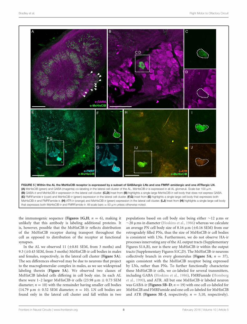
FIGURE 5 | Within the AL the MsHisClB receptor is expressed by a subset of GABAergic LNs and one FMRF-amidergic and one ATRergic LN.(A) MsHisClB (green) and GABA (magenta) co-labeling in the lateral cell cluster of the AL. MsHisClB-ir is expressed in all AL glomeruli. Scale bar 100 μm.(B) GABA-ir and MsHisClB-ir expression in the lateral cell cluster. (C,D) Inset from (B) highlights a single large MsHisClB-ir cell body that does not express GABA.(E) FMRFamide-ir (cyan) and MsHisClB-ir (green) expression in the lateral cell cluster. (F,G) Inset from (E) highlights a single large cell body that expresses bothMsHisClB-ir and FMRFamide-ir. (H) ATR-ir (orange) and MsHisClB-ir (green) expression in the lateral cell cluster. (I,J) Inset from (H) highlights a single large cell bodythat expresses both MsHisClB-ir and FMRFamide-ir. All scale bars = 50 μm unless otherwise noted.
the immunogenic sequence (Figures 1G,H; n = 6), making itunlikely that this antibody is labeling additional proteins. Itis, however, possible that the MsHisClB-ir reflects distributionof the MsHisClB receptor during transport throughout thecell as opposed to distribution of the receptor at functionalsynapses.
In the AL we observed 11 (±0.81 SEM, from 3 moths) and9.3 (±0.43 SEM, from 3 moths) MsHisClB-ir cell bodies in malesand females, respectively, in the lateral cell cluster (Figure 5A).The sex differences observed may be due to neurons that projectto the macroglomerular complex in males, as we see widespreadlabeling therein (Figure 5A). We observed two classes ofMsHisClB labeled cells differing in cell body size. In each ALthere were 1–2 larger MsHisClB-ir cells (23.98 μm ± 0.73 SEMdiameter; n = 10) with the remainder having smaller cell bodies(14.79 μm ± 0.52 SEM diameter; n = 10). LN cell bodies arefound only in the lateral cell cluster and fall within in two
populations based on cell body size being either ∼12 μms or∼20 μms in diameter (Hoskins et al., 1986) whereas we calculatean average PN cell body size of 8.16 μm (±0.16 SEM) from ourretrogradely filled PNs, thus the size of MsHisClB-ir cell bodiesis consistent with LNs. Furthermore, we do not observe HA-irprocesses innervating any of the AL output tracts (SupplementaryFigures S1A,B), nor is there any MsHisClB-ir within the outputtracts (Supplementary Figures S1C,D). TheMsHisClB-ir neuronscollectively branch in every glomerulus (Figure 5A; n = 37),again consistent with the MsHisClB receptor being expressedby LNs, rather than PNs. To further functionally characterizethese MsHisClB-ir cells, we co-labeled for several transmitters,including GABA (Hoskins et al., 1986), FMRFamide (Homberget al., 1990), and ATR. All but one MsHisClB-ir labeled neuronwas GABA-ir (Figures 5B–D; n= 19) with one cell co-labeled forMsHisClB and FMRFamide and one cell co-labeled for MsHisClBand ATR (Figures 5E–J, respectively; n = 5,10, respectively).
Frontiers in Neural Circuits | www.frontiersin.org 8 February 2016 | Volume 10 | Article 5

Bradley et al. Flight Motor to Olfactory Circuit
Together these results suggest that any influence of the MDHnson AL processing is exerted via a population of GABAergic andpeptidergic LNs. The expression of the MsHisClB receptor by ALneurons and the MDHn being the sole source of HA-ir in the ALsuggests that the MDH neurons provide some form of input tothe AL. This does not, however, imply that the MDH neuronsdo not also provide input to circuitry within the MsG. MsHisClBreceptor is also expressed within the MsG (Supplementary FigureS2), however, both the MDHns and HA-ir neurons from themetathoracic and abdominal ganglion (Figure 2F) innervate theMsG, suggesting that HA also plays a role in network functionwithin the MsG.
MDHns Survive Metamorphosis but theLAC lacks MsHisClB ExpressionThere are many neurons that survive metamorphosis, oftenbeing repurposed to take on new tasks to match the dramaticchanges in behavioral demands between the larval and adultlife stage. In Manduca, motor neurons survive metamorphosis,but their morphology and biophysical properties are altereddramatically to allow them to take on life-stage specific tasks,for instance, transitioning from participating in walking motorprograms as larvae to flying motor programs as adults (Duchand Levine, 2000). Given that odor-guided flight is an adultspecific behavior, we predicted that the MDHns would eithernot be present or the MsHisClB-ir would not be expressed inthe LAC. Similar to adults (see Figure 3A), fifth instar larvaehave a pair of large HA-ir cells in the MsG that ascend to the
FIGURE 6 | The MDHns survive metamorphosis, but the MsHisClBreceptor is not expressed in the LAC. (A) Horizontal view of HA-ir in thefifth instar larval MsG shows highly similar cell morphology and radiationpatterns of fine processes as in the adult MsG. (B) HA-ir in the larval brain(green) shows extensive branching in the tritocerebrum (dash-dot line), butvery little innervation in the LAC (dashed line). Syto-59 (magenta) highlights theboundary of the tritocerebrum and LAC. (C) MsHisClB-ir (green) is present inthe tritocerebrum, but not in the LAC. LAC and tritocerebrum highlighted withSyto-59 (magenta) as in (B). All scale bars = 100 μm.
brain (Figure 6A). As in adults, the cell bodies are also locatedventrally near the intersection of the sagittal and horizontalmidlines of theMsG, with a single axon ipsilaterally projecting upeach connective. Furthermore, the HA-ir processes also radiatein all directions in the dorsal MsG as in the adult. Becausethe LAC does not express BRP-ir, we used Syto-59 to label thenuclei of cell bodies that surround the LAC (Figures 6B,C) asa means of highlighting the boundaries of this brain region. Inthe larval brain, HA-ir is most abundant in the tritocerebrum(Figure 6B; dash line) just ventral and lateral to the larval LAC(small dotted line) with a small amount of HA-ir entering theLAC (n = 17). This suggests that the MDHns are present andproject to the olfactory system of larval Manduca. However,there are no MsHisClB-ir neurons within the LAC, despitethe presence of MsHisClB-ir collaterals in the tritocerebrum(Figure 6C; n = 6). This suggests that while the MDHnsprovide sparse innervation of the LAC, they likely do not playa functional role in the larval olfactory system, at least via theMsHisClB receptor, although it is possible that the MsHisClAreceptor is expressed there. What role this circuit would playin the larval olfactory system is not clear as the larva donot fly, but there could be information pertaining to walkingpatterns.
DISCUSSION
Animals use a variety of behavioral strategies to optimize internalrepresentations of the external world, including repetitivemotor patterns that alter stimulus structure. Nervous systemshave concurrently evolved circuits that provide informationto sensory systems about impending behaviors that will affectsensory input. Although this has been well-documented inmany sensory systems, very little is known about neuralcircuits projecting from neural centers governing odor-guidedbehaviors to olfactory networks. The goal of this study wasto characterize a novel sensory-motor to olfactory circuit thatprojects from flight sensory-motor centers to the primaryolfactory processing center in insects. We found that theMDH circuit provides the only source of HA to the AL andaffects a small but diverse population of widely projectingLNs in adult Manduca (Figure 7). Our data suggest thatthe MDHns provide histaminergic inhibitory input to theAL that could modify olfactory processing within the contextof flight or other MsG mediated activity such as walkingpatterns.
The MDHn processes project laterally across the MsG(Figure 3A), yet are most dense in the dorsal MsG (Figure 3B),suggesting that while they may integrate information fromboth sides of the animal, they are likely to interact withcells that are restricted to the dorsal aspect of the MsG.The MsG contains wing and leg motor neurons, sensoryafferents, CPG components, and modulatory neurons someof which occupy specific MsG regions. The dendritic fieldsof wing elevator and depressor motor neurons are locatedin the dorsal region of the MsG in Manduca (Rind, 1983)whereas most of the sensory afferents from the wings are
Frontiers in Neural Circuits | www.frontiersin.org 9 February 2016 | Volume 10 | Article 5

Bradley et al. Flight Motor to Olfactory Circuit
FIGURE 7 | Schematic of the proposed MDHn circuit. (A) Manduca with overlaid nervous system cartoon. (B) Schematic of the MDHn cells from the thoracicganglia to the AL. Only one cell is shown in detail with processes radiating in the MsG, a small process in the prothoracic ganglion, projecting up the cervicothoracicconnective, a branch to the AMMC, and bilateral projections to each AL. (C) MDHn projection entering the ventral AL (green) along with the proposed AL circuitry.For the sake of simplicity, only the processes from MsHisClB-ir expressing neurons (green outline) are shown. MsHisClB-ir GABAergic (pink with green outline) andpeptidergic (cyan or orange with green outline for FMRFamide and ATR, respectively) LNs ramify each glomerulus. Other cell types are also present including PNs(open circles), GABAergic LNs (pink circles with black outlines), ATR LNs (orange circles with black outline), and FMRF LNs (blue circles with black outline). AL,antennal lobe; oe, esophageal foramen; SEZ, subesophageal zone; CTC, cervicothoracic connective; PtG, prothoracic ganglion; MsG, mesothoracic ganglion; MtG,metathoracic ganglion; ab1, abdominal ganglion 1; ab2, abdominal ganglion 2.
localized in both the dorsal and ventral MsG in a closelyrelated species of hawkmoth, Agrius convolvuli (Ando et al.,2011). In addition, there are a population of non-spiking,GABAergic LNs that project to the dorsal side of the MsGof the locust (Watson and Burrows, 1987), and populationsof octopaminergic (Stevenson et al., 1992), serotonergic anddopaminergic neurons (Claassen and Kammer, 1986) that projectthroughout the MsG. The extensive branching of the MDHnsin the MsG suggests that these neurons interact with oneor more components of the MsG. The potential cumulativeeffect of multiple inputs onto MDHns makes understanding theinput to this neural circuit challenging. Single neurons releasingmultiple neurotransmitters alone can have state dependent effectson network output (Swensen and Marder, 2000; Nusbaumet al., 2001). Furthermore, this complexity is compounded whenconsidering the MDHns impact a heterogeneous population ofAL LNs.
Arthropod HA receptors are ligand gated Cl− channels(McClintock and Ache, 1989; Hardie, 1989) sharing ∼45%amino acid similarity to the alpha3 subunit of the humanglycine receptor (Zheng et al., 2002), thus the effect of HAon MsHisClB expressing LNs is likely inhibitory in nature.Within the AL there are ∼300 LNs that belong to a diverseset of subtypes based on morphology, neurotransmitter contentand physiological response properties (Chou et al., 2010;Reisenman et al., 2011). These LNs mediate diverse processingmechanisms such as lateral inhibition for gain control (Olsenand Wilson, 2008). In addition, these widely branching LNsactivate metabotropic receptors whose effects occur on longerand more variable time scales than ionotropic receptors.Therefore the overall network effect of MDHn activity isvariable in both the spatial and temporal domain makingthis circuit difficult to characterize. One potential mechanismwould be suppression of GABA, FMRFamide and ATR releaseby select LNs within the AL. Theoretically, decreasing the
influence of these predominantly inhibitory LNs could actto disinhibit the inhibitory AL local network, which couldlead to a refinement of PN activity. While the role thisrefinement has on AL output activity is not clear, it couldbe in response to the rapid oscillatory nature of the stimulusexperience which is driven in part by wing-beating (Sane andJacobson, 2006). Finally, while invertebrate sensory-motor tosensory circuits typically function to filter reafferent stimuli,we suggest that it is unlikely that the MDHns function inthis manner because non-olfactory responses persist in fullyintact preparations (Tripathy et al., 2010). Therefore, it maybe that MDHn activity indirectly refines PN spatiotemporalresponse patterns to modify the information output to higherorder processing centers during flight. Indeed evidence suggeststhat the fine temporal structure of AL/OB output patternscontain substantial information about odor identity (Daly et al.,2004; Staudacher et al., 2009; Rebello et al., 2014). However,future studies investigating both the activity patterns of MDHnsduring flight behavior and the consequences of HA releaseon AL response properties are necessary to confirm thishypothesis.
Many active sampling behaviors rapidly sample the sensoryfield providing discrete epochs of input to a sensory system; forexample, micro-saccadic eye movements mentioned above. Inaddition, the details of temporally structured reafference maybe dependent on the behavior of the animal. For instance,when exposed to a novel stimulus mice and rats increasetheir sniff frequencies (Kepecs et al., 2007; Wesson et al.,2008a,b) and sniff frequency modulation is dependent onthe specifics of the behavioral task such as free exploration,detection, and discrimination. Insects also show stereotypedactive sampling behaviors that are temporally structured. Bombyxmori require wing beating to track pheromone plumes despitetheir inability to fly (Obara, 1979) and male oriental fruit mothscontinue to fan their wings as they track a calling female
Frontiers in Neural Circuits | www.frontiersin.org 10 February 2016 | Volume 10 | Article 5

Bradley et al. Flight Motor to Olfactory Circuit
even though their final approach is on foot (Baker and Carde,1979).
From a whole nervous system perspective, it is perhaps notsurprising that network-specific processing of information mustbe adjusted based on inputs from many disparate networks.It is becoming increasingly apparent that networks receiveinput from a large number of different sources and thusmust integrate a variety of ongoing contexts. The mammalianRaphe nuclei provide widespread serotonergic input, yet theyalso receive input from many other brain areas (Dorocicet al., 2014; Liu et al., 2014; Weissbourd et al., 2014). Morespecifically, the olfactory systems of animals receive a varietyof inputs from other brain regions including serotonergic(Kent et al., 1987; McLean and Shipley, 1987; Dacks et al.,2006), dopaminergic (Dacks et al., 2012), cholinergic (Macrideset al., 1981; Mandairon et al., 2006), octopaminergic (Dackset al., 2005; Sinakevitch et al., 2005; Sinakevitch and Strausfeld,2006; Dacks and Nighorn, 2011), and GABAergic (Gracia-Llanes et al., 2010; Nunez-Parra et al., 2013) cells all ofwhich modify sensory processing within different, sometimescompeting contexts. Our data support the hypothesis thatolfactory processing in Manduca may also be adjusted withinthe context of ongoing activity in the MsG via the histaminergicMDHns.
AUTHOR CONTRIBUTIONS
SB, PC, KL, KD, and AD designed research. SB, PC, and KLperformed research. SB, PC, KL, KD, and AD analyzed the data.SB, KD, and AD wrote the paper.
FUNDING
This work was supported by RO3DC13997-01 to AMD, and RO1DC009417 to KCD.
ACKNOWLEDGMENTS
We would like to thank Sarah Farris for technical advice andfor editing the manuscript, Jackie Metheny for performing theWestern Blot for the MsHisClB antibody.
SUPPLEMENTARY MATERIAL
The Supplementary Material for this article can be found onlineat: http://journal.frontiersin.org/article/10.3389/fncir.2016.00005
REFERENCESAdrian, E. D. (1942). Olfactory reactions in the brain of the hedgehog. J. Physiol.
100, 459–473. doi: 10.1113/jphysiol.1942.sp003955Ando, N., Wang, H., Shirai, K., and Kanzaki, R. (2011). Central projections of
the wing afferents in the hawkmoth, Agrius convolvuli. J. Insect. Physiol. 57,1518–1536. doi: 10.1016/j.jinsphys.2011.08.002
Baker, T. C., and Carde, R. T. (1979). Analysis of pheromone-mediated behaviorsin male Grapholitha molesta, the oriental fruit moth (Lepidoptera: Tortricidae).Environ. Entomol. 8, 956–968. doi: 10.1093/ee/8.5.956
Bell, R. A., and Joachim, F. G. (1976). Techniques for rearing laboratory colonies oftobacco hornworms and pink bollworms. Ann. Entomol. Soc. Am. 69, 365–373.doi: 10.1093/aesa/69.2.365
Brainard, M. S., and Doupe, A. J. (2000). Interruption of a basal ganglia-forebraincircuit prevents plasticity of learned vocalizations. Nature 404, 762–766. doi:10.1038/35008083
Chalfie, M., Sulston, J. E., White, J. G., Southgate, E., Thomson, J. N., andBrenner, S. (1985). The neural circuit for touch sensitivity in Caenorhabditiselegans. J. Neurosci. 5, 956–964.
Chou, Y. H., Spletter, M. L., Yaksi, E., Leong, J. C., Wilson, R. I., and Luo, L. (2010).Diversity and wiring variability of olfactory local interneurons in the Drosophilaantennal lobe.Nat. Neurosci. 13, 439–449. doi: 10.1038/nn.2489
Christensen, T. A., Waldrop, B. R., and Hildebrand, J. G. (1998). Multitaskingin the olfactory system: context-dependent responses to odors reveal dualGABA-regulated coding mechanisms in single olfactory projection neurons.J. Neurosci. 18, 5999–6008.
Claassen, D. E., and Kammer, A. E. (1986). Effects of octopamine, dopamine, andserotonin on production of flight motor output by thoracic ganglia ofManducasexta. J. Neurobiol. 17, 1–14. doi: 10.1002/neu.480170102
Crapse, T. B., and Sommer, M. A. (2008). Corollary discharge across the animalkingdom. Nat. Rev. Neurosci. 9, 587–600. doi: 10.1038/nrn2457
Dacks, A. M., Christensen, T. A., Agricola, H. J., Wollweber, L., andHildebrand, J. G. (2005). Octopamine-immunoreactive neurons in the brainand subesophageal ganglion of the hawkmothManduca sexta. J. Comp. Neurol.488, 255–268. doi: 10.1002/cne.20556
Dacks, A. M., Christensen, T. A., and Hildebrand, J. G. (2006). Phylogenyof serotonin-immunoreactive neuron in the primary olfactory center
of the insect brain. J. Comp. Neurol. 498, 727–746. doi: 10.1002/cne.21076
Dacks, A. M., and Nighorn, A. J. (2011). The organization of the antennallobe correlates not only with phylogenetic relationship, but also lifehistory: a Basal hymenopteran as exemplar. Chem. Senses 36, 209–220. doi:10.1093/chemse/bjq121
Dacks, A.M., Reisenman, C. E., Paulk, A. C., and Nighorn, A. J. (2010). Histamine-immunoreactive local neurons in the antennal lobes of the hymenoptera.J. Comp. Neurol. 518, 2917–2933. doi: 10.1002/cne.22371
Dacks, A. M., Riffell, J. A., Martin, J. P., Gage, S. L., and Nighorn, A. J.(2012). Olfactory modulation by dopamine in the context of aversive learning.J. Neurophysiol. 108, 539–550. doi: 10.1152/jn.00159.2012
Daly, K. C., Galan, R. F., Peters, O. J., and Staudacher, E. M. (2011). Detailedcharacterization of local field potential oscillations and their relationship tospike timing in the antennal lobe of the mothManduca sexta. Front. Neuroeng.4:12. doi: 10.3389/fneng.2011.00012
Daly, K. C., Kalwar, F., Hatfield, M., Staudacher, E. M., and Bradley, S. P. (2013).Odor detection in Manduca sexta is optimized when odor stimuli are pulsedat a frequency matching the wing beat during flight. PLoS ONE 8:e81863. doi:10.1371/journal.pone.0081863
Daly, K. C., Wright, G. A., and Smith, B. H. (2004). Molecular features of odorantssystematically influence slow temporal responses across clusters of coordinatedantennal lobe units in the moth Manduca sexta. J. Neurophysiol. 92, 236–254.doi: 10.1152/jn.01132.2003
Dorocic, I. P., Fürth, D., Xuan, Y., Johansson, Y., Pozzi, L., Silberberg, G., et al.(2014). A whole-brain atlas of inputs to serotonergic neurons of the dorsaland median raphe nuclei. Neuron 83, 663–678. doi: 10.1016/j.neuron.2014.07.002
Duch, C., and Levine, R. B. (2000). Remodeling of membrane properties anddendritic architecture accompanies the postembryonic conversion of a slowinto a fast motoneuron. J. Neurosci. 20, 6950–6961.
Fouquet, W., Owald, D., Wichmann, C., Mertel, S., Depner, H., Dyba, M., et al.(2009). Maturation of active zone assembly by Drosophila Bruchpilot. J. CellBiol. 186, 129–145. doi: 10.1083/jcb.200812150
Gisselmann, G., Pusch, H., Hovemann, B. T., and Hatt, H. (2002). Two cDNAscoding for histamine-gated ion channels in D. melanogaster. Nat. Neurosci. 5,11–12. doi: 10.1038/nn787
Frontiers in Neural Circuits | www.frontiersin.org 11 February 2016 | Volume 10 | Article 5

Bradley et al. Flight Motor to Olfactory Circuit
Gracia-Llanes, F. J., Crespo, C., Blasco-Ibáñez, J. M., Nacher, J., Varea, E.,Rovira-Esteban, L., et al. (2010). GABAergic basal forebrain afferents innervateselectively GABAergic targets in the main olfactory bulb. Neuroscience 170,913–922. doi: 10.1016/j.neuroscience.2010.07.046
Guerenstein, P. G., Christensen, T. A., and Hildebrand, J. G. (2004). Sensoryprocessing of ambient CO2 information in the brain of the moth Manducasexta. J. Comparat. Physiol. A 190, 707–725. doi: 10.1007/s00359-004-0529-0
Halpern, B. P. (1983). Tasting and smelling as active, exploratory sensory processes.Am. J. Otolaryngol. 4, 246–249. doi: 10.1016/S0196-0709(83)80066-0
Hardie, R. C. (1989). A histamine-activated chloride channel involved inneurotransmission at a photoreceptor synapse. Nature 339, 704–706. doi:10.1038/339704a0
Homberg, U. (1994). Distribution of Neurotransmitters in the Insect Brain. Jena:Gustav Fischer Verlag.
Homberg, U., and Hildebrand, J. G. (1991). Histamine-immunoreactive neurons inthe midbrain and suboesophageal ganglion of the sphinx mothManduca sexta.J. Comparat. Neurol. 307, 647–657. doi: 10.1002/cne.903070410
Homberg, U., Kingan, T. G., and Hildebrand, J. G. (1990). Distributionof FMRFamide-like immunoreactivity in the brain and suboesophagealganglion of the sphinx moth Manduca sexta and colocalization with SCPB-,BPP-, and GABA-like immunoreactivity. Cell Tissue Res. 259, 401–419. doi:10.1007/BF01740767
Hoskins, S. G., Homberg, U., Kingan, T. G., Christensen, T. A., and Hildebrand,J. G. (1986). Immunocytochemistry of GABA in the antennal lobes of the sphinxmothManduca sexta. Cell Tissue Res. 244, 243–252. doi: 10.1007/BF00219199
Houot, B., Burkland, R., Tripathy, S., and Daly, K. C. (2014). Antennal loberepresentations are optimizedwhen olfactory stimuli are periodically structuredto simulate natural wing beat effects. Front. Cell Neurosci. 8:159. doi:10.3389/fncel.2014.00159
Jones, A. K., Bera, A. N., Kees, K., and Sattelle, D. B. (2010). The cys-loop ligand-gated ion channel gene superfamily of the parasitoid wasp, Nasonia vitripennis.Heredity 3, 247–259. doi: 10.1038/hdy.2009.97
Kent, K. S., Hoskins, S. G., and Hildebrand, J. G. (1987). A novel serotonin-immunoreactive neuron in the antennal lobe of the sphinx moth Manducasexta persists throughout postembryonic life. Dev. Neurobiol. 18, 451–465. doi:10.1002/neu.480180506
Kepecs, A., Uchida, N., and Mainen, Z. F. (2007). Rapid and precise control ofsniffing during olfactory discrimination in rats. J. Neurophysiol. 98, 205–213.doi: 10.1152/jn.00071.2007
Kittel, R. J., Wichmann, C., Rasse, T. M., Fouquet, W., Schmidt, M.,Schmid, A., et al. (2006). Bruchpilot promotes active zone assembly,Ca2+ channel clustering, and vesicle release. Science 312, 1051–1054. doi:10.1126/science.1126308
Liu, Z., Zhou, J., Li, Y., Hu, F., Lu, Y., Ma, M., et al. (2014). Dorsal rapheneurons signal reward through 5-HT and glutamate. Neuron 81, 1360–1374.doi: 10.1016/j.neuron.2014.02.010
Lizbinski, K. M., Metheny, J. D., Bradley, S. P., Kesari, A., and Dacks, A. M.(2015). The anatomical basis for modulatory convergence in the antennal lobeof Manduca sexta. J. Comp. Neurol. doi: 10.1002/cne.23926 [Epub ahead ofprint].
Loudon, C., Best, B., and Koehl, M. (1994). When does motion relative toneighboring surfaces alter the flow through arrays of hairs? J. Exp. Biol. 193,233–254.
Loudon, C., and Koehl, M. A. (2000). Sniffing by a silkworm moth: wing fanningenhances air penetration through and pheromone interception by antennae.J. Exp. Biol. 203, 2977–2990.
Macrides, F., Davis, B. J., Youngs, W. M., Nadi, N. S., and Margolis, F. L.(1981). Cholinergic and catecholaminergic afferents to the olfactory bulb in thehamster: a neuroanatomical, biochemical, and histochemical investigation. J.Comp. Neurol. 203, 495–514. doi: 10.1002/cne.902030311
Mandairon, N., Ferretti, C. J., Stack, C. M., Rubin, D. B., Cleland, T. A., andLinster, C. (2006). Cholinergic modulation in the olfactory bulb influencesspontaneous olfactory discrimination in adult rats. Eur. J. Neurosci. 24,3234–3244. doi: 10.1111/j.1460-9568.2006.05235.x
Marder, E., Calabrese, R. L., Nusbaum, M. P., and Trimmer, B. (1987).Distribution and partial characterization of FMRFamide-like peptides in thestomatogastric nervous systems of the rock crab, Cancer borealis, and the
spiny lobster, Panulirus interruptus. J. Comp. Neurol. 259, 150–163. doi:10.1002/cne.902590111
Martinez-Conde, S., Macknik, S. L., Troncoso, X. G., and Dyar, T. A. (2006).Microsaccades counteract visual fading during fixation. Neuron 49, 297–305.doi: 10.1016/j.neuron.2005.11.033
McClintock, T. S., and Ache, B. W. (1989). Histamine directly gates a chloridechannel in lobster olfactory receptor neurons. Proc. Natl. Acad. Sci. U.S.A. 86,8137–8141. doi: 10.1073/pnas.86.20.8137
McLean, J. H., and Shipley, M. T. (1987). Serotonergic afferents to the rat olfactorybulb: I. Origins and laminar specificity of serotonergic inputs in the adult rat.J. Neurosci. 7, 3016–3028.
Melzig, J., Buchner, S., Wiebel, F., Wolf, R., Burg, M., Pak, W. L., et al.(1996). Genetic depletion of histamine from the nervous system of Drosophilaeliminates specific visual andmechanosensory behavior. J. Comp. Physiol. A 179,763–773. doi: 10.1007/BF00207355
Mohr, C., Roberts, P. D., and Bell, C. C. (2003). The mormyromast region ofthe mormyrid electrosensory lobe. II. Responses to input from central sources.J. Neurophysiol. 90, 1211–1223. doi: 10.1152/jn.00211.2003
Nunez-Parra, A., Maurer, R. K., Krahe, K., Smith, R. S., and Araneda, R. C. (2013).Disruption of centrifugal inhibition to olfactory bulb granule cells impairsolfactory discrimination. Proc. Natl. Acad. Sci. U.S.A. 110, 14777–14782. doi:10.1073/pnas.1310686110
Nusbaum, M. P., Blitz, D. M., Swensen, A. M., Wood, D., and Marder, E. (2001).The roles of co-transmission in neural network modulation. Trends Neurosci.24, 146–154. doi: 10.1016/S0166-2236(00)01723-9
Obara, T. (1979). Bombyx mori mating dance: an essential in locating the female.Appl. Entomol. Zool. 14, 130–132.
Olsen, S. R., and Wilson, R. I. (2008). Lateral presynaptic inhibition mediates gaincontrol in an olfactory circuit. Nature 452, 956–960. doi: 10.1038/nature06864
Pantazis, A., Sezaran, A., Liu, C.-H., Nikolaev, A., Rister, J., Thum, A. S.,et al. (2008). Distinct roles for two histamine receptors (hclA and hclB)at the Drosophila photoreceptor synapse. J. Neurosci. 28, 7250–7259. doi:10.1523/JNEUROSCI.1654-08.2008
Peng, H., Ruan, Z., Long, F., Simpson, J. H., and Myers, E. W. (2010). V3D enablesreal-time 3D visualization and quantitative analysis of large-scale biologicalimage data sets. Nat. Biotechnol. 28, 348–353. doi: 10.1038/nbt.1612
Rebello, M. R., McTavish, T. S., Willhite, D. C., Short, S. M., Shepherd, G. M.,and Verhagen, J. V. (2014). Perception of odors linked to precise timing in theolfactory system. PLoS Biol. 12:e1002021. doi: 10.1371/journal.pbio.1002021
Reisenman, C. E., Dacks, A. M., and Hildebrand, J. G. (2011). Local interneurondiversity in the primary olfactory center of the moth Manduca sexta. J. Comp.Physiol. A 6, 653–665. doi: 10.1007/s00359-011-0625-x
Rind, C. F. (1983). The organization of flight motoneurones in the moth,Manducasexta. J. Exp. Biol. 102, 239–251.
Ross, J., Concetta Morrone, M., Goldberg, M. E., and Burr, D. C. (2001). Changesin visual perception at the time of saccades. Trends Neurosci. 24, 113–121. doi:10.1016/S0166-2236(00)01685-4
Sane, S. P., and Jacobson, N. P. (2006). Induced airflow in flying insects II.Measurement of induced flow. J. Exp. Biol. 209, 43–56. doi: 10.1242/jeb.01957
Sievers, F., Wilm, A., Dineen, D. G., Gibson, T. J., Karplus, K., Li, W., et al.(2011). Fast, scalable generation of high-quality protein multiple sequencealignments using Clustal Omega. Mol. Syst. Biol. 7, 1–6. doi: 10.1038/msb.2011.75
Sinakevitch, I., Niwa, M., and Strausfeld, N. J. (2005). Octopamine-likeimmunoreactivity in the honey bee and cockroach: comparable organizationin the brain and subesophageal ganglion. J. Comp. Neurol. 488, 233–254. doi:10.1002/cne.20572
Sinakevitch, I., and Strausfeld, N. J. (2006). Comparison of octopamine-likeimmunoreactivity in the brains of the fruit fly and blow fly. J. Comp. Neurol.494, 460–475. doi: 10.1002/cne.20799
Sommer, M. A., and Wurtz, R. H. (2002). A pathway in primate brainfor internal monitoring of movements. Science 296, 1480–1482. doi:10.1126/science.1069590
Staudacher, E. M., Huetteroth, W., Schachtne, J., and Daly, K. C. (2009). A 4-dimensional representation of antennal lobe output based on an ensembleof characterized projection neurons. J. Neurosci. Methods 180, 208–223. doi:10.1016/j.jneumeth.2009.03.019
Frontiers in Neural Circuits | www.frontiersin.org 12 February 2016 | Volume 10 | Article 5

Bradley et al. Flight Motor to Olfactory Circuit
Stevenson, P. A., Pflüger, H. J., Eckert, M., and Rapus, J. (1992). Octopamineimmunoreactive cell populations in the locust thoracic-abdominal nervoussystem. J. Comp. Neurol. 315, 382–397. doi: 10.1002/cne.903150403
Stuart, A. E. (1999). From fruit flies to barnacles, histamine is the neurotransmitterof arthropod photoreceptors. Neuron 22, 431–433. doi: 10.1016/S0896-6273(00)80699-6
Swensen, A. M., and Marder, E. (2000). Multiple peptides converge to activatethe same voltage-dependent current in a central pattern-generating circuit.J. Neurosci. 20, 6752–6759.
Tripathy, S. J., Peters, O. J., Staudacher, E. M., Kalwar, F. R., Hatfield, M. N., andDaly, K. C. (2010).Odors pulsed at wing beat frequencies are tracked by primaryolfactory networks and enhance odor detection. Front. Cell Neurosci. 4:1. doi:10.3389/neuro.03.001.2010
Utz, S., Huetteroth, W., Vömel, M., and Schactner, J. (2008). Mas-allatotropinin the developing antennal lobe of the sphinx moth Manduca sexta:distribution, time course, developmental regulation, and colocalization withother neuropeptides.Dev. Neurobiol. 68, 123–142. doi: 10.1002/dneu.20579
Veenstra, J. A., and Hagedorn, H. H. (1993). Sensitive enzyme immunoassay forManduca allatotropin and the existence of an allatotropin-immunoreacitvepeptide in Periplaneta americana.Arch. Insect Biochem. Physiol. 23, 99–109. doi:10.1002/arch.940230302
Verhagen, J. V., Wesson, D. W., Netoff, T. I., White, J. A., and Wachowiak, M.(2007). Sniffing controls an adaptive filter of sensory input to the olfactory bulb.Nat. Neurosci. 10, 631–639. doi: 10.1038/nn1892
Wagh, D. A., Rasse, T. M., Asan, E., Hofbauer, A., Schwenkert, I., Dürrbeck, H.,et al. (2006). Bruchpilot, a proteinwith homology to ELKS/CAST, is required forstructural integrity and function of synaptic active zones in Drosophila. Neuron49, 833–844. doi: 10.1016/j.neuron.2006.02.008
Watson, A. H., and Burrows, M. (1987). Immunoctochemical and pharmacologicalevidence for GABAergic spiking local interneurons in the locust. J. Neurosci. 7,1741–1751.
Weissbourd, B., Ren, J., DeLoach, K. E., Guenthner, C. J., Miyamichi, K.,and Luo, L. (2014). Presynaptic partners of dorsal raphe serotonergic
and GABAergic neurons. Neuron 83, 645–662. doi: 10.1016/j.neuron.2014.06.024
Wesson, D. W., Carey, R. M., Verhagen, J. V., and Wachowiak, M. (2008a).Rapid encoding and perception of novel odors in the rat. PLoS Biol. 6:82. doi:10.1371/journal.pbio.0060082
Wesson, D. W., Donahou, T. N., Johnson, M. O., and Wachowiak, M. (2008b).Sniffing behavior of mice during performance in odor-guided tasks. Chem.Senses 33, 581–596. doi: 10.1093/chemse/bjn029
Witten, J. L., and Truman, J. W. (1996). Developmental plasticity ofneuropeptide expression in motoneurons of the moth, Manduca sexta:steroid hormone regulation. J. Neurobiol. 29, 99–114. doi: 10.1002/(SICI)1097-4695(199601)29:1<99::AID-NEU8>3.3.CO;2-4
Woo, S. H., Stumpfova, M., Jensen,U. B., Lumpkin, E. A., andOwens, D.M. (2010).Identification of epidermal progenitors for the Merkel cell lineage.Development23, 3965–3971. doi: 10.1242/dev.055970
Zaretsky, M., and Rowell, C. H. F. (1979). Saccadic suppression by corollarydischarge in the locust. Nature 280, 583–585. doi: 10.1038/280583a0
Zheng, Y., Hirschberg, B., Yuan, J., Wang, A. P., Hunt, D. C., Ludmerer, S.W., et al.(2002). Identification of two novel Drosophila melanogaster histamine-gatedchloride channel subunits expressed in the eye. J. Biol. Chem. 3, 2000–2005. doi:10.1074/jbc.M107635200
Conflict of Interest Statement: The authors declare that the research wasconducted in the absence of any commercial or financial relationships that couldbe construed as a potential conflict of interest.
Copyright © 2016 Bradley, Chapman, Lizbinski, Daly and Dacks. This is an open-access article distributed under the terms of the Creative Commons AttributionLicense (CC BY). The use, distribution or reproduction in other forums is permitted,provided the original author(s) or licensor are credited and that the originalpublication in this journal is cited, in accordance with accepted academic practice.No use, distribution or reproduction is permitted which does not comply with theseterms.
Frontiers in Neural Circuits | www.frontiersin.org 13 February 2016 | Volume 10 | Article 5