CHAPTER 17 SENSORY SYSTEM. OLFACTION Sense of smell Olfactory organs in nasal cavity Olfactory...
43
CHAPTER 17 SENSORY SYSTEM
-
Upload
august-ferguson -
Category
Documents
-
view
230 -
download
1
Transcript of CHAPTER 17 SENSORY SYSTEM. OLFACTION Sense of smell Olfactory organs in nasal cavity Olfactory...
C H A P T E R 1 7
SENSORY SYSTEM
OLFACTION
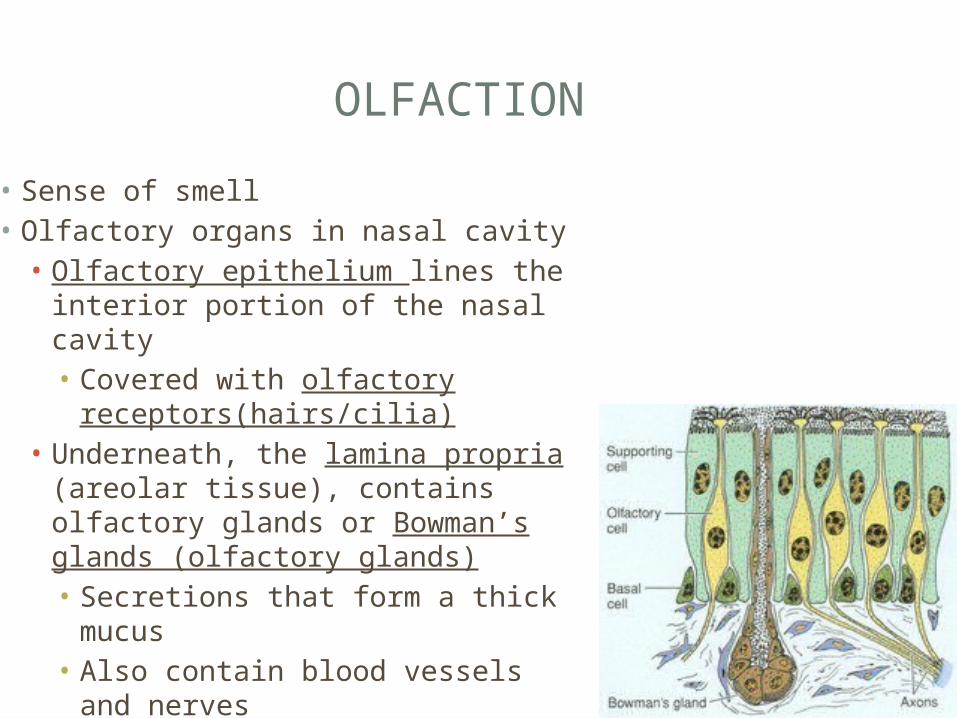
• Sense of smell• Olfactory organs in nasal cavity• Olfactory epithelium lines the
interior portion of the nasal cavity• Covered with olfactory
receptors(hairs/cilia)• Underneath, the lamina propria
(areolar tissue), contains olfactory glands or Bowman’s glands (olfactory glands)• Secretions that form a thick
mucus• Also contain blood vessels and
nerves
OLFACTORY RECEPTORS
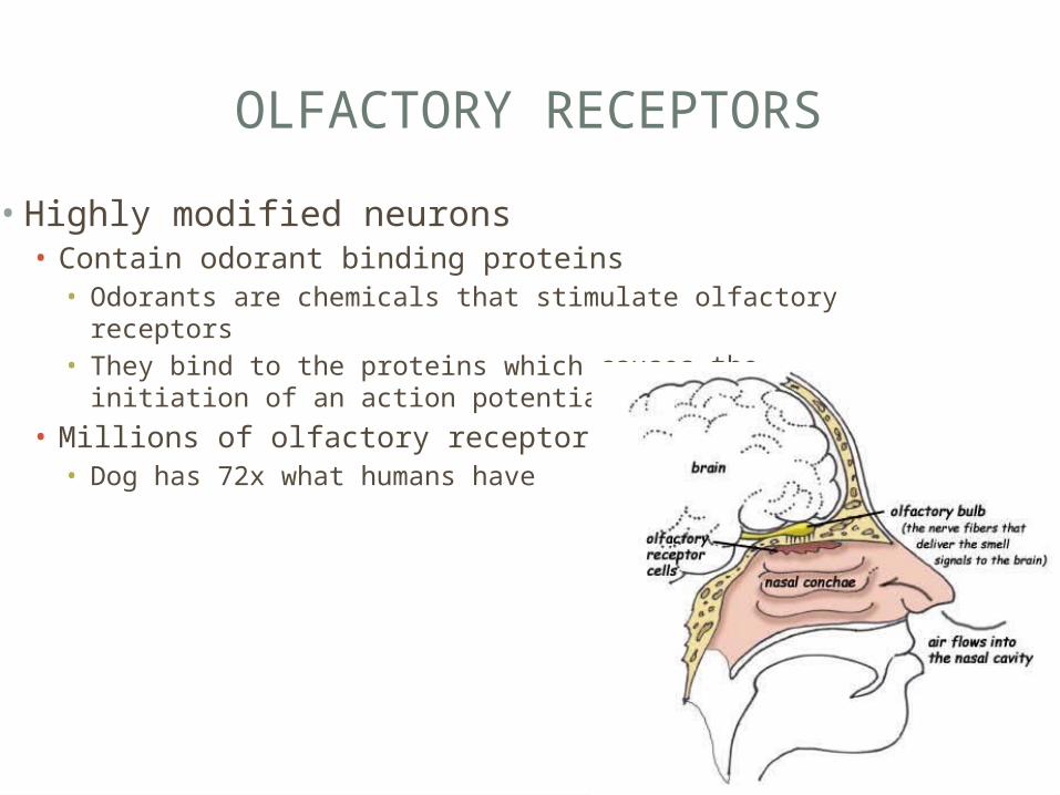
• Highly modified neurons• Contain odorant binding proteins • Odorants are chemicals that stimulate olfactory receptors• They bind to the proteins which causes the initiation of an
action potential to the CNS
• Millions of olfactory receptors • Dog has 72x what humans have
OLFACTORY PATHWAYS
• Very sensitive• 4 molecules of odorants can activate an olfactory receptor• Message must be sent to the olfactory cortex within the
temporal lobe before you’re aware of it
• Odorants stimulate Olfactory Receptors action potential initiated pass through the Ethmoid bone to the olfactory bulbs within the cerebrum message passed to the olfactory nerves hypothalamus (remember emotions) which trigger emotional/behavior responses
• perfumes
GUSTATION
• Taste-provides information about foods/liquids consumed• Gustatory Receptors = taste receptors, on superior
surface of tongue• Gustatory Receptors + specialized epithelial cells =
taste buds• Approx. 3000 per adult• Contain about approx. 40 gustatory receptors each• Projections or taste hairs that collect chemicals
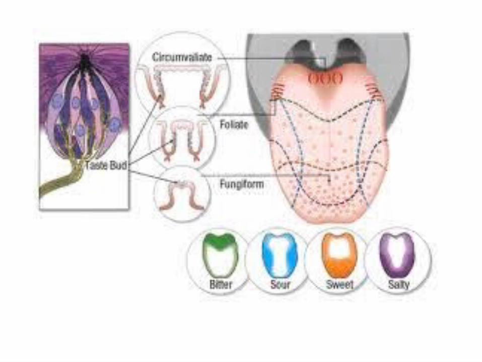
LINGUAL PAPILLAE
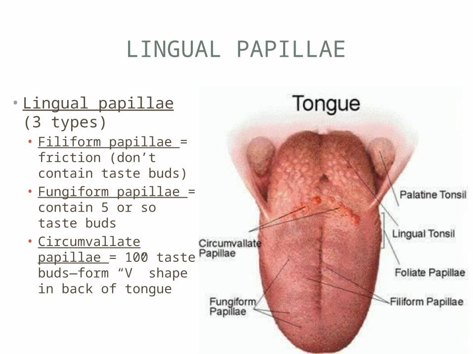
• Lingual papillae (3 types)• Filiform papillae =
friction (don’t contain taste buds)
• Fungiform papillae = contain 5 or so taste buds
• Circumvallate papillae = 100 taste buds—form “V” shape in back of tongue
GUSTATORY PATHWAY
• Taste buds are controlled by the facial, glossopharyngeal, and vagus nerves. • Conscious perception of taste is processed with
other sensory data• Texture of food• Taste
• Afferent Neural connection between Olfaction and Gustation• More sensitive to taste when your sense of olfaction is
working properly• When your ill, airborne molecules cannot reach the olfactory
receptors = meals taste dull

GUSTATORY PATHWAY
• Dissolved chemicals contact taste hairs depending upon the chemical, various protein channels will open initiates an immediate release of neurotransmitter to the postsynaptic cell postsynaptic cell generates action potential CNS• Primary taste sensations• Sweet• Salt• Sour• Bitter• Umami = amino acids, peptides found in beef/chicken products• Water = in pharynx, hold water in mouth triggers hormone
secretion leading to water loss in kidneys
TASTE LAB
• Get with a partner (need a notebook and pencil)• On the paper, draw a simple illustration of the tongue
• Grab 2 q-tips per partner
• Dip the q-tip into the unknown solution and roll it on various regions of your tongue• See if you can identify regions of hypersensitivity and the
type of taste being stimulated• Once identified, pencil them in on your tongue diagram
C H A P T E R 1 7
VISION/EQUILIBRIUM & HEARING
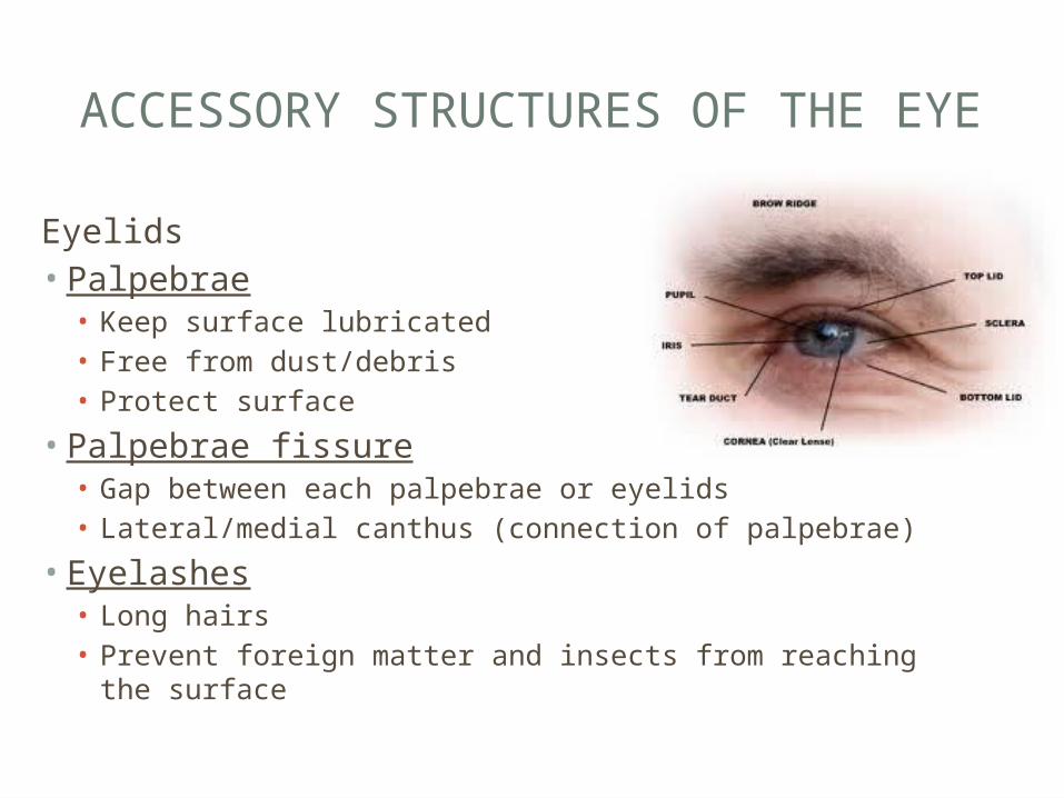
ACCESSORY STRUCTURES OF THE EYE
Eyelids• Palpebrae• Keep surface lubricated• Free from dust/debris• Protect surface
• Palpebrae fissure• Gap between each palpebrae or eyelids• Lateral/medial canthus (connection of palpebrae)
• Eyelashes• Long hairs• Prevent foreign matter and insects from reaching the
surface
ACCESSORY STRUCTURES OF THE EYE
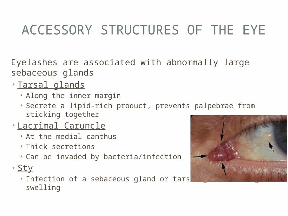
Eyelashes are associated with abnormally large sebaceous glands• Tarsal glands• Along the inner margin• Secrete a lipid-rich product, prevents palpebrae from sticking
together
• Lacrimal Caruncle• At the medial canthus• Thick secretions• Can be invaded by bacteria/infection
• Sty• Infection of a sebaceous gland or tarsal gland causing swelling
ACCESSORY STRUCTURES OF THE EYE
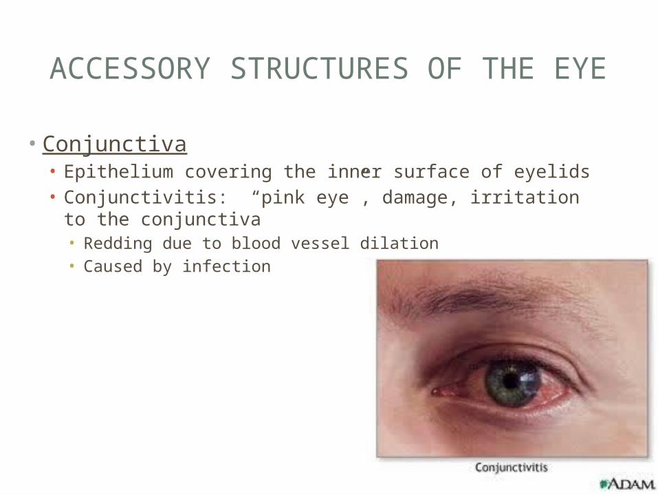
• Conjunctiva• Epithelium covering the inner surface of eyelids• Conjunctivitis: “pink eye”, damage, irritation to the
conjunctiva• Redding due to blood vessel dilation• Caused by infection
ACCESSORY STRUCTURES OF THE EYE
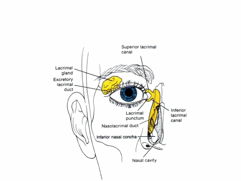
• The Lacrimal Apparatus• Produces, distributes, and removes tears• Reduce friction, remove debris, prevent bacterial infections,
provide nutrients to that tissue
• Parts• Lacrimal gland: secretes the tear substance with a lysozyme,
to attack bacteria• Lacrimal puncta: 2 small pores that drain tears to canaliculi• Lacrimal Canaliculi: small canals that lead to the sac for
disposal of tears• Lacrimal Sac: collects a portion of the tears before
depositing them into the duct• Nasolacrimal duct/canal: found in the lacrimal bone, delivers
the tears to nasal cavity
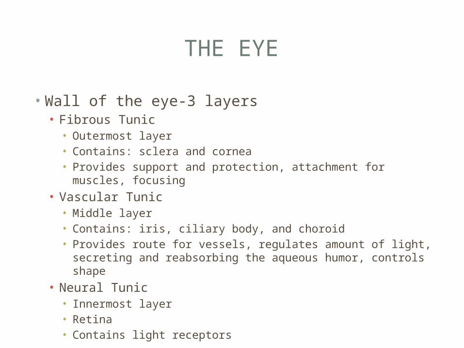
THE EYE
• Wall of the eye-3 layers• Fibrous Tunic• Outermost layer• Contains: sclera and cornea• Provides support and protection, attachment for muscles,
focusing
• Vascular Tunic• Middle layer• Contains: iris, ciliary body, and choroid• Provides route for vessels, regulates amount of light,
secreting and reabsorbing the aqueous humor, controls shape
• Neural Tunic• Innermost layer• Retina• Contains light receptors
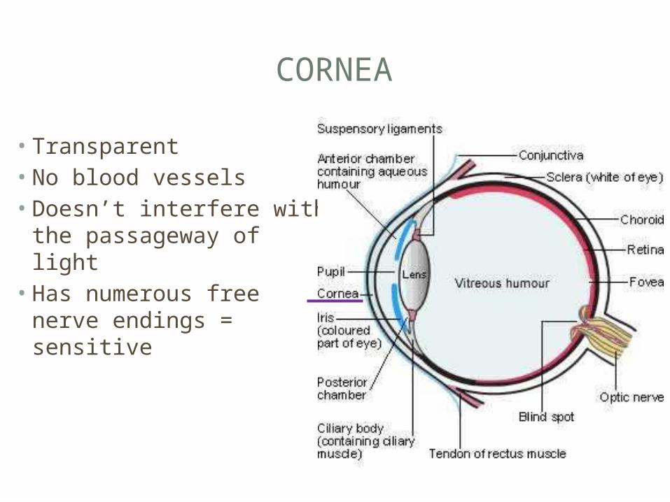
CORNEA
• Transparent• No blood vessels• Doesn’t interfere with the
passageway of light• Has numerous free nerve
endings = sensitive
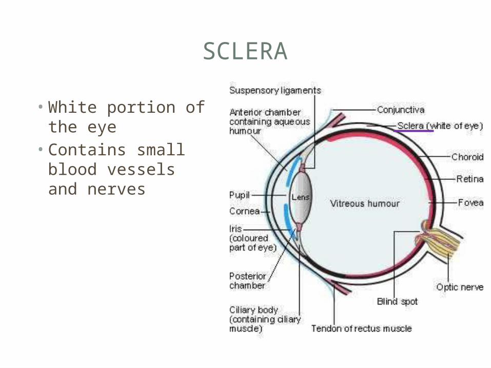
SCLERA
• White portion of the eye• Contains small
blood vessels and nerves
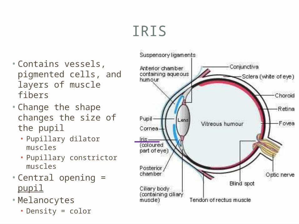
IRIS
• Contains vessels, pigmented cells, and layers of muscle fibers• Change the shape
changes the size of the pupil• Pupillary dilator muscles• Pupillary constrictor
muscles
• Central opening = pupil• Melanocytes• Density = color
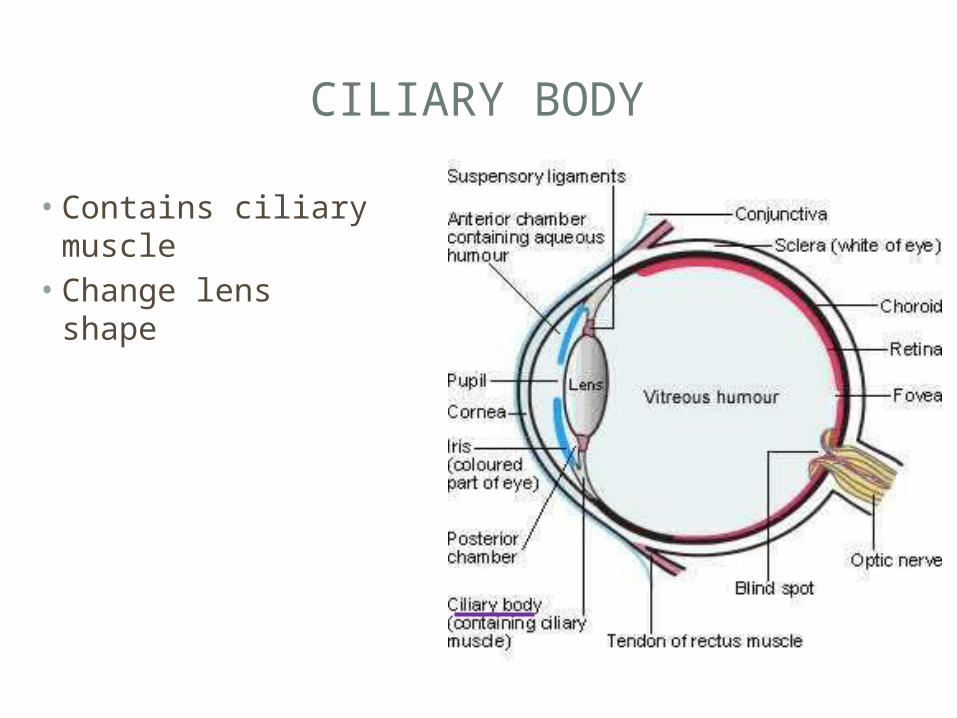
CILIARY BODY
• Contains ciliary muscle• Change lens shape
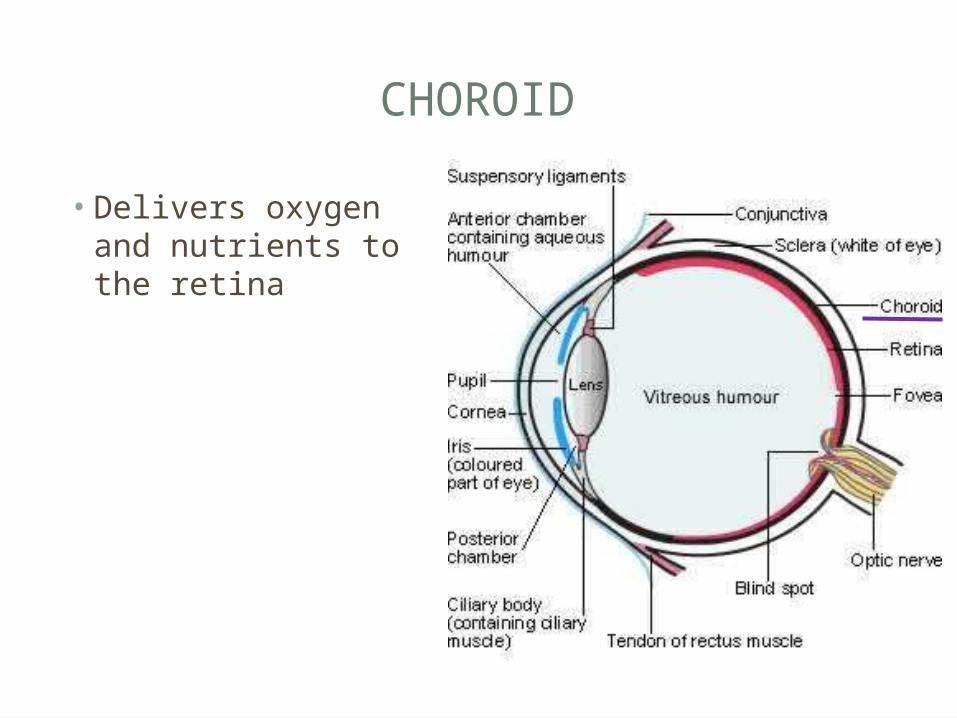
CHOROID
• Delivers oxygen and nutrients to the retina
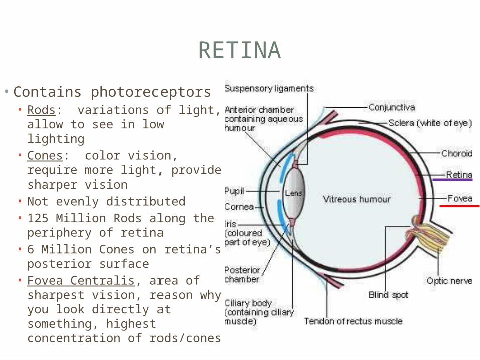
RETINA
• Contains photoreceptors• Rods: variations of light,
allow to see in low lighting• Cones: color vision, require
more light, provide sharper vision
• Not evenly distributed• 125 Million Rods along the
periphery of retina• 6 Million Cones on retina’s
posterior surface• Fovea Centralis, area of
sharpest vision, reason why you look directly at something, highest concentration of rods/cones
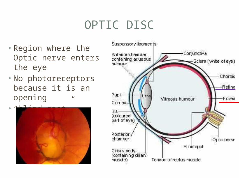
OPTIC DISC
• Region where the Optic nerve enters the eye• No photoreceptors
because it is an opening• “blind spot”
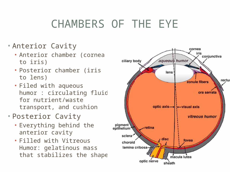
CHAMBERS OF THE EYE
• Anterior Cavity• Anterior chamber (cornea
to iris)• Posterior chamber (iris to
lens)• Filed with aqueous humor :
circulating fluid for nutrient/waste transport, and cushion
• Posterior Cavity• Everything behind the
anterior cavity• Filled with Vitreous Humor:
gelatinous mass that stabilizes the shape
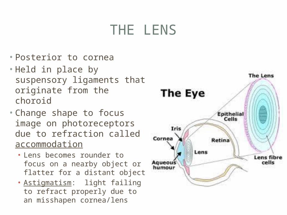
THE LENS
• Posterior to cornea• Held in place by
suspensory ligaments that originate from the choroid• Change shape to focus
image on photoreceptors due to refraction called accommodation• Lens becomes rounder to
focus on a nearby object or flatter for a distant object
• Astigmatism: light failing to refract properly due to an misshapen cornea/lens
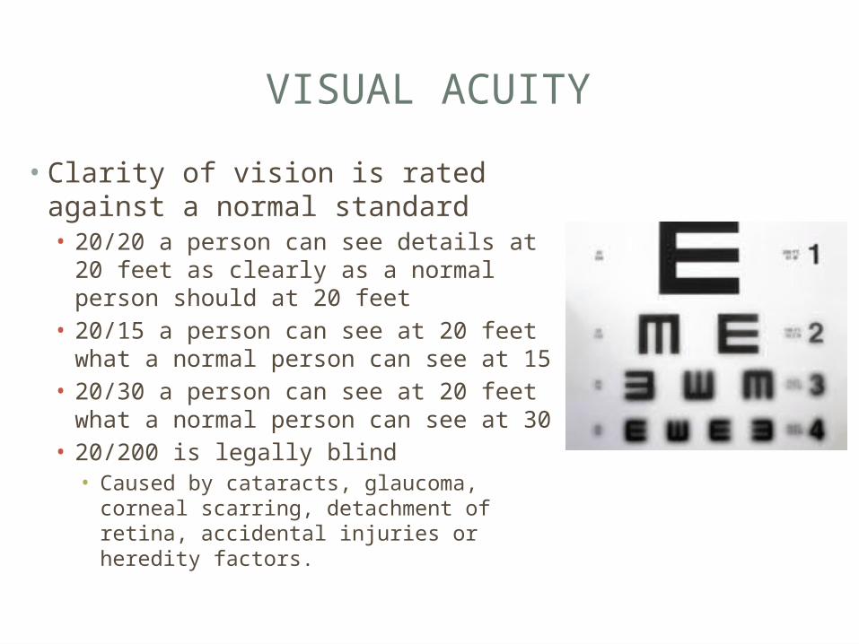
VISUAL ACUITY
• Clarity of vision is rated against a normal standard• 20/20 a person can see details at 20
feet as clearly as a normal person should at 20 feet
• 20/15 a person can see at 20 feet what a normal person can see at 15
• 20/30 a person can see at 20 feet what a normal person can see at 30
• 20/200 is legally blind• Caused by cataracts, glaucoma, corneal
scarring, detachment of retina, accidental injuries or heredity factors.
SIGHT LAB
Groups of 3• 2 groups Visual Acuity • 2 groups Peripheral vision disc• 1 group Astigmatism/blind spot• 1 group Page 455 in packet part B• 1 group study with models

EMOTIONAL TEARS
• proteins carrying hormones found in tears• Prolactin• Hormone in greater levels in women than men
• When you have an emotional response, it is due to the onset production of hormones• These hormones are produced in the lacrimal gland
which then are released through tears
• Prolactin levels increase tremendously during breastfeeding, stimulates lactation• Women have a strong emotional responses while breast
feeing
LASIK SURGERY
• Operation• Patient is awake, given anesthetic eye drops• Step 1: Remove the conjunctiva tissue like a flap• Step 2: Remodel the cornea because the uneven amounts
push on the lens causing a change in its shape or excess causes blurred vision
• http://www.youtube.com/watch?v=O4kDC4sZ5Jg
VISUAL DISORDERS
• Cataracts• Clouding or discoloration of the lens• Due to a precise balance of structural and biochemical
characteristics• Symptoms: blurry vision, faded colors, glare, poor night
vision, double vision• Lens transplants to fix• Develops as you age
• Glaucoma• Intraocular pressure rises because the aqueous humor
can’t be filter properly• Pressure distorts the soft tissues of the eye • Treated with drugs, surgery to enable drainage

COLOR VISION
• Anatomy of Rods and cones• Rod/cone refer to the outer segment’s
shape
• 3 types of cones• Blue Cones (16%)• Green Cones (10%)• Red Cones (74%)• If all 3 are stimulated, you perceive the color
white• Combination is what allows us to understand
different colors/shades
COLOR BLINDNESS
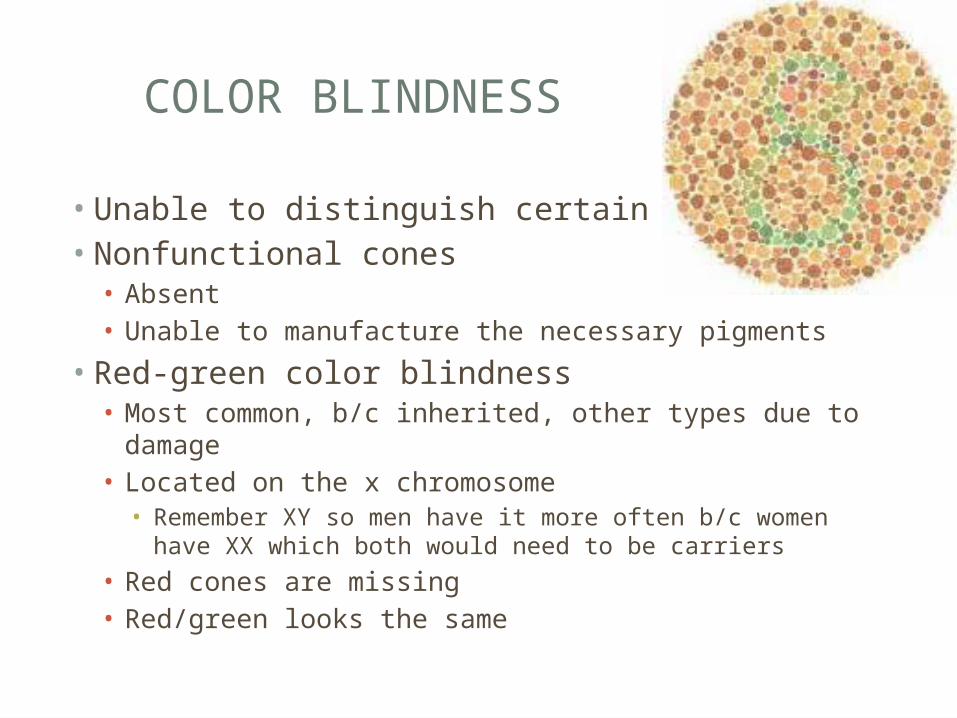
• Unable to distinguish certain colors• Nonfunctional cones• Absent• Unable to manufacture the necessary pigments
• Red-green color blindness• Most common, b/c inherited, other types due to damage• Located on the x chromosome• Remember XY so men have it more often b/c women have XX
which both would need to be carriers
• Red cones are missing• Red/green looks the same
COW EYE DISSECTION
SOUND
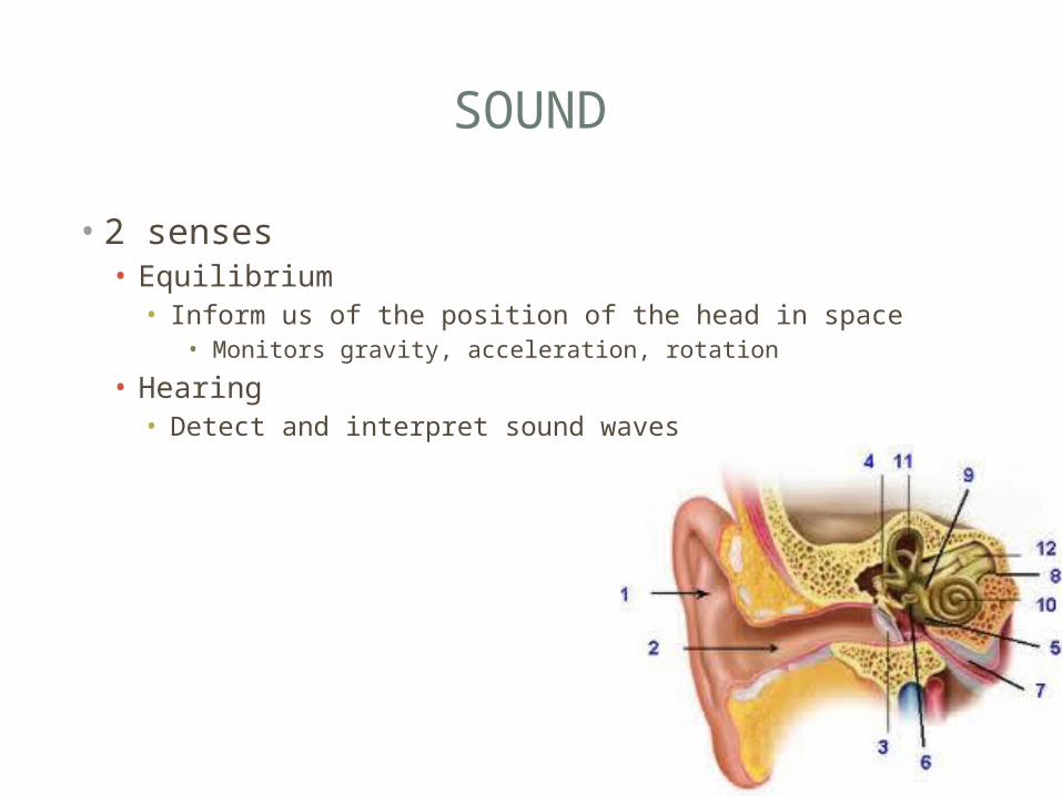
• 2 senses• Equilibrium• Inform us of the position of the head in space
• Monitors gravity, acceleration, rotation
• Hearing• Detect and interpret sound waves
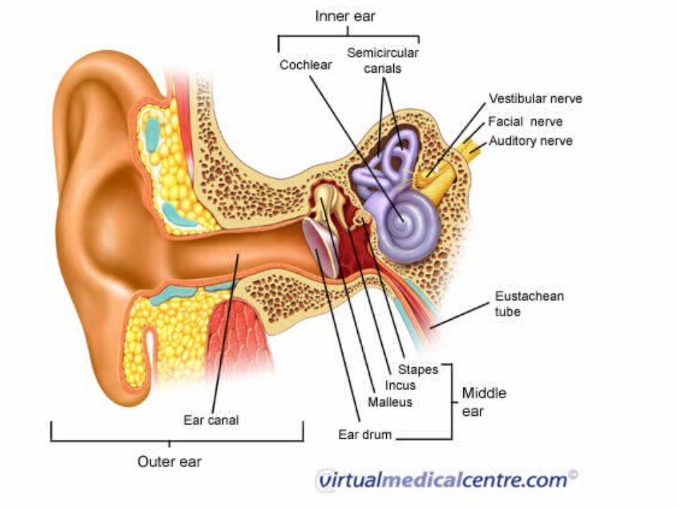
ANATOMY OF THE EAR
• 3 main Regions• External Ear• Visible portion• Collects/directs sound waves and directs them to the middle
ear
• Middle Ear• Chamber within the temporal bone• Collect sound waves and transmit them to the inner ear
• Inner Ear• Contains sensory organs for hearing and equilibrium
EXTERNAL EAR
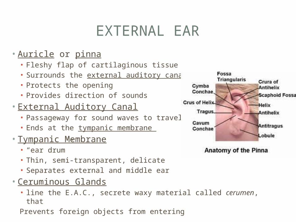
• Auricle or pinna• Fleshy flap of cartilaginous tissue• Surrounds the external auditory canal • Protects the opening• Provides direction of sounds
• External Auditory Canal• Passageway for sound waves to travel• Ends at the tympanic membrane
• Tympanic Membrane• “ear drum”• Thin, semi-transparent, delicate• Separates external and middle ear
• Ceruminous Glands• line the E.A.C., secrete waxy material called cerumen, thatPrevents foreign objects from entering
MIDDLE EAR
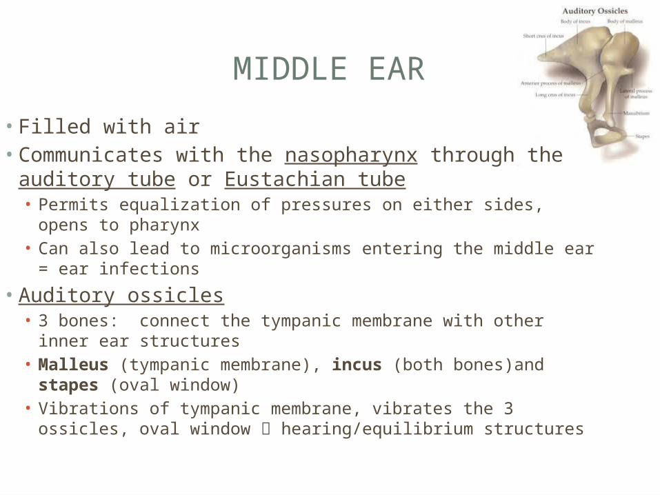
• Filled with air• Communicates with the nasopharynx through the
auditory tube or Eustachian tube• Permits equalization of pressures on either sides, opens to
pharynx• Can also lead to microorganisms entering the middle ear = ear
infections
• Auditory ossicles• 3 bones: connect the tympanic membrane with other inner
ear structures• Malleus (tympanic membrane), incus (both bones)and stapes (oval window)
• Vibrations of tympanic membrane, vibrates the 3 ossicles, oval window hearing/equilibrium structures
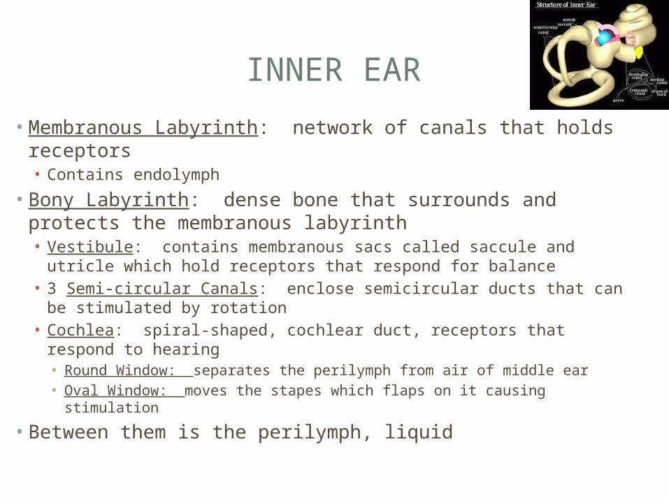
INNER EAR
• Membranous Labyrinth: network of canals that holds receptors• Contains endolymph
• Bony Labyrinth: dense bone that surrounds and protects the membranous labyrinth• Vestibule: contains membranous sacs called saccule and utricle
which hold receptors that respond for balance• 3 Semi-circular Canals: enclose semicircular ducts that can be
stimulated by rotation• Cochlea: spiral-shaped, cochlear duct, receptors that respond to
hearing• Round Window: separates the perilymph from air of middle ear• Oval Window: moves the stapes which flaps on it causing stimulation
• Between them is the perilymph, liquid


PATHWAY OF SOUND
• Sound waves external auditory canal and make the tympanic membrane vibrate.
• Tympanic membrane vibration causes movement of auditory ossicles, sound waves amplified
• Stapes moves against oval window, pressure waves generated
• Travel from oval window and through the scala vestibuli (area of cochlea)
• Hair cells in the cochlea detect movement and convert stimulus into nerve impulse which travels to the cochlear nerve.
• Excess waves exit through the round window
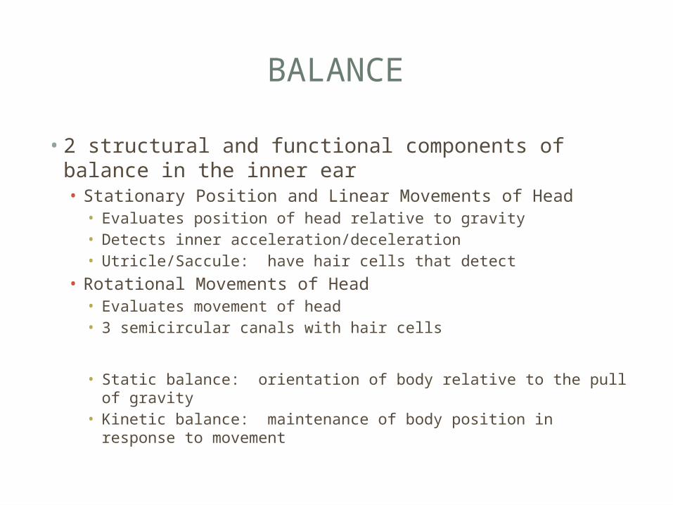
BALANCE
• 2 structural and functional components of balance in the inner ear• Stationary Position and Linear Movements of Head• Evaluates position of head relative to gravity• Detects inner acceleration/deceleration• Utricle/Saccule: have hair cells that detect
• Rotational Movements of Head• Evaluates movement of head• 3 semicircular canals with hair cells
• Static balance: orientation of body relative to the pull of gravity
• Kinetic balance: maintenance of body position in response to movement