A Clinicopathological Analysis of Granulomatous Dermatitis ... Article.pdf · 2. Inoue Y, Suqa M....
4
JK SCIENCE 22 www.jkscience.org Vol. 19 No. 1, Jan.-March 2017 ORIGINAL ARTICLE A Clinicopathological Analysis of Granulomatous Dermatitis : 4 Year Retrospective Study Jyotsna Suri, Subhash Bhardwaj, Rita Kumari, Shailija Kotwal Granuloma is defined as a focal chronic inflammatory response to tissue injury, characterised by focal, compact collection of inflammatory cells, principally of the activated histiocytes, modified epitheloid macrophages & multinucleate giant cells that may or may not be rimmed by lymphocytes and show central necrosis. (1) Granulomas are formed as an end result of complex interplay of inflammatory cells and chemical mediators in hypersensitive response to nondegradable product or antigen resulting in prolonged antigenaemia. Various chemoattractants secreted by activated macrophages, T cells, B cells attract monocytes macrophages which undergo transformation to form epitheloid cells and many fuse to form multinucleate giant cells. (2, 3) CD4 helper cells and lymphocytes are necessary for development of the granulomas. The helper cells produce IL2, IFN gamma & TNF beta upon stimulation with antigen and delayed type of hypersensitivity reaction. Granulomas resulting from different infections have different immunoregulatory mechanisms governing their formation and resolution. On the basis of aetiology, granulomatous inflammation is classified into bacterial, fungal, viral, cat scratch fever, lymhogranuloma venereum, helminthic, foreign body type and unknown cause. In the dermatology and dermatopathology granulomatous dermatitis presents a diagnostic challenge, many a times. The granulomatous dermatitis comprise a large family sharing the common histological denominator of granulomas formation. Rightly said that in granulomatous dermatitis, an identical histologic picture may be produced by several causes & conversely a single cause may produce varied histologic patterns. (3) This makes it cumbersome to classify granulomatous dermatitis in a satisfactory way. The present study was carried out with the aim of classifying granulomatous dermatitis on the basis of aetiology and morphology and to study the incidence of different aetiologies of granulomatous dermatitis in hospital based population of Jammu region. Material and Methods A retrospective 4 year analysis of skin biopsies reported in dermatopathology section of the department of pathology GMC Jammu and the cases diagnosed as one or other form of granulomatous dermatitis were retrieved. Detailed information's like age, sex and clinical diagnosis were recorded. In each case, haematoxylin and eosin stained paraffin sections along with special stains like PAS, Ziehl-Neelsen (ZN), Giemsa, etc. were studied. Results In the four year retrospective study, out of total 1081 cases reported in the dermatopathology section of Introduction Abstract The present study was carried out with an attempt to study the incidence of granulomatous dermatitis in hospital based population and to classify and compare the granulomatous dermatitis on the basis of histopathology and find the etiology. This is a four year retrospective study done on the data available in the dermatopathology section of department of pathology. The cases diagnosed as granulomatous dermatitis were retrieved, clinical data and the histopathological features compared to know the incidence of various etiologies of GD. Out of 310 cases of GD with male to female ratio 2.03:1, leprosy comprised major reported etiology (n-244) followed by tuberculosis (n-44), sarcoidosis (n-4), Leshmania Donovani (n-14)) granuloma annulare (n-1) and 3 granulomatous lesions not further classified. Infections form the commonest form of GD out of which leprosy forms the major group. Role of histopathology(H&E and special stains) is very important in confirming the diagnosis of granulomatous dermatitis . Key Words Granuloma, Dermatitis, Inflammatory, Tissue Injury From the Department of Pathology, Govt. Medical College Jammu- J&K India 180001 Correspondence to : Dr Jyotsna Suri , Associate Professor, Department of Pathology , Government Medical College, Jammu J&K India 180001
Transcript of A Clinicopathological Analysis of Granulomatous Dermatitis ... Article.pdf · 2. Inoue Y, Suqa M....

JK SCIENCE
22 www.jkscience.org Vol. 19 No. 1, Jan.-March 2017
ORIGINAL ARTICLE
A Clinicopathological Analysis of GranulomatousDermatitis : 4 Year Retrospective Study
Jyotsna Suri, Subhash Bhardwaj, Rita Kumari, Shailija Kotwal
Granuloma is defined as a focal chronic inflammatoryresponse to tissue injury, characterised by focal, compactcollection of inflammatory cells, principally of the activatedhistiocytes, modified epitheloid macrophages &multinucleate giant cells that may or may not be rimmedby lymphocytes and show central necrosis. (1)
Granulomas are formed as an end result of complexinterplay of inflammatory cells and chemical mediatorsin hypersensitive response to nondegradable product orantigen resulting in prolonged antigenaemia. Variouschemoattractants secreted by activated macrophages, Tcells, B cells attract monocytes macrophages whichundergo transformation to form epitheloid cells and manyfuse to form multinucleate giant cells. (2, 3)
CD4 helper cells and lymphocytes are necessary fordevelopment of the granulomas. The helper cells produceIL2, IFN gamma & TNF beta upon stimulation withantigen and delayed type of hypersensitivity reaction.Granulomas resulting from different infections havedifferent immunoregulatory mechanisms governing theirformation and resolution. On the basis of aetiology,granulomatous inflammation is classified into bacterial,fungal, viral, cat scratch fever, lymhogranuloma venereum,helminthic, foreign body type and unknown cause. In thedermatology and dermatopathology granulomatous
dermatitis presents a diagnostic challenge, many a times.The granulomatous dermatitis comprise a large familysharing the common histological denominator ofgranulomas formation. Rightly said that in granulomatousdermatitis, an identical histologic picture may be producedby several causes & conversely a single cause mayproduce varied histologic patterns. (3) This makes itcumbersome to classify granulomatous dermatitis in asatisfactory way. The present study was carried out withthe aim of classifying granulomatous dermatitis on thebasis of aetiology and morphology and to study theincidence of different aetiologies of granulomatousdermatitis in hospital based population of Jammu region.Material and Methods
A retrospective 4 year analysis of skin biopsiesreported in dermatopathology section of the departmentof pathology GMC Jammu and the cases diagnosed asone or other form of granulomatous dermatitis wereretrieved. Detailed information's like age, sex and clinicaldiagnosis were recorded. In each case, haematoxylin andeosin stained paraffin sections along with special stainslike PAS, Ziehl-Neelsen (ZN), Giemsa, etc. were studied.Results
In the four year retrospective study, out of total 1081cases reported in the dermatopathology section of
Introduction
AbstractThe present study was carried out with an attempt to study the incidence of granulomatous dermatitis inhospital based population and to classify and compare the granulomatous dermatitis on the basis ofhistopathology and find the etiology. This is a four year retrospective study done on the data available in thedermatopathology section of department of pathology. The cases diagnosed as granulomatous dermatitiswere retrieved, clinical data and the histopathological features compared to know the incidence of variousetiologies of GD. Out of 310 cases of GD with male to female ratio 2.03:1, leprosy comprised majorreported etiology (n-244) followed by tuberculosis (n-44), sarcoidosis (n-4), Leshmania Donovani (n-14))granuloma annulare (n-1) and 3 granulomatous lesions not further classified. Infections form the commonestform of GD out of which leprosy forms the major group. Role of histopathology(H&E and special stains)is very important in confirming the diagnosis of granulomatous dermatitis .
Key WordsGranuloma, Dermatitis, Inflammatory, Tissue Injury
From the Department of Pathology, Govt. Medical College Jammu- J&K India 180001Correspondence to : Dr Jyotsna Suri , Associate Professor, Department of Pathology , Government Medical College, Jammu J&K India 180001
JK SCIENCE
Vol. 19 No. 1, Jan.-March 2017 www.jkscience.org 23
department of pathology, 310 cases of granulomatousdermatitis were retrieved. The age of the patients rangedfrom 4-80 years with male to female ratio of2.03:1(208:102).
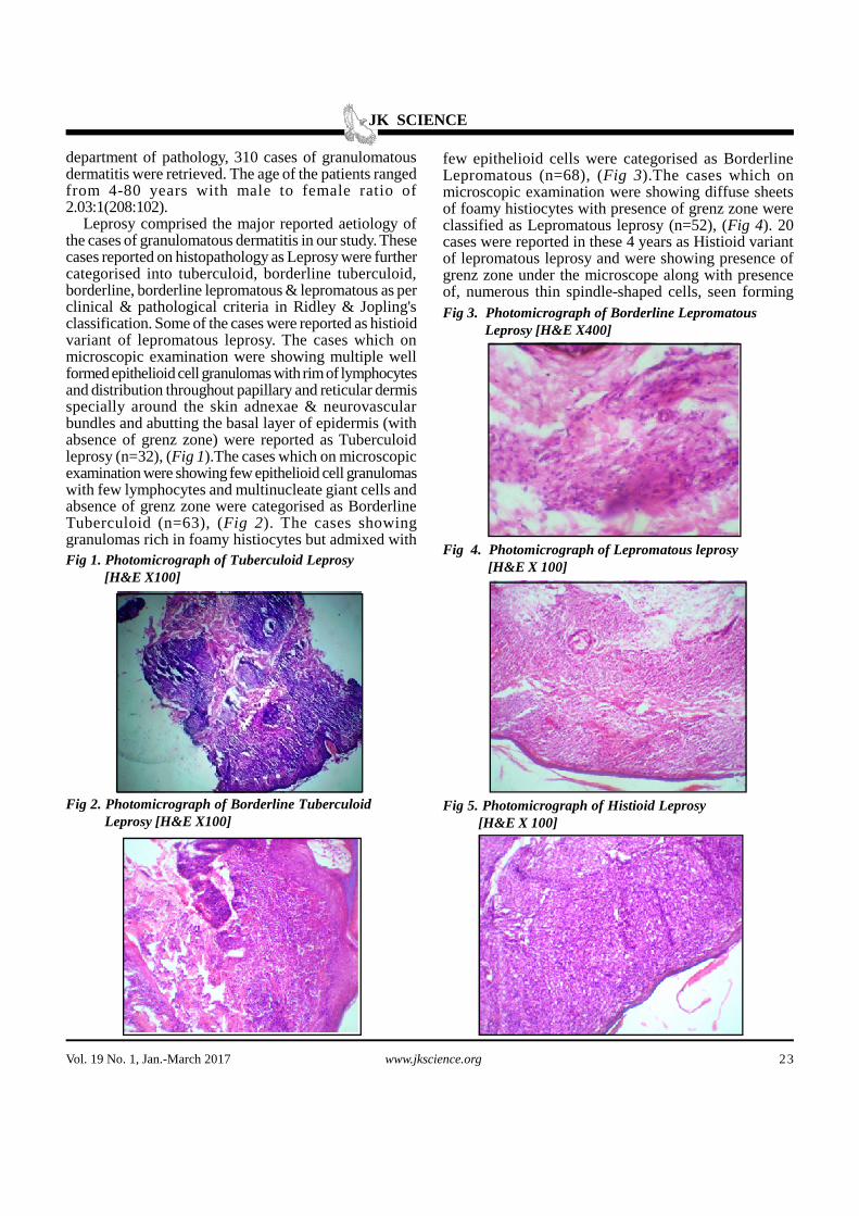
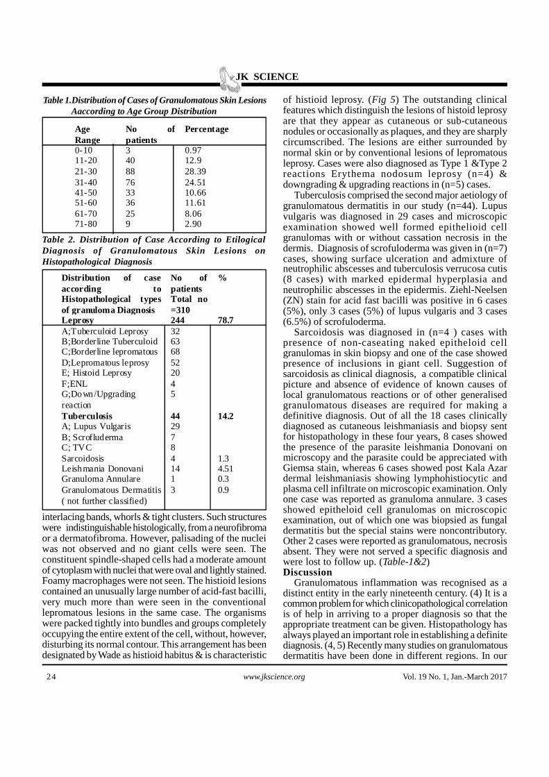
Leprosy comprised the major reported aetiology ofthe cases of granulomatous dermatitis in our study. Thesecases reported on histopathology as Leprosy were furthercategorised into tuberculoid, borderline tuberculoid,borderline, borderline lepromatous & lepromatous as perclinical & pathological criteria in Ridley & Jopling'sclassification. Some of the cases were reported as histioidvariant of lepromatous leprosy. The cases which onmicroscopic examination were showing multiple wellformed epithelioid cell granulomas with rim of lymphocytesand distribution throughout papillary and reticular dermisspecially around the skin adnexae & neurovascularbundles and abutting the basal layer of epidermis (withabsence of grenz zone) were reported as Tuberculoidleprosy (n=32), (Fig 1).The cases which on microscopicexamination were showing few epithelioid cell granulomaswith few lymphocytes and multinucleate giant cells andabsence of grenz zone were categorised as BorderlineTuberculoid (n=63), (Fig 2). The cases showinggranulomas rich in foamy histiocytes but admixed with
few epithelioid cells were categorised as BorderlineLepromatous (n=68), (Fig 3).The cases which onmicroscopic examination were showing diffuse sheetsof foamy histiocytes with presence of grenz zone wereclassified as Lepromatous leprosy (n=52), (Fig 4). 20cases were reported in these 4 years as Histioid variantof lepromatous leprosy and were showing presence ofgrenz zone under the microscope along with presenceof, numerous thin spindle-shaped cells, seen forming
Fig 1. Photomicrograph of Tuberculoid Leprosy [H&E X100]
Fig 2. Photomicrograph of Borderline Tuberculoid Leprosy [H&E X100]
Fig 3. Photomicrograph of Borderline Lepromatous Leprosy [H&E X400]
Fig 4. Photomicrograph of Lepromatous leprosy [H&E X 100]
Fig 5. Photomicrograph of Histioid Leprosy [H&E X 100]
JK SCIENCE
24 www.jkscience.org Vol. 19 No. 1, Jan.-March 2017
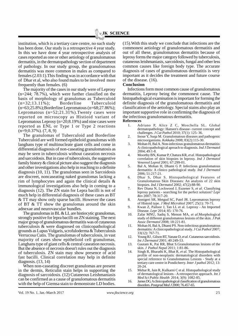
Table 1.Distribution of Cases of Granulomatous Skin Lesions Aaccording to Age Group Distribution
interlacing bands, whorls & tight clusters. Such structureswere indistinguishable histologically, from a neurofibromaor a dermatofibroma. However, palisading of the nucleiwas not observed and no giant cells were seen. Theconstituent spindle-shaped cells had a moderate amountof cytoplasm with nuclei that were oval and lightly stained.Foamy macrophages were not seen. The histioid lesionscontained an unusually large number of acid-fast bacilli,very much more than were seen in the conventionallepromatous lesions in the same case. The organismswere packed tightly into bundles and groups completelyoccupying the entire extent of the cell, without, however,disturbing its normal contour. This arrangement has beendesignated by Wade as histioid habitus & is characteristic
neutrophilic abscesses and tuberculosis verrucosa cutis(8 cases) with marked epidermal hyperplasia andneutrophilic abscesses in the epidermis. Ziehl-Neelsen(ZN) stain for acid fast bacilli was positive in 6 cases(5%), only 3 cases (5%) of lupus vulgaris and 3 cases(6.5%) of scrofuloderma.
Sarcoidosis was diagnosed in (n=4 ) cases withpresence of non-caseating naked epitheloid cellgranulomas in skin biopsy and one of the case showedpresence of inclusions in giant cell. Suggestion ofsarcoidosis as clinical diagnosis, a compatible clinicalpicture and absence of evidence of known causes oflocal granulomatous reactions or of other generalisedgranulomatous diseases are required for making adefinitive diagnosis. Out of all the 18 cases clinicallydiagnosed as cutaneous leishmaniasis and biopsy sentfor histopathology in these four years, 8 cases showedthe presence of the parasite leishmania Donovani onmicroscopy and the parasite could be appreciated withGiemsa stain, whereas 6 cases showed post Kala Azardermal leishmaniasis showing lymphohistiocytic andplasma cell infiltrate on microscopic examination. Onlyone case was reported as granuloma annulare. 3 casesshowed epitheloid cell granulomas on microscopicexamination, out of which one was biopsied as fungaldermatitis but the special stains were noncontributory.Other 2 cases were reported as granulomatous, necrosisabsent. They were not served a specific diagnosis andwere lost to follow up. (Table-1&2)Discussion
Granulomatous inflammation was recognised as adistinct entity in the early nineteenth century. (4) It is acommon problem for which clinicopathological correlationis of help in arriving to a proper diagnosis so that theappropriate treatment can be given. Histopathology hasalways played an important role in establishing a definitediagnosis. (4, 5) Recently many studies on granulomatousdermatitis have been done in different regions. In our
of histioid leprosy. (Fig 5) The outstanding clinicalfeatures which distinguish the lesions of histoid leprosyare that they appear as cutaneous or sub-cutaneousnodules or occasionally as plaques, and they are sharplycircumscribed. The lesions are either surrounded bynormal skin or by conventional lesions of lepromatousleprosy. Cases were also diagnosed as Type 1 &Type 2reactions Erythema nodosum leprosy (n=4) &downgrading & upgrading reactions in (n=5) cases.
Tuberculosis comprised the second major aetiology ofgranulomatous dermatitis in our study (n=44). Lupusvulgaris was diagnosed in 29 cases and microscopicexamination showed well formed epithelioid cellgranulomas with or without cassation necrosis in thedermis. Diagnosis of scrofuloderma was given in (n=7)cases, showing surface ulceration and admixture of
AgeRange
No ofpatients
Percentage
0-10 3 0.9711-20 40 12.921-30 88 28.3931-40 76 24.5141-50 33 10.6651-60 36 11.6161-70 25 8.0671-80 9 2.90
Distribution of caseaccording toHistopathological typesof granuloma Diagnosis
No ofpatientsTotal no=310
%
LeprosyA;Tuberculoid LeprosyB;Borderline TuberculoidC;Borderline lepromatousD;Lepromatous leprosyE; Histoid LeprosyF;ENLG;Down/Upgradingreaction
244326368522045
78.7
TuberculosisA; Lupus VulgarisB; ScrofludermaC; TVC
442978
14.2
Sarcoidosis 4 1.3Leishmania Donovani 14 4.51Granuloma Annulare 1 0.3Granulomatous Dermatitis( not further classified)
3 0.9
Table 2. Distribution of Case According to EtilogicalDiagnosis of Granulomatous Skin Lesions onHistopathological Diagnosis
JK SCIENCE
Vol. 19 No. 1, Jan.-March 2017 www.jkscience.org 25
1. Adriano P, Alice Z C, Moschella SL. Globaldermatopathology: Hansen's disease- current concept andchallenges. J Cut Pathol 2010; 37(1): 125- 36.
2. Inoue Y, Suqa M. Granulomatous diseases and pathogenicmicroorganisms. Kekkaku 2008; 83(2):115-30.
3. Mohan H, Bal A. Non-infectious granulomatous dermatitis:A clinicopathological aproach to diagnosis. Ind J Dermatol2004; 49:1-8.
4. Moorthy BN, Kumar P, Chatura KR, et al. Histopathologicalcorrelation of skin biopsies in leprosy. Ind J DermatolVenereol Leprol 2001; 67:299-01.
5. Bal A, Mohan H, Dhami G P. Infectious granulomatousdermatitis: A clinico pathological study. Ind J Dermatol2006; 51:217-21.
6. Dhar S, Dhar S. Histopathological Features ofGranulomatous Skin Diseases: An analysis of 22 skinbiopsies. Ind J Dermatol 2002; 47(2):88-90.
7. Rev Diana N, Lockwood J, Euzener S, et al. Classifyingleprosy patients - searching for the perfect solution? LeprRev 2007; 78:317-20.
8. Annigeri SR, Metgud SC, Patel JR. Lepromatous leprosyof Histoid type. J Med Microbiol 2007; 25(1): 70-71.
9. Kwan Z, Pailoor J, Tan LL et al. Leprosy - An ImportedDisease. Lepr 2014; 85: 170-76.
10. Zafar MNU, Sadiq S, Menon MA, et al.Morphologicalstudy of different granulomatous lesions of the skin. J PakAssoc Dermatol 2008; 18:221-81.
11. Mohan H, Bal A, Dhami PS. Non-infectious granulomatousdermatitis: A clinicopathological study. J Cut Pathol 2007;33(12): 767-71.
12. Young RJ , Gilson RT, Yanase D, et al. Cutaneous sarcoidosis.Int J Dermatol 2001; 40:249-53.
13. Gautam K, Pai RR, Bhat S.Granulomatous lesions of theskin. J Pathol Nepal 2011; 1:81-86.
14. Singh R, Bharathi K, Bhat R, et al. The Histopathologicalprofile of non-neoplastic dermatological disorders withspecial reference to Granulomatous Lesions - Study at atertiary care centre in Pondicherry. Inter J pathol 2012; 13:20-24.
15. Mehar R, Jain R, Kulkarni C et al. Histopathological studyof dermatological lesions - A retrospective approach. Int JMed Sci Public Health 2014; 3(9): 1082-85.
16. James DG. A clinicopathological classification of granulomatousdisorders. Postgrad Med J2000; 76.457-65.
References
institution, which is a tertiary care centre, no such studyhas been done. Our study is a retrospective 4 year study.In this we have done 4 year retrospective analysis ofcases reported as one or other aetiology of granulomatousdermatitis, in the dermatopathology section of departmentof pathology. In our study group, the granulomatousdermatitis was more common in males as compared tofemales (2.03:1).This finding was in accordance with thatof Dhar et al, who also found males to be involved morefrequently than females. (6)
The majority of the cases in our study were of Leprosy(n=244; 78.7%), which were further classified on thebasis of morphology of granulomas as Tuberculoid(n=32;13.11%); Borderline Tuberculoid(n=63;25.8%);Borderline Lepromatous (n=68;27.86%);Lepromatous (n=52;21.31%).Twenty cases werereported on microscopy as Histioid variant ofLepromatous Leprosy (n=20;8.19%) and nine cases werereported as ENL or Type 1 or Type 2 reactions(n=9;0.37%). (7, 8, 9)
The granulomas of Tuberculoid and BorderlineTuberculoid are well formed epitheloid granulomas withlanghans type of multinucleate giant cells and come indifferential diagnosis of non-caseating granulomatosis asmay be seen in tuberculosis without caseation necrosisand sarcoidosis. But in case of tuberculosis, the suggestivefamily history & clinical picture also suggest the diagnosisand other investigations also help in reaching to a definitediagnosis (10, 11). The granulomas seen in Sarcoidosisare discreet, noncaseating naked granulomas lacking arim of lymphocytes and again the clinical details &immunological investigations also help in coming to adiagnosis (12). The ZN stain for Lepra bacilli is not ofmuch help in differential diagnosis of these cases as BT& TT may show only sparse bacilli. However the casesof BT & TT show the granulomas around the skinadnexae and neurovascular bundles.
The granulomas in BL & LL are histiocytic granulomas,strongly positive for lepra bacilli on ZN staining. The nextmajor group of granulomatous dermatitis was of cutaneoustuberculosis & were diagnosed on clinicopathologicalgrounds as Lupus Vulgaris, scrofuloderma & TuberculosisVerrucosa Cutis. The granulomas of tuberculosis, in vastmajority of cases show epithelioid cell granulomas,Langhans type of giant cells & central caseation necrosis.But the absence of necrosis doesn't rules out the diagnosisof tuberculosis. ZN stain may show presence of acidfast bacilli. Clinical correlation may help in definitediagnosis. (13, 14)
When non-caseating discreet granulomas are presentin the dermis, Reticulin stain helps in supporting thediagnosis of sarcoidosis. (12) Cutaneous Leishmaniasiscan be confirmed as a cause of granulomatous dermatitiswith the help of Giemsa stain to demonstrate LD bodies.
(15).With this study we conclude that infections are thecommonest aetiology of granulomatous dermatitis andout of all these, granulomatous dermatitis because ofleprosy forms the major category followed by tuberculosis,cutaneous leishmaniasis, sarcoidosis, fungal and other lesscommon causes like foreign body type. The accuratediagnosis of cases of granulomatous dermatitis is veryimportant as it decides the treatment and future courseof the disease. (16).Conclusion
Infections form most common cause of granulomatousdermatitis, Leprosy being the commonest cause. Thehistopathological examination is important for forming thedefinite diagnosis of the granulomatous dermatitis andclassification of the aetiology. Special stains also play animportant supportive role in confirming the diagnosis ofthe infectious granulomatous dermatitis.