
Granulomatous Lymphadenitis
16
Review Article Granulomatous Lymphadenitis Shigeyuki Asano In this review, representative types of granulomatous lymphadenitis (GLA) are described. GLA can be classified as noninfectious GLA and infectious GLA. Noninfectious GLA includes sarcoidosis and sarcoid-like reaction. The cause of sarcoidosis remains unknown, but it has good prognosis. Sarcoid-like reaction, which is considered to be a biological defense mechanism, is observed in regional lymph nodes with many underlying diseases. Infectious GLA can be classified as suppura- tive lymphadenitis (LA) and nonsuppurative LA. Suppurative LA generally shows follicular hyperplasia and sinus histiocytosis in the early phase. In tularemia and cat scratch disease, monocytoid B lymphocytes (MBLs) with T cells and macrophages contribute to the formation of granuloma. However, none of the epithelioid cell granulomas of Yersinia LA contains MBLs like in cat scratch disease. In addition, almost all have a central abscess in granulomas induced by Gram-negative bacteria. In terms of the lymph nodes, tularemia and cat scratch disease are apt to affect the axillary and cervical regions while Yersinia LA affects the mesenteric lymph node. Nonsuppurative LA includes tuberculosis and BCG-histiocytosis. These are induced by delayed allergic reaction of M. tuberculosis. Tuberculosis LA mainly appears in the cervical lymph node. Organisms are histologically detected by Ziehl-Neelsen staining in the necrotic area. Toxoplasmosis is also a nonsuppurative protozoan infection (Toxoplasma gondii). In toxoplasma LA, MBLs can also be seen, but round and organized, well-formed granulomas are not found in this disease. Furthermore, necrosis is not induced and there are no accompanying neutrophils, eosinophils and fibrosis. GLA described above is associated with characteristic histological findings. An accurate pathological diagnosis using the above findings can lead to precise treatment. 〔J Clin Exp Hematopathol 52(1) : 1-16, 2012〕 Keywords: granulomatous lymphadenitis, granuloma, abscess, macrophage, monocytoid B lymphocyte INTRODUCTION Granulomatous lymphadenitis can be classified into non- infectious and infectious types 1 (Table 1). Noninfectious granulomatous lymphadenitis includes berylliosis, Hodgkin’s lymphoma, non-Hodgkin’s lymphoma, lymph node draining neoplasms (sarcoid-like reaction), lymph node draining Crohn’s disease and sarcoidosis. These rarely have abscesses and necrosis in the center of granulomas. Infectious granulomatous lymphadenitis can be classified into suppurative lymphadenitis and nonsuppurative lymph- adenitis. The former is exemplified by tularemia, cat scratch disease, Yersinia lymphadenitis and lymphogranuloma vene- reum. These almost all have central abscesses and necrosis in granulomas induced by Gram-negative bacteria and chlamy- dia. In terms of the lymph nodes, tularemia and cat scratch disease are apt to affect the axillary and cervical regions, Yersinia lymphadenitis affects mesenteric lymph node and lymphogranuloma venereum affects inguinal lymph nodes. 1 J Clin Exp Hematopathol Vol. 52, No. 1, May 2012 Received : November 14, 2011 Revised : January 13, 2012 Accepted : January 31, 2012 Department of Pathology, Iwaki Kyoritsu General Hospital, Iwaki, Japan Address correspondence and reprint requests to : Shigeyuki Asano, M.D., Department of Pathology, Iwaki Kyoritsu General Hospital, 16 Kusehara, Mimaya-machi, Uchigo, Iwaki 973-8555, Japan E-mail : [email protected] Table 1. Granulomatous lymphadenitis 1. Noninfectious granulomatous disorders 1) Sarcoidosis lymphadenitis 2) Sarcoid-like lymphadenitis 3) Berylliosis 2. Infectious granulomatous disorders A. Suppurative 1) Tularemia lymphadenitis 2) Cat scratch lymphadenitis 3) Yersinia lymphadenitis 4) Lymphogranuloma venereum 5) Fungal infection B. Non-suppurative 1) Tuberculous lymphadenitis 2) Atypical mycobacterial infection 3) BCG-lymphadenitis 4) Toxoplasma lymphadenitis (Piringer-Kuchinka lymphadenopathy) 5) Lepra 6) Syphilis 7) Brucellosis 8) Fungal infection (Cryptococcus, Histoplasma, Coccidioidomycosis, Pneumocystis)
Transcript of Granulomatous Lymphadenitis

01_Asano.mcd Page 1 v4.21
Review Article
Granulomatous Lymphadenitis
Shigeyuki Asano
In this review, representative types of granulomatous lymphadenitis (GLA) are described. GLA can be classified asnoninfectious GLA and infectious GLA. Noninfectious GLA includes sarcoidosis and sarcoid-like reaction. The cause ofsarcoidosis remains unknown, but it has good prognosis. Sarcoid-like reaction, which is considered to be a biological defensemechanism, is observed in regional lymph nodes with many underlying diseases. Infectious GLA can be classified as suppura-tive lymphadenitis (LA) and nonsuppurative LA. Suppurative LA generally shows follicular hyperplasia and sinus histiocytosisin the early phase. In tularemia and cat scratch disease, monocytoid B lymphocytes (MBLs) with T cells and macrophagescontribute to the formation of granuloma. However, none of the epithelioid cell granulomas of Yersinia LA contains MBLs likein cat scratch disease. In addition, almost all have a central abscess in granulomas induced by Gram-negative bacteria. In termsof the lymph nodes, tularemia and cat scratch disease are apt to affect the axillary and cervical regions while Yersinia LA affectsthe mesenteric lymph node. Nonsuppurative LA includes tuberculosis and BCG-histiocytosis. These are induced by delayedallergic reaction of M. tuberculosis. Tuberculosis LA mainly appears in the cervical lymph node. Organisms are histologicallydetected by Ziehl-Neelsen staining in the necrotic area. Toxoplasmosis is also a nonsuppurative protozoan infection(Toxoplasma gondii). In toxoplasma LA, MBLs can also be seen, but round and organized, well-formed granulomas are notfound in this disease. Furthermore, necrosis is not induced and there are no accompanying neutrophils, eosinophils and fibrosis.GLA described above is associated with characteristic histological findings. An accurate pathological diagnosis using the abovefindings can lead to precise treatment. 〔J Clin Exp Hematopathol 52(1) : 1-16, 2012〕
Keywords: granulomatous lymphadenitis, granuloma, abscess, macrophage, monocytoid B lymphocyte
INTRODUCTION
Granulomatous lymphadenitis can be classified into non-infectious and infectious types1 (Table 1). Noninfectiousgranulomatous lymphadenitis includes berylliosis, Hodgkin’slymphoma, non-Hodgkin’s lymphoma, lymph node drainingneoplasms (sarcoid-like reaction), lymph node drainingCrohn’s disease and sarcoidosis. These rarely have abscessesand necrosis in the center of granulomas.
Infectious granulomatous lymphadenitis can be classifiedinto suppurative lymphadenitis and nonsuppurative lymph-adenitis. The former is exemplified by tularemia, cat scratchdisease, Yersinia lymphadenitis and lymphogranuloma vene-reum. These almost all have central abscesses and necrosis ingranulomas induced by Gram-negative bacteria and chlamy-dia. In terms of the lymph nodes, tularemia and cat scratch
disease are apt to affect the axillary and cervical regions,Yersinia lymphadenitis affects mesenteric lymph node andlymphogranuloma venereum affects inguinal lymph nodes.
1
J Clin Exp Hematopathol
Vol. 52, No. 1, May 2012
Received : November 14, 2011Revised : January 13, 2012Accepted : January 31, 2012Department of Pathology, Iwaki Kyoritsu General Hospital, Iwaki, JapanAddress correspondence and reprint requests to : Shigeyuki Asano, M.D.,Department of Pathology, Iwaki Kyoritsu General Hospital, 16 Kusehara,Mimaya-machi, Uchigo, Iwaki 973-8555, JapanE-mail : [email protected]
Table 1. Granulomatous lymphadenitis
1. Noninfectious granulomatous disorders
1) Sarcoidosis lymphadenitis2) Sarcoid-like lymphadenitis3) Berylliosis
2. Infectious granulomatous disorders
A. Suppurative1) Tularemia lymphadenitis2) Cat scratch lymphadenitis3) Yersinia lymphadenitis4) Lymphogranuloma venereum5) Fungal infection
B. Non-suppurative1) Tuberculous lymphadenitis2) Atypical mycobacterial infection3) BCG-lymphadenitis4) Toxoplasma lymphadenitis (Piringer-Kuchinka lymphadenopathy)5) Lepra6) Syphilis7) Brucellosis8) Fungal infection (Cryptococcus, Histoplasma, Coccidioidomycosis,
Pneumocystis)

01_Asano.mcd Page 2 v4.21
The latter, that is, nonsuppurative lymphadenitis, includestuberculosis and Bacil lus Calmette-Guérin (BCG)-lymphadenitis. These have nonsuppurative hypersensitivity-type granulomas induced by mycobacterium. Tuberculouslymphadenitis mainly appears in the cervical lymph node.Predominant histiocytes with smaller numbers of T cells,dendritic cells and peripheral B cells are recruited for granulo-ma formation. Organisms are histologically detected byZiehl-Neelsen staining in the necrotic area. Toxoplasmosis isalso a nonsuppurative protozoan infection (Toxoplasma gon-dii).
Here we clinicopathologically describe eight representa-tive types of granulomatous lymphadenitis, namely, sarcoido-sis, sarcoid-like reaction, tularemia, cat scratch disease,Yersinia lymphadenitis, tuberculosis, BCG-lymphadenitis andtoxoplasmosis.
SARCOIDOSIS
Sarcoidosis means “meaty chunk-like lesions” in Latin.The cause of the disease remains unknown, but it is non-hereditary with a good clinical course. The disease involvesmultiple organs, such as pulmonary hilar lymph nodes, lungs,eyes and skin.2 The nodes show variously sized non-caseousepithelioid granulomas. Blindness, difficulty breathing andheart failure, which are complications of sarcoidosis, mayinterfere with daily life. In Japan, anaerobic bacteria such asPropionibacterium acne (P. acne) and P. granulosum areconsidered among the causative agents of sarcoidosis becausegenes of these bacteria have been detected in the lungs andlymph nodes in such cases. On the other hand, in Europe,associations between sarcoidosis and Mycobacterium tubercu-losis (M. tuberculosis), virus and autoimmune disease havebeen reported.3-6
Epidemiology
Sarcoidosis is found worldwide, affecting people of allraces and ages. Seasonal clustering occurs during winter andearly spring.7 Sarcoidosis occurs twice as frequently in wom-en as in men, but rarely in children.8,9 Adults between 20 and40 years of age are usually affected. In Japan, there are noseasonal and regional biases, but those of both sexes in theirtwenties are most often affected, with very few symptoms.Recently, cases in women after their forties (fifties and six-ties, in particular) have also been seen. In addition, the globalannual incidence is approximately eight cases per 100,000peoples, but 3.1 cases in Japan. Susceptible organs are thelungs and hilar lymph nodes (80%), eyes (50%), skin (20%)and other lymph nodes, namely, cervical, axilla and inguinallymph nodes.
History
In terms of the history of sarcoidosis, Hutchinson (1877)was the first to describe such macroscopic findings of the skinand Boeck (1897) histologically examined macular rash of theskin in this disease. In Japan, around 1950, sarcoidosis wasrecognized as pulmonary hilar lymphadenopathy.
Clinical Findings
Sarcoidosis involves multiple organs, most commonly thelungs, skin and eyes, and the symptoms depend on the af-fected organs. Ocular involvement presents blurred vision,photophobia and floaters, dermatologic involvement presentsnodules and plaque, while respiratory lesions are associatedwith cough and breathing discomfort. Cardiac involvement ismore common and causes arrhythmia.
Sudden death occurs in 5% to 10% of patients withsarcoidosis.10 Lymphadenopathy is frequently presented,most commonly in pulmonary hilar lymph nodes (93.5%).Cervical (12.2%), axillary (5.2%) and inguinal (3.3%) lymphnodes are also affected.
The useful factors for diagnosis of sarcoidosis are as fol-lows : bilateral hilar lymphadenopathy for X-p (Fig. 1a) andcomputed tomography, increased angiotensin-converting en-zyme, hypercalcemia, negative on tuberculin test, bronchio-alveolar liquid (BAL) cytology and increased CD4+ /CD8+
ratio, and positive on the Kveim test.The granulomas of sarcoidosis can usually be distin-
guished from tuberculosis, fungal infection, silicosis, beryllio-sis and Hodgkin’s lymphoma by their characteristic sharpdemarcation, lack of central necrosis and special staining,such as acid-fast and silver impregnation staining.6
Histopathology of Lymph Nodes
In the early phase, follicular hyperplasia and sinus histio-cytosis appear like nonspecific lymphadenitis. Subsequently,small epithelioid cell nodules appear in the cortex after adecrease of histiocytes.
In the peak phase, well-demarcated granulomas composedof epithelioid cells with scattered multinucleated giant cellsare observed throughout the lymph node. Granulomas mayoccasionally coalesce (Fig. 1b, 1c).
In the late phase, increased collagen fibers result in fibro-sis and hyalinization.6 This type of granulomatous diseasehas no neutrophils, and small foci of central fibrinoid andcoagulation necrosis are uncommon. CD4/CD8 ratio, usuallyover 3.5, is increased, which indicates a state of cell-mediated hyperreactivity in sarcoidosis. Furthermore, T lym-phocytes, dendritic cells and macrophages are important com-ponents for granuloma formation.11 There are variousinclusion bodies such as asteroids (Fig. 1d), Schaumann or
Asano S
2

01_Asano.mcd Page 3 v4.21
Hamazaki-Wesenberg bodies.7,12
SARCOID-LIKE REACTION (SARCOID-LIKE
GRANULOMA)
Epithelioid granuloma resembling sarcoidosis is occasion-ally observed in regional lymph nodes with an underlyingdisease, but it is not indicative of systemic sarcoidosis. Thisis called a sarcoid-like reaction.13 There are many underlyingdiseases as follows : carcinoma, toxoplasmosis, fungal infec-tion, tuberculosis, atypical mycobacterial disease, pneumoco-niosis, immunocompromised status (Crohn's disease, primarybiliary cirrhosis and Sjögren's syndrome), extrinsic allergicinflammatory alveolitis (farmer's lung) and anticancer chemo-therapy, as well as associations with chemicals (beryllium,zirconium, silicon, starch granules and pine pollen).14
Carcinomas of uterus, breast, lung and stomach are associatedwith this type of reaction.13,15-17 It is also known that ad-
vanced cancer accompanied by sarcoid-like reaction has goodprognosis.18,19
This reaction is considered to be a long-term biologicaldefense mechanism of persistently stimulated lymph nodesagainst metabolites and breakdown products of malignanttumors.20,21
History
Wolbach (1911)22 and Brincker (1986)13 reported that4.4% of cancers, 7.3% of non-Hodgkin’s lymphomas and13.8% of Hodgkin's lymphoma are accompanied by sarcoid-like reaction. More recently, these reactions have been ob-served in the lymph nodes in various diseases.
Clinical Findings
Symptoms often depend on the underlying disease and
Granulomatous lymphadenitis
3
Fig. 1. Sarcoidosis lymphadenitis. (1a) Chest X-p shows bilateral hilar lymph node swel-ling. (1b) Lymph node is occupied by numerous granulomas that have not coalesced witheach other. There are many plasma cells around each granuloma. (1c) Granuloma is com-posed of epithelioid cells and multinucleated giant cells. (1d) Asteroid body is observed inthe cytoplasm of multinucleated giant cell.
1a 1b
1c 1d

01_Asano.mcd Page 4 v4.21
there are no specific clinical symptoms. In chest X-ray, thereare no signs of hilar lymphadenopathy. Results of tuberculinskin and Kveim test are also negative.
Histopathology of Lymph Nodes
Scattered small epithelioid granulomas, composed ofsparsely arranged epithelioid cells, are accompanied by lym-phocyte infiltration among granuloma cells. In addition, theborder of granulomas is often obscure (Fig. 2a, 2b). T lym-phocytes, dendritic cells and macrophages are important com-ponents for granuloma formation. A CD4/CD8 ratio of 0.8 to2.25 is observed in confluent-type rather than solitary-typegranuloma.23 The clinical course and laboratory data areuseful for differential diagnosis between sarcoidosis andsarcoid-like reaction.
TULAREMIA (OHARA’S DISEASE)
Tularemia is a zoonosis caused by the Gram-negativecoccobacillus Francisella tularensis (F. tularensis), which isthe etiologic agent of this acute infectious disease.
This disease is generally spread among wild animals suchas rodent (hare, mouse), bird and dog, and it is lethal to
animals. There are several modes of infection to humans,such as direct contact, eating of raw meat of an infectedanimal, bite by infective arthropods, ingestion of contami-nated water or food and inhalation of infective aerosols.
F. tularensis invades not only via mucous membranes butalso via skin, and bacteria grow at the site of invasion. Then,F. tularensis moves to the lymph node via lymphatic drainageand induces infection during its stay there. There is no trans-mission from person to person.
The genus Francisella consists of two species, F. tularen-sis and F. philomiragia.24 F. philomiragia is a bacteriumcausing opportunistic infections, which may cause diseases inaccident victims and immunocompromised patients, for ex-ample. F. tularensis is currently divided into three subspecies,namely, subspecies (subsp.) tularensis, subsp. holarctica andsubsp. mediasiatica. Among these, only subsp. tularensis
(type A) in North America shows high virulence, while subsp.holarctica (type B) is distributed in Eurasia from Europe toJapan and shows attenuated virulence.25-27 The third subspe-cies, mediasiatica, has only been isolated in Central Asia andhas virulence similar to that of subsp. holarctica.
Epidemiology
Subsp. holarctica is widely distributed throughout theNorthern Hemisphere around a latitude of 30 degrees north,including in Japan. Tularemia is likely to occur during thehunting season (November to January) and during the seasonwhen bloodsucking insects such as ticks are particularly ac-tive (April to June).27,28 In Japan, parts of the Kanto regionand the whole of the Tohoku region (east and north of Japan)are particularly affected by tularemia.29 In Japan, there were1,372 reported cases from 1924, when records began, until1994. Since then, there have been reports of one case inChiba Prefecture in 1999, and one case each in FukushimaPrefecture and Chiba Prefecture in 2008. The number of casereports of this disease has decreased year by year.
History
Soken Honma, a physician in Mito, Ibaraki Prefecture,Japan, described tularemia as “hare meat poisoning” (1837)30
in the oldest report of the disease. In California, USA,McCoy (1911)31 reported a plague-like disease affectingsquirrels. Francis30,32 confirmed its transmissibility to hu-mans. Francisella tularensis was named after Francis and thetown in which the bacterium was isolated : Tulare,California, USA (1921).31 In Japan, Hachiro Ohara estab-lished the Ohara Institute in Fukushima, Japan, for the activestudy of tularemia (1925).30 Japanese armed forces (1932-1945) and the U.S. Army (1950-1960) undertook studies todevelop tularemia bacteria for use as a biological weapon.3,5,9
After the bioterrorism attacks with anthrax in 2001, the
Asano S
4
Fig. 2. Sarcoid-like lymphadenitis. (gastriccarcinoma) (2a) Irregularly shaped epithelioidgranulomas are observed in regional lymphnodes accompanied by sinus and follicularhyperplasia. (2b) Lymphocytes are inter-mingled with epithelioid cells in granuloma.
2a
2b
01_Asano.mcd Page 5 v4.21
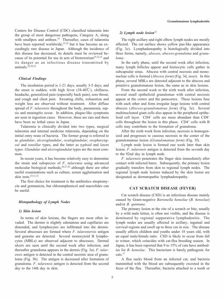
Centers for Disease Control (CDC) classified tularemia intothe group of most dangerous pathogens, Category A, alongwith smallpox and anthrax.33 Thereafter, cases of tularemiahave been reported worldwide,25,34 but it has become an ex-ceedingly rare disease in Japan. Although the incidence ofthis disease has decreased, its details must be reviewed be-cause of its potential for use in acts of bioterrorism25,32,35 andits danger as an infectious disease transmitted byanimals.24,30,32
Clinical Findings
The incubation period is 1-21 days, usually 3-5 days, andthe onset is sudden, with high fever (38-40℃), chillness,headache, generalized pain (especially back pain), sore throat,and cough and chest pain. Sweating chills, exhaustion andweight loss are observed without treatment. After diffusespread of F. tularensis throughout the body, pneumonia, sep-sis and meningitis occur. In addition, plague-like symptomsare seen in ingestion cases. However, these are rare and therehave been no lethal cases in Japan.
Tularemia is clinically divided into two types, surgicaltularemia and internal medicine tularemia, depending on theinitial entry route of bacteria. The former group is referred toas glandular, ulceroglandular, oculoglandular, oropharyng-eal and tonsillar types, and the latter as typhoid and latent
types. Glandular and ulceroglandular types are the most com-mon.
In recent years, it has become relatively easy to determinethe strain and subspecies of F. tularemia using advancedmolecular biological methods,26,27,36 although there are otheruseful examinations such as culture, serum agglutination andskin tests.25,31,32
The first choice for treatment is the antibiotics streptomy-cin and gentamicin, but chloramphenicol and macrolides canbe useful.
Histopathology of Lymph Nodes
1) Skin lesion
In terms of skin lesions, the fingers are most often in-vaded. The dermis is slightly edematous and capillaries aredistended, and lymphocytes are infiltrated into the dermis.Several abscesses are formed where F. tularentersis antigenand genome are detected. Several monocytoid B lympho-cytes (MBLs) are observed adjacent to abscesses. Dermalulcers are seen until the second week after infection, andthereafter granuloma appears in the dermis (Fig. 3a). F. tular-ensis antigen is detected in the central necrotic area of granu-loma (Fig. 3b). The antigen is decreased after formation ofgranuloma. F. tularensis antigen is detected from the secondday to the 14th day in skin.
2) Lymph node lesion32
The right axillary and right elbow lymph nodes are mostlyaffected. The cut surface shows yellow pus-like appearance(Fig. 3c). Lymphadenopathy is histologically divided intothree forms, namely, abscess, abscess-granuloma and granu-loma.
In the early phase, until the second week after infection,many lymph follicles appear and histiocytic cells gather insubcapsular sinus. Abscess with central necrosis and mono-nuclear cells is formed (Abscess form) (Fig. 3d, inset). In thisphase, several MBLs are detected adjacent to the abscess andprimitive granulomatous lesion, the same as in skin lesions.
From the second week to the sixth week after infection,several small epithelioid granulomas with central necrosisappear at the cortex and the paracortex. These lesions fusewith each other and form irregular large lesions with centralabscess (Abscess-granulomatous form) (Fig. 3e). Severalmultinucleated giant cells also appear in the peripheral epithe-lioid cell layer. CD4+ cells are more abundant than CD8+
cells throughout the lesion in this phase. CD4+ cells with Bcells may contribute to the formation of granuloma.
After the sixth week from infection, necrosis is homogen-ized and progresses to caseous necrosis in the center of thegranulomatous lesion (Granulomatous form) (Fig. 3f).
Lymph node lesion is formed one week later than skinlesion. F. tularensis antigen is detected from the seventh dayto the 92nd day in lymph node.
F. tularensis penetrates the finger skin immediately aftercontact with infected hares. Subsequently, the primary lesiongradually transfers from skin to regional lymph nodes. Theregional lymph node lesions induced by the skin lesion aredesignated as dermatopathic lymphadenopathy.
CAT SCRATCH DISEASE (FEVER)
Cat scratch disease (CSD) is an infectious disease mainlycaused by Gram-negative Bartonella henselae (B. henselae)and/or B. quintana.
The primary lesion at the site of a scratch or bite, usuallyby a wild male kitten, is often not visible, and the disease isdominated by regional suppurative lymphadenitis. Thelymph nodes are usually affected in axillary, inguinal andcervical regions and swell up to three cm in size. The diseaseusually affects children and youths under 18 years old, withan equal male/female ratio. CSD is likely to occur from fallto winter, which coincides with cat-flea breeding season. InJapan, it has been reported that 9 to 15% of cats have antibod-ies for B. henselae. This bacterium is barely pathogenic forcats.37
A flea sucks blood from an infected cat, and bacteriatransmitted with the blood are subsequently excreted in thefeces of the flea. Thereafter, bacteria attached to a tooth or
Granulomatous lymphadenitis
5

01_Asano.mcd Page 6 v4.21
nail of the cat invade the human skin via the scratch or bitewound. The annual incidence of CSD has been estimated at9.3 cases per 100,000 people per year in the United States,although there are no national statistics for Japan.38
History
Parinaud first described only a small subset of CSD ;subsequently, Greer and Keefer published the first report onCSD in the American literature, in which they described abroad spectrum of CSD manifestations.39 Patients with CSD
have a high titer of seropositivity for Rochalimaea henselae,as in AIDS patients, but low titer for Afipia felis.40 Aftergenotypic evaluation, the nomenclature, R. henselae, was re-placed by B. henselae, which was identified as the causativebacterium.41
Clinical Findings
Erythema (20 to 50%), papules, blisters (vesicular) andabscess are observed at the injury site within three to ten days
Asano S
6
Fig. 3. Tularemia lymphadenitis. (3a) Variously sized granulomas at the ulcer baseof finger skin (14 days after infection). (3b) Immunostaining of skin lesion. F. tularen-sis antigens are detected mainly in abscess and necrotic area (14 days after infection)(anti-F. tularensis antibody). (3c) Markedly swollen axillary lymph node on a cutsection. (3d) Abscess form (12 days after infection). Abscess and necrosis lesionwithout epithelioid granuloma in lymph node. Inset : F. tularensis antigens are de-tected mainly in abscess and necrotic area. (3e) Abscess granulomatous form. Centralabscess and necrosis lesions are surrounded by thickened epithelioid cells. Thesegranulomas are apt to fuse with each other (27 days after infection). (3f)Granulomatous form. Large granuloma with central homogenized necrosis and withradiative pattern of epithelioid cells (81 days after infection).
3a 3b
3c 3d
3e 3f
01_Asano.mcd Page 7 v4.21
of cat scratch. The upper extremities are mostly affected,followed by the cervical and facial regions. After one to threeweeks, regional lymphadenopathy with continued pain, usu-ally in the cervical (33%), axillary (27%) and inguinal regions(18%), appears. Most patients visit a doctor complaining ofsystemic signs such as fever, malaise, headache and lymphnode swelling.39-42 Other rare complications such as Parinaudsyndrome (periauricular lymphadenopathy and conjunctivi-tis), encephalitis, endocarditis, hepatitis and thrombocyto-penic purpura have been reported.
Kaposi’s sarcoma-like granuloma (epithelioid angiomato-sis) arising on the skin is reported in immunocompromisedpatients (AIDS).40
The diagnosis of CSD is made on the basis of the follow-ing features and procedures42 : 1) history of contact with a cat(on rare occasions, a dog), 2) checking of injury site, 3)regional lymph node swelling, 4) characteristic histology oflymph node and 5) skin test for CSD.
In recent years, immunohistochemical staining and poly-merase chain reaction (PCR) have been recommended fordiagnosis of CSD instead of skin test.43-46
Histopathology of Lymph Nodes47
1) Early phase (non-specific reaction)
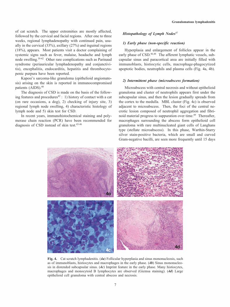
Hyperplasia and enlargement of follicles appear in theearly phase of CSD.46,48 The afferent lymphatic vessels, sub-capsular sinus and paracortical area are initially filled withimmunoblasts, histiocytic cells, macrophage-phagocytizedapoptotic bodies, neutrophils and plasma cells (Fig. 4a, 4b).
2) Intermittent phase (microabscess formation)
Microabscess with central necrosis and without epithelioidgranuloma and cluster of neutrophils appears first under thesubcapsular sinus, and then the lesion gradually spreads fromthe cortex to the medulla. MBL cluster (Fig. 4c) is observedadjacent to microabscess. Then, the foci of the central ne-crotic lesion composed of neutrophil aggregation and fibri-noid material progress to suppuration over time.49 Thereafter,macrophages surrounding the abscess form epithelioid cellgranuloma with rare multinucleated giant cells of Langhanstype (stellate microabscess). In this phase, Warthin-Starrysilver stain-positive bacteria, which are small and curvedGram-negative bacilli, are seen more frequently until 15 days
Granulomatous lymphadenitis
7
Fig. 4. Cat scratch lymphadenitis. (4a) Follicular hyperplasia and sinus mononucleosis, suchas of immunoblasts, histiocytes and macrophages in the early phase. (4b) Sinus mononucleo-sis in distended subcapsular sinus. (4c) Imprint feature in the early phase. Many histiocytes,macrophages and monocytoid B lymphocytes are observed (Giemsa staining). (4d) Largeepithelioid cell granuloma with central abscess and necrosis.
4a
4d4c
4b

01_Asano.mcd Page 8 v4.21
after onset or in the very early lesions.43,50,51
Immunohistochemical staining with antibodies to B. hen-selae in formalin-fixed paraffin-embedded tissue sections canspecifically identify the bacillus of CSD.44,52 In addition,rapid assay for the detection of B. henselae in formalin-fixedparaffin-embedded lymph nodes and aspirated pus fromlymph nodes is available by PCR.43,45,46
3) Late phase (granulomatous abscess)
In the late phase, coalescence of stellate microabscessesleads to formation of an irregularly shaped large abscess(geographic abscess) (Fig. 4d). Furthermore, the capsule oflymph node often shows fibrosis or marked inflammation.The detection rate of bacteria becomes lower than that in theprevious phase.
YERSINIA LYMPHADENITIS (MESENTERIC
LYMPHADENITIS)
Bacteria of the genus Yersinia are not detected in normalhuman gut flora, but Yersinia enterocolitica (Y. ent.) andYersinia pseudotuberculosis (Y. pseud.), which are Gram-negative small bacilli, are important human pathogens.
The disease Yersinia lymphadenitis is characterized byacute febrile gastroenteritis, with symptoms that mimic ap-pendicitis, and occurs predominantly in infants, children andyouths. The route of infection is orally, involving infectedmeat, drinking water or other food.53
Yersinia bacteria arrive at the mesenteric lymph nodes vialymphatic vessels of the intestine after ingestion of infectedmaterial, and form the characteristic lesion. The lymph nodeenlarges due to an inflammatory process, resulting in abdomi-nal pain.
In this disease, there are no specific findings except forswelling of mesenteric lymph nodes, even though acute ap-pendicitis or terminal ileitis can be shown.
History
This group of bacteria was described under differentnames, such as Bacterium enterocoliticum,54 Pasteurella X55
and germe X,56 from 1939 to the early 1960s. The first Y. ent.infections in humans were described in 194957 as infectionswith Pasteurella pseudotuberculosis. Since then, there havebeen many reports on Y. ent. infection in Western Europe andthe United States.58-60
Clinical Findings
The incubation period is 24 to 48 hours after the onset ofingestion. The most common symptoms of Y. ent. infectionare diarrhea (80%), lower right quadrant pain (50%), nausea,
vomiting and fever (38~39℃) with flu-like symptoms, whichare somewhat correlated with patient age. Thereafter, diar-rhea is likely to develop in infants, but terminal ileitis, appen-dicitis and mesenteric lymphadenitis occur in children andyouths.58,59 Given the abdominal findings described above,these cases are often diagnosed as appendicitis, mesentericlymphadenitis and terminal ileitis. Other symptoms such asarthritis, erythema nodosum, sepsis, Reiter’s syndrome, men-ingitis and cutaneous lesions appear in older patients.58,59 Onthe other hand, Y. pseud. infection is seen in infants and hassymptoms similar to those of Y. ent. infection. The patientsare younger than those with appendicitis. The diagnosis ismade by culture of stool, blood, vomit, resected appendix andlymph node. Furthermore, diagnosis also requires serum anti-body titer. Therapy is usually not necessary, but tetracyclineis useful, and amikacin, chloramphenicol and gentacin arealso effective.61
Histopathology of Lymph Nodes
1) Lymph nodes
Yersinia lymphadenitis is caused by the bacteria Y. ent.and Y. pseud.53 In this disease, lymph nodes of the ileocecalregion are often enlarged up to 1.5 cm in size. They do notcoalesce with each other and show an elastic-soft appearancewithout distinctive necrosis on cut sections.62
In the early phase of lymphadenopathy by Y. ent., lympho-cytes, immunoblasts and plasma cells are packed within di-lated sinuses. In addition, hyperplastic follicles are oftenobserved in cortex and paracortex with increases in the cellsdescribed above. These findings coincide with nonspecificreactions.
In progression and later phases, thickened edematous cap-sule occasionally contains lymphocytes, immunoblasts andplasma cells. In addition, the same kinds of cells are ob-served in extended sinuses (Fig. 5a, 5b). Later, many epithe-lioid cell granulomas (EPGs) emerge in the germinal centersand they are predominantly nonsuppurative (Fig. 5c, 5d).63
Some EPGs show suppuration of the centers of granulomas(central microabscesses) (Fig. 5e). These lesions eventuallyenlarge to form round microabscesses, but no giant cells.EPGs are composed of histiocytes with or without epithelioidcell features along with scattered small T lymphocytes andplasmacytoid monocytes.64 EPGs of this type of lymphadeni-tis do not contain MBLs like in cat scratch disease and lym-phogranuloma venereum.65 Gram-negative acid-fast diplo-bacilli may be identified in the lesions.53,58,59,62,66
On the other hand, lymphadenopathy induced by Y. pseud.is quite similar to that of Y. ent. infection in the initial phase.53
In the progression phase, Y. pseud.-infected lymph nodesshow massive neutrophil infiltration, and later, there are scat-tered microabscess lesions with central pus formation sur-
Asano S
8

01_Asano.mcd Page 9 v4.21
rounded by plump histiocytes (suppurative granulomas) iden-tical to those seen in cat scratch disease,53,58,66,67 a feature thatis rare with Y. ent.
2) Appendix and terminal ileum
In this disease, the wall of the appendix is thickened andthe mucosa is edematous. Appendical lesions contain trans-mural, mixed inflammatory infiltrates with numerous lym-phoid follicles and EPGs. EPGs are predominantly non-suppurative. In addition, they are usually surrounded bysmall T lymphocytes and plasmacytoid monocytes. EPGs donot contain MBLs like regional lymph nodes.64 However,some cases show mild hemorrhage and catarrh, although re-
gional lymph nodes show typical EPGs.
TUBERCULOUS LYMPHADENITIS
Tuberculosis is a chronic airborne infectious disease in-duced by M. tuberculosis. In Japan recently, tuberculosis hasbeen ranked as only the 20th most common cause of mortal-ity, so it has been relatively ignored. However, cases are stillconspicuous and outbreaks are occasionally reported. Themajor problem for tuberculosis is the severity of illness inassociation with the global spread of AIDS. Tuberculosis hasbecome remarkable as a reemerging infectious disease notonly in developing countries but worldwide.68,69
Granulomatous lymphadenitis
9
Fig. 5. Yersinia lymphadenitis. (5a & 5b) Edematous thickening capsule (C) and markedlydilated subcapsular sinuses (SS) contain innumerable lymphocytes, immunoblasts and plasmacells. (5c) Many epithelioid cell granulomas appear in the germinal centers and are predomi-nantly nonsuppurative. (5d) Enlargement of granuloma with nonsuppurative lesion. (5e)Granuloma with microabscess formation.
5c
5e
5d
01_Asano.mcd Page 10 v4.21
Epidemiology
According to global statistics, two billion people are af-fected by tuberculosis globally. At present, there are eightmillion new patients and three million deaths every year, andalmost all patients reside in the developing countries.70,71
Meanwhile, in Japan, the rate of mortality from tuberculosisamong the elderly has been gradually decreasing. The reduc-tion of mortality slowed down in the 1980s, and it then beganto increase again. Given these circumstances, in 1999, anemergency declaration to thwart the spread of tuberculosiswas issued.70,72
Clinical Findings
In the case of highly virulent M. tuberculosis or individu-als with weakened resistance against tuberculosis during pri-mary infection, lymphadenitis occurs at pulmonary hilar andcervical lymph nodes. Tuberculous meningitis and militarytuberculosis occur by lymphogenous spread of M. tuberculo-sis. In the case of longstanding multifocal adhesive lympha-denitis, lymph node biopsy, tuberculin reaction and PCR73 arerecommended for diagnosis of tuberculous lymphadenitis.74,75
Histopathology of Lymph Nodes
M. tuberculosis grows within alveolar macrophages andconsequently forms a well-established primary lesion in thelungs. It mostly occurs subjacent to the pleura of the upperlobe. Some bacteria arrive at the bronchopulmonary lymphnode via lymphatic vessels and form lymphadenopathy, clas-sical Ghon complex.
The primary infection begins with inhalation of M. tuber-culosis and ends with T cell-mediated immune response thatinduces hypersensitivity against the organism. The inhaledorganism is phagocytized by alveolar macrophages and trans-ported by these cells to hilar lymph nodes. After a fewweeks, T cell-mediated immunity develops in two ways. Oneis formation of epithelioid cell granuloma by CD4+ cells andthe other is formation of caseating granuloma by CD8+ cells.75
Thereafter, epithelioid granulomas are encapsulated and prog-ress to central caseous necrosis, eventually resulting in heal-ing.
This lymphadenitis is induced by M. tuberculosis and canbe seen as a part of the primary complex or secondary (organ)tuberculosis. About 90% of tuberculous lymphadenitismainly appears in the cervical lymph node and others are inthe mediastinal node. Sometimes it is difficult to distinguishamong tuberculous lymphadenitis, malignant lymphoma andmetastatic tumors.
Asano S
10
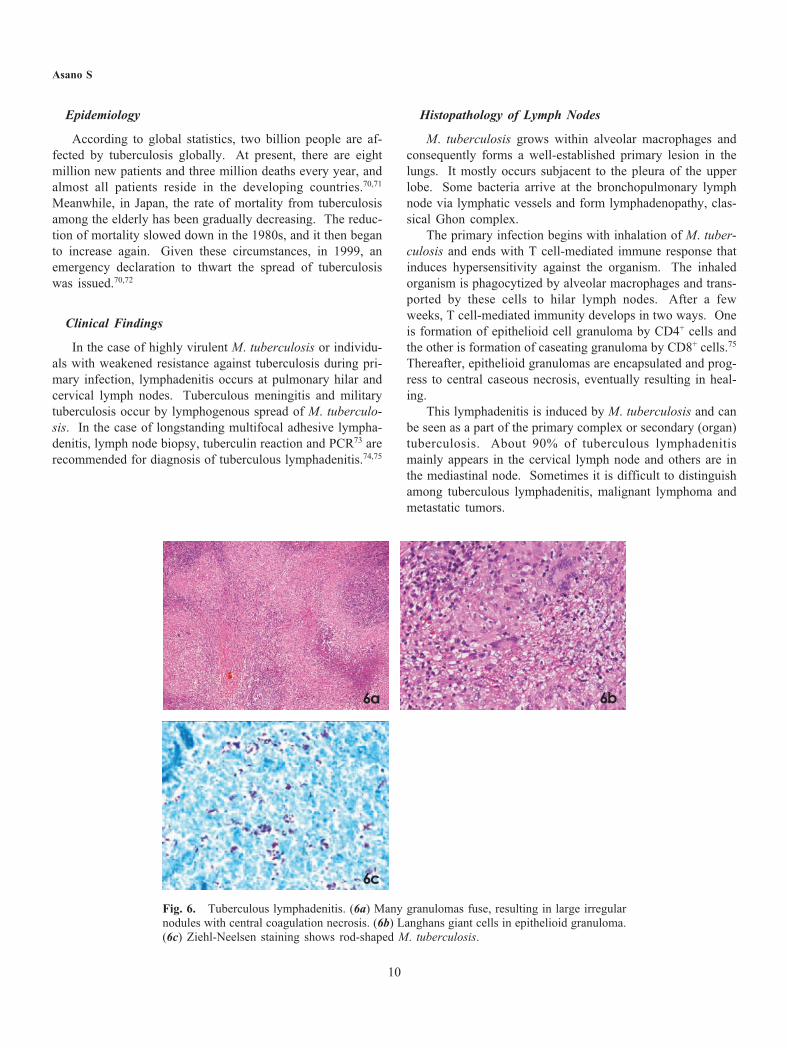
Fig. 6. Tuberculous lymphadenitis. (6a) Many granulomas fuse, resulting in large irregularnodules with central coagulation necrosis. (6b) Langhans giant cells in epithelioid granuloma.(6c) Ziehl-Neelsen staining shows rod-shaped M. tuberculosis.
6a 6b
6c

01_Asano.mcd Page 11 v4.21
In the early phase of tuberculous lymphadenitis, thelymph node shows elastic hard tumor like nonspecific lym-phadenitis. Inflammatory cells gradually infiltrate and peri-adenitis appears over time.
After the formation of abscesses in the lymph node, theyenlarge and gradually change to caseous necrosis and soften.The histology of tuberculous lymphadenitis is characterizedby central caseous necrosis surrounded by epithelioid celllayer and sporadic Langhans giant cells (Fig. 6a, 6b). Inaddition, in the outermost layer can be seen lymphocytes andfibroblasts, but no plasma cells are observed. This node iscalled tuberculous nodule, type IV allergic reactions againstM. tuberculosis. Tuberculous lymphadenitis is distinguishedfrom sarcoidosis lymphadenitis by the presence of centralcaseous necrosis.
In this phase, Ziehl-Neelsen staining occasionally showsshort rod-shaped bacteria (M. tuberculosis) in the coagulationnecrosis (Fig. 6c). Organisms are now most easily detectedby PCR. Finally, healing occurs with calcification.76
BCG-LYMPHADENITIS (BCG-HISTIOCYTOSIS)
Immunization of BCG (Bacillus Calmette-Guérin) vac-cine, as an immunostimulant, is very useful for preventing thedissemination of Mycobacterium tuberculosis from the pri-mary complex. BCG is conventionally re-injected fortuberculin-test-negative patients, but according to the revisionof the Tuberculosis Prevention Law, from 2005, infants under6 months should be immunized only once without tuberculintest. BCG is a version of Mycobacterium bovis with attenu-ated virulence, and is the weakest strain globally. After vac-cination of BCG, the bacillus moves to regional lymph nodeand arrives at systemic organs within several hours.Although tuberculous lesions in systemic organs such as re-gional lymph nodes, liver and spleen can appear, there may bespontaneous healing without specific lesions.77,78 Rednessand suppuration at the inoculation site within ten days ofvaccination may indicate tuberculosis infection. About threemonths later, the lesion heals with desquamation, crusting andscar.
Clinical Findings
The side effects of BCG-vaccination can include localizedulcer, abscess formation and regional lymph node enlarge-ment. In terms of the lymph nodes, they are usually affectedin axillary and cervical regions and swell up to two to threecm in size. Side effects are observed more frequently in casesof intradermal vaccination than percutaneous one.79
The incidence of side effects is 0.73 to 2.2% in vaccinatedchildren. Younger children are more susceptible to infection,and lymphadenopathy occurs from one week to 9 monthsafter vaccination.80,81 BCG lymphadenopathy of over 3 cm
must be surgically resected, but follow up is necessary forthose smaller than 3 cm.
In children with congenital immune deficiencies82 and inpatients with HIV infection and AIDS, suppurative lymphade-nitis and tuberculosis sometimes develop.80
Histopathology of Lymph Nodes83
BCG-lymphadenopathy is usually smaller than tubercu-lous lymphadenopathy. Follicular hyperplasia and sinus his-tiocytes are observed as in nonspecific lymphadenitis in theearly phase. Later, micronodules of epithelioid granulomaswithout necrosis and epithelioid cell granuloma with centralcoagulation necrosis are observed (Fig. 7a, 7b). Langhansgiant cells rarely appear.
TOXOPLASMA LYMPHADENITIS (PIRINGER-
KUCHINKA LYMPHADENITIS)
Toxoplasmosis is a common zoonosis caused byToxoplasma gondii ; it is found worldwide, and is prevalentin warm and humid climates, but infrequent in cold and dryareas.84
Granulomatous lymphadenitis
11
7a
7b
Fig. 7. BCG-lymphadenit is . (7a)Epithelioid cell granulomas with central co-agulation necrosis are observed. (7b) Oldgranuloma with central liquefaction necrosis.

01_Asano.mcd Page 12 v4.21
Epidemiology
In the United States, toxoplasmosis is the most commonparasitic infection and over 60 million people are infected.Fifty percent of Americans have antibodies, but most exhibitasymptomatic infection. In Western Europe, there are occa-sional infections through the ingestion of bradyzoit, a type oftrophozyte of Toxoplasma gondii, from contaminated under-cooked or close-to-raw meat.85 In France, the prevalence ofantibodies is 85%.86 Similarly, the antibody titers are high inGermany, the Netherlands and Brazil, but the prevalence inJapanese adults is around 20~30%.
Clinical Findings
Toxoplasma parasitizes a wide range of warm-bloodedanimals that include humans and rodents as intermediatehosts, and cats as the definitive host.83
The symptoms of toxoplasmosis resemble acute infectiousmononucleosis with slight fever in the general population.There are three predominant sources of infection in humans,namely, ingestion of undercooked meat containing cysts, oo-
cysts in cat stool and transplacental transfer from mother tofetus. Toxoplasma gondii mainly exists in humans in twoforms, namely, tachyzoites within macrophages in the circula-tion and bradyzoites within intracellular cysts. Oocysts arefound only in cat stool. If humans ingest oocysts or cysts,symptoms appear after 5 to 20 days, and 10 days to severalweeks, respectively, although almost all patients are asympto-matic.
In immune-deficient patients, such as those with leuke-mia, lymphoma, acquired immunodeficiency syndrome(AIDS) and undergoing immunosuppressive therapy, as wellas in fetuses, Toxoplasma gondii infection becomes acutelydisseminated and results in death from myocarditis, pneumo-nitis, chorioretinitis and encephalitis.87 The damage to thefetus is serious when transmission occurs in early pregnancy.
Histopathology of Lymph Nodes
Toxoplasmic lymphadenitis is diagnosed histologicallyand confirmed by serologic assays.88-90 In immunologicallynormal adults, localized posterior cervical lymphadenopathywith fever is a common symptom.91
Asano S
12
Fig. 8. Toxoplasma lymphadenitis (Piringer-Kuchinka lymphadenopathy). (8a) The charac-teristic features such as reactive follicular hyperplasia, cluster of epithelioid cells and patchesof monocytoid B lymphocyte (MBL) proliferation are seen in lymph node. (8b) Enlargedfollicles with reactive germinal centers include increased centroblasts, mitosis and macro-phages containing tingible bodies. (8c) Imprint smear shows MBLs with clear cytoplasm anddarkly stained nuclei are observed (Giemsa staining). (8d) Mononuclear cells, macrophagesand elongated epithelioid cells are observed by imprint smear (Giemsa staining).
8a 8b
8c 8d

01_Asano.mcd Page 13 v4.21
The characteristic main three histological findings oflymph node are florid reactive follicular hyperplasia, clusterof epithelioid cells and patches of MBL proliferation (Fig. 8a-8c). The follicles are enlarged by the intensely reactive ger-minal centers, including increased number of centroblasts,mitosis and macrophages containing tingible bodies (Fig. 8b).MBLs with clear cytoplasm and darkly stained nuclei (Fig.8c) are observed in distended sinuses and adjacent to bloodvessels. MBLs can also be seen in cat scratch disease51 andhuman immunodeficiency virus (HIV) infection,92 althoughthey are more common in toxoplasmic lymphadenitis. Thereare ill-defined aggregations of epithelioid cells scatteredthroughout the cortex, paracortex and occasionally withingerminal centers. They do not form round, organized, well-formed granulomas (Fig. 8a, 8d). They also do not inducenecrosis and are not accompanied by neutrophils, eosinophilsand fibrosis.
Toxoplasmic lymphadenitis can be diagnosed using reli-able serologic testing and the three main histological findingswith immunohistochemistry rather than PCR.93,94
CONCLUSION
Histopathologically, infectious granulomatous lymphade-nitis generally shows follicular hyperplasia and sinus histiocy-tosis in the early phase. In tularemia and cat scratch disease,numerous neutrophils, MBLs, histiocytic cells, T cells anddendritic cells gradually appear around granulomas. MBLsare found adjacent to granulomatous foci. It may be consid-ered that MBLs recruit neutrophils and promote necrosis, andMBLs with T cells and macrophages contribute to the forma-tion of granuloma. However, none of the EPGs of Y. ent.lymphadenitis contains MBLs like in cat scratch disease andlymphogranuloma venereum. Furthermore, Y. ent. lymphade-nitis EPGs differ from other types of suppurative lymphadeni-tis and the EPGs emerge in the germinal centers and arepredominantly nonsuppurative.
Granuloma without suppuration of tuberculosis and BCG-lymphadenitis are induced by delayed allergic reaction of M.tuberculosis. These types of lymphadenitis and sarcoidosisinvolve B-cell-negative hypersensitivity. In toxoplasma lym-phadenitis, MBLs can also be seen and ill-defined aggrega-tions of epithelioid cells are scattered throughout the lymphnode. However, they do not form round, organized, well-formed granulomas. Furthermore, they do not induce ne-crosis and are not accompanied by neutrophils, eosinophilsand fibrosis.
Organisms are easily detectable by some staining, immu-nohistochemical and molecular biological methods such asPCR. Accurate pathological diagnosis by using the aboveprocedures can lead to precise treatment.
ACKNOWLEDGEMENT
This work was supported by a grant for Research onEmerging and Reemerging Infectious Disease (H22 Shinko-Ippann-6) from the Ministry of Health, Labour, and Welfare,Japan. The author appreciates support for the figures from Dr.Masaru Kojima, Dokkyo University School of Medicine,Tochigi, Japan.
REFERENCES
1 Chang KL, Arber DA, Gaal KK, Weiss LM: Lymph nodes and
spleen. In : Silverberg SG, DeLellis RA, Frable WJ, Livolsi VA,
Wick MR (eds) : Silverberg’s principles and practice of surgical
pathology and cytopathology. 4th ed, Philadelphia, Churchill
LivingStone, pp.508-607, 2006
2 Siltzbach LE: Sarcoidosis : clinical features and management.
Med Clin North Am 51:483-502, 1967
3 Moller DR: Etiology of sarcoidosis. Clin Chest Med 18:695-706,
1997
4 Bocart D, Lecossier D, Lassence AD, Valeyre D, Battesti JP, et
al.: A search for mycobacterial DNA in granulomatous tissues
from patients with sarcoidosis using polymerase chain reaction.
Am Rev Respir Dis 145:1142-1148, 1992
5 Popper HH, Klemen H, Hoefler G, Winter E: Presence of myco-
bacterial DNA in sarcoidosis. Hum Pathol 28:796-800, 1997
6 Newman LS, Rose CS, Maier LA: Sarcoidosis. N Engl J Med
336:1224-1234, 1997
7 Bardinas F, Morera J, Fité E, Plasencia A: Seasonal clustering of
sarcoidosis. Lancet 2:455-456, 1989
8 Cunningham JA: Sarcoidosis. Pathol Annu 2:31-47, 1967
9 James DG, Neville E: Pathobiology of sarcoidosis. Pathobiol
Annu 7:31-63, 1977
10 Ioachim HL, Medeiros LJ: Sarcoidosis Lymphadenopathy. In :
Ioachim HL, Medeiros LJ (eds) : Ioachim’s Lymph node pathol-
ogy. 4th ed, Philadelphia, Lippincott Williams & Wilkins, pp.
203-212, 2009
11 Ota M, Amakawa R, Uehira K, Ito T, Yagi Y, et al.: Involvement
of dendritic cells in sarcoidosis. Thorax 59:408-413, 2004
12 Rosen Y, Vuletin JC, Pertschunk LP, Silverstein E: Sarcoidosis :
from the pathologist’s vantage point. Pathol Annu 14 (Pt 1):405-
439, 1979
13 Brincker H: Sarcoid reactions in malignant tumours. Cancer Treat
Rev 13:147-156, 1986
14 James DG: Editorial : Modern concept of sarcoidosis. Chest 64:
675-677, 1973
15 Kojima M, Nakamura S, Fujisaki M, Hirahata S, Hasegawa H, et
al.: Sarcoid-like reaction in the regional lymph nodes and spleen
in gastric carcinoma : a clinicopathologic study of five cases. Gen
Diagn Pathol 142:347-352, 1997
16 Virgili A, Maranini C, Califano A: Granulomatous lesions of the
homolateral limb after previous mastectomy. Br J Dermatol 146:
891-894, 2002
Granulomatous lymphadenitis
13

01_Asano.mcd Page 14 v4.21
17 Gregorie HB Jr, Othersen HB Jr, Moore MP Jr: The significance
of sarcoid-like lesions in association with malignant neoplasms.
Am J Surg 104:577-586, 1962
18 Yamamoto T, Tateishi H, Nishimura Y, Watanabe M, Ukyo S, et
al.: A study of gastric cancer with sarcoid reaction as observed in
the regional lymph node (author's transl). Nihon Shokakibyo
Gakkai Zasshi 77:1555-1561, 1980 [in Japanese with English
abstract] .
19 Takeuchi H, Suchi T, Suzuki R, Sato T: Histological study of
immune parameters of regional lymph nodes of gastric cancer
patients. Gann 73:420-428, 1982
20 Gorton G, Linell F: Malignant tumours and sarcoid reactions in
regional lymph nodes. Acta Radiol 47:381-392, 1957
21 Gherardi GJ: Localized lymph node sarcoidosis associated with
carcinoma of the bile ducts ; report of a case. AMA Arch Pathol
49:163-168, 1950
22 Wolbach SB: A new type of cell inclusion, not parasitic, associ-
ated with disseminated granulomatous lesions. J Med Res 24:243-
258, 1911
23 Kurata A, Terado Y, Schulz A, Fujioka Y, Franke FE:
Inflammatory cells in the formation of tumor-related sarcoid reac-
tion. Hum Pathol 35:546-554, 2005
24 Harris S: Japanese biological warfare research on humans : a case
study of microbiology and ethics. Ann N Y Acad Sci 666:21-52,
1992
25 Denis DT, Inglesby TV, Henderson DA, Bartlett JG, Ascher MS,
et al.: Tularemia as a biological weapon : Medical and public
health management. JAMA 285:2763-2773, 2001
26 Johansson A, Ibrahim A, Goeransson I, Eriksson U, Gurycova D,
et al.: Evaluation of PCR-based methods for discrimination of
Francisella species and development of a specific PCR that distin-
guished the two major subspecies of Francisella tularensis. J Clin
Microbiol 38:4180-4185, 2000
27 Vogler AJ, Birdsell D, Price LB, Bowers JR, Beckstrom-
Sternberg SM, et al.: Phylogeography of Francisella tularensis :
Global expansion of a highly fit clone. J Bacteriol 191:2474-2484,
2009
28 Boyce JM: Recent trends in the epidemiology of tularemia in the
United States. J Infect Dis 131:197-199, 1975
29 Ohara S, Hoshishima K: Diagnosis of Yato-byo (Ohara disease,
tularemia in Japan). Fukushima J Med Sci 4:51-61, 1957
30 Ohara S, Ichikawa K: The distribution of Yato-byo and its clinical
observations. Ohara Nenpou 11:1-22, 1962 (in Japanese)
31 Francis E: Tularemia. JAMA 84:1243-1250, 1925
32 Kitamura S, Fukuda M, Takeda H, Ouchi S, Nakano S, et al.:
Pathology of tularemia. Acta Pathol Jap 6:719-753, 1956
33 Inglesby TV, O’Toole T, Henderson DA, Bartlett JG, Ascher MS,
et al.; Working Group on Civilian Biodefense : Anthrax as a
biological weapon, 2002 : updated recommendations for manage-
ment. JAMA 287:2236-2252, 2002
34 Syrjälä H, Kujala P, Myllylä V, Salminen A: Airborne transmis-
sion of tularemia in farmers. Scand J Infect Dis 17:371-375, 1985
35 Christopher GW, Cieslak TJ, Pavlin JA, Eitzen EM Jr: Biological
warfare. A histological perspective. JAMA 278:412-417, 1997
36 Versage, JL, Severin DD, Chu MC, Petersen JM: Development of
a multitarget real-time TaqMan PCR assay for enhanced detection
of Francisella tularensis in complex specimens. J Clin Microbiol
41:5492-5499, 2003
37 Tsukahara M: Cat scratch disease in Japan. J Infect Chemother 8:
321-325, 2002
38 Jackson LA, Perkins BA, Wenger JD: Cat scratch disease in the
United States : an analysis of three national databases. Am J
Public Health 83:1707-1711, 1993
39 Greer WE, Keefer CS: Cat-scratch fever ; a disease entity. N Eng
J Med 244:545-548, 1951
40 Regnery RL, Anderson BE, Clarridge JE 3rd, Rodriguez-Barradas
MC, Jones DC, et al.: Characterization of a novel Rochalimaea
species, R. henselae sp. nov., isolated from blood of a febrile,
human immunodeficiency virus-positive patient. J Clin Microbiol
30:265-274, 1992
41 Dauga C, Miras I, Grimont PA: Identification of Bartonella
henselae and B. quintana 16s rDNA sequences by branch-, genus-
and species-specific amplification. J Med Microbiol 45:192-199,
1996
42 Wear DJ, Margileth AM, Handfield TL, Fischer GW, Schlagel CJ,
et al.: Cat scratch disease : a bacterial infection. Science
221:1403-1405, 1983
43 Anderson B, Sims K, Regnery RL, Robinson L, Schmidt MJ, et
al.: Detection of Rochalimaea henselae DNA in specimens from
cat scratch disease patients by PCR. J Clin Microbiol 32:942-948,
1994
44 Min KW, Reed JA, Welch DF: Morphologically variable bacilli
of cat scratch disease are identified by immunocytochemical label-
ing with antibodies to Rochalimaea henselae. Am J Clin Pathol
101:607-610, 1994
45 Scott MA, McCurley TL, Vnencak-Jones CL, Hager C, McCoy
JA, et al.: Cat scratch disease : detection of Bartonella henselae
DNA in archival biopsies from patients with clinically, serologi-
cally, and histologically defined disease. Am J Pathol 149:2161-
2167, 1996
46 Mouritsen CL, Litwin CM, Maiese RL, Segal SM, Segal GH:
Rapid polymerase chain reaction-based detection of the causative
agent of cat scratch disease (Rochalimaea henselae) in formalin-
fixed, paraffin embedded-samples. Hum Pathol 28:820-826, 1997
47 Naji AF, Carbonell F, Barker HJ: Cat scratch disease. A report of
three new cases, review of the literature, and classification of the
pathologic changes in the lymph nodes during various stages of
the disease. Am J Clin Pathol 38:513-521, 1962
48 Dorfman RF, Warnke R: Lymphadenopathy simulating the malig-
nant lymphoma. Hum Pathol 5:519-550, 1974
49 Wear D, English C, Margileth A, Banks I: Histopathologic
changes in the lymph nodes of over 500 patients with cat scratch
disease. Lab Invest 58:100A, 1988
50 Ioachim HL, Medeiros LJ: Cat-scratch Lymphadenopathy. In :
Ioachim HL, Medeiros LJ (eds) : Ioachim’s Lymph node pathol-
ogy. 4th ed, Philadelphia, Lippincott Williams & Wilkins, pp.
Asano S
14

01_Asano.mcd Page 15 v4.21
110-114, 2009
51 Kojima M, Nakamura S, Hosomura Y, Shimizu K, Kurabayashi Y,
et al.: Abscess-forming granulomatous lymphadenitis : histologi-
cal typing of suppurative granulomas and clinicopathological find-
ings with special reference to cat scratch disease. Acta Pathol Jpn
43:11-17, 1993
52 Regnery R, Martin M, Olson J: Naturally occurring
“Rochalimaea henselae” infection in domestic cat. Lancet 340:
557-558, 1992
53 Schapers RF, Reif R, Lennert K, Knapp W: Mesenteric lym-
phadenitis due to Yersinia enterocolitica. Virchows Arch A Pathol
Anat Histol 390:127-138, 1981
54 Schleifstein J, Coleman M: An unidentified microorganism re-
sembling B. lignieri and Pasteurella pseudotuberculosis and
pathogenic for man. NY State J Med 39:1749-1753, 1939
55 Daniels JJHM: Untersuchungen an als Pasteurella pseudotuber-
culosis diagnostizierten Stämmen von Chinchillas. Zentralbl Vet
Med 10:413-419, 1963 (in German)
56 Mollaret HH, Destombes P: Les germes “X” en pathologie hu-
mane. Presse Med 72:2913-2915, 1964 (in French)
57 Hässig A, Karrer J, Pusterla F: Über Pseudotuberculose beim
Menschen. Schweiz Med Wochenschr 79:971-973, 1949
58 Finlayson NB, Fagundes B: Pasteurella pseudotuberculosis infec-
tion : three cases in the United States. Am J Clin Pathol 55:24-29,
1971
59 Gleason TH, Patterson SD: The pathology of Yersinia enterocolitica
ileocolitica. Am J Surg Pathol 6:347-355, 1982
60 Winblad S: Studies on O-antigen factors of “Yersinia enterocolitica”.
Symp Series Immunobiol Stand 9:337-343, 1968
61 Knapp W, Lysy J, Knapp C, Stille W, Goll U: Enterale Infektion
beim Menschen durch Yersinia enterocolitica und ihre Diagnose.
Infection 1:113-125, 1973 (in German)
62 Koizumi F, Sakai T: Yersinia lymphadenitis. Byori to Rinsyou,
12:341-345, 1994 (in Japanese)
63 Lamps LW, Madhusudhan KT, Greenson JK, Pierce RH, Massoll
NA, et al.: The role of Yersinia enterocolitica and Yersinia pseu-
dotuberculosis in granulomatous appendicitis : a histologic and
molecular study. Am J Surg Pathol 25:508-515, 2001
64 Kojima M, Morita Y, Shimizu K, Yoshida T, Yamada I, et al.:
Immunohistological findings of suppurative granulomas of
Yersinia enterocolitica appendicitis : A report of two cases.
Pathol Res Pract 203:115-119, 2007
65 Chan JKC: Rendering a definitive diagnosis of lymphogranuloma
venereum lymphadenitis by morphologic assessment and approach
to diagnosis of suppurative granulomatous lymphadenitis. Adv
Anat Pathol 4:28-39, 1997
66 Maßhoff W: Eine neuartige Form der mesenterialen
Lymphadenitis. Dtsch Med Wochenschr 78:532-535, 1953 (in
German)
67 El-Maraghi NR, Mair NS: The histopathology of enteric infection
with Yersinia pseudotuberculosis. Am J Clin Pathol 71:631-639,
1979
68 Sunderam G, MacDonald RJ, Maniatis T, Oleske J, Kapila R, et
al.: Tuberculosis as a manifestation of the acquired immunodefi-
ciency syndrome (AIDS). JAMA 256:362-366, 1986
69 Ramaswamy G, Rao SV, Reddy SA, et al.: Mycobacterial infec-
tion and acquired immunodeficiency syndrome : a New York City
hospital experience. Lab Med 19:652-654, 1988
70 Global tuberculosis programme. In global tuberculosis control.
WHO report 1998. Geneva, World Health Organization, 237, 1998
71 Ravigllione MC, Snider DE Jr, Kochi A: Global epidemiology of
tuberculosis : morbidity and mortality of worldwide epidemic.
JAMA 273:220-226, 1995
72 Sepkowitz KA: One disease two epidemics. AIDS at 25. N Engl J
Med 354:2411-2414, 2006
73 Marchetti G, Gori A, Catozzi L, Vago L, Nebuloni M, et al.:
Evaluation of PCR in detection of Mycobacterium tuberculosis
from formalin-fixed, paraffin-embedded tissues : comparison of
four amplification assays. J Clin Microbiol 36:1512-1517, 1998
74 Polesky A, Grove W, Bhatia G: Peripheral tuberculous lymphade-
nitis : epidemiology, diagnosis, treatment, and outcome. Medicine
84:350-362, 2005
75 Samuelson J, von Lichtenberg F: Infectious disease. In : Cotran
RS, Kumar V, Robbins SL (eds) : Pathologic basis of disease, 5th
ed, Philadelphia, W. B. Saunders, pp.305-377, 1994
76 Ioachim HL: Granulomatous lesions of lymph nodes. In :
Ioachim HL (ed) : Pathology of granulomas. New York, Raven
Press, pp.151-187, 1983
77 Gormsen H: On the occurrence of the epithelioid cell granulomas
in the organs of BCG-vaccinated human beings. Acta Pathol
Microbiol Scand Suppl 39 (Suppl 111):117-120, 1956
78 Horwitz O, Meyer J: The safety record of BCG vaccination and
untoward reactions observed after vaccination. Bibl Tuberc 13:
245-271, 1957
79 Watanabe K, Segawa M, Hukuhara M, Tatsumi K, Takayanagi S,
et al.: A case of subcutaneous tuberculous granuloma induced
BCG immunization. Shounika Rinnsyou 39:1313-1316, 1986 (in
Japanese)
80 Al-Bhlal AL: Pathologic findings for bacille Calmette-Guérin in-
fections in immunocompetent and immunocompromised patients.
Am J Clin Pathol 113:703-708, 2000
81 Ali-el Dein B, Nabeeh A: Tuberculous iliac lymphadenitis : a
rare complication of intravesical bacillus Calmette-Guerin and
cause of tumor over staging. J Uro 156:1766-1767, 1996
82 Abramowsky C, Gonzalez B, Sorensen RU: Disseminated bacil-
lus Calmette-Guérin infections in patients with primary immuno-
deficiencies. Am J Clin Pathol 100:52-56, 1993
83 Symmers W St C: Lymphadenitis following injection of bacillus
Calmette-Guérin. Systemic pathology. 5th ed, Edinburgh, Churchil
Livingstone, pp.370-371, 1992
84 Remington JS, Jacobs L, Kaufman HE: Toxoplasmosis in the
adult. N Engl J Med 262:180-186, 1960 & 262:237-241, 1960
85 Krick JA, Remington JS: Toxoplasmosis in the adult : an over-
view. N Engl J Med 298:550-553, 1978
86 Ioachim HL, Medeiros LJ: Toxoplasma Lymphadenitis. In :
Ioachim HL, Medeiros LJ (eds) : Ioachim’s Lymph node pathol-
Granulomatous lymphadenitis
15

01_Asano.mcd Page 16 v4.21
ogy. 4th ed, Philadelphia, Lippincott Williams & Wilkins, pp.
159-164, 2009
87 Gleason TH, Hamlin WB: Disseminated toxoplasmosis in the
compromised host. A report of five cases. Arch Intern Med 134:
1059-1062, 1974
88 Piringer-Kuchinka A, Martin I, Thalhammer O: Über die vorzu-
glich cerviconuchale Lymphadenitis mit kleinherdiger Epithelioid
zellwucherung. Virchows Arch A Pathol Anat Histopathol 331:
522-535, 1958 (in German)
89 Saxen E, Saxen L, Gronroos P: Glandular toxoplasmosis. A report
on 23 histologically diagnosed cases. Acta Pathol Microbiol Scand
44:319-328, 1958
90 Miettinen M: Histologic differential diagnosis between lymph
node toxoplasmosis and other benign lymph node hyperplasia.
Histopathology 5:205-216, 1981
91 Gray GF Jr, Kimball AC, Kean BH: The posterior cervical lymph
node in toxoplasmosis. Am J Pathol 69:349-358, 1972
92 Ioachim HL, Cronin W, Roy M, Maya M: Persistent lymphadeno-
pathies in people at high risk for HIV infection. Clinicopathologic
correlations and long-term follow-up in 79 cases. Am J Pathol 93:
208-218, 1990
93 Stansfeld AG: The histologic diagnosis of toxoplasmic lymphade-
nitis. J Clin Pathol 14:565-573, 1961
94 Andres TL, Dorman SA, Winn WC Jr, Trainer TD, Perl DP, et al.:
Immunohistochemical demonstration of Toxopasma gondii. Am J
Clin Pathol 75:431-434, 1981
Asano S
16


![Accepted: Published: Positivity in Tubercular ISSN ... · with caseous necrosis) in 49.3% [5]. Few studies reported granulomatous lymphadenitis in 57.8% cases [6]. But they did not](https://static.fdocuments.in/doc/165x107/5fc3fd1695fbe21b461044b6/accepted-published-positivity-in-tubercular-issn-with-caseous-necrosis-in.jpg)


