A Case of Secondary Syphilis with the Extragenital Chancre ...
3
Case Report A Case of Secondary Syphilis with the Extragenital Chancre on the Nipple Xiao Yan Wang, Juliandri, Zi Jing Liu, Jia Wen Zhang, Yun Yi Liu, Da-Guang Wang and Yang Xu Department of Dermatology, e First Affiliated Hospital of Nanjing Medical University, Nanjing 210029, China Correspondence should be addressed to Yang Xu; [email protected] Received 24 March 2019; Accepted 27 June 2019; Published 11 February 2020 Academic Editor: Ravi Krishnan Copyright © 2020 Xiao Yan Wang et al. is is an open access article distributed under the Creative Commons Attribution License, which permits unrestricted use, distribution, and reproduction in any medium, provided the original work is properly cited. Syphilis is a sexually transmitted disease caused by Treponema pallidum. e signs and symptoms of syphilis vary depending on which of the four stages it presents. e primary stage of syphilis classically presents with a painless ulcer (chancre). We report a case of the extragenital chancre on the nipple which is examined from skin biopsy and immunohistochemistry. is case showed that it is important to identify the special site’s pruritus erythema by pathology and serological examination. 1. Introduction Syphilis is a common sexually transmitted or blood con- tracted, congenital transmission disease. It can be divided into primary syphilis, secondary syphilis, tertiary syphilis, and neurosyphilis. e diagnosis is based on the clinical manifes- tations and serological methods. In this article, we report a case of secondary syphilis with extragenital chancre on the nipple. 2. Case Presentation A 56-year-old male was presented to our dermatology depart- ment on June 12, 2018, with complaints of the right nipple erosion and discomfort for a week. Diagnosis of “eczema” was made and the patient was treated with topical zinc oxide oil and traditional Chinese herbal cream. Physical examination showed local erythematous erosion with a few exudations on the right nipple (Figure 1) and a skin biopsy was performed. e histopathological results showed epidermal hyperplasia, cutaneous protrusion, migration of small amount of inflam- matory cells, infiltration of large number of cells around the blood vessels of the dermis, and some of the nucleus were large, with scattered cells such as the lymphocytes and plasma cells (Figure 2). Immunohistochemistry was recommended to confirm the diagnosis. e results revealed the (the right papillary skin) proliferation of lymphocytes: CD20(+), CD3(+), CD5(+), Pax-5(+), CD38(+), Bc1-2(+), CD10(−), Bcl-6(−), MUM1(−), Cyclin D1(−), CD163(+), Igκ(+), and Igλ(+), combined with hematoxylin-eosin (H-E) staining, showed numerous lymphoplasmacytes infiltration in the cuta- neous dermal layer (Figure 3). On the follow-up, the patient complained of painless itchy erythema on bilateral soles. Physical examination showed that bilateral palms and soles had multiple copper-red macula and papules, some of which were desquamated on the surface (Figure 4). e syphilis serol- ogy examination showed that TPPA was positive, RPR was positive, and the titer was >1 : 32, so the diagnosis was con- firmed for syphilis. en the patient was treated with IM injec- tion of 2.4 million units of benzathine penicillin. Following a week later for follow up, the papillary erosion scab desquama- tion and the erythema subsided, the bilateral palms and the soles of dark erythema also subsided. And the 2.4 million units of benzathine penicillin were administered intramuscularly once a week for three weeks totally. e patient is still in fur- ther follow-up. 3. Discussion Syphilis is a sexually transmitted disease caused by Treponema pallidum [1]. e primary-stage of syphilis is mainly charac- terized by painless ulcers (chancre), which occur mostly in the genitals, anus, oral cavity [2], lips, pharyngeal, and nipple-areola [3]. ere have been several reports of primary syphilis Hindawi Case Reports in Dermatological Medicine Volume 2020, Article ID 2391907, 3 pages https://doi.org/10.1155/2020/2391907
Transcript of A Case of Secondary Syphilis with the Extragenital Chancre ...
Case ReportA Case of Secondary Syphilis with the Extragenital Chancre on the Nipple
Xiao Yan Wang, Juliandri, Zi Jing Liu, Jia Wen Zhang, Yun Yi Liu, Da-Guang Wang and Yang Xu
Department of Dermatology, �e First Affiliated Hospital of Nanjing Medical University, Nanjing 210029, China
Correspondence should be addressed to Yang Xu; [email protected]
Received 24 March 2019; Accepted 27 June 2019; Published 11 February 2020
Academic Editor: Ravi Krishnan
Copyright © 2020 Xiao Yan Wang et al. �is is an open access article distributed under the Creative Commons Attribution License, which permits unrestricted use, distribution, and reproduction in any medium, provided the original work is properly cited.
Syphilis is a sexually transmitted disease caused by Treponema pallidum. �e signs and symptoms of syphilis vary depending on which of the four stages it presents. �e primary stage of syphilis classically presents with a painless ulcer (chancre). We report a case of the extragenital chancre on the nipple which is examined from skin biopsy and immunohistochemistry. �is case showed that it is important to identify the special site’s pruritus erythema by pathology and serological examination.
1. Introduction
Syphilis is a common sexually transmitted or blood con-tracted, congenital transmission disease. It can be divided into primary syphilis, secondary syphilis, tertiary syphilis, and neurosyphilis. �e diagnosis is based on the clinical manifes-tations and serological methods. In this article, we report a case of secondary syphilis with extragenital chancre on the nipple.
2. Case Presentation
A 56-year-old male was presented to our dermatology depart-ment on June 12, 2018, with complaints of the right nipple erosion and discomfort for a week. Diagnosis of “eczema” was made and the patient was treated with topical zinc oxide oil and traditional Chinese herbal cream. Physical examination showed local erythematous erosion with a few exudations on the right nipple (Figure 1) and a skin biopsy was performed. �e histopathological results showed epidermal hyperplasia, cutaneous protrusion, migration of small amount of inflam-matory cells, infiltration of large number of cells around the blood vessels of the dermis, and some of the nucleus were large, with scattered cells such as the lymphocytes and plasma cells (Figure 2). Immunohistochemistry was recommended to confirm the diagnosis. �e results revealed the (the right papillary skin) proliferation of lymphocytes: CD20(+),
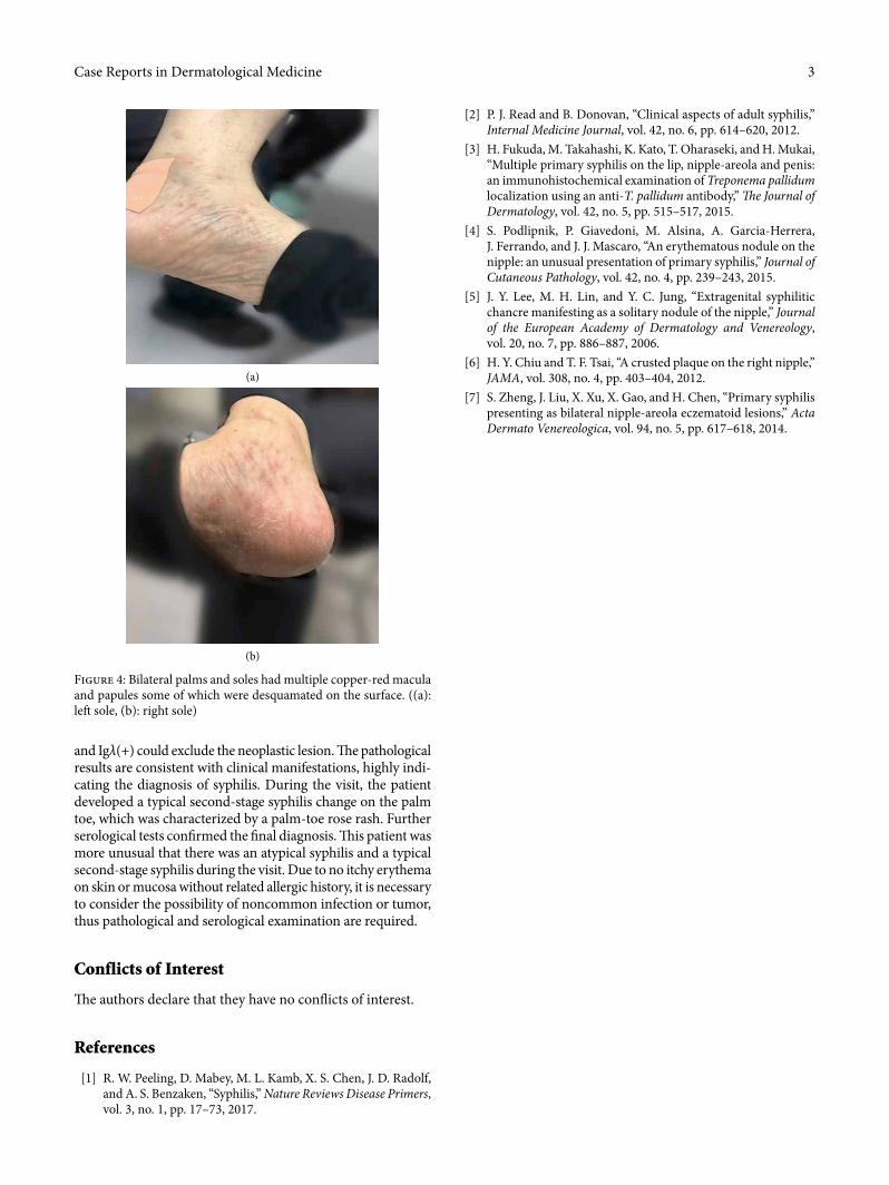
CD3(+), CD5(+), Pax-5(+), CD38(+), Bc1-2(+), CD10(−), Bcl-6(−), MUM1(−), Cyclin D1(−), CD163(+), Igκ(+), and Igλ(+), combined with hematoxylin-eosin (H-E) staining, showed numerous lymphoplasmacytes infiltration in the cuta-neous dermal layer (Figure 3). On the follow-up, the patient complained of painless itchy erythema on bilateral soles. Physical examination showed that bilateral palms and soles had multiple copper-red macula and papules, some of which were desquamated on the surface (Figure 4). �e syphilis serol-ogy examination showed that TPPA was positive, RPR was positive, and the titer was >1 : 32, so the diagnosis was con-firmed for syphilis. �en the patient was treated with IM injec-tion of 2.4 million units of benzathine penicillin. Following a week later for follow up, the papillary erosion scab desquama-tion and the erythema subsided, the bilateral palms and the soles of dark erythema also subsided. And the 2.4 million units of benzathine penicillin were administered intramuscularly once a week for three weeks totally. �e patient is still in fur-ther follow-up.
3. Discussion
Syphilis is a sexually transmitted disease caused by Treponema pallidum [1]. �e primary-stage of syphilis is mainly charac-terized by painless ulcers (chancre), which occur mostly in the genitals, anus, oral cavity [2], lips, pharyngeal, and nipple-areola [3]. �ere have been several reports of primary syphilis
HindawiCase Reports in Dermatological MedicineVolume 2020, Article ID 2391907, 3 pageshttps://doi.org/10.1155/2020/2391907

Case Reports in Dermatological Medicine2
occurring on the nipple and, its clinical manifestations vary [4–7], such as erythema nodules on the nipple, swelling, ero-sion, indolent ulcers, asymptomatic scaly erythematous or crusted plaque, etc. �is case is a unilateral nipple erythematous exudation change, which can be easily misdiagnosed as eczema, Paget’s breast disease and so on. Histopathological and immu-nohistochemical results suggested that lymphocytes and plasma cells infiltrate the dermis, and the diseases of intradermal plasma cell infiltration are common in plasmacytosis, plasma-cytoma, syphilis, and fungal infections. �e dermis may have plasma cells, neutrophils, and lymphocytes infiltrated in pri-mary syphilis. In immunohistochemistry Pax5, CD20 are
expressed on the surface of B cells, CD3, are CD5 are the T cells markers. CD163 is expressed by the macrophages, CD38 is most strongly expressed in plasmocytes; however, Bc1-2(+), CD109(−), Bcl-6(−), MUM1(−), Cyclin D1(−), Alk(−), Igκ(+),
Figure 1: An erythematous erosion with a few exudation on the right nipple before treatment.
(a)
(b)
Figure 2: Biopsy of the nipple showed that epidermal hyperplasia, cutaneous protrusion, a migration of small amount of inflammatory cells, an infiltration of large number of cells around the blood vessels of the dermis (a). �e inflammatory cells in the dermis were majorly lymphocytes and plasma cells (b). (H-E staining, original magnification: (a) ×100; (b) ×400)
(a)
(b)
(c)
(d)
Figure 3: Immunohistochemical staining showed that lym phocplas-mocytes infiltration in the cutaneous dermal layer. (a: CD20(+), b: CD38(+), c: lgκ(+), d: Igλ(+), original magnification: ×400)
3Case Reports in Dermatological Medicine
and Igλ(+) could exclude the neoplastic lesion. �e pathological results are consistent with clinical manifestations, highly indi-cating the diagnosis of syphilis. During the visit, the patient developed a typical second-stage syphilis change on the palm toe, which was characterized by a palm-toe rose rash. Further serological tests confirmed the final diagnosis. �is patient was more unusual that there was an atypical syphilis and a typical second-stage syphilis during the visit. Due to no itchy erythema on skin or mucosa without related allergic history, it is necessary to consider the possibility of noncommon infection or tumor, thus pathological and serological examination are required.
Conflicts of Interest
�e authors declare that they have no conflicts of interest.
References
[1] R. W. Peeling, D. Mabey, M. L. Kamb, X. S. Chen, J. D. Radolf, and A. S. Benzaken, “Syphilis,” Nature Reviews Disease Primers, vol. 3, no. 1, pp. 17–73, 2017.
[2] P. J. Read and B. Donovan, “Clinical aspects of adult syphilis,” Internal Medicine Journal, vol. 42, no. 6, pp. 614–620, 2012.
[3] H. Fukuda, M. Takahashi, K. Kato, T. Oharaseki, and H. Mukai, “Multiple primary syphilis on the lip, nipple-areola and penis: an immunohistochemical examination of Treponema pallidum localization using an anti-T. pallidum antibody,” �e Journal of Dermatology, vol. 42, no. 5, pp. 515–517, 2015.
[4] S. Podlipnik, P. Giavedoni, M. Alsina, A. Garcia-Herrera, J. Ferrando, and J. J. Mascaro, “An erythematous nodule on the nipple: an unusual presentation of primary syphilis,” Journal of Cutaneous Pathology, vol. 42, no. 4, pp. 239–243, 2015.
[5] J. Y. Lee, M. H. Lin, and Y. C. Jung, “Extragenital syphilitic chancre manifesting as a solitary nodule of the nipple,” Journal of the European Academy of Dermatology and Venereology, vol. 20, no. 7, pp. 886–887, 2006.
[6] H. Y. Chiu and T. F. Tsai, “A crusted plaque on the right nipple,” JAMA, vol. 308, no. 4, pp. 403–404, 2012.
[7] S. Zheng, J. Liu, X. Xu, X. Gao, and H. Chen, “Primary syphilis presenting as bilateral nipple-areola eczematoid lesions,” Acta Dermato Venereologica, vol. 94, no. 5, pp. 617–618, 2014.
(a)
(b)
Figure 4: Bilateral palms and soles had multiple copper-red macula and papules some of which were desquamated on the surface. ((a): le� sole, (b): right sole)