A BSTRACT ID - IRIA - 1164 TITLE : TO COMPARE X-RAY AND NCCT SCAN WITH MRI FOR SCREENING OF...
20
ABSTRACT ID- IRIA - 1164 TITLE : TO COMPARE X-RAY AND NCCT SCAN WITH MRI FOR SCREENING OF PARANASAL SINUS DISEASE
-
Upload
phillip-bowes -
Category
Documents
-
view
223 -
download
1
Transcript of A BSTRACT ID - IRIA - 1164 TITLE : TO COMPARE X-RAY AND NCCT SCAN WITH MRI FOR SCREENING OF...

ABSTRACT ID- IRIA - 1164
TITLE : TO COMPARE X-RAY AND NCCT SCAN WITH MRI FOR SCREENING OF PARANASAL SINUS DISEASE

AIMS AND OBJECTIVES
To compare x-ray and NCCT scan with MRI for screening of paranasal sinus disease

NEED FOR STUDY
Radiograph PNS Water`s view is the most commonly requisitioned imaging modality for screening of paranasal sinus disease
However it has very poor sensitivity and specificity
CT scan is the present gold standard for diagnosis of paranasal sinus disease but it involves radiation hazard
MRI can offer radiation free alternative, with the development of fast sequences, it can be a rapid tool for detecting PNS pathology

MATERIALS AND METHODS
Sample Size: 25 patients referred for X-ray and NCCT PNS to radiology dept
Radiograph PNS and NCCT of the PNS were performed as requested by the referring doctor, and thereafter the subjects underwent MRI PNS with T2 and T1 weighted sequences
Study duration: Apr 2013 to Mar 2014

INCLUSION CRITERIA: Patients referred for imaging of PNS with X-ray and NCCT
scan, who volunteer to undergo MRI of PNS were included in the study
EXCLUSION CRITERIA: Patients having contraindication to X- rays and MRI such
as : Pregnancy Electromagnetic /ferromagnetic implant/ cochlear implant Claustrophobia
CONSENT: Informed consent obtained from the patients/ guardians/parents

MATERIALS AND METHODS:IMAGING PROTOCOL
Plain Radiography In our study only PNS
water`s view was included as it is the most commonly requisitioned view
Equpment : Mindray 500 mA static machine with CR cassette
kVp of 60-70 mAs of 50-60

IMAGING PROTOCOL: CT SCAN
Equipment: Philips brilliance 16 slice helical Multidetector CT
Scan protocol : Patient head first supine with hard palate parallel to scan plane
Collimation : 0.8 mm FOV : Anterior walls of the frontal sinuses to
the posterior wall of the sphenoid sinus kVp : 120 -130 mAs : 80 -100 Scan acquisition done in axial plane with
multiplanar reconstruction

IMAGING PROTOCOL : MRI Position: Head first supine with
head and neck coil Localiser: Laser beam localizer
was placed over glabella. Three plane localizer was taken
Following T1 and T2 weighted sequences were performend on first two index cases for selection of best and rapid sequence for screening of PNS disease
T1 FLASH and T2 TSE 3mm 0 distance were selected
Only coronal acquisition was carried out
T2 WI: T2 TSE 3mm T2 TSE 2mm HASTE PDFS GRE
T1 WI: T1 TSE T1 FLASH T1 3D MPR T1 VIBE FS

REPORTING CHECKLIST
1. DNS to Left / Right 2. Bony spur 3. Crista Galli 4. Frontal sinus 5. Frontal cells 6. Ager Nasi 7. Ethmoid roof 8. Maxillary sinus
9. Uncinate process 10. Middle turbinate 11. Ethmoid bulla 12. Hiatus semilunaris 13. Lamina papyracea 14. Haller cells 15. Sinus lateralis 16. Posterior ethmoid air
cells 17. Sphenoethmoidal
recess

OBSERVATION AND RESULTS
Age group: 7-67 yrs 68 % males 32 % females GENDER DISTRI-
BUTION
MALESFEMALES

RESULTS : ANATOMICAL VARIATIONS
19 of the 25 patients were detected to have various anatomical variation
MRI detected 18 of these cases
Only one case could be detected on radiography
Sensitivity = 94.7 % (CI: 0.718 to 0.997)
Specificity = 100% (CI: 0.516 to 1). Series1
0
10
20
30
40
50
60
70
80
CT MRI PLAIN RA-DIOGRAPHS

COMPARATIVE ANALYSIS: DETECTION OF MUCOSAL PATHOLOGY
20 patients detected to have mucosal pathologies on NCCT
MRI detected all these cases along with a false positive
Radiographs detected only 5 of these cases
Sensitivity of MRI= 100% (CI = 0.799547-1.0), specificity = 80% (CI = 0.298792- 0.989)
Series10
10
20
30
40
50
60
70
80
90
CT MRI PLAIN RA-DIOGRAPHS
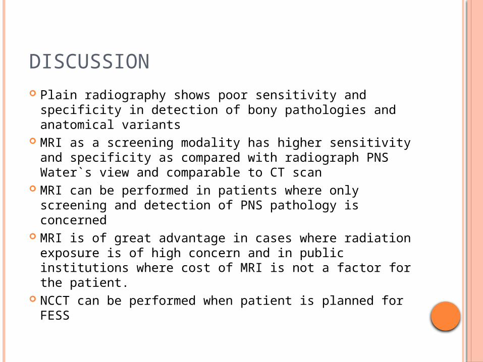
DISCUSSION Plain radiography shows poor sensitivity and
specificity in detection of bony pathologies and anatomical variants
MRI as a screening modality has higher sensitivity and specificity as compared with radiograph PNS Water`s view and comparable to CT scan
MRI can be performed in patients where only screening and detection of PNS pathology is concerned
MRI is of great advantage in cases where radiation exposure is of high concern and in public institutions where cost of MRI is not a factor for the patient.
NCCT can be performed when patient is planned for FESS

CONCLUSION
MRI is more sensitive and specific for screening of paranasal sinus disease as compared to X rays and is almost comparable to NCCT for detection of mucosal disease
With a fast MRI screening protocol (T2 coronal and T1 FLASH) it is possible to replace X-ray PNS as the screening modality
CT scan can be performed when FESS is planned to delineate the bony landmarks.

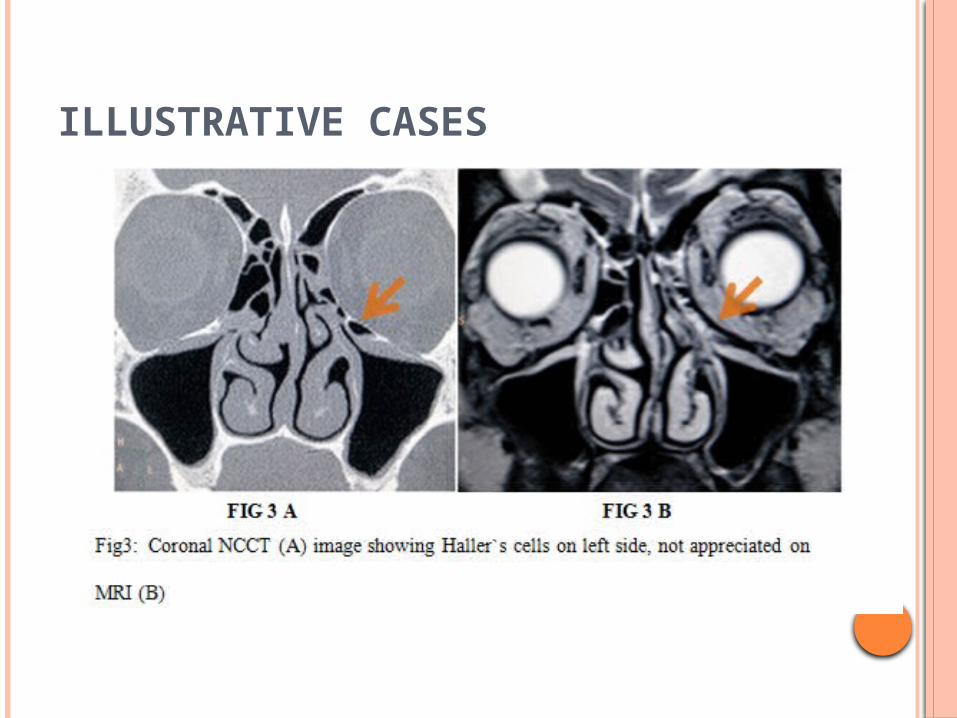
ILLUSTRATIVE CASES

ILLUSTRATIVE CASES
ILLUSTRATIVE CASES

ILLUSTRATIVE CASES

ILLUSTRATIVE CASES

REFRENCES 1. Pete S.Batra, et al. Radiologic imaging in rhinosinusitis. Cleveland clinic journal
of medicine. November 2004;71 :886-888
2. Davidson TM, Brahme FJ, Gallagher ME. Radiographic evaluation for nasal dysfunction: computed tomography versus plain films. Head Neck 1989; 11:405–409.
3. Konen E, Faibel M, Kleinbaum Y, et al. The value of occipitomental (Waters’) view in diagnosis of sinusitis: a comparative study with computed tomography. Clin Radiol 2000; 55:856–860.
4. Girish M. Fatterpekar, Bradleyn.Delman,Peterm.Som. Imaging the paranasal sinuses: Where we are and where we are going. The anatomical record 2008; 291:1564–1572
5. Lazar RH, Younis RT, Parvey LS.Comparison of plain radiographs, coronal CT, and intraoperative findings in children with chronic sinusitis. Otolaryngol Head Neck Surg. 1992;107 (1):29-34.
6. Germiller JA, Monin DL, Sparano AM, Tom LW. Intracranial complications of sinusitis in children and adolescents and their outcomes. Arch Otolaryngol Head Neck Surg. 2006;132(9):969-976