a-Amylase from mung beans (Vigna radiata) – Correlation of.pdf
9
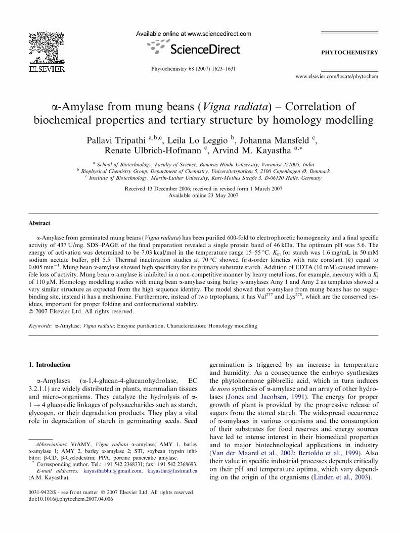
a-Amylase from mung beans (Vigna radiata) – Correlation of biochemical properties and tertiary structure by homology modelling Pallavi Tripathi a,b,c , Leila Lo Leggio b , Johanna Mansfeld c , Renate Ulbrich-Hofmann c , Arvind M. Kayastha a, * a School of Biotechnology, Faculty of Science, Banaras Hindu University, Varanasi 221005, India b Biophysical Chemistry Group, Department of Chemistry, Universitetsparken 5, 2100 Copenhagen Ø, Denmark c Institute of Biotechnology, Martin-Luther University, Kurt-Mothes Straße 3, D-06120 Halle, Germany Received 13 December 2006; received in revised form 1 March 2007 Available online 23 May 2007 Abstract a-Amylase from germinated mung beans (Vigna radiata) has been purified 600-fold to electrophoretic homogeneity and a final specific activity of 437 U/mg. SDS–PAGE of the final preparation revealed a single protein band of 46 kDa. The optimum pH was 5.6. The energy of activation was determined to be 7.03 kcal/mol in the temperature range 15–55 °C. K m for starch was 1.6 mg/mL in 50 mM sodium acetate buffer, pH 5.5. Thermal inactivation studies at 70 °C showed first-order kinetics with rate constant (k) equal to 0.005 min 1 . Mung bean a-amylase showed high specificity for its primary substrate starch. Addition of EDTA (10 mM) caused irrevers- ible loss of activity. Mung bean a-amylase is inhibited in a non-competitive manner by heavy metal ions, for example, mercury with a K i of 110 lM. Homology modelling studies with mung bean a-amylase using barley a-amylases Amy 1 and Amy 2 as templates showed a very similar structure as expected from the high sequence identity. The model showed that a-amylase from mung beans has no sugar- binding site, instead it has a methionine. Furthermore, instead of two trptophans, it has Val 277 and Lys 278 , which are the conserved res- idues, important for proper folding and conformational stability. Ó 2007 Elsevier Ltd. All rights reserved. Keywords: a-Amylase; Vigna radiata; Enzyme purification; Characterization; Homology modelling 1. Introduction a-Amylases (a-1,4-glucan-4-glucanohydrolase, EC 3.2.1.1) are widely distributed in plants, mammalian tissues and micro-organisms. They catalyze the hydrolysis of a- 1 ! 4 glucosidic linkages of polysaccharides such as starch, glycogen, or their degradation products. They play a vital role in degradation of starch in germinating seeds. Seed germination is triggered by an increase in temperature and humidity. As a consequence the embryo synthesizes the phytohormone gibbrellic acid, which in turn induces de novo synthesis of a-amylase and an array of other hydro- lases (Jones and Jacobsen, 1991). The energy for proper growth of plant is provided by the progressive release of sugars from the stored starch. The widespread occurrence of a-amylases in various organisms and the consumption of their substrates for food reserves and energy sources have led to intense interest in their biomedical properties and to major biotechnological applications in industry (Van der Maarel et al., 2002; Bertoldo et al., 1999). Also their value in specific industrial processes depends critically on their pH and temperature optima, which vary depend- ing on the origin of the organisms (Linden et al., 2003). 0031-9422/$ - see front matter Ó 2007 Elsevier Ltd. All rights reserved. doi:10.1016/j.phytochem.2007.04.006 Abbreviations: VrAMY, Vigna radiata a-amylase; AMY 1, barley a-amylase 1; AMY 2, barley a-amylase 2; STI, soybean trypsin inhi- bitor; b-CD, b-Cyclodextrin; PPA, porcine pancreatic amylase. * Corresponding author. Tel.: +91 542 2368331; fax: +91 542 2368693. E-mail addresses: [email protected], [email protected] (A.M. Kayastha). www.elsevier.com/locate/phytochem Phytochemistry 68 (2007) 1623–1631 PHYTOCHEMISTRY
-
Upload
firyal-a-gista -
Category
Documents
-
view
87 -
download
1
description
Biochemistry
Transcript of a-Amylase from mung beans (Vigna radiata) – Correlation of.pdf
www.elsevier.com/locate/phytochem
Phytochemistry 68 (2007) 1623–1631
PHYTOCHEMISTRY
a-Amylase from mung beans (Vigna radiata) – Correlation ofbiochemical properties and tertiary structure by homology modelling
Pallavi Tripathi a,b,c, Leila Lo Leggio b, Johanna Mansfeld c,Renate Ulbrich-Hofmann c, Arvind M. Kayastha a,*
a School of Biotechnology, Faculty of Science, Banaras Hindu University, Varanasi 221005, Indiab Biophysical Chemistry Group, Department of Chemistry, Universitetsparken 5, 2100 Copenhagen Ø, Denmark
c Institute of Biotechnology, Martin-Luther University, Kurt-Mothes Straße 3, D-06120 Halle, Germany
Received 13 December 2006; received in revised form 1 March 2007Available online 23 May 2007
Abstract
a-Amylase from germinated mung beans (Vigna radiata) has been purified 600-fold to electrophoretic homogeneity and a final specificactivity of 437 U/mg. SDS–PAGE of the final preparation revealed a single protein band of 46 kDa. The optimum pH was 5.6. Theenergy of activation was determined to be 7.03 kcal/mol in the temperature range 15–55 �C. Km for starch was 1.6 mg/mL in 50 mMsodium acetate buffer, pH 5.5. Thermal inactivation studies at 70 �C showed first-order kinetics with rate constant (k) equal to0.005 min�1. Mung bean a-amylase showed high specificity for its primary substrate starch. Addition of EDTA (10 mM) caused irrevers-ible loss of activity. Mung bean a-amylase is inhibited in a non-competitive manner by heavy metal ions, for example, mercury with a Ki
of 110 lM. Homology modelling studies with mung bean a-amylase using barley a-amylases Amy 1 and Amy 2 as templates showed avery similar structure as expected from the high sequence identity. The model showed that a-amylase from mung beans has no sugar-binding site, instead it has a methionine. Furthermore, instead of two trptophans, it has Val277 and Lys278, which are the conserved res-idues, important for proper folding and conformational stability.� 2007 Elsevier Ltd. All rights reserved.
Keywords: a-Amylase; Vigna radiata; Enzyme purification; Characterization; Homology modelling
1. Introduction
a-Amylases (a-1,4-glucan-4-glucanohydrolase, EC3.2.1.1) are widely distributed in plants, mammalian tissuesand micro-organisms. They catalyze the hydrolysis of a-1! 4 glucosidic linkages of polysaccharides such as starch,glycogen, or their degradation products. They play a vitalrole in degradation of starch in germinating seeds. Seed
0031-9422/$ - see front matter � 2007 Elsevier Ltd. All rights reserved.
doi:10.1016/j.phytochem.2007.04.006
Abbreviations: VrAMY, Vigna radiata a-amylase; AMY 1, barleya-amylase 1; AMY 2, barley a-amylase 2; STI, soybean trypsin inhi-bitor; b-CD, b-Cyclodextrin; PPA, porcine pancreatic amylase.
* Corresponding author. Tel.: +91 542 2368331; fax: +91 542 2368693.E-mail addresses: [email protected], [email protected]
(A.M. Kayastha).
germination is triggered by an increase in temperatureand humidity. As a consequence the embryo synthesizesthe phytohormone gibbrellic acid, which in turn inducesde novo synthesis of a-amylase and an array of other hydro-lases (Jones and Jacobsen, 1991). The energy for propergrowth of plant is provided by the progressive release ofsugars from the stored starch. The widespread occurrenceof a-amylases in various organisms and the consumptionof their substrates for food reserves and energy sourceshave led to intense interest in their biomedical propertiesand to major biotechnological applications in industry(Van der Maarel et al., 2002; Bertoldo et al., 1999). Alsotheir value in specific industrial processes depends criticallyon their pH and temperature optima, which vary depend-ing on the origin of the organisms (Linden et al., 2003).
1624 P. Tripathi et al. / Phytochemistry 68 (2007) 1623–1631
In general a-amylases are classified based on theirsequence into family-13 of glycoside hydrolases (Henrissat,1991; CAZY server, www.cazy.org), with related enzymesclassified in families 70 and 77. Many X-ray crystal struc-tures have been reported in case of a-amylase such asAspergillus oryzae TAKA a-amylase (Matsuura et al.,1984; Swift et al., 1991), porcine pancreas PPA (Qianet al., 1993), Aspergillus niger a-amylase (Boel et al.,1990; Brady et al., 1991), barley a-amylase isozyme 2(AMY 2), (Kadziola et al., 1994), barley a-amylase isozyme1 (Robert et al., 2003), and from Alteromonas haloplanctis
(Aghajari et al., 1998). The structures consist of single poly-peptide chains folded into three domains (A, B and C). Thecatalytic domain A consists of (b/a)8-barrel. Domains Band C are located roughly at opposite sides of this TIM-barrel. Domain B is probably responsible for the differ-ences in substrate specificity and stability among the a-amylases (Svensson, 1994). Domain C constitutes the C-terminal part of the sequence and contains a Greek keymotif and its functional role is yet to be established (Neil-sen et al., 2004). With nearly all a-amylases, X-ray struc-tures contain at least one conserved calcium ion, which islocated at the interface between the A and B domains (Boelet al., 1990; Machius et al., 1995; Robert et al., 2002). Oneor more additional calcium ions have been found in severalstructures. It has been suggested that the role of the cal-cium ions is mainly structural (Gilles et al., 1996) and theconserved regions are involved in the architecture of theCa2+ binding site and of the active site. The essential roleof calcium ion has been explained by the observation thatits ligands belong to domains A and B, the active site cleftis stabilized by inducing an ionic bridge between domain Aand B (Buisson et al., 1987). A hyperthermophilic a-amy-lase with a novel (Ca, Zn) two metal centres has also beenreported and it does not require the addition of metal ionsfor its full activity (Linden et al., 2003).
In the present study, we report purification of a-amylasefrom mung beans (Vigna radiata), its biochemical charac-terization and computer modelling studies using AMY 1and AMY 2, as templates.
2. Results and discussion
2.1. Purification and biochemical properties
The specific activity of a-amylase from VrAmy increasedto a maximum in 24 h following imbibition. Thereafter,
Table 1Purification of a-amylase from germinated mung beans (300 g)
Steps Total activity (U) Total protein (mg)
Crude extract 11,000 15,000(NH4)2SO4 (30–50%) 9900 1620Affinity chromatography 2000 6.4Superdex 75 1000 1.6
a Fold purification calculated with respect to the specific activity of the crud
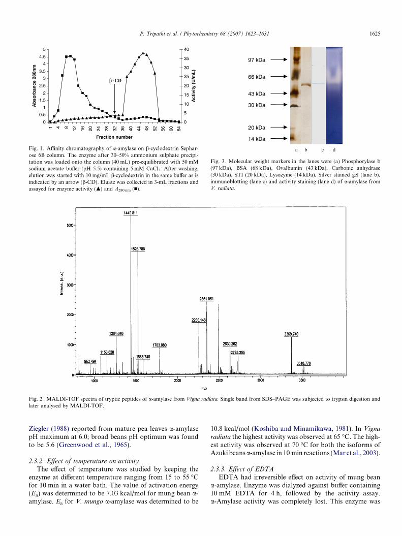
a-amylase specific activity showed a marked decline(results not shown). As outlined in Table 1, a-amylase from24 h germinated mung beans was purified 599-fold to afinal specific activity of 437 U/mg (Fig. 1) and an overallyield of 9%. Koshiba and Minamikawa (1981) purified a-amylase from V. mungo with specific activity of 203 U/mg. Mar et al. (2003) recently purified two isoforms(VAAmy1 and VAAmy2) from Azuki beans (V. angularis)
with specific activities of 75 and 63 U/mg, respectively. Zie-gler (1988) reported a-amylase from mature pea leavesusing affinity chromatography with a yield about 20%;another report from shoots and cotyledons of pea seedlingsshowed fold purification of 6700 and 850, respectively(Beers and Duke, 1990). Greenwood et al. (1965) reporteda-amylase from broad beans (Vicia faba) with specificactivity of 360 U/mg. In the b-cyclodextrin Sepharose col-umn chromatography of kidney beans (Phaseolus vulgaris)cotyledons, a small peak of starch hydrolysing activity wasfirst eluted by a lower concentration of b-cyclodextrin,while most of the enzyme activity was more tightly boundto the column (Mori et al., 1995). According to Mori et al.(1995) the two isoforms may be common in leguminousplant a-amylase.
2.2. MALDI-TOF mass spectrometry and electrophoresis
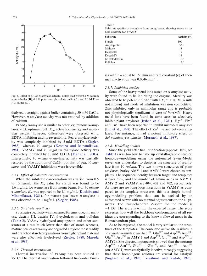
MALDI-TOF analysis of the trypsin digested singleband of molecular mass 46 kDa (from SDS–PAGE)revealed a set of peptides, which matched with mass dataof a-amylase from Vigna angularis from the available data-base with 98% sequence homology with V. radiata (Fig. 2)(Koizuka et al., 1990; Mar et al., 2003).
Coomassie Blue/silver staining showed a single bandcorresponding to 46 kDa on polyacrylamide gel electro-phoresis. The identification of this band as a-amylase wasfurther confirmed by immunoblotting with antibodiesagainst barley Amy2 and activity staining (Fig. 3).
2.3. Kinetic studies
2.3.1. Effect of pH on activity
The effect of pH on purified a-amylase was examined inthe pH range 3.6–9.0. The a-amylase from mung beansshowed maximal activity at pH 5.6 in 0.1 M sodium acetatebuffer (Fig. 4). Koshiba and Minamikawa (1981) reportedpH optima at pH 6.0 from V. mungo a-amylase; Mar et al.(2003) reported optimum pH from Azuki beans at pH 5.3and 5.2 for VAAmy1 and VAAmy2 isoforms, respectively;
Specific activity (U/mg) Purification (-fold)a Yield (%)
0.73 – –6.1 8.4 90
312 427 18437 599 9
e extract.
0
0.5
1
1.5
2
2.5
3
3.5
4
4.5
51 4 8 12 16 20 24 28 32 36 40 44 48 52 56 60 64
Fraction number
Ab
sorb
ance
280
nm
0
5
10
15
20
25
30
35
40
Act
ivit
y (U
/mL
)
β -CD
Fig. 1. Affinity chromatography of a-amylase on b-cyclodextrin Sephar-ose 6B column. The enzyme after 30–50% ammonium sulphate precipi-tation was loaded onto the column (40 mL) pre-equilibrated with 50 mMsodium acetate buffer (pH 5.5) containing 5 mM CaCl2. After washing,elution was started with 10 mg/mL b-cyclodextrin in the same buffer as isindicated by an arrow (b-CD). Eluate was collected in 3-mL fractions andassayed for enzyme activity (m) and A280 nm (n).
Fig. 2. MALDI-TOF spectra of tryptic peptides of a-amylase from Vigna radiata. Single band from SDS–PAGE was subjected to trypsin digestion andlater analysed by MALDI-TOF.
97 kDa
66 kDa
43 kDa
30 kDa
20 kDa
14 kDa
a b c d
Fig. 3. Molecular weight markers in the lanes were (a) Phosphorylase b(97 kDa), BSA (68 kDa), Ovalbumin (43 kDa), Carbonic anhydrase(30 kDa), STI (20 kDa), Lysozyme (14 kDa), Silver stained gel (lane b),immunoblotting (lane c) and activity staining (lane d) of a-amylase fromV. radiata.
P. Tripathi et al. / Phytochemistry 68 (2007) 1623–1631 1625
Ziegler (1988) reported from mature pea leaves a-amylasepH maximum at 6.0; broad beans pH optimum was foundto be 5.6 (Greenwood et al., 1965).
2.3.2. Effect of temperature on activity
The effect of temperature was studied by keeping theenzyme at different temperature ranging from 15 to 55 �Cfor 10 min in a water bath. The value of activation energy(Ea) was determined to be 7.03 kcal/mol for mung bean a-amylase. Ea for V. mungo a-amylase was determined to be
10.8 kcal/mol (Koshiba and Minamikawa, 1981). In Vigna
radiata the highest activity was observed at 65 �C. The high-est activity was observed at 70 �C for both the isoforms ofAzuki beans a-amylase in 10 min reactions (Mar et al., 2003).
2.3.3. Effect of EDTA
EDTA had irreversible effect on activity of mung beana-amylase. Enzyme was dialyzed against buffer containing10 mM EDTA for 4 h, followed by the activity assay.a-Amylase activity was completely lost. This enzyme was
pH
3 4 5 6 7 8 9 10
Act
ivity
(U
/ mL)
0
5
10
15
20
25
30
35
40
Fig. 4. Effect of pH on a-amylase activity. Buffer used were: 0.1 M sodiumacetate buffer (d), 0.1 M potassium phosphate buffer (n), and 0.1 M Tris–HCl buffer (s).
Table 2Substrate specificity a-amylase from mung beans, showing starch as thebest substrate for VrAMY
Substrate Activity (%)
Starch 100Amylopectin 53.8Maltose 18DextrinIII 17DextrinIV 14b-Cyclodextrin 6.3Pullulan 5.0
1626 P. Tripathi et al. / Phytochemistry 68 (2007) 1623–1631
dialyzed overnight against buffer containing 50 mM CaCl2.However, a-amylase activity was not restored by additionof calcium.
VrAMy a-amylase is similar to other leguminous a-amy-lases w.r.t. optimum pH, Km, activation energy and molec-ular weight; however, differences were observed w.r.t.EDTA inhibition and its reversibility. Pea a-amylase activ-ity was completely inhibited by 5 mM EDTA (Ziegler,1988), whereas V. mungo (Koshiba and Minamikawa,1981), VrAMY and V. angularis a-amylase activity wascompletely inhibited by 10 mM EDTA (Mar et al., 2003).Interestingly, V. mungo a-amylase activity was partiallyrestored by the addition of CaCl2, but that of pea, V. ang-
ularis and VrAMY inhibitions were irreversible.
2.3.4. Effect of substrate concentration
When the substrate concentration was varied from 0.5to 10 mg/mL, the Km value for starch was found to be1.6 mg/mL for a-amylase from mung beans. For V. mungo
a-amylase, Km was reported to be 1.1 mg/mL (Koshiba andMinamikawa, 1981), for mature pea leaves a-amylase itwas observed to be 1 mg/mL (Ziegler, 1988).
2.3.5. Substrate specificity
Substrate specificity was measured for amylopectin, malt-ose, dextrin III, dextrin IV, b-cyclodextrin and pullulan(Table 2). VrAmy hydrolyzed amylopectin and other sub-strates at a rate much lower than that of starch whereas,mature pea leaves a-amylase degraded amylose most readilyand branched starch preparations from higher plant materialwere also effectively hydrolyzed (Ziegler, 1988; Masudaet al., 1987).
2.3.6. Thermal inactivationThermal inactivation of VrAmy has been studied at
75 �C. The thermal inactivation followed first-order kinet-
ics with t1/2 equal to 150 min and rate constant (k) of ther-mal inactivation was 0.0046 min�1.
2.3.7. Inhibition studies
Some of the heavy metal ions tested on a-amylase activ-ity were found to be inhibiting the enzyme. Mercury wasobserved to be potent inhibitor with a Ki of 110 lM (resultsnot shown) and mode of inhibition was non competitive.Zinc inhibited only in millimolar range and is probablynot physiologically significant in case of VrAMY. Heavymetal ions have been found in some cases to selectivelyinhibit plant amylases (Irshad et al., 1981). Hg2+, Pb2+
and Cu2+ have been reported to inhibit microbial amylases(Lin et al., 1998). The effect of Zn2+ varied between amy-lases. For instance, it had a potent inhibitory effect onSchwanniomyces alluvius (Moranelli et al., 1987).
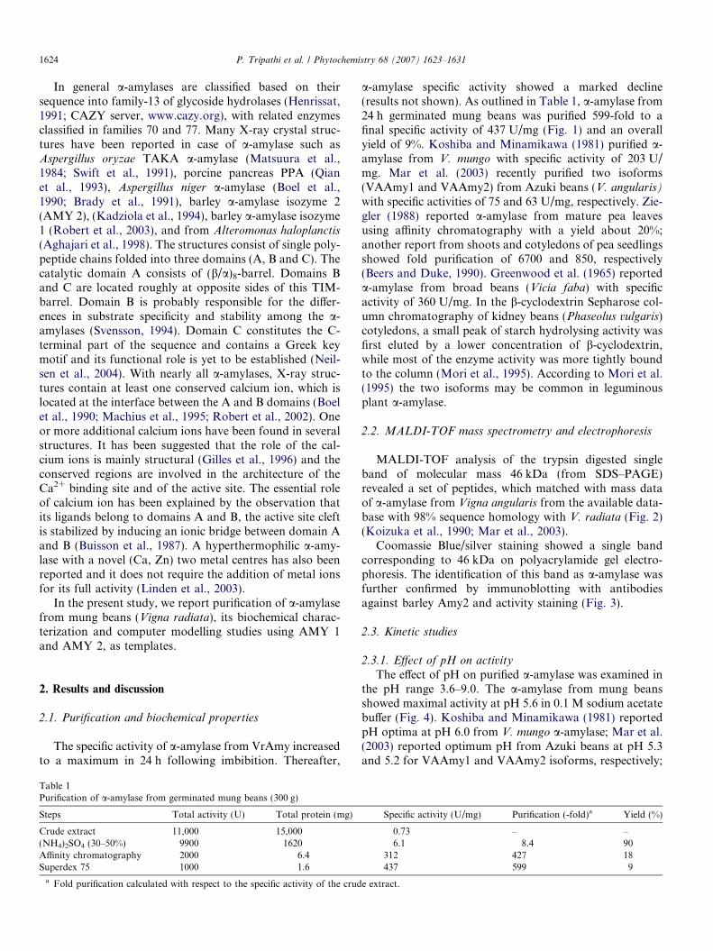
2.3.8. Modelling studies
Since the yield after final purification (approx. 10%, seeTable 1) was too low to take up crystallographic studies,homology-modelling using the automated Swiss-Modelserver was undertaken to decipher the structure of a-amy-lase from V. radiata. The two known structures of plantamylases, barley AMY 1 and AMY 2 were chosen as tem-plates. The sequence identity between target and templatesis over 65%, and the number of amino acids in AMY 1,AMY 2 and VrAMY are 404, 402 and 402, respectively.As there are no long loop insertions in VrAMY as com-pared to the template structures, this is a simple homol-ogy-modelling problem that can be tackled by anautomated server with no manual adjustments to the align-ments. The Ramachandran Z-score for the model is�1.132. The score is within the expected range. The scoreexpresses how well the backbone conformations of all res-idues are corresponding to the known allowed areas in theRamachandran plot.
As to be expected, the model is very similar to the struc-tures of the templates. The conserved active site residues inV. radiata a-amylase are Asp182, Glu207 and Asp290 (Asp180,Glu205, Asp291 in AMY 1 and Asp179, Glu204 and Asp289 inAMY2). Site directed mutagenesis showed that the mutantsAsp180! Asn180, Glu205! Gln205, and Asp291! Asn291
(on AMY 1) were catalytically inactive, strongly suggestingthat these homologous residues are crucial for catalysis(Søgaard et al., 1993; Terashima and Katoh, 1996).
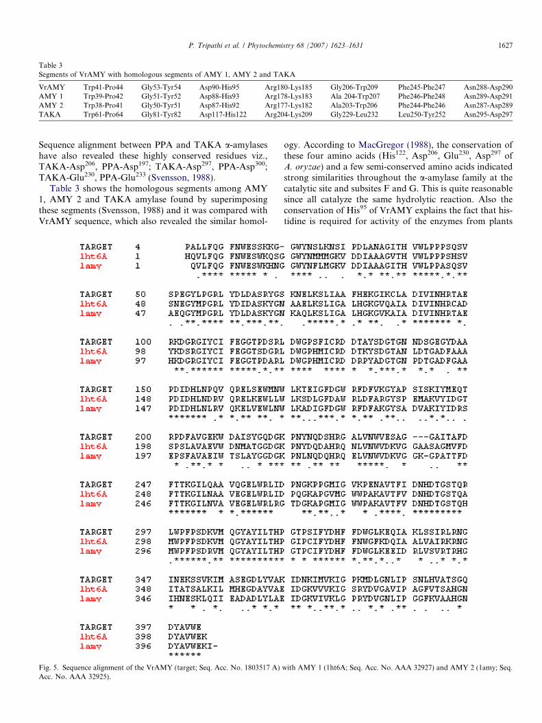
Table 3Segments of VrAMY with homologous segments of AMY 1, AMY 2 and TAKA
VrAMY Trp41-Pro44 Gly53-Tyr54 Asp90-His95 Arg180-Lys185 Gly206-Trp209 Phe245-Phe247 Asn288-Asp290AMY 1 Trp39-Pro42 Gly51-Tyr52 Asp88-His93 Arg178-Lys183 Ala 204-Trp207 Phe246-Phe248 Asn289-Asp291AMY 2 Trp38-Pro41 Gly50-Tyr51 Asp87-His92 Arg177-Lys182 Ala203-Trp206 Phe244-Phe246 Asn287-Asp289TAKA Trp61-Pro64 Gly81-Tyr82 Asp117-His122 Arg204-Lys209 Gly229-Leu232 Leu250-Tyr252 Asn295-Asp297
P. Tripathi et al. / Phytochemistry 68 (2007) 1623–1631 1627
Sequence alignment between PPA and TAKA a-amylaseshave also revealed these highly conserved residues viz.,TAKA-Asp206, PPA-Asp197; TAKA-Asp297, PPA-Asp300;TAKA-Glu230, PPA-Glu233 (Svensson, 1988).
Table 3 shows the homologous segments among AMY1, AMY 2 and TAKA amylase found by superimposingthese segments (Svensson, 1988) and it was compared withVrAMY sequence, which also revealed the similar homol-
Fig. 5. Sequence alignment of the VrAMY (target; Seq. Acc. No. 1803517 A)Acc. No. AAA 32925).
ogy. According to MacGregor (1988), the conservation ofthese four amino acids (His122, Asp206, Glu230, Asp297 ofA. oryzae) and a few semi-conserved amino acids indicatedstrong similarities throughout the a-amylase family at thecatalytic site and subsites F and G. This is quite reasonablesince all catalyze the same hydrolytic reaction. Also theconservation of His95 of VrAMY explains the fact that his-tidine is required for activity of the enzymes from plants
with AMY 1 (1ht6A; Seq. Acc. No. AAA 32927) and AMY 2 (1amy; Seq.
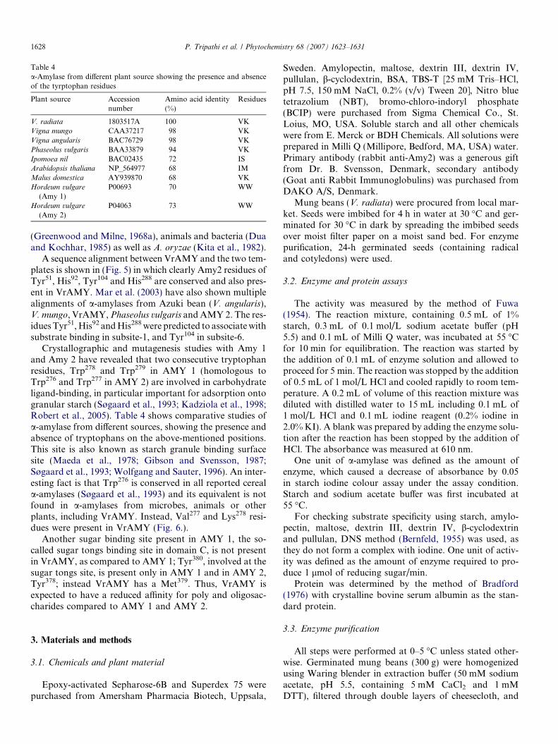
Table 4a-Amylase from different plant source showing the presence and absenceof the tyrptophan residues
Plant source Accessionnumber
Amino acid identity(%)
Residues
V. radiata 1803517A 100 VKVigna mungo CAA37217 98 VKVigna angularis BAC76729 98 VKPhaseolus vulgaris BAA33879 94 VKIpomoea nil BAC02435 72 ISArabidopsis thaliana NP_564977 68 IMMalus domestica AY939870 68 VKHordeum vulgare
(Amy 1)P00693 70 WW
Hordeum vulgare
(Amy 2)P04063 73 WW
1628 P. Tripathi et al. / Phytochemistry 68 (2007) 1623–1631
(Greenwood and Milne, 1968a), animals and bacteria (Duaand Kochhar, 1985) as well as A. oryzae (Kita et al., 1982).
A sequence alignment between VrAMY and the two tem-plates is shown in (Fig. 5) in which clearly Amy2 residues ofTyr51, His92, Tyr104 and His288 are conserved and also pres-ent in VrAMY. Mar et al. (2003) have also shown multiplealignments of a-amylases from Azuki bean (V. angularis),V. mungo, VrAMY, Phaseolus vulgaris and AMY 2. The res-idues Tyr51, His92 and His288 were predicted to associate withsubstrate binding in subsite-1, and Tyr104 in subsite-6.
Crystallographic and mutagenesis studies with Amy 1and Amy 2 have revealed that two consecutive tryptophanresidues, Trp278 and Trp279 in AMY 1 (homologous toTrp276 and Trp277 in AMY 2) are involved in carbohydrateligand-binding, in particular important for adsorption ontogranular starch (Søgaard et al., 1993; Kadziola et al., 1998;Robert et al., 2005). Table 4 shows comparative studies ofa-amylase from different sources, showing the presence andabsence of tryptophans on the above-mentioned positions.This site is also known as starch granule binding surfacesite (Maeda et al., 1978; Gibson and Svensson, 1987;Søgaard et al., 1993; Wolfgang and Sauter, 1996). An inter-esting fact is that Trp276 is conserved in all reported cereala-amylases (Søgaard et al., 1993) and its equivalent is notfound in a-amylases from microbes, animals or otherplants, including VrAMY. Instead, Val277 and Lys278 resi-dues were present in VrAMY (Fig. 6.).
Another sugar binding site present in AMY 1, the so-called sugar tongs binding site in domain C, is not presentin VrAMY, as compared to AMY 1; Tyr380, involved at thesugar tongs site, is present only in AMY 1 and in AMY 2,Tyr378; instead VrAMY has a Met379. Thus, VrAMY isexpected to have a reduced affinity for poly and oligosac-charides compared to AMY 1 and AMY 2.
3. Materials and methods
3.1. Chemicals and plant material
Epoxy-activated Sepharose-6B and Superdex 75 werepurchased from Amersham Pharmacia Biotech, Uppsala,
Sweden. Amylopectin, maltose, dextrin III, dextrin IV,pullulan, b-cyclodextrin, BSA, TBS-T [25 mM Tris–HCl,pH 7.5, 150 mM NaCl, 0.2% (v/v) Tween 20], Nitro bluetetrazolium (NBT), bromo-chloro-indoryl phosphate(BCIP) were purchased from Sigma Chemical Co., St.Loius, MO, USA. Soluble starch and all other chemicalswere from E. Merck or BDH Chemicals. All solutions wereprepared in Milli Q (Millipore, Bedford, MA, USA) water.Primary antibody (rabbit anti-Amy2) was a generous giftfrom Dr. B. Svensson, Denmark, secondary antibody(Goat anti Rabbit Immunoglobulins) was purchased fromDAKO A/S, Denmark.
Mung beans (V. radiata) were procured from local mar-ket. Seeds were imbibed for 4 h in water at 30 �C and ger-minated for 30 �C in dark by spreading the imbibed seedsover moist filter paper on a moist sand bed. For enzymepurification, 24-h germinated seeds (containing radicaland cotyledons) were used.
3.2. Enzyme and protein assays
The activity was measured by the method of Fuwa(1954). The reaction mixture, containing 0.5 mL of 1%starch, 0.3 mL of 0.1 mol/L sodium acetate buffer (pH5.5) and 0.1 mL of Milli Q water, was incubated at 55 �Cfor 10 min for equilibration. The reaction was started bythe addition of 0.1 mL of enzyme solution and allowed toproceed for 5 min. The reaction was stopped by the additionof 0.5 mL of 1 mol/L HCl and cooled rapidly to room tem-perature. A 0.2 mL of volume of this reaction mixture wasdiluted with distilled water to 15 mL including 0.1 mL of1 mol/L HCl and 0.1 mL iodine reagent (0.2% iodine in2.0% KI). A blank was prepared by adding the enzyme solu-tion after the reaction has been stopped by the addition ofHCl. The absorbance was measured at 610 nm.
One unit of a-amylase was defined as the amount ofenzyme, which caused a decrease of absorbance by 0.05in starch iodine colour assay under the assay condition.Starch and sodium acetate buffer was first incubated at55 �C.
For checking substrate specificity using starch, amylo-pectin, maltose, dextrin III, dextrin IV, b-cyclodextrinand pullulan, DNS method (Bernfeld, 1955) was used, asthey do not form a complex with iodine. One unit of activ-ity was defined as the amount of enzyme required to pro-duce 1 lmol of reducing sugar/min.
Protein was determined by the method of Bradford(1976) with crystalline bovine serum albumin as the stan-dard protein.
3.3. Enzyme purification
All steps were performed at 0–5 �C unless stated other-wise. Germinated mung beans (300 g) were homogenizedusing Waring blender in extraction buffer (50 mM sodiumacetate, pH 5.5, containing 5 mM CaCl2 and 1 mMDTT), filtered through double layers of cheesecloth, and
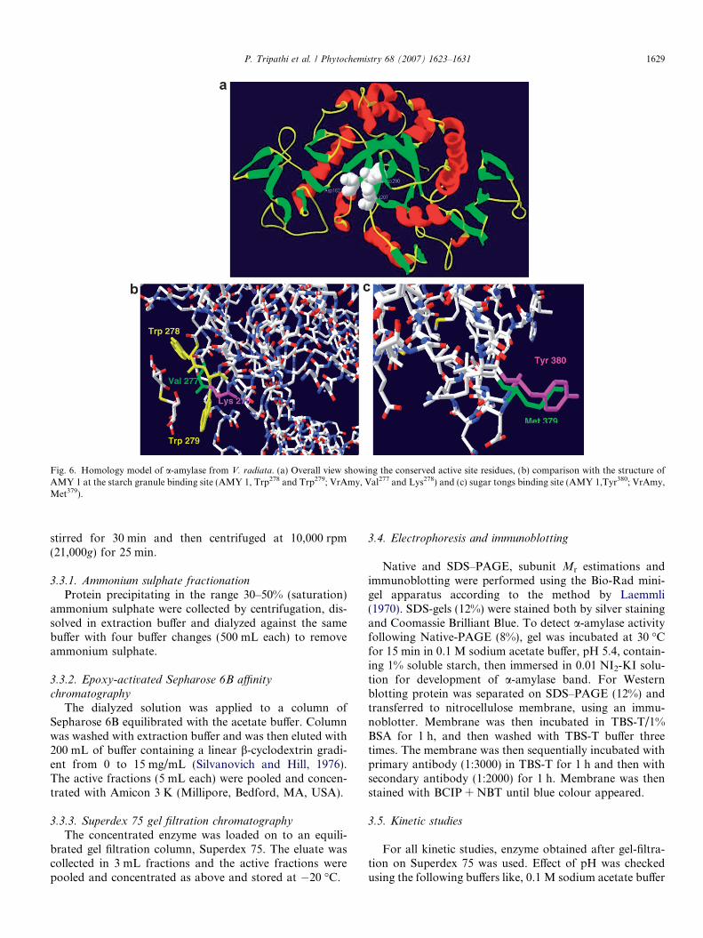
Fig. 6. Homology model of a-amylase from V. radiata. (a) Overall view showing the conserved active site residues, (b) comparison with the structure ofAMY 1 at the starch granule binding site (AMY 1, Trp278 and Trp279; VrAmy, Val277 and Lys278) and (c) sugar tongs binding site (AMY 1,Tyr380; VrAmy,Met379).
P. Tripathi et al. / Phytochemistry 68 (2007) 1623–1631 1629
stirred for 30 min and then centrifuged at 10,000 rpm(21,000g) for 25 min.
3.3.1. Ammonium sulphate fractionation
Protein precipitating in the range 30–50% (saturation)ammonium sulphate were collected by centrifugation, dis-solved in extraction buffer and dialyzed against the samebuffer with four buffer changes (500 mL each) to removeammonium sulphate.
3.3.2. Epoxy-activated Sepharose 6B affinity
chromatography
The dialyzed solution was applied to a column ofSepharose 6B equilibrated with the acetate buffer. Columnwas washed with extraction buffer and was then eluted with200 mL of buffer containing a linear b-cyclodextrin gradi-ent from 0 to 15 mg/mL (Silvanovich and Hill, 1976).The active fractions (5 mL each) were pooled and concen-trated with Amicon 3 K (Millipore, Bedford, MA, USA).
3.3.3. Superdex 75 gel filtration chromatography
The concentrated enzyme was loaded on to an equili-brated gel filtration column, Superdex 75. The eluate wascollected in 3 mL fractions and the active fractions werepooled and concentrated as above and stored at �20 �C.
3.4. Electrophoresis and immunoblotting
Native and SDS–PAGE, subunit Mr estimations andimmunoblotting were performed using the Bio-Rad mini-gel apparatus according to the method by Laemmli(1970). SDS-gels (12%) were stained both by silver stainingand Coomassie Brilliant Blue. To detect a-amylase activityfollowing Native-PAGE (8%), gel was incubated at 30 �Cfor 15 min in 0.1 M sodium acetate buffer, pH 5.4, contain-ing 1% soluble starch, then immersed in 0.01 NI2-KI solu-tion for development of a-amylase band. For Westernblotting protein was separated on SDS–PAGE (12%) andtransferred to nitrocellulose membrane, using an immu-noblotter. Membrane was then incubated in TBS-T/1%BSA for 1 h, and then washed with TBS-T buffer threetimes. The membrane was then sequentially incubated withprimary antibody (1:3000) in TBS-T for 1 h and then withsecondary antibody (1:2000) for 1 h. Membrane was thenstained with BCIP + NBT until blue colour appeared.
3.5. Kinetic studies
For all kinetic studies, enzyme obtained after gel-filtra-tion on Superdex 75 was used. Effect of pH was checkedusing the following buffers like, 0.1 M sodium acetate buffer
1630 P. Tripathi et al. / Phytochemistry 68 (2007) 1623–1631
(pH 3.6–5.6), 0.1 M phosphate buffer (pH 6.0–7.6), 0.1 MTris–HCl buffer (pH 7.2–9.0). Enzyme activity was assayedas described above except that the starch solution was in theappropriate buffers. The activation energy (Ea) of the mungbean a-amylase was determined by an Arrhenius plot inthe temperature range 15–55 �C. For thermal inactivation,the enzyme was maintained at the specified temperature±1 �C in a water bath (Multitemp Bath, LKB, Sweden).Small aliquots were withdrawn at different time intervals;chilled immediately and tested for enzyme activity at37 �C, using routine assay.
Substrate specificity was checked with starch and sixother substrates. All parameters are the mean of triplicatesdeterminations from three independent preparations of thepurified enzyme and are reproducible to within ±10% SEof the mean value.
3.6. MALDI-TOF mass spectrometry
Identification and determination of molecular masseswas performed by matrix assisted laser desorption massspectrometry (Bruker-Daltonik, Bremen, Germany). Inorder to establish the identity of each fragment in-gel tryp-tic digest was employed. Single band of VrAMY wereexcised from the SDS–PAGE gel, washed and incubatedwith MALDI grade trypsin, digested overnight and ana-lysed. The peptides obtained were matched with thesequence available in the database.
3.7. Modelling studies
Modelling studies were performed using Swiss Modelserver (http://swissmodel.expasy.org/) (Schwede et al.,2003). The sequence used was taken from a-amylase frommung beans (Koizuka et al., 1990), accession number1803517A. The chosen templates were the only a-amylasesfrom plant origin for which X-ray structure information isavailable, isozyme 1 and isozyme 2 of a-amylase from bar-ley, sharing 70% and 73% sequence identity with the V.
radiata a-amylase. The highest resolution structures avail-able at the time were chosen as templates correspondingto pdb codes 1ht6 for AMY 1 (1.5 A resolution) (Robertet al., 2003) and 1 amy for AMY 2 (2.8 A resolution) (Kad-ziola et al., 1994). A higher resolution structure for AMY 2is available in the PDB, but since it is in complex with aproteinaceous inhibitor, this structure was not chosen astemplate. SWISS-MODEL was used in first approachmode using default parameters. Structures were visualizedusing Swiss-PDBViewer (Guex and Peitsch, 1997).
Acknowledgements
Financial assistance of the Council of Scientific andIndustrial Research, New Delhi (Junior Research Fellow-ship to PT) is thankfully acknowledged. One of us (PT)would also like to thank Boehringer Ingelheim Fonds
(Foundation for Basic Research in Medicine) and DAAD(Sandwich Model Fellowship), Germany for funding tocarry out a part of research work in Denmark and Ger-many. L.L.L. acknowledges financial support from theDanish Natural Sciences Research Council (SNF). We alsothank Dr. A. Schierhorn (Martin-Luther University Halle-Wittenberg, Germany) for performing Mass spectrometrymeasurements.
References
Aghajari, N., Feller, G., Gerday, C., Haser, R., 1998. Crystal structures ofthe psychrophilic a-amylase from Alteromonas haloplanctis in its nativeform and complexed with an inhibitor. Protein Sci. 7, 564–572.
Beers, E.P., Duke, S.H., 1990. Characterization of a-amylase from shootsand cotyledons of pea (Pisum sativum L.) seedlings. Plant Physiol.(USA) 92, 1154–1163.
Bernfeld, P., 1955. Amylases a and b. In: Colowick, I.S.P., Kaplan, N.O.(Eds.), . In: Methods Enzymol, vol. 1. Academic Press, Inc., NewYork, pp. 149–158.
Bertoldo, C., Duffner, F., Jorgensen, P.L., Antranikian, G., 1999.Pullulanase type I from Fervidobacterium pennavorans Ven 5: cloning,sequencing and expression of the gene and biochemical characteriza-tion of the recombinant enzyme. Appl. Environ. Microbiol. 65, 2084–2091.
Boel, E., Brady, L., Brzozowski, A.M., Derewenda, Z., Dodson, G.G.,Jensen, V.J., Petersen, S.B., Swift, H., Thim, L., Woldike, H.F., 1990.Calcium binding in a-amylases: an X-ray diffraction study at 2.1 Aresolution of two enzymes from Aspergillus. Biochemistry 29, 6244–6249.
Bradford, M., 1976. A rapid and sensitive method for the quantitation ofmicrogram quantities of protein utilizing the principle of protein-dyebinding. Anal. Biochem. 72, 248–254.
Brady, R.L., Brzozowski, A.M., Derewenda, Z.S., Dodson, E.J., Dodson,G.G., 1991. Solution of the structure of Aspergillus niger acid a-amylase by combined molecular replacement and multiple isomor-phous replacement methods. Acta Crystal. B. 47, 527–535.
Buisson, G., Duee, E., Haser, R., Payan, F., 1987. Three-dimensionalstructure of porcine pancreatic a-amylase at 2.9 A resolution. Role ofcalcium in structure and activity. EMBO J. 6, 3909–3916.
Dua, R.D., Kochhar, S., 1985. Active site studies on Bacillus amyloliq-
uifaciens a-amylase. Mol. Cell Biochem. 66, 13–20.Fuwa, H., 1954. A new method for microdetermination of amylase
activity by the use of amylose as the substrate. J. Biochem. (Tokyo) 41,583–603.
Gibson, R.M., Svensson, B., 1987. Identification of tryptophanyl residuesinvolved in binding of carbohydrate ligands to barley a-amylase 2.Carlsberg Res. Commun. 52, 373–379.
Gilles, C., Astier, J.P., Marchis-Mouren, G., Cambillau, C., Payan, F.,1996. Crystal structure of pig pancreatic a-amylase isoenzyme II, incomplex with the carbohydrate inhibitor acarbose. Eur. J. Biochem.238, 561–569.
Greenwood, C.T., MacGregor, A.W., Milne, E.A., 1965. The a-amylasefrom broad beans. Purification and properties. Arch. Biochem.Biophys. 112, 459–465.
Greenwood, C.T., Milne, E.A., 1968a. Studies on starch-degradingenzymes. VIII. A comparison of a-amylases from different sources:their properties and action patterns. Starch 20, 101–107.
Guex, R.E. F., Peitsch, M.C., 1997. Swiss Model and Swiss Pdb Viewer:an environment for comparative protein modelling. Electrophoresis18, 2714–2723.
Henrissat, B., 1991. A classification of glycosyl hydrolases based on aminoacid sequence similarities. Biochem. J. 280, 309–316.
Irshad, M., Goel, M., Sharma, C.B., 1981. Effect of Zn2+ on plant a-amylases in vitro. Phytochemistry 20, 2123–2126.
P. Tripathi et al. / Phytochemistry 68 (2007) 1623–1631 1631
Jones, R.L., Jacobsen, J.V., 1991. Regulation of synthesis and transport ofsecreted proteins in cereal aleurone. Int. Rev. Cytol. 126, 49–88.
Kadziola, A., Abe, J., Svensson, B., Haser, R., 1994. Crystal andmolecular structure of barley a-amylase. J. Mol. Biol. 231, 104–121.
Kadziola, A., Søgaard, M., Svensson, B., Haser, R., 1998. Molecularstructure of barley a-amylase-inhibitor complex: Implications forstarch binding and catalysis. J. Mol. Biol. 278, 205–217.
Koizuka, N., Tanaka, Y., Morohashi, Y., 1990. Isolation of a cDNAclone for a-amylase in mung beans cotyledons. Plant Physiol. (USA)94, 1488–1491.
Kita, Y., Sakaguchi, S., Nitta, Y., Watanabe, T., 1982. Kinetic studies onchemical modification of TAKA amylase II. Ethoxycarbonylation ofhistidine residues. J. Biochem. 92, 1499–1504.
Koshiba, T., Minamikawa, T., 1981. Purification by affinity chromatog-raphy of a-amylase – a major amylase in cotyledons of germinatingVigna mungo seeds. Plant Cell Physiol. 22, 979–987.
Laemmli, U.K., 1970. Cleavage of the structure proteins during theassembly of the head of bacteriophage T4. Nature 227, 680–685.
Lin, L.-L., Chyau, C.-C., Hsu, W.-H., 1998. Production and properties ofa raw-starch-degrading amylase from the thermophilic and alkaliphilicBacillus sp. TS-23. Biotechnol. Appl. Biochem. 28, 61–68.
Linden, A., Mayans, O., Meyer-Klaucke, W., Antranikian, G., Wilmanns,M., 2003. Differential regulation of a hyperthermophilic a-amylasewith a novel (Ca, Zn). Two metal center by zinc. J. Biol. Chem. 278,9875–9884.
Machius, M., Wiegand, G., Huber, R., 1995. Crystal structure of calcium-depleted Bacillus licheniformis a-amylase at 2.2 A resolution. J. Mol.Biol. 246, 545–559.
MacGregor, E.A., 1988. a-Amylase structure and activity. J. ProteinChem. 7, 399–415.
Maeda, I., Kiribuchi, S., Nakamura, M., 1978. Digestion of barley starchgranules by the combined action of a- and b-amylases purified frombarley and barley malt. Agric. Biol. Chem. 42, 259–267.
Mar, S.S., Mori, H., Lee, H.J., Fukuda, K., Saburi, W., Fukuhara, A.,Okuyama, M., Chiba, S., Kimura, A., 2003. Purification, character-ization and sequence analysis of two a-amylase isoforms from azukibeans, Vigna angularis, showing different affinity towards b-cyclodex-trin. Biosci. Biotechnol. Biochem. 67, 1080–1093.
Masuda, H., Takahashi, T., Sugawara, S., 1987. Purification andproperties of starch hydrolyzing enzymes in mature roots of sugarbeets. Plant Physiol. (USA) 84, 361–365.
Matsuura, Y., Kusunoki, M., Harada, W., Kakudo, M., 1984. Structureand possible catalytic residues of taka-amylase A. J. Biochem. (Tokyo)95, 697–702.
Moranelli, F., Yaguchi, M., Calleja, G.B., Nasim, A., 1987. Purificationand characterization of the extracellular alpha amylase activity of theyeast Schwanniomyces alluvius. Biochem. Cell. Biol. 65, 899–908.
Mori, H., Tatematsu, A., Saito, A., Matsui, H., Kimura, A., Chiba, S.,1995. Substrate specificity and subsite affinities of a-amylase from
germinating cotyledons of Phaseolus vulgaris l.cv Toramame Oyo
Toshitsu Kagaku. J. Appl. Glycosci. 42, 387–394.Neilsen, P.K., Bonsager, B.C., Fukuda, K., Svensson, B., 2004. Barley a-
amylase/subtilisin inhibitor: structure, biophysics and protein engi-neering. Biochim. Biophys. Acta 1696, 157–164.
Qian, M., Haser, R., Payan, F., 1993. Structure and molecular modelrefinement of pig pancreatic a-amylase at 2.1 A resolution. J. Mol.Biol. 231, 785–799.
Robert, X., Haser, R., Gottschalk, T.E., Svensson, B., Aghajari, N., 2003.The structure of barley a-amylase isozyme 1 reveals a novel role ofdomain C in substrate recognition and binding: a pair of sugar tongs.Structure 11, 973–984.
Robert, X., Gottschalk, T.E., Haser, R., Svensson, B., Aghajari, N., 2002.Expression, purification and preliminary crystallographic studies of a-amylase isozyme1 from barley seeds. Acta Cryst. D58, 683–686.
Robert, X., Haser, R., Mori, H., Svensson, B., Aghajari, N., 2005.Oligosaccharide binding to barley a-amylase 1. J. Biol. Chem. 280,32968–32978.
Silvanovich, M.P., Hill, R.D., 1976. Affinity chromatography of cereal a-amylase. Anal. Biochem. 73, 430–433.
Søgaard, M., Kadziola, A., Haser, R., Svensson, B., 1993. Site directedmutagenesis of histidine 93, aspartic acid 180, glutamic acid 180,glutamic acid 205, histidine 290 and aspartic acid 291 at the active siteand tryptophan 279 at the raw starch-binding site in barley a-amylase1. J. Biol. Chem. 268, 22480–22484.
Svensson, B., 1988. Regional distant sequence homology between amy-lases, a-glucosidases and transglucanosylases. FEBS Lett. 230, 72–76.
Svensson, B., 1994. Protein engineering in the a-amylase family: catalyticmechanism, substrate specificity, and stability. Plant Mol. Biol. 25,141–157.
Swift, H.J., Brady, L., Derewenda, Z.S., Dodson, E.J., Dodson, G.G.,Turkenburg, J.P., Wilkinson, A.J., 1991. Structure and molecularmodel refinement of Aspergillus oryzae (TAKA) a-amylase: anapplication of the stimulated-annealing method. Acta Cryst. B47,535–544.
Schwede, T., Kopp, J., Guex, R.E.F., Peitsch, M.C., 2003. SWISSMODEL: An automated protein homology-modelling server. Nuc.Acid Res. 31, 3381–3385.
Terashima, M., Katoh, S., 1996. Modification of a-amylase function byprotein engineering. Ann. NY Acad. Sci. 799, 65–69.
Van der Maarel, J.E.C., van der Veen, B., Uitdehaag, C.M.J., Leemhuis,H., Dijkhuizen, L., 2002. Properties and applications of starch-converting enzymes of the a-amylase family. J. Biotechnol. 94, 137–155.
Wolfgang, W., Sauter, J.J., 1996. Purification and properties of a starchgranule degrading a-amylase from potato tuber. J. Exp. Bot. 47, 1789–1795.
Ziegler, P., 1988. Partial purification and characterization of the majorendoamylase of mature pea leaves. Plant Physiol. (USA) 86, 659–666.