91W10 Advanced Individual Training Course - MEDTRNGmedtrng.com/handbook1/Handbook_Invasive...
37
91W10 Advanced Individual Training Course Invasive Procedures Handbook Department of the Army Academy of Health Sciences Fort Sam Houston, Texas 78234
-
Upload
phunghuong -
Category
Documents
-
view
221 -
download
0
Transcript of 91W10 Advanced Individual Training Course - MEDTRNGmedtrng.com/handbook1/Handbook_Invasive...
91W10 Advanced Individual
Training Course
Invasive Procedures Handbook
Department of the Army Academy of Health Sciences
Fort Sam Houston, Texas 78234
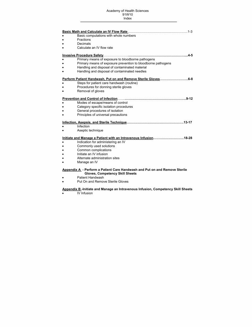
Academy of Health Sciences 91W10 Index
Basic Math and Calculate an IV Flow Rate ………………………………………………1-3 • Basic computations with whole numbers • Fractions • Decimals • Calculate an IV flow rate Invasive Procedure Safety…………………..……………………………………………....4-5 • Primary means of exposure to bloodborne pathogens • Primary means of exposure prevention to bloodborne pathogens • Handling and disposal of contaminated material • Handling and disposal of contaminated needles Perform Patient Handwash, Put on and Remove Sterile Gloves……………………..6-8 • Steps for patient care handwash (routine) • Procedures for donning sterile gloves • Removal of gloves Prevention and Control of Infection…….………………………………………………..9-12 • Modes of escape/means of control • Category specific isolation procedures • General procedures of isolation • Principles of universal precautions Infection, Asepsis, and Sterile Technique…………………………………………….13-17 • Infection • Aseptic technique Initiate and Manage a Patient with an Intravenous Infusion……………………….18-28 • Indication for administering an IV • Commonly used solutions • Common complications • Initiate an IV infusion • Alternate administration sites • Manage an IV Appendix A - Perform a Patient Care Handwash and Put on and Remove Sterile
Gloves, Competency Skill Sheets • Patient Handwash • Put On and Remove Sterile Gloves Appendix B -Initiate and Manage an Intravenous Infusion, Competency Skill Sheets • IV Infusion
Academy of Health Sciences 91W10
Basic Math and Calculate an IV Flow Rate
1
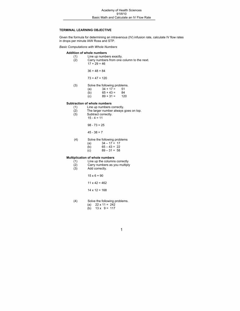
TERMINAL LEARNING OBJECTIVE Given the formula for determining an intravenous (IV) infusion rate, calculate IV flow rates in drops per minute IAW Ross and STP.
Basic Computations with Whole Numbers
Addition of whole numbers (1) Line up numbers exactly. (2) Carry numbers from one column to the next.
17 + 29 = 46
36 + 48 = 84
73 + 47 = 120 (3) Solve the following problems.
(a) 34 + 17 = 51 (b) 65 + 43 = 84 (c) 89 + 31 = 120
Subtraction of whole numbers
(1) Line up numbers correctly. (2) The larger number always goes on top. (3) Subtract correctly.
15 - 4 = 11 98 - 73 = 25 45 - 38 = 7
(4) Solve the following problems (a) 34 – 17 = 17 (b) 65 – 43 = 22 (c) 89 – 31 = 58
Multiplication of whole numbers
(1) Line up the columns correctly (2) Carry numbers as you multiply (3) Add correctly.
15 x 6 = 90 11 x 42 = 462 14 x 12 = 168
(4) Solve the following problems. (a) 22 x 11 = 242 (b) 13 x 9 = 117
Academy of Health Sciences 91W10
Basic Math and Calculate an IV Flow Rate
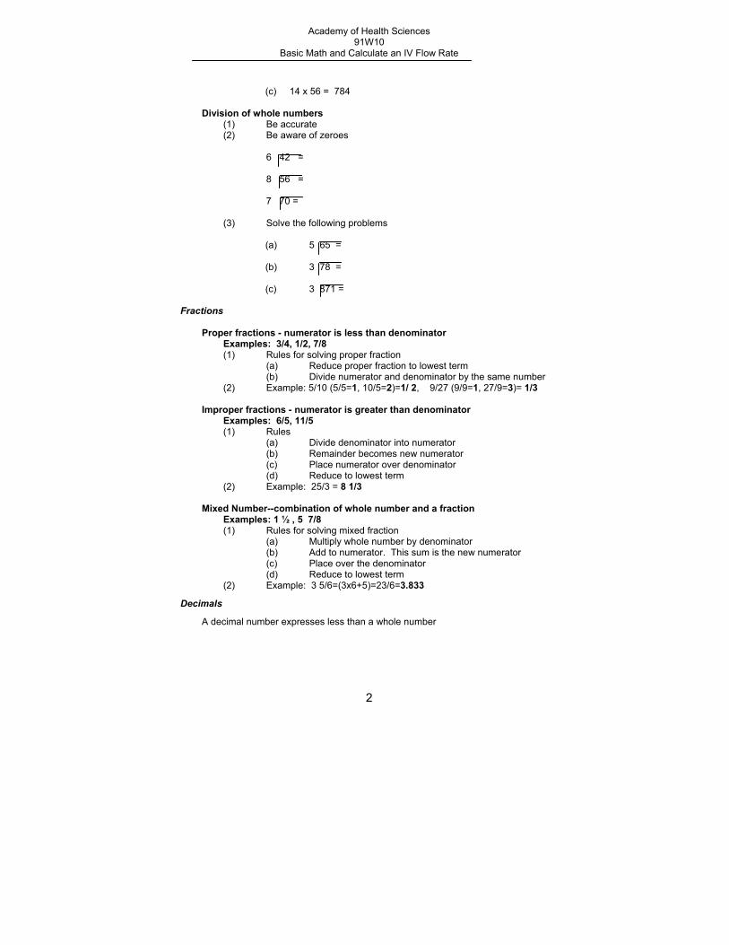
(c) 14 x 56 = 784
Division of whole numbers (1) Be accurate (2) Be aware of zeroes
6 42 =
2
8 56 =
7 70 =
(3) Solve the following problems
(a) 5 65 = (b) 3 78 = (c) 3 871 =
Fractions
Proper fractions - numerator is less than denominator Examples: 3/4, 1/2, 7/8 (1) Rules for solving proper fraction
(a) Reduce proper fraction to lowest term (b) Divide numerator and denominator by the same number
(2) Example: 5/10 (5/5=1, 10/5=2)=1/ 2, 9/27 (9/9=1, 27/9=3)= 1/3
Improper fractions - numerator is greater than denominator Examples: 6/5, 11/5 (1) Rules
(a) Divide denominator into numerator (b) Remainder becomes new numerator (c) Place numerator over denominator (d) Reduce to lowest term
(2) Example: 25/3 = 8 1/3
Mixed Number--combination of whole number and a fraction Examples: 1 ½ , 5 7/8 (1) Rules for solving mixed fraction
(a) Multiply whole number by denominator (b) Add to numerator. This sum is the new numerator (c) Place over the denominator (d) Reduce to lowest term
(2) Example: 3 5/6=(3x6+5)=23/6=3.833
Decimals
A decimal number expresses less than a whole number
Academy of Health Sciences 91W10
Basic Math and Calculate an IV Flow Rate
3
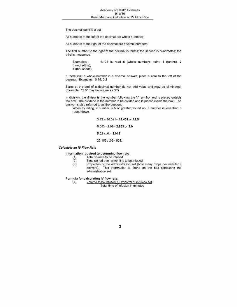
The decimal point is a dot All numbers to the left of the decimal are whole numbers All numbers to the right of the decimal are decimal numbers The first number to the right of the decimal is tenths; the second is hundredths; the third is thousands
Examples: 5.125 is read 5 (whole number); point; 1 (tenths), 2 (hundredths), 5 (thousands)
If there isn't a whole number in a decimal answer, place a zero to the left of the decimal. Examples: 0.75, 0.2 Zeros at the end of a decimal number do not add value and may be eliminated. (Example: "2.0" may be written as "2") In division, the divisor is the number following the "/" symbol and is placed outside the box. The dividend is the number to be divided and is placed inside the box. The answer is also referred to as the quotient.
When rounding, if number is 5 or greater, round up; if number is less than 5 round down.
3.43 + 16.021= 19.451 or 19.5
5.053 - 2.09= 2.963 or 3.0
5.02 x .6 = 3.012
25.155 / .05= 503.1
Calculate an IV Flow Rate
Information required to determine flow rate (1) Total volume to be infused (2) Time period over which it is to be infused (3) Properties of the administration set (how many drops per milliliter it
delivers). This information is found on the box containing the administration set.
Formula for calculating IV flow rate:
(1) Volume to be infused X Drops/ml of infusion set Total time of infusion in minutes
Academy of Health Sciences 91W10
Invasive Procedure Safety
4
TERMINAL LEARNING OBJECTIVE Given list of exposure methods, select the primary means of exposure to bloodborne pathogens, exposure prevention to bloodborne pathogens and select the correct procedure for handling and disposal of contaminated material and needles IAW cited references.
Primary Means of Exposure to Bloodborne Pathogens
Injection under the skin by puncture wounds or cuts from contaminated sharps Contaminated blood entering non-intact skin; cuts, abrasions, burns, rashes, lacerations, ulcerations, or chapped/cracked hands. Splashes from contaminated blood entering the mucous membranes of the eyes, nose, and mouth
Primary Means of Exposure Prevention to Bloodborne Pathogens
Wash hands Protect and shield all potentially contaminated body areas with personal protective equipment Complete all invasive procedures in a controlled, confident manner Dispose of potentially contaminated materials immediately and carefully in provided biohazard receptacles Use universal precautions with the following potentially infectious materials:
(1) Blood (2) Semen (3) Vaginal secretions (4) Cerebrospinal fluid (5) Synovial fluid (6) Pleural fluid (7) Any body fluid with visible blood (8) Any unidentifiable body fluid (9) Saliva from dental procedures
Handling and Disposal of Contaminated Material and Needles
Contaminated material handling and disposal (1) Wear disposable gloves in areas that you may come into contact
with blood or other potentially infectious materials (a) Immediately wash hands and replace gloves when torn or
punctured (b) Immediately wash hands and replace gloves when gloves
are covered with blood or other potentially infectious materials
Academy of Health Sciences 91W10
Invasive Procedure Safety
5
(2) Dispose of all disposable materials such as gloves, gauze sponges, alcohol pads, and/or chux pads in regular trash. If they are grossly contaminated, dispose in a leak proof, red "Biohazard" bag.
NOTE: When gathering and disposing contaminated materials, always take extra time to look closely for any needles, ampules, or other sharp objects which may be hidden.
(3) Dispose of blood specimens in a container that prevents leakage
(red "sharps" container) (4) Dispose of non-contaminated items such as wrappers, needle
covers, etc., in regular waste containers (5) If a blood spill occurs, use decontamination procedures listed within
the Bloodborne Pathogen SOP (6) Specimens of blood will be placed in a container (such as a sharps
container) which prevents leakage during handling, storage and transport
(7) Contaminated waste bags must be kept under lock at the AMEDDC&S and turned into AMEDDC&S CMS for disposal at the end of final testing for invasive procedures
Handling and disposal of contaminating needles
Contaminated needles or other contaminated sharps must not be bent or recapped.
(1) Wear gloves (2) Dispose of needle and/or syringe immediately, by gently dropping it
into a biohazard sharps container from a height no greater than six inches
Do not leave contaminated needle/syringe in area with chux and other disposables as this may increase the likelihood of needle sticks.
(3) A sharps container should be at every bedside in a hospital and next
to each gurney during invasive procedures (4) DO NOT place your hands into sharps container (5) Close lid of sharps container when 3/4 full. DO NOT overfill. (6) Dispose of sharps containers IAW Bloodborne Pathogen SOP (7) If needles are to be removed from the syringe, use a hemostat or
one-handed technique (8) DO NOT attempt to push out a vacutainer needle using your fingers.
The posterior needle is covered with a latex sheath that will puncture your finger if pressed.
Academy of Health Sciences 91W10
Perform Patient Care Handwash & Put On and Remove Sterile Gloves
6
TERMINAL LEARNING OBJECTIVE Given the appropriate materials and sterile gloves, put on and remove sterile gloves without contaminating the gloves or self IAW cited references.
Steps for Patient Care Handwash (Routine)
Equipment (1) Running water (sink and faucets) (2) Handsoap (usually a germicidal agent in dispensers) IAW local SOP
Remove jewelry
CAUTION: Wearing rings in a patient care area should be minimized to reduce possible bacteria locations. If rings are worn, they should be plain and washed when hands are washed.
Stand in front of sink Avoid leaning against sink. Turn on water and adjust temperature.
NOTE: Knee or foot pedals may be available on some sinks. Warm
water is preferable to cold because it avoids chapping skin and removes skin oils.
Wash hands
(1) Thoroughly wet hands and forearms under running water (2) Apply soap (3) Wash hands, wrists, and lower forearm, using a circular scrubbing
motion. Interlace fingers and rub hands back and forth. (4) Given particular attention to creases and folds in the skin where
microorganisms are difficult to dislodge. The duration of the patient care handwash should be a minimum of 10-15 seconds.
NOTE: Depending on reference used, duration will be from 10 seconds
to as long as 2 minutes or longer, depending on the potential for contamination with microorganisms.
Rinse hands, wrists, and forearms
(1) Do not touch any part of the contaminated sink or faucets (2) Rinse thoroughly with hands and wrists lower than elbow so that
water runs from fingers to elbow
Dry hands, wrists, and forearms (1) Dry thoroughly using clean paper towels (2) Dry arm from the fingertips toward the elbows without retracing to
keep the fingers and hands free of recontamination. Repeat the process for the other arm using another towel.
(3) Keep hands pointed upward (4) Do not go back toward fingertips
Academy of Health Sciences 91W10
Perform Patient Care Handwash & Put On and Remove Sterile Gloves
7
(5) Dispose of the towels properly without dropping the hands below waist level
Dispose of drying material IAW local SOP - use trash container
Apply hand lotion (optional)--hand lotion aids in preventing skin from chapping
Procedures for Donning Sterile Gloves
Select and obtain proper size package of sterile gloves
Inspect the glove package for signs of contamination. Discard if you find any of the following:
(1) Water spots or moisture (2) Tears (3) Any other evidence of damage or contamination
Perform a patient care handwash
Open the sterile package
(1) Place the package on a flat, clean, dry surface in the area where the gloves are to be worn
(2) Peel the outer wrapper open to completely expose the inner package
Position inner package (1) Remove the inner package touching only the folded side of the
wrapper (2) Position the package so that the cuff end is nearest to you
Unfold the inner package - Open the package to a fully flat position without touching the gloves
Expose both gloves
(1) Grasp the lower inside corners or designated areas on the folder (2) Pull gently to the side without touching the gloves
Put on the first glove
(1) Grasp the cuff at the folded edge and remove it from the wrapper with one hand
(2) Step away from the table or tray (3) Keeping the hands above the waist, insert the fingers of the other
hand into the glove (4) Pull the glove on touching only the exposed inner surface of the
glove
NOTE: If there is difficulty in getting the fingers fully fitted into the glove fingers, make the adjustments after both gloves are on.
Put on the second glove
Academy of Health Sciences 91W10
Perform Patient Care Handwash & Put On and Remove Sterile Gloves
8
(1) Insert the fingertips of the gloved hand under the edge of the folded over cuff
NOTE: The gloved thumb may be kept up and away from the cuff area
or may be inserted under the edge of the folded over cuff with the fingertips.
(2) Keeping the hands above the waist, insert the fingers of the
ungloved hand into the glove (3) Pull the glove on (4) Do Not contaminate either glove NOTE: Anything below the waist is considered contaminated.
Adjust the gloves to fit properly
(1) Grasp and pick up the glove surfaces on the individual fingers to adjust them
(2) Pick up the palm surfaces and work the fingers and hands into the gloves
(3) Interlock the gloved fingers and work the gloved hands until the gloves are firmly on the fingers
CAUTON: Avoid dropping your hands below waist level once your
gloves are on.
Putting on gloves with a sterile gown (Closed gloving) (1) Hands are covered with gown sleeves as another nurse or soldier
medic opens the inner sterile gloves package (2) With dominant hand inside gown cugg, pick up glove from dominant
hand by grasping folded cuff (3) Extend dominant forearm palm up and place palm of glove against
palm of dominant hand. Glove fingers will point toward elbow. (4) Grasp back of glove cuff with nondominant hand and turn glove cuff
over end of dominant hand and gown cuff (5) Grasp top of glove and underlying gown sleeve with covered
nondominant hand (6) Carefully extend fingers into glove, being sure glove's cuff covers
gown's cuff (7) Glove nondominant hand in same manner. Use gloved right hand to
pull on the glove. Keep hand inside sleeve.
Removal of Gloves
Grasp one glove at the heel of the hand with the other gloved hand Peel off glove, retaining it in the palm of the gloved hand Reach under the cuff of the remaining glove with one or two fingers of the ungloved hand Peel off the glove over the glove being held in the palm Do not contaminate self Discard the gloves according to local SOP. Wash hands.
Academy of Health Sciences 91W10
Prevention and Control of Infection
9
TERMINAL LEARNING OBJECTIVE
Given a scenario, choose the appropriate patient management procedures associated with breaking the cycle of infection IAW cited references. Performed appropriate prevention and control of infection.
Modes of Escape/Means of Control
Respiratory tract (examples: Tuberculosis, common cold) (1) Breathing out (coughing, sneezing, etc.) (2) Means of control--covering nose and mouth
Gastrointestinal tract (examples: Hepatitis and Cholera) (1) Body secretions and excretions
(a) Secretions - Release of chemical substances manufactured by cells of glandular organs
(b) Excretions - Process of eliminating, shedding, or getting rid of substances by body organs or tissues, as part of a natural metabolic activity
(2) Means of control--personal hygiene
Skin (1) Wound drainage (2) Means of control--personal hygiene, wound care
Genitourinary tract (examples: Syphilis, Gonorrhea, Urinary tract infections) (1) Body secretions and excretions (2) Means of control--personal hygiene gloves
Susceptible Hosts Any person whose resistance to infection is insufficient to combat the invading infectious organism High-risk individuals--persons who are very young, very old, malnourished, chronically diseased, receiving chemotherapy, ill, in shock, and persons with open wounds
Means of control
(1) Standard precautions (a) Handwashing (b) Gloves (c) Mask, eye protection, face shield (d) Gown
(2) Miscellaneous guidelines (a) Ensure sharps are placed in a biohazard needle box (b) Spills of blood or body fluids are cleaned up with 1:10
solution of bleach and water (c) All soiled linen is placed in laundry bag
Academy of Health Sciences 91W10
Prevention and Control of Infection
10
(d) Soldier medics with exudative (draining) lesions should refrain from direct patient care and from handling patient care equipment
(e) Separation of high-risk person from those with known or potential infections
(f) Separation of high-risk person from those with known or potential infections
Category-Specific Isolation Precautions
NOTE: In most cases when a private room is required, patients infected with the same organism may share a room
Strict isolation
(1) Prevent transmission of highly communicable disease spread by contact and airborne routes. Most restrictive of isolation measures
(2) Disease example–chickenpox, diphtheria, herpes (3) Equipment--private room, mask, gown, and gloves will always be
worn
Contact isolation (1) Prevents serious diseases, infectious and/or conditions caused by
highly contagious organisms that do not require strict isolation. Diseases are primarily spread by close or direct contact with patient or items used by the patient.
(2) Disease example–pediculosis, scabies, acute respiratory infections in infants and young children
(3) Equipment--private room, mask for close contact, gowns if there is potential for soiling, gloves if contact with infective material is likely
Respiratory isolation
(1) Reduces transmission of infectious diseases over short distances through the air by sneezing, coughing, or exhaling pathogens
(2) Disease example–measles, mumps, pneumonia, meningitis, or pertussis
(3) Equipment--private room and mask for close contact
Acid-fast bacillus (Tuberculosis) isolation (1) Indicated for patients who have been diagnosed with tuberculosis (2) Disease example–pulmonary or pharyngeal tuberculosis (3) Equipment--private room (negative pressure room in level 3
hospital), mask with special filter, gown to prevent gross contamination
Enteric precautions
(1) Prevents infections that are transmitted by direct or indirect contact with feces
(2) Disease example--Hepatitis A, Salmonellosis, infectious gastroenteritis
Academy of Health Sciences 91W10
Prevention and Control of Infection
11
(3) Equipment--private room if hygiene is poor, gowns if there is potential for soiling, gloves if contact with infective material is likely
Drainage and secretions precautions
(1) Prevent infections that are transmitted by direct or indirect contact with material or drainage from infectious body sites
(2) Disease examples–skin infection, wound, burn, abscess, conjunctivitis
(3) Equipment--gowns if there is potential for soiling, gloves if contact with infective material is likely
Blood and body fluid precautions
(1) Prevent infections that are transmitted by direct contact with infected blood or body fluids.
(2) Disease example–AIDS, hepatitis B and C, malaria (3) Equipment--private room if hygiene is poor, mask if contact with
blood or body fluids is likely, gowns if contact with splashes of blood or body fluids is likely, gloves if contact with blood or body fluids is likely
Protective (reverse) isolation
(1) Used for patients who are susceptible to disease, due to their low immunity status, low white blood cell counts. Measures are taken to prevent transmission of pathogens from the outside to the susceptible patient and his surroundings
(2) Disease example--leukemia, cancer chemotherapy, AIDS
Equipment--private room, mask, gown, gloves, and hair coverings
NOTE: Equipment needed will depend on type of isolation and specific order or hospital SOP.
NOTE: Isolation equipment is needed to protect hospital staff, visitors,
and patients. NOTE: Placards are placed on the door to the patient's room and
contain specific instructions for hospital staff and visitors to follow prior to entering isolation area.
General Procedures of Isolation
Handwashing (1) The single most important precaution to take to eliminate the spread
of disease (2) Before and after contact with each patient (3) Use antiseptic soap/detergent. Keep non-disposable
equipment (i.e., thermometers, B/P cuffs, etc) in isolation areas
Patient's charts are NOT taken into STRICT ISOLATION areas
Academy of Health Sciences 91W10
Prevention and Control of Infection
12
Double bag all contaminated items when leaving isolation areas Visitors
(1) Keep to minimum (2) Explain isolation precautions to take before, during, and after visit (3) Ensure precautions are followed at all times
Inform patients about potential of spreading their disease(s) Principles of Universal Precautions
General precautions
(1) Wear gloves (See C191W054) (a) When touching blood or body tissue, body fluids containing
blood and body fluids (semen, vaginal secretions, cerebrospinal fluid, synovial fluid, pleural fluid, peritoneal fluid, and amniotic fluid)
(b) Handling items or surfaces soiled with blood or body fluids (c) Performing venipuncture and other vascular access
procedures
NOTE: Blood is the single most important source of HIV, hepatitis B virus and blood-borne pathogens in the health care facility.
(2) Use protective barriers (mask, protective eyewear, face shields,
gowns or aprons) when performing procedures that may produce blood or body fluids splashes when conditions permit
(3) Wash hands and skin surfaces immediately if contaminated with blood or body fluids.
(4) Take precautions to prevent injuries from needles, scalpels, and other sharp objects during use, clean up, or disposal(5) Used needles should NEVER be recapped!
(6) Health care workers with lesions or dermatitis should refrain from all direct patient care and from handling patient-care equipment until the condition resolves
Protective equipment
(1) Gloves (2) Mask (3) Protective eyewear, face shield (4) Gowns or aprons.

Academy of Health Sciences 91W10
Infection, Asepsis, and Sterile Technique
13
TERMINAL LEARNING OBJECTIVE Given a scenario, choose the appropriate patient management procedures associated with medical and surgical asepsis IAW cited references.
Infection
Process of Infection (1) Involves three stages
(a) Invasion (b) Localization/Containment (c) Resolution
(2) Infection may revert back or become worse at any stage of the process
(3) Identification of the infecting agent is essential so that specific antibiotic therapy may be initiated if appropriate
(4) Invasion - introduction of pathogenic microorganisms into the tissue (a) May be result of violating aseptic or sterile technique
during wound preparation or medical procedure. (b) Poor skin/ wound preparation of a contaminated wound (c) Other routes
(5) Localization/Containment (a) The inflammatory response is the body's initial defense
directed toward localization and containment of the infecting organism (i) RBC’S, WBC’S, and Macrophages infiltrate the
tissue with possible abscess formation (ii) The body attempts to ward off the abscess by
building a membrane encapsulating the tissue or cells
(6) Resolution (a) Depends on immunological responses capable of
overcoming the infectious process (b) Associated with drainage and removal of foreign material,
including debris of bacteria and cells, lysis (disintegration) of microorganisms, reabsorption of exudate, and sloughing of necrotic tissue
Classification of infection
(1) Infections are classified by source and etiology (a) Source
(i) Community acquired - natural disease process that develop or were incubating prior to admission to the hospital
(ii) Nosocomial - infection in hospitalized patients that were not present or incubating when the patient was admitted
(b) Etiology (i) Bacterial - caused by bacteria (ii) Nonbacterial - All other causes such as viruses,
fungi, athropod, etc.
Academy of Health Sciences 91W10
Infection, Asepsis, and Sterile Technique
14
Factors that influence wound healing
(1) General health (a) Diabetes, alcoholism, malignancy and other chronic
conditions upset normal physiology, delaying wound healing and making the patient more susceptible to infection
(b) Remote focus infections (c) Smoking contributes to respiratory complications and
causes change in mucosa and decreases blood flow (2) Age -- premature infants and geriatric patients are especially prone
to infection due to deficient immune systems (3) Nutritional status
(a) Wound healing is impaired by deficiencies in proteins, carbohydrates, zinc, and vitamins A, B, and K
(b) Body weight--loss greater than 10% and protein energy malnutrition, compromise wound healing and increase the risk of infection
(c) Patients with extensive burns, and multiple injuries will have increased caloric requirements
(4) Obesity (a) Fat is the most vulnerable of all tissues to trauma and
infection because of its poor vascularity (b) Patients with more than 100 lbs over ideal body weight will
have some degree of cardiac decompensation and respiratory insufficiency
(5) Fluid and electrolyte balance (a) Fever associated with infection will affect the bodies ability
to maintain a normal balance (b) Fever can raise fluid requirements as much as 15% for
each 1.5 degrees Fahrenheit of body temperature
Factors that contribute to infection -- Infection results from the interaction between three elements: organisms, tissues, and host defenses
(1) Organism - size and virulence have to do with the microbes ability to cause disease
(2) Tissue - the condition of the tissues is significant; necrotic, devitalized, avascular tissue or the presence of blood or foreign bodies provide an excellent media for pathogenic growth
(3) Host defense - the general health of the patient influences resistance to microbial invasion
Aseptic Technique
Contact contamination plays a major role in bacterial spread. Barriers are established to control the spread of microorganisms by:
(1) Protecting sterile areas (2) Isolating surgical wounds (3) Keeping free microbes to a minimum
Academy of Health Sciences 91W10
Infection, Asepsis, and Sterile Technique
15
Types of barriers (1) Skin
(a) Washing with soap (antimicrobial) before and after patient contact
(b) Donning surgical attire NOTE: It is important to note that under emergency surgical
conditions, all steps necessary to maintain asepsis are taken.
(c) Covering abrasions and cuts on hands with gloves
(2) Hair -- caps should be worn (3) Mouth and nose
(a) A mask should be worn (b) People with respiratory tract infections should not work
with open wounds (c) Talking should be kept to a minimum
(4) Fomites - nonliving material such as bed linen that may transmit microorganisms (a) Should be packaged and stored properly (b) Clean and soiled supplies should be physically separated (c) Prompt decontamination of used equipment and reusable
supplies
Housekeeping
NOTE: Depending on the tactical or clinical environment, cleaning and disinfection should be according to local infection control policies.
(1) Surfaces should not remain wet (2) Organic debris should be promptly removed and surfaces disinfected (3) Housekeeping equipment should be kept clean and dry (4) Appropriate waste receptacles should be used
Isolation precautions (1) Purpose
(a) To prevent transmission of pathogenic microorganisms from the patient, and personnel to the patient
(b) Isolation techniques separate infected patients and noninfected susceptible patients
(c) When employing isolation techniques, hand washing remains the most important control measure
(2) Methods of isolation (a) Category-specific isolation -- for patients with suspected or
confirmed infectious disease transmitted by droplets via airborne route or enteric excretions, drainage, and secretions.
(b) Disease-specific isolation -- precautions for contact with patients known to be infected with blood borne pathogen, such as HBV.

Academy of Health Sciences 91W10
Infection, Asepsis, and Sterile Technique
16
(c) Body-substance isolation (BSI)-- incorporating universal precautions for contact with all moist body substances, including blood, urine, feces, saliva, sputum, tears, wound drainage, etc., body substance isolation is interaction driven, rather than diagnosis driven.
NOTE: Hand washing cannot be overemphasized; this means vigorous scrubbing with lathered soap followed by rinsing under a stream of water.
Sterile Technique
NOTE: Aseptic techniques control microorganisms in the environment, sterile techniques prevent transfer of microorganisms into the body tissues. Need for sterile technique
(1) Freshly incised or traumatized tissue is easily infected (2) Intact skin is the body’s first line of defense against infection (3) Any break in the integrity of the skin is a potential route of entry for
infection (4) Items coming into contact with the skin or mucous membrane are a
potential hazard (a) Critical -- items entering the body tissues underlying skin
and mucous membrane must be sterile; they are handled to maintain sterility.
(b) Semicritical -- sterility is less critical for items used on intact skin and mucous membrane, these items may have been cleaned, disinfected, or even sterilized prior to use, but sterility is not maintained.
(c) Noncritical -- sterility is not maintained on these items, they will be used on intact skin and mucous membrane only. These items will be terminally cleaned and shelved until the next use.
Principles of sterile technique
(1) The patient is the center of the sterile field. This includes the surface the patient is lying on, furniture, and properly attired personnel.
(2) Sterile staff have scrubbed, gowned, and gloved (3) Only sterile items may be used within the sterile field
(a) Items may be obtained commercially sterilized, or be locally sterilized by CMS
(b) Each person that dispenses a sterile item must be sure of its sterility and protect its sterility until it is used
NOTE: If you have a question about the sterility of an item, consider it unsterile! When in doubt, throw it out!
(4) Gowns are considered sterile only from the waist to the shoulder-
level in the front, and the sleeves (5) Sterile people keep their hands in sight and above waist level (6) Hands are kept away from the face, elbows are kept at the sides (7) Items dropped below waist level will be considered unsterile
Academy of Health Sciences 91W10
Infection, Asepsis, and Sterile Technique
17
(8) Tables are considered sterile at table level only (a) Only the top of a sterile draped table is considered sterile
(edges and sides are not) (b) Anything falling or extending over the edge of the table is
considered unsterile (c) Outer 1 inch edge of table top is considered unsterile
(9) Only persons that are sterile touch sterile items (10) Unsterile persons do not reach over a sterile field; sterile persons
avoid leaning over a sterile field. (11) The sterile field is created as close as possible to the time of use.
The degree of contamination is proportional to the time the sterile items are exposed to the environment.
(12) Sterile areas are continuously kept in view. Avoid turning your back to a sterile field, or walking between two sterile fields.
(13) Integrity of the sterile package is destroyed if it is perforated, punctured, or contaminated with moisture
(14) Microorganisms must be kept to an irreducible minimum (a) Skin cannot be sterilized and is a potential source of
contamination. Scrubbing, gowning, and gloving reduce the possibility of contamination to a minimum.
(b) Where some areas cannot be scrubbed (i.e., mouth, nose, throat), masking reduces the risk of contamination
(c) Air is contaminated by dust and droplets. Environmental control measures must be employed to control this source of contamination.
Academy of Health Sciences 91W10
Initiating and Managing a Patient with an Intravenous Infusion
18
TERMINAL LEARNING OBJECTIVE Given a fully stock CMVS or M5 Bag, initiate an intravenous infusion (IV) IAW cited references.
Identify indications for administering an Intravenous (IV) infusion
Dehydration - when oral replacement is inadequate or impossible To replace blood and blood products To maintain or replace electrolytes Administer medications and dilute poisons in the blood To provide a source of nutrients To administer water-soluble vitamins
Identify commonly used IV solutions
Fluid replacement (1) Whole blood (2) Packed red blood cells (3) Fresh frozen plasma (4) Colloids - contain protein or other high molecular weight molecules
(a) Plasma Protein Fraction (b) Salt-poor albumin (c) Dextran (d) Hetastarch
(5) Crystalloids (a) Isotonic solutions - Will not cause a significant fluid or
electrolyte shift (b) Hypertonic solutions
(i) Higher solute concentration (ii) Tend to cause fluid shift out of intracellular
compartment into extracellular compartment (c) Hypotonic solutions
(i) Less solute concentration when compared to plasma
(ii) Tend to cause movement from extracellular compartment into the intracellular compartment
Commonly used intravenous solutions
(1) Normal saline solution (NS, 0.9% NaCl) (a) Description
(i) Isotonic solution (contains same amounts of sodium and chloride found in plasma)
(ii) Contains 90 grams of sodium chloride per 100 ml of water
(b) Indications (i) Solution of choice to be used in conjunction with
a blood transfusion (ii) Indicated for restoring the loss of body fluids
(2) Ringer's Solution or Lactated Ringer's (LR) (a) Description
Academy of Health Sciences 91W10
Initiating and Managing a Patient with an Intravenous Infusion
19
(i) Isotonic solution (replaces electrolytes in amounts similarly found in plasma)
(ii) Contains sodium chloride, potassium chloride, calcium chloride, and sodium lactate.
(b) Indications (i) Solution of choice for burns (ii) Most cases of dehydration (iii) Supportive treatment of trauma
(3) Five percent dextrose and water (D5W) (a) Description
(i) Isotonic solution (the glucose is metabolized quickly, leaving a solution of dilute water)
(ii) Contains 5 grams of dextrose per 100 ml of water
(c) Indications (i) Provide a source of calorie replacement (ii) Solution of choice when glucose is needed for
metabolism (hypoglycemia)
Identify common complications of IV therapy
Infiltration - Accumulation of fluid in the tissue surrounding an IV needle site (1) Cause - penetration of the vein wall by the needle/catheter or later
dislodgment (2) Signs and symptoms
(a) Flow rate may or may not be slow or no flow of solution (b) Infusion site is cool and hard to the touch (c) Infusion site or extremity is pale and swollen (d) Patient complaints of pain, tenderness, burning, or
irritation at infusion site (e) Fluid leaking around infusion site
(3) Corrective action (a) Stop IV infusion immediately and remove needle or
catheter (b) Elevate extremity with IV (c) If noticed within 30 minutes of onset, apply ice to swelling (d) If noted later than 30 minutes of onset, apply warm
compresses to encourage absorption (e) Notify supervisor of infiltration (f) Document observations and actions (g) Restart IV in another location, if directed
(4) Preventive measures (a) Tape catheter hub and tubing securely to limb (b) Stabilize extremity in use by applying arm board if
necessary
Phlebitis - Inflammation of the wall of the vein
(1) Causes (a) Injury to the vein during puncture
Academy of Health Sciences 91W10
Initiating and Managing a Patient with an Intravenous Infusion
20
(b) From later needle movement (c) Irritation to vein as result of
(i) Long-term therapy (vein overuse) (ii) Irritating or incompatible additive (iii) Using large-bore catheters (iv) Using lower extremities as IV sites (v) Infection
(2) Signs and symptoms (a) Sluggish flow rate (b) Swelling around the infusion site (c) Patient complaints of pain and tenderness (d) Redness and warmth along the vein
(3) Corrective action (a) Stop IV infusion immediately (b) Report observations to supervisor (c) Treat affected limb like an infiltration (d) Document observations/actions
(4) Preventive measures (a) Keep infusion flowing at prescribed rate (b) Select large vein when irritating drugs/fluids are given (c) Change tubing every 24 to 48 hours or IAW local SOP (d) Change solutions and dressings every 24 to 48 hours or
IAW local SOP (e) Change IV site PRN or IAW local SOP
Air embolism - The obstruction of a blood vessel (usually occurring in the lungs or heart) by air carried via the bloodstream
WARNING: The minimum quantity of air that may be fatal to humans is not known. Animal experimentation indicates that fatal volumes of air are much larger than the quantity present in the entire length of IV tubing. Average IV tubing holds about 5 ml of air, an amount not ordinarily considered dangerous.
(1) Causes
(a) Failure to remove air from the tubing (b) Allowing the solution to run dry (c) Disconnected IV tubing
(2) Signs and symptoms (a) Abrupt drop in blood pressure (b) Weak, rapid pulse (c) Cyanosis (d) Chest pain
(3) Corrective action (a) Notify supervisor and physician immediately (b) Immediately place patient on left side with feet elevated to
allow the pulmonary artery to absorb small air bubbles (c) Administer oxygen as needed
(4) Preventive measures (a) Clear all air from tubing before attaching it to the patient
Academy of Health Sciences 91W10
Initiating and Managing a Patient with an Intravenous Infusion
21
(b) Monitor solution levels closely and change before they are empty
(c) Check to see that all connections are secure
Circulatory overload - an increased blood volume resulting from excessive IV fluid being infused too rapidly into the vein
CAUTION: Use extreme caution when administering IV fluid to pediatric or geriatric patients and to patients experiencing CHF, pulmonary edema or head trauma. All of these types of patients are at increased risk of circulatory overload. (1) Causes
(a) Fluid delivered too fast (b) Reduced kidney function (c) Congestive heart failure or cardiac insufficiency
(2) Signs and symptoms (a) Elevated blood pressure (b) Distended neck veins (c) Rapid breathing, shortness of breath, tachycardia (d) Fluid intake is much greater than urinary output
(3) Corrective action (a) Decrease flow rate to keep vein open (TKO) (b) Place the patient in the semi-Fowler's position to facilitate
breathing (c) Notify supervisor immediately (d) Record observations and actions taken
(4) Preventive measures - frequently check flow rate to maintain desired rate
Infection
(1) Causes (a) Use of contaminated equipment (b) Poor aseptic venipuncture technique (c) Contaminated site or IV equipment not changed regularly
(2) Signs and symptoms (a) Redness, swelling, and soreness around IV site (b) Sudden rise in temperature and pulse (c) Drainage from IV site
(3) Corrective action (a) Notify supervisor immediately (b) Discontinue IV by removing the catheter tip and take a
culture of wound to identify pathogens present (c) Use strict aseptic technique when cleaning and dressing
the wound CAUTION: If a culture of the site is ordered, it must be done before cleaning the site.
(d) Document all corrective actions taken
Academy of Health Sciences 91W10
Initiating and Managing a Patient with an Intravenous Infusion
22
(4) Preventive measures (a) Use complete aseptic technique when starting an IV (b) Clean site thoroughly when IV is initiated and then
periodically IAW local SOP to prevent infection (c) Anchor catheter and tubing securely (d) Check site at least daily for signs of inflammation IAW
local SOP
Initiate an IV
Complete the following steps, in sequence, to initiate an IV (1) Obtain a physician's order. May be standing order if in combat unit. (2) Perform a patient care hand wash (3) Gather equipment
(a) Correct IV solution and amount (bag or bottle) (b) IV administration set and needle or catheter
(i) Size of the catheter or needle varies from a 28 g to a 14 g
(ii) Size will depend on the size of the vein or the amount and rate of fluid administration, the viscosity (thickness) of the solution to be administered, and the size and condition of the patient. (e.g., administration of blood requires a large bore catheter, 18 g or larger due to the thickness of blood)
(c) Tape, constricting band, sterile gauze, antiseptic sponges (Betadine is commonly used for cleaning the skin)
(d) Latex gloves (4) Identify patient and explain the procedure (ask about known allergies
such as betadine, alcohol, medications, ask if they have had an IV before)
(5) Inspect and assemble equipment (a) IV bag - Expiration date, clarity, leaks
CAUTION: Any doubt regarding clarity, leaks, defect or contamination of any equipment or supplies, check with supervisor prior to starting the IV.
(b) Packages (gloves, gauze) - tears or rips, water marks (c) Tubing - remove infusion set from box. Inspect for any
holes, cracks or other signs of defects and tighten the clamp 6 to 8 inches below the drip chamber.
(d) Remove protective covers from the spike of the drip chamber and from the outlet (long spout) of the IV container without contaminating them.
(e) Needle or catheter - inspect for barbs or nicks and discard in sharps container if flawed
(f) Insert the spike into the container (i) If using a bag, push the spike firmly into the
container's outlet tube (ii) If using a bottle, push the spike firmly through
the container's diaphragm
Academy of Health Sciences 91W10
Initiating and Managing a Patient with an Intravenous Infusion
23
(6) Hang the container at least 2 feet above the level of the patient's heart if possible and squeeze the drip chamber until it is half full of solution.
(7) Remove air from tubing as follows (a) Hold end of tubing above the level of the bottom of the IV
container (b) Loosen protective cover on needle adapter to allow air to
escape (c) Release the clamp on tubing (d) Gradually lower the tubing until the solution reaches the
end of the needle adapter
CAUTION: If small air bubbles remain in tubing, tap tubing with finger from bottom to top allowing air bubbles to rise.
(e) Clamp the tubing (f) Retighten protective cover (g) Loop tubing over IV stand
(8) Cut several strips of tape and hang them in a readily accessible location
(9) Select the infusion site (a) Choose the most distal and accessible vein of an uninjured
arm or hand (b) Avoid veins that are infected or injured and irritated areas
WARNING: Use lower extremities only if there is no accessible site in upper extremity.
(c) Avoid sites over joints because the catheter is difficult to
stabilize. Dislodgment and infiltration can occur and flow may increase or decrease with joint movement.
(d) Use the nondominant hand or arm, whenever possible (e) Select a vein large enough to accommodate the size of
needle/catheter to be used (10) Prepare infusion site
(a) Apply constricting band about 2 inches above venipuncture site, tight enough to stop venous flow but not so tight that the radial pulse cannot be felt.
CAUTION: Do not leave the constricting band in place for more than 2 minutes.
(b) Instruct patient to open and close his/her fist several times
to increase circulation (c) Select and palpate a prominent vein (d) Clean skin with antiseptic sponge in a circular motion from
the center outward (e) Inspect IV catheter and loosen catheter/needle by rotating
catheter
Academy of Health Sciences 91W10
Initiating and Managing a Patient with an Intravenous Infusion
24
CAUTION: If patient is allergic to betadine, use only the alcohol pad.
(11) Put on gloves for self-protection against transmission of contaminants
(12) Hold the catheter with your dominant hand and remove protective cover without contaminating the needle
(13) Hold flash chamber with thumb and forefinger directly above the vein or slightly to one side of the vein
(14) Draw the skin below the cleaned area downward to hold the skin taut over the site of venipuncture
(15) Position the needlepoint, bevel-up, parallel to the vein and about 2 inch below the site of venipuncture
(16) Hold the needle at approximately a 20-30 degree angle and pierce skin
CAUTION: Ensure that thumb-holding skin taut does not contaminate catheter/needle or recontaminate cleansed skin area.
(17) Decrease the angle until almost parallel to skin surface and direct it
toward the vein. Continue advancing the needle/catheter until the vein wall is pierced.
CAUTION: A faint "give" will be felt as the vein wall is pierced.
(18) Check for blood in the flash chamber
(a) If blood is not noted in the flash chamber, pull needle back slightly (but not above skin surface) and attempt to redirect the needlepoint into the vein.
(b) If still unsuccessful, release the constricting band, withdraw catheter/needle, and use the alcohol swab or a small sterile gauze to put pressure on the site.
(c) Notify your supervisor before attempting a venipuncture at another site
CAUTION: If constricting band is not released, patient will bleed excessively when needle is removed.
(19) Advance catheter/needle units approximately 1/8 inch further to
ensure placement of catheter is into the vein (20) Stabilize flash chamber with dominant hand, grasp catheter hub with
nondominant hand and thread catheter into vein, to catheter hub
CAUTION: Pressing lightly on skin over catheter tip is necessary to decrease or stop blood flow from catheter hub after needle is removed.
(21) Remove flash chamber/needle and lay aside (dispose of needle
properly after IV site has been secured)
Academy of Health Sciences 91W10
Initiating and Managing a Patient with an Intravenous Infusion
25
CAUTION: After removal of stylet needle - Never reinsert stylet needle into catheter; a portion of the catheter sheath could be sheared off causing an embolus.
(22) With dominant hand, remove the protective cover from needle
adapter on tubing and quickly connect adapter into the catheter hub, while maintaining stabilization of the hub with nondominant hand.
(23) Tell the patient to unclench the fist and then release the constricting band
(24) Unclamp IV tubing and adjust flow rate to appropriate drip rate (TKO or per doctor's orders) (a) TKO or KVO means to keep vein open (b) Maintained at a flow rate of 30 ml's per hour or about 7-10
drops per minute (25) Examine infusion site for infiltration and discontinue if infiltration is
present (26) Clean the area of blood if necessary and secure hub of catheter with
tape, leaving hub and tubing connection visible CAUTION: Do not release hold on catheter hub/catheter connection until secured with at least one piece of tape. (27) Apply a sterile dressing over the puncture site or IAW local SOP (28) Loop the IV tubing on extremity and secure with tape (29) Splint the arm loosely on a padded splint, if necessary, to reduce
movement (30) Print the date, gauge of the catheter, time the IV was started and
initials of the person initiating the IV on a piece of tape. Secure the tape to the dressing.
(31) Print patient's identification, drip rate, date, time the IV infusion was initiated and initials of the person initiating the IV on a piece of tape. Secure the tape to the IV container.
(32) Print the date and time the tubing was put in place and the initials of the person initiating the IV on a piece of tape and wrap the tape around the tubing, leaving a tab.
(33) Re-examine site for infiltration (34) Remove gloves and perform a patient care hand wash (35) Record the procedure on the appropriate form
Identify alternate administration sites
Femoral veins (1) Indication - no suitable peripheral veins can be accessed (2) Site
(a) Medially adjacent to pulsation of femoral artery (b) Find midpoint of line drawn between the symphysis pubis
and superior iliac crest (3) Complications
(a) Hematoma from vein or adjacent femoral artery (b) Thrombosis
Academy of Health Sciences 91W10
Initiating and Managing a Patient with an Intravenous Infusion
26
(c) Phlebitis (d) Thrombosis or phlebitis may extend to iliac veins or inferior
vena cava
Intraosseous infusion (1) Indications
(a) The pediatric or adult patient who is in cardiac arrest and in whom you cannot quickly obtain peripheral venous access
(b) Hypovolemic pediatric patient who faces a prolonged transport time and in whom you cannot quickly and easily start a peripheral IV
(2) Site - proximal tibia, one fingerbreadth below the tibial tuberosity either midline or slightly medial to the midline
(3) Steps in the procedure (a) Prep the skin with betadine (very important) (b) Prepare bone marrow needle or 16 gauge spinal needle
for proper depth during insertion. Insert needle pointing away from epiphyseal plate, advancing to periosteum
(c) Use a screwing motion to penetrate the bone until decreased resistance is felt (see diagram 5)
(d) Once you have penetrated the bone and decreased resistance in the bone marrow cavity
(e) Remove the stylet (see diagram 6 and 7) (f) Aspirate bone marrow into a saline filled syringe. (Bone
marrow may not always be aspirated) (g) Infuse saline by syringe to ensure placement and to
remove clots (h) Secure needle with tape, although the needle is usually
well stabilized by bone (i) Attach standard IV tubing and fluids to infuse under gravity
or pressure as prescribed by medical control (4) Complications
(a) Epiphyseal plate injury (b) Osteomyelitis (c) Sepsis (d) Tibial fracture if needle too large (e) Marrow damage (f) Fat embolism
Manage an IV
Replace the solution container (only). Every 24 hours when running a slow infusion. CAUTION: Do not allow solution container to run completely out before changing containers.
(1) Perform a patient care hand wash
Academy of Health Sciences 91W10
Initiating and Managing a Patient with an Intravenous Infusion
27
(2) Select or prepare the new solution container by removing the protective cover from the outlet tube
(3) Clamp IV tubing shut (4) Remove the used container from the IV hanger, and remove the
spike from it
CAUTION: The old tubing is connected to the catheter. Care must be taken to maintain sterility. To prevent the back flow of blood, keep the spike and tubing elevated. (5) Insert the IV spike into new IV container (6) Hang the new container (7) Adjust the infusion rate (8) Label the solution container and prepare a timing label (9) Record the amount of solution received from the previous container,
and time, type, and amount of new solution.
Change the dressings every 24 hours or IAW local SOP (1) Perform patient care hand wash and don gloves (2) Remove the tape and the old dressing without dislodging the
catheter
NOTE: Tubing should remain taped in place to reduce the chance of accidental dislodgment of the catheter.
(3) Clean the area around the infusion site IAW local SOP (4) Examine the site for signs and symptoms of infiltration (5) Cover the infusion site with sterile gauze and secure with tape, or
dress IAW local SOP (6) Secure the dressing to the site without encircling the wrist or arm (7) Label the dressing
Replace the solution container and tubing every 48 hours or IAW local SOP CAUTION: Changing the tubing should coincide with the time the solution container will be changed. (1) Perform patient care hand wash (2) Spike the new tubing into a new solution container and hang it from
the IV pole (3) Prime the tubing and clamp it (4) Clamp old tubing shut (5) Connect the new tubing to the catheter hub
WARNING: Wear gloves for self-protection and to protect the patient against transmission of contaminants whenever handling body fluids.
(a) Loosen the tape on the old tubing without dislodging the catheter
Academy of Health Sciences 91W10
Initiating and Managing a Patient with an Intravenous Infusion
28
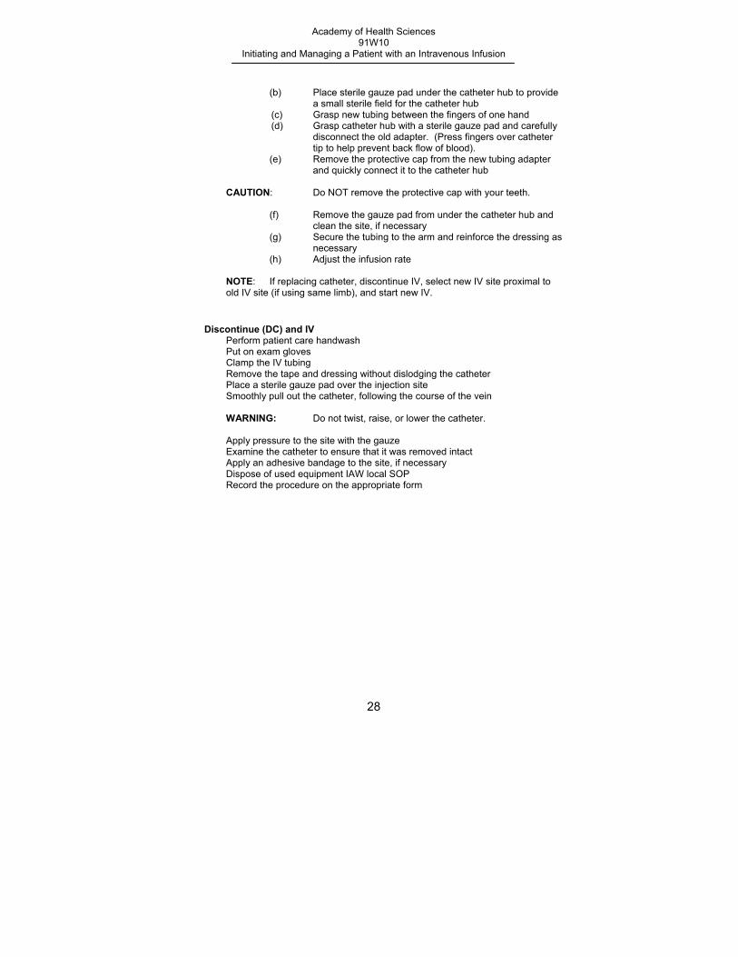
(b) Place sterile gauze pad under the catheter hub to provide a small sterile field for the catheter hub
(c) Grasp new tubing between the fingers of one hand (d) Grasp catheter hub with a sterile gauze pad and carefully
disconnect the old adapter. (Press fingers over catheter tip to help prevent back flow of blood).
(e) Remove the protective cap from the new tubing adapter and quickly connect it to the catheter hub
CAUTION: Do NOT remove the protective cap with your teeth.
(f) Remove the gauze pad from under the catheter hub and
clean the site, if necessary (g) Secure the tubing to the arm and reinforce the dressing as
necessary (h) Adjust the infusion rate
NOTE: If replacing catheter, discontinue IV, select new IV site proximal to old IV site (if using same limb), and start new IV.
Discontinue (DC) and IV Perform patient care handwash Put on exam gloves Clamp the IV tubing Remove the tape and dressing without dislodging the catheter Place a sterile gauze pad over the injection site Smoothly pull out the catheter, following the course of the vein
WARNING: Do not twist, raise, or lower the catheter.
Apply pressure to the site with the gauze Examine the catheter to ensure that it was removed intact Apply an adhesive bandage to the site, if necessary Dispose of used equipment IAW local SOP Record the procedure on the appropriate form

Perform a Patient Care Handwash & Put On and Remove Sterile Glvoes Appendix A
Competency Skill Sheets
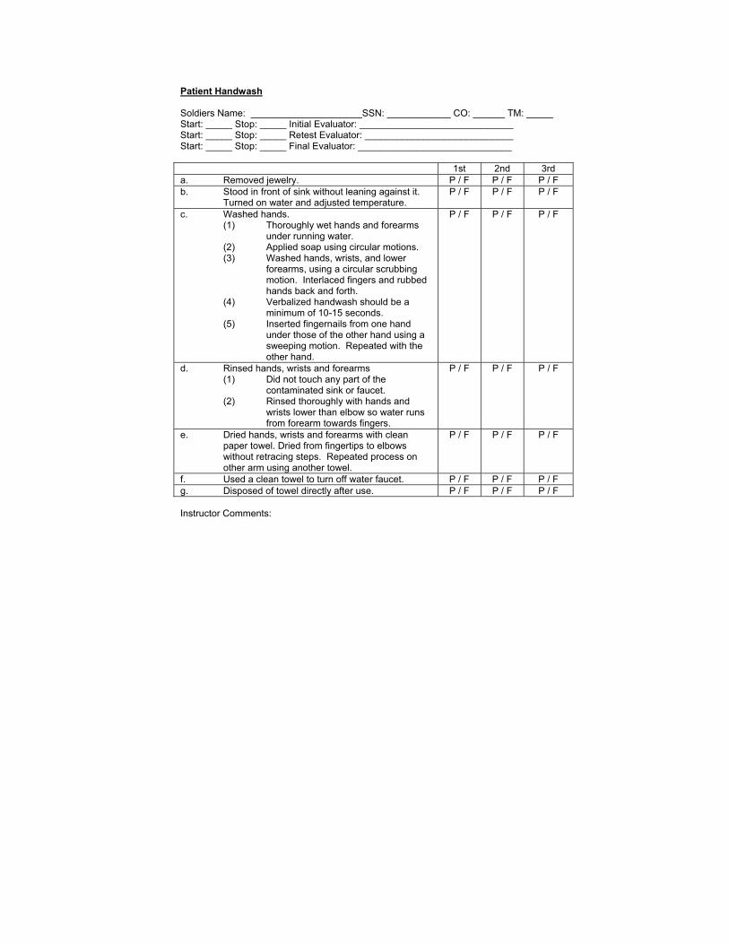
Patient Handwash Soldiers Name: _____________________SSN: ____________ CO: ______ TM: _____ Start: _____ Stop: _____ Initial Evaluator: _____________________________ Start: _____ Stop: _____ Retest Evaluator: ____________________________ Start: _____ Stop: _____ Final Evaluator: _____________________________ 1st 2nd 3rd a. Removed jewelry. P / F P / F P / F b. Stood in front of sink without leaning against it.
Turned on water and adjusted temperature. P / F P / F P / F
c. Washed hands. (1) Thoroughly wet hands and forearms
under running water. (2) Applied soap using circular motions. (3) Washed hands, wrists, and lower
forearms, using a circular scrubbing motion. Interlaced fingers and rubbed hands back and forth.
(4) Verbalized handwash should be a minimum of 10-15 seconds.
(5) Inserted fingernails from one hand under those of the other hand using a sweeping motion. Repeated with the other hand.
P / F P / F P / F
d. Rinsed hands, wrists and forearms (1) Did not touch any part of the
contaminated sink or faucet. (2) Rinsed thoroughly with hands and
wrists lower than elbow so water runs from forearm towards fingers.
P / F P / F P / F
e. Dried hands, wrists and forearms with clean paper towel. Dried from fingertips to elbows without retracing steps. Repeated process on other arm using another towel.
P / F P / F P / F
f. Used a clean towel to turn off water faucet. P / F P / F P / F g. Disposed of towel directly after use. P / F P / F P / F Instructor Comments:
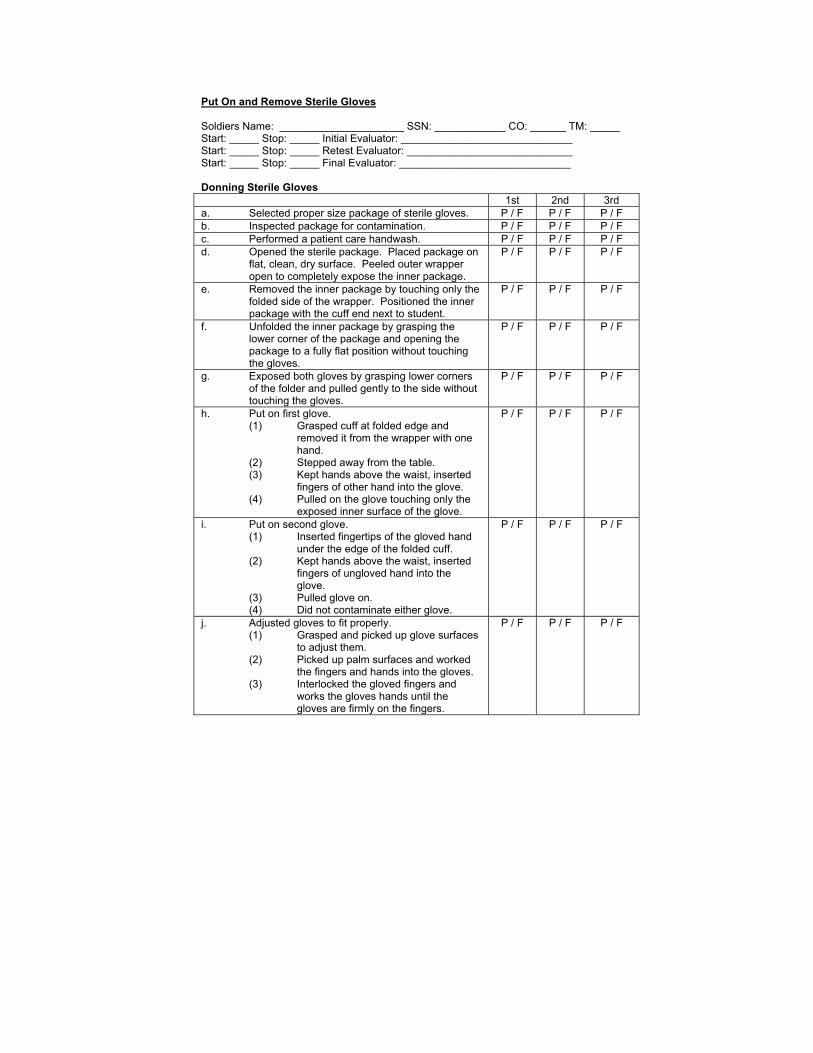
Put On and Remove Sterile Gloves
Soldiers Name: _____________________ SSN: ____________ CO: ______ TM: _____ Start: _____ Stop: _____ Initial Evaluator: _____________________________ Start: _____ Stop: _____ Retest Evaluator: ____________________________ Start: _____ Stop: _____ Final Evaluator: _____________________________ Donning Sterile Gloves 1st 2nd 3rd a. Selected proper size package of sterile gloves. P / F P / F P / F b. Inspected package for contamination. P / F P / F P / F c. Performed a patient care handwash. P / F P / F P / F d. Opened the sterile package. Placed package on
flat, clean, dry surface. Peeled outer wrapper open to completely expose the inner package.
P / F P / F P / F
e. Removed the inner package by touching only the folded side of the wrapper. Positioned the inner package with the cuff end next to student.
P / F P / F P / F
f. Unfolded the inner package by grasping the lower corner of the package and opening the package to a fully flat position without touching the gloves.
P / F P / F P / F
g. Exposed both gloves by grasping lower corners of the folder and pulled gently to the side without touching the gloves.
P / F P / F P / F
h. Put on first glove. (1) Grasped cuff at folded edge and
removed it from the wrapper with one hand.
(2) Stepped away from the table. (3) Kept hands above the waist, inserted
fingers of other hand into the glove. (4) Pulled on the glove touching only the
exposed inner surface of the glove.
P / F P / F P / F
i. Put on second glove. (1) Inserted fingertips of the gloved hand
under the edge of the folded cuff. (2) Kept hands above the waist, inserted
fingers of ungloved hand into the glove.
(3) Pulled glove on. (4) Did not contaminate either glove.
P / F P / F P / F
j. Adjusted gloves to fit properly. (1) Grasped and picked up glove surfaces
to adjust them. (2) Picked up palm surfaces and worked
the fingers and hands into the gloves. (3) Interlocked the gloved fingers and
works the gloves hands until the gloves are firmly on the fingers.
P / F P / F P / F

Removal of Sterile Gloves a. Grasped one glove at the heel of the hand with the other gloved hand. P / Fb. Peeled off glove, retaining it in the palm of the gloved hand. P / Fc. Reached under the cuff of the remaining glove with one or two fingers of the
ungloved hand. P / F
d. Peeled off glove over the glove being held in the palm. P / Fe. Did not contaminate himself. P / Ff. Discarded the gloves. P / Fg. Performed a patient care handwash. P / F Instructor Comments:

Initiate and Manage a Patient with an Intravenous Infusion Appendix B
Competency Skill Sheets
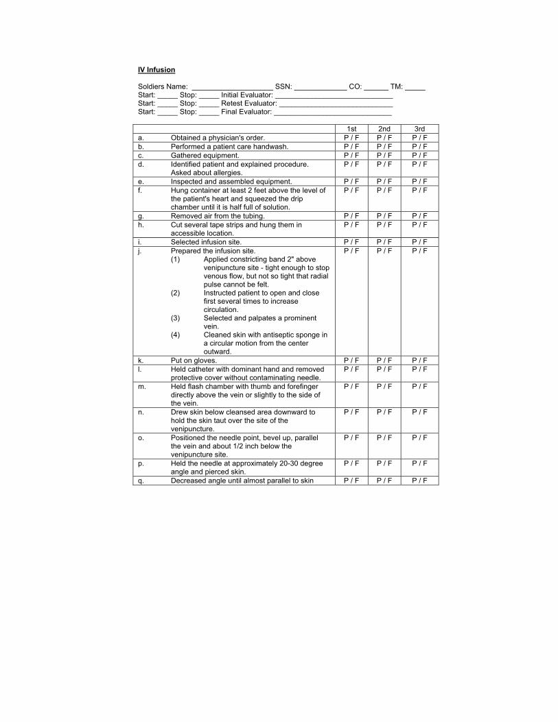
IV Infusion Soldiers Name: ____________________ SSN: _____________ CO: ______ TM: _____ Start: _____ Stop: _____ Initial Evaluator: _____________________________ Start: _____ Stop: _____ Retest Evaluator: ____________________________ Start: _____ Stop: _____ Final Evaluator: _____________________________ 1st 2nd 3rd a. Obtained a physician's order. P / F P / F P / F b. Performed a patient care handwash. P / F P / F P / F c. Gathered equipment. P / F P / F P / F d. Identified patient and explained procedure.
Asked about allergies. P / F P / F P / F
e. Inspected and assembled equipment. P / F P / F P / F f. Hung container at least 2 feet above the level of
the patient's heart and squeezed the drip chamber until it is half full of solution.
P / F P / F P / F
g. Removed air from the tubing. P / F P / F P / F h. Cut several tape strips and hung them in
accessible location. P / F P / F P / F
i. Selected infusion site. P / F P / F P / F j. Prepared the infusion site.
(1) Applied constricting band 2" above venipuncture site - tight enough to stop venous flow, but not so tight that radial pulse cannot be felt.
(2) Instructed patient to open and close first several times to increase circulation.
(3) Selected and palpates a prominent vein.
(4) Cleaned skin with antiseptic sponge in a circular motion from the center outward.
P / F P / F P / F
k. Put on gloves. P / F P / F P / F l. Held catheter with dominant hand and removed
protective cover without contaminating needle. P / F P / F P / F
m. Held flash chamber with thumb and forefinger directly above the vein or slightly to the side of the vein.
P / F P / F P / F
n. Drew skin below cleansed area downward to hold the skin taut over the site of the venipuncture.
P / F P / F P / F
o. Positioned the needle point, bevel up, parallel the vein and about 1/2 inch below the venipuncture site.
P / F P / F P / F
p. Held the needle at approximately 20-30 degree angle and pierced skin.
P / F P / F P / F
q. Decreased angle until almost parallel to skin P / F P / F P / F
surface and direct it toward the vein. Continued advancing the needle/catheter until vein is pierced.
r. Checked for blood in the flash chamber. P / F P / F P / F s. Advanced catheter/needle unit approximately 1/8
inch further to insure catheter placement in the vein.
P / F P / F P / F
t. Stabilized flash chamber with dominant hand, grasped catheter hub with non-dominant hand and treaded catheter into vein to catheter hub.
P / F P / F P / F
u. Removed flash chamber/needle and laid aside. P / F P / F P / F v. With dominant hand, removed protective cover
from needle adapter on tubing and quickly connected adapter into the catheter hub, while maintaining stabilization of the hub with non-dominant hand.
P / F P / F P / F
w. Told patient to unclench fist and released constricting band.
P / F P / F P / F
x. Unclamped IV tubing and adjusted flow rate to appropriate drip rate.
P / F P / F P / F
y. Examined infusion site for infiltration and discontinued if infiltration is present.
P / F P / F P / F
z. Cleaned the area of blood, if necessary, and secured hub of catheter with tape, leaving hub and tubing connection visible.
P / F P / F P / F
aa. Applied a sterile dressing over the puncture site. P / F P / F P / F bb. Looped the IV tubing in extremity and secured
with tape. P / F P / F P / F
cc. Splinted the arm loosely on a padded splint, if necessary, to reduce movement.
P / F P / F P / F
dd. Printed the date, gauge of the catheter, and time IV was started and initials of person starting IV on a piece of tape and secured the tape to the dressing.
P / F P / F P / F
ee. Printed patient's identification, drip rate, date and time the IV infusion was initiated and the person initiating the IV on a piece of tape and secured the tape to the IV container.
P / F P / F P / F
ff. Printed the date and time the tubing was put in place and the initials of the person initiating the IV on a piece of tape and wrapped the tape around the tubing, leaving a tab.
P / F P / F P / F
gg. Re-examined the IV site for infiltration. P / F P / F P / F hh. Removed gloves and performed a patient care
handwash. P / F P / F P / F
ii. Recorded the procedure on the appropriate form. P / F P / F P / F Instructor Comments: