8.5 no vids
41
8.5 Selected Synovial Joints • Synovial joints are diverse • All have general features, but some also have unique structural features, abilities, and weaknesses • Five main synovial joints – Knee – Shoulder – Elbow – Hip – Jaw © 2016 Pearson Education, Inc.
-
Upload
amo-oliverio -
Category
Education
-
view
701 -
download
0
Transcript of 8.5 no vids

8.5 Selected Synovial Joints
• Synovial joints are diverse • All have general features, but some also have unique structural
features, abilities, and weaknesses• Five main synovial joints
– Knee– Shoulder– Elbow– Hip– Jaw
© 2016 Pearson Education, Inc.

Knee Joint
• Largest, most complex joint of body• Consists of three joints surrounded by single cavity
1. Femoropatellar joint • Plane joint• Allows gliding motion during knee flexion
2. Lateral joint and 3. Medial joint• Lateral and medial joints together are called tibiofemoral joint• Joint between femoral condyles and lateral and medial menisci of tibia• Hinge joint that allows flexion, extension, and some rotation when knee
partly flexed
© 2016 Pearson Education, Inc.

Figure 8.7a The knee joint.
Femur
ArticularcapsulePosteriorcruciateligament
Lateral meniscus
Anteriorcruciateligament
Tibia
Tendon ofquadricepsfemorisSuprapatellarbursaPatellaSubcutaneousprepatellar bursa
Synovial cavityLateral meniscusInfrapatellar fat padDeep infrapatellarbursaPatellar ligament
Sagittal section through the right knee joint© 2016 Pearson Education, Inc.

Figure 8.7b The knee joint.
Anterior
Anteriorcruciateligament
Articularcartilageon medialtibialcondyle
Medialmeniscus
Posteriorcruciate ligament
Superior view of the right tibia in the knee joint,showing the menisci and cruciate ligaments
Articularcartilage onlateral tibialcondyle
Lateralmeniscus
© 2016 Pearson Education, Inc.

Knee Joint (cont.)
• Joint capsule is thin and absent anteriorly• Anteriorly, quadriceps tendon gives rise to three broad ligaments
that run from patella to tibia– Medial and lateral patellar retinacula that flank the patellar
ligament• Doctors tap patellar ligament to test knee-jerk reflex
• At least 12 bursae associated with knee joint
© 2016 Pearson Education, Inc.

Figure 8.7c The knee joint.
Quadricepsfemoris muscle
Tendon ofquadricepsfemoris musclePatella
LateralpatellarretinaculumFibularcollateralligament
Fibula
Medial patellarretinaculum
Tibial collateralligament
PatellarligamentTibia
Anterior view of right knee© 2016 Pearson Education, Inc.

Knee Joint (cont.)
• Capsular, extracapsular, or intracapsular ligaments act to stabilize knee joint
• Capsular and extracapsular ligaments help prevent hyperextension of knee– Fibular and tibial collateral ligaments: prevent rotation when
knee is extended– Oblique popliteal ligament: stabilizes posterior knee joint– Arcuate popliteal ligament: reinforces joint capsule posteriorly
© 2016 Pearson Education, Inc.

Figure 8.7d The knee joint.
Tendon ofadductormagnus
Medial head ofgastrocnemiusmuscle
Popliteusmuscle (cut)
Tibialcollateralligament
Tendon ofsemimembranosusmuscle
Femur
Articular capsule
Oblique poplitealligament
Lateral head ofgastrocnemiusmuscle
BursaFibular collateralligamentArcuate poplitealligament
Tibia
Posterior view of the joint capsule, including ligaments© 2016 Pearson Education, Inc.

Knee Joint (cont.)
• Intracapsular ligaments reside within capsule, but outside synovial cavity
• Help to prevent anterior-posterior displacement– Anterior cruciate ligament (ACL)
• Attaches to anterior tibia• Prevents forward sliding of tibia and stops hyperextension of knee
– Posterior cruciate ligament• Attaches to posterior tibia• Prevents backward sliding of tibia and forward sliding of femur
© 2016 Pearson Education, Inc.

Figure 8.7e The knee joint.
Posteriorcruciateligament
Fibularcollateralligament
MedialcondyleTibialcollateralligament
AnteriorcruciateligamentMedialmeniscus
Patellarligament
Patella
Quadricepstendon
Lateralcondyleof femur
Lateralmeniscus
Tibia
Fibula
Anterior view of flexed knee, showing thecruciate ligaments (articular capsuleremoved, and quadriceps tendon cut andreflected distally)
© 2016 Pearson Education, Inc.

Figure 8.7f The knee joint.
Medial femoralcondyleAnterior cruciateligamentMedial meniscuson medial tibialcondyle
Patella
Photograph of an opened knee joint;view similar to (e)
© 2016 Pearson Education, Inc.

Clinical – Homeostatic Imbalance 8.1
• Knee absorbs great amount of vertical force; however, it is vulnerable to horizontal blows– Common knee injuries involved the 3 C’s:
• Collateral ligaments• Cruciate ligaments• Cartilages (menisci)
– Lateral blows to extended knee can result in tears in tibial collateral ligament, medial meniscus, and anterior cruciate ligament
– Injuries affecting just ACL are common in runners who change direction, twisting ACL
– Surgery usually needed for repairs
© 2016 Pearson Education, Inc.

Figure 8.8 The “unhappy triad:” ruptured ACL, ruptured tibial collateral ligament, and torn meniscus.
Lateral
Hockey puckPatella(outline)
Medial
Tibialcollateralligament(torn)
Medialmeniscus(torn)
Anteriorcruciateligament(torn)
© 2016 Pearson Education, Inc.

Shoulder (Glenohumeral) Joint
• Most freely moving joint in body• Stability is sacrificed for freedom of movement• Ball-and-socket joint
– Large, hemispherical head of humerus fits in small, shallow glenoid cavity of scapula• Like a golf ball on a tee
• Articular capsule enclosing cavity is also thin and loose– Contributes to freedom of movement
© 2016 Pearson Education, Inc.

Figure 8.9a The shoulder joint.
Acromion of scapulaCoracoacromial ligament
Subacromial bursaFibrous layer ofarticular capsule
Tendon sheath
Tendon of long headof biceps brachii muscle
Synovial cavity ofthe glenoid cavitycontaining synovialfluidArticular cartilage
Synovial membrane
Fibrous layer ofarticular capsule
Humerus
Frontal section through right shoulder joint
© 2016 Pearson Education, Inc.

Figure 8.9b The shoulder joint.
Synovial cavityof the glenoidcavity containingsynovial fluid
Articular cartilage
Fibrous layer ofarticular capsule
Humerus
Cadaver photo corresponding to (a)
© 2016 Pearson Education, Inc.

Shoulder (Glenohumeral) Joint (cont.)
• Glenoid labrum: fibrocartilaginous rim around glenoid cavity – Helps to add depth to shallow cavity– Cavity still only holds one-third of head of humerus
• Reinforcing ligaments– Primarily on anterior aspect– Coracohumeral ligament
• Helps support weight of upper limb– Three glenohumeral ligaments
• Strengthen anterior capsule, but are weak support
© 2016 Pearson Education, Inc.

Figure 8.9c The shoulder joint.
AcromionCoracoacromial ligament
Subacromial bursa
Coracohumeralligament
Transverse humeralligament
Tendon sheathTendon of long headof biceps brachiimuscle
Coracoid processArticular capsulereinforced byglenohumeralligamentsSubscapularbursaTendon of thesubscapularismuscleScapula
Anterior view of right shoulder joint capsule
© 2016 Pearson Education, Inc.

Figure 8.9d The shoulder joint.
Acromion
Coracoid process
Articular capsuleGlenoid cavityGlenoid labrum
Tendon of long headof biceps brachii muscleGlenohumeral ligaments
Tendon of thesubscapularis muscleScapula
Posterior Anterior
Lateral view of socket of right shoulder joint,humerus removed
© 2016 Pearson Education, Inc.

Shoulder (Glenohumeral) Joint (cont.)
• Reinforcing muscle tendons contribute most to joint stability– Tendon of long head of biceps brachii muscle is “superstabilizer”
• Travels through intertubercular sulcus • Secures humerus to glenoid cavity
– Four rotator cuff tendons encircle the shoulder joint• Subscapularis• Supraspinatus• Infraspinatus• Teres minor
© 2016 Pearson Education, Inc.

Figure 8.9c The shoulder joint.
AcromionCoracoacromial ligament
Subacromial bursa
Coracohumeralligament
Transverse humeralligament
Tendon sheathTendon of long headof biceps brachiimuscle
Coracoid processArticular capsulereinforced byglenohumeralligamentsSubscapularbursaTendon of thesubscapularismuscleScapula
Anterior view of right shoulder joint capsule
© 2016 Pearson Education, Inc.

Figure 8.9d The shoulder joint.
Acromion
Coracoid process
Articular capsuleGlenoid cavityGlenoid labrum
Tendon of long headof biceps brachii muscleGlenohumeral ligaments
Tendon of thesubscapularis muscleScapula
Posterior Anterior
Lateral view of socket of right shoulder joint,humerus removed
© 2016 Pearson Education, Inc.

Figure 8.9e The shoulder joint.
Acromion (cut)
Glenoid cavityof scapula
Glenoid labrum
Rotator cuffmuscles(cut)
Capsule ofshoulder joint(opened)
Head of humerus
Posterior view of an opened right shoulder joint
© 2016 Pearson Education, Inc.

Elbow Joint
• Humerus articulates with radius and ulna• Hinge joint formed primarily from trochlear notch of ulna
articulating with trochlea of humerus– Allows for flexion and extension only
• Anular ligament surrounds head of radius• Two capsular ligaments restrict side-to-side movement
– Ulnar collateral ligament– Radial collateral ligament
© 2016 Pearson Education, Inc.

Figure 8.10a The elbow joint.
ArticularcapsuleSynovialmembrane
Humerus
Fat pad
Tendon oftriceps muscle
Bursa
TrochleaArticular cartilageof the trochlearnotch
Median sagittal section through right elbow(lateral view)
Synovial cavityArticular cartilageCoronoid processTendon ofbrachialis muscleUlna
© 2016 Pearson Education, Inc.
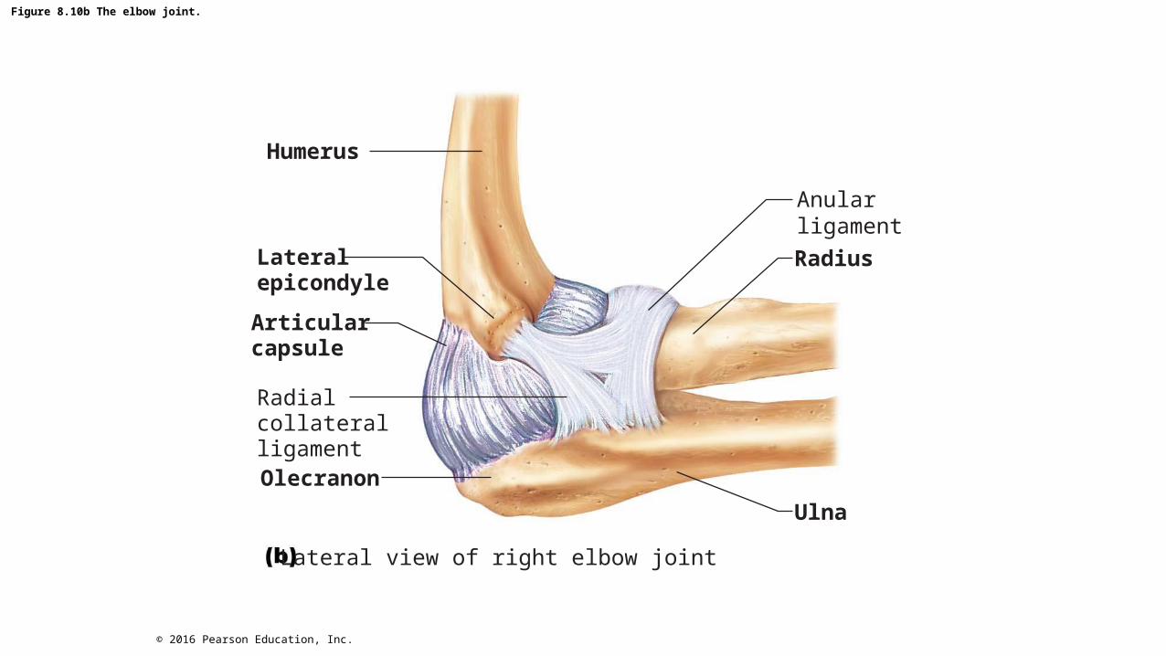
Figure 8.10b The elbow joint.
Humerus
Anularligament
Lateralepicondyle
Articularcapsule
RadialcollateralligamentOlecranon
Ulna
Lateral view of right elbow joint
Radius
© 2016 Pearson Education, Inc.

Figure 8.10c The elbow joint.
Humerus
Anularligament
Medialepicondyle
RadiusArticularcapsule
Coronoidprocessof ulnaUlna
Ulnarcollateralligament
Cadaver photo of medial view of right elbow
© 2016 Pearson Education, Inc.

Figure 8.10d The elbow joint.
ArticularcapsuleAnularligamentCoronoidprocess
MedialepicondyleUlnarcollateralligament
Humerus
Radius
Ulna
Medial view of right elbow
© 2016 Pearson Education, Inc.

Hip (Coxal) Joint
• Ball-and-socket joint• Large, spherical head of the femur articulates with deep cup-
shaped acetabulum• Good range of motion, but limited by the deep socket
– Acetabular labrum: rim of fibrocartilage that enhances depth of socket (hip dislocations are rare)
© 2016 Pearson Education, Inc.

Figure 8.11a The hip joint.
Articularcartilage
Acetabularlabrum
Femur
Hip (coxal) boneLigament of thehead of the femur(ligamentum teres)
Synovial cavityArticular capsule
Frontal section through the right hip joint
© 2016 Pearson Education, Inc.

Figure 8.11b The hip joint.
Acetabularlabrum
SynovialmembraneLigament of thehead of the femur(ligamentum teres)
Head of femur
Articular capsule(cut)
Photo of the interior of the hip joint, lateral view
© 2016 Pearson Education, Inc.

Hip (Coxal) Joint (cont.)
• Reinforcing ligaments include:– Iliofemoral ligament– Pubofemoral ligament– Ischiofemoral ligament– Ligament of head of femur (ligamentum teres)
• Slack during most hip movements, so not important in stabilizing• Does contain artery that supplies head of femur
• Greatest stability comes from deep ball-and-socket joint
© 2016 Pearson Education, Inc.

Figure 8.11c The hip joint.
IschiumIliofemoralligamentIschiofemoralligament
Greatertrochanterof femur
Posterior view of right hip joint,capsule in place
© 2016 Pearson Education, Inc.

Figure 8.11d The hip joint.
Anteriorinferior iliacspineGreatertrochanter
IliofemoralligamentPubofemoralligament
Anterior view of right hip joint, capsule in place
© 2016 Pearson Education, Inc.

Temporomandibular Joint (TMJ)
• Jaw joint is a modified hinge joint• Mandibular condyle articulates with temporal bone
– Posterior temporal bone forms mandibular fossa, while anterior portion forms articular tubercle
• Articular capsule thickens into strong lateral ligament
© 2016 Pearson Education, Inc.

Temporomandibular Joint (TMJ) (cont.)
• Two types of movement– Hinge: depression and elevation of mandible– Gliding: side-to-side (lateral excursion) grinding of teeth
• Most easily dislocated joint in the body
© 2016 Pearson Education, Inc.

Figure 8.12a The temporomandibular (jaw) joint.
Mandibular fossaArticular tubercleZygomatic processInfratemporal fossa
Externalacousticmeatus
ArticularcapsuleRamus ofmandible
Lateralligament
Location of the joint in the skull© 2016 Pearson Education, Inc.

Figure 8.12b The temporomandibular (jaw) joint.
Articular discMandibularfossa
Articular tubercleSuperior jointcavity
Articularcapsule
Synovialmembranes
Condylarprocess ofmandible
Ramus ofmandible
Inferior jointcavity
Enlargement of a sagittal section through the joint© 2016 Pearson Education, Inc.

Figure 8.12c The temporomandibular (jaw) joint.
Superior view
Outline ofthe mandibularfossa
Lateral excursion: lateral (side-to-side) movements of the mandible
© 2016 Pearson Education, Inc.

Clinical – Homeostatic Imbalance 8.2
• Dislocation of TMJ is most common because of shallow socket of joint
• Almost always dislocates anteriorly, causing mouth to remain open– To realign, physician must push mandible back into place
© 2016 Pearson Education, Inc.

Clinical – Homeostatic Imbalance 8.2
• Symptoms: ear and face pain, tender muscles, popping sounds when opening mouth, joint stiffness
• Usually caused by grinding teeth, but can also be due to jaw trauma or poor occlusion of teeth– Treatment for grinding teeth includes bite plate – Relaxing jaw muscles helps
© 2016 Pearson Education, Inc.


![backthepack flyer VS_2 8.5 x 8.5[1][1]](https://static.fdocuments.in/doc/165x107/587fcb331a28ab3b158b7027/backthepack-flyer-vs2-85-x-8511.jpg)