58 The Pupils - Famona Site H. SPECTOR Definition The normal ... error, suppression amblyopia,...
5
58 ThePupils ROBERTH .SPECTOR Definition Thenormalpupilsizeinadultsvariesfrom2to4mmin diameterinbrightlightto4to8mminthedark .Thepupils aregenerallyequalinsize .Theyconstricttodirectillumi- nation(directresponse)andtoilluminationoftheopposite eye(consensualresponse) .Thepupildilatesinthedark . Bothpupilsconstrictwhentheeyeisfocusedonanear object(accommodativeresponse) .Thepupilisabnormalif itfailstodilatetothedarkorfailstoconstricttolightor accommodation . ThepopularacronymPERRLA-pupilsequal,round, andreactivetolightandaccommodation-isaconvenient butincompletedescriptionofpupillomotorfunction .Itspe- cificallyomitsimportantclinicaldatasuchastheactualsize andshapeofeachpupil,thespeedandextentofpupillary constriction,andtheresultsofdetermininganafferentpu- pillarydefect . Technique Theexaminerfirstmustcheckthesize,shape,equality,and positionofthepupils,andtheirresponsetoabrightlight . Becausethesephenomenaarebesttestedwiththepupils inasemidilatedstate,clinicalobservationsshouldbemade inadimlylightedroom .Patientsshouldbeencouragedto fixatevisuallyonadistantobject,becauseiftheyinadver- tentlylookatyournoseortheflashlight,theattemptto convergewillreflexlyevokemiosis,andcertainsignsmay beoverlooked(e.g ., anisocoria,light-neardissociation,ora subtleMarcusGunnsign .Forthesamereasons,trynotto startleortouchpatientswithyourhandsorinstruments,as psychosensorystimulationinducesmydriasis,hippus,and relativelyhyperactivepupils . Toassesspupillarysizeinadarkenedroom,illuminate thefacefrombelow .Slowlymovethelightuptothepatient's eyelevelandcheckthepupillaryresponsetothebright lightoneachsideseveraltimes .Gradetheseresponsesfrom 1+to4+ .Next,lookattheamountofpupillaryconstric- tionthatoccurswhenthepatientisforcedtofocusona nearobject,suchasathumbheld15to20cmabovethe eyes .Recordthesedatasothattheyareeasytoreadand recall .Belowisanexampleofonemethod : Normally,theconvergencereactionisasbriskandas extensiveasthelightreaction .Theextentofconstriction dependsalsoontheconditionoftheiris .Abrowniriscon- tractslessthanablueiris .Inoldpeopleandinpatientswith irisatrophy,thesphincterbecomesrigid,hencethelight reactiondiminishesinextent . BasicScience Thesizeofthepupiliscontrolledbytheactivitiesoftwo muscles :thecircumferentialsphinctermusclefoundinthe marginoftheiris,innervatedbytheparasympatheticner- voussystem ;andtheirisdilatormuscle,runningradially fromtheirisroottotheperipheralborderofthesphincter . Theirisdilatorfiberscontaina-adrenergicsympatheticre- ceptorsthatrespondtochangesinsympathetictonusand changesinthebloodlevelofcirculatingcatecholamines . Thepupillarylightreflexarcbeginsintheretina(Figure 58 .1) .Considerableevidenceexiststhatthevisualcellsof theretina,thatis,therodsandcones,alsoserveaslight receptorscontrollingpupillomotoractivity .Fibersoriginat- ingfromthenasalneuroreceptorcellsdecussateintheoptic chiasmtotheoppositeoptictract,whereasthetemporal fiberscontinueinthehomolateraloptictract ."Pupillary fibers"frombotheyeswithintheoptictractpassviathe superiorquadrigeminalbrachiumandthesuperiorcolli- culustothemesencephalicpretectumandpretectalnuclei . Axonsfromeachpretectalnucleuspassipsilaterallyand contralaterallytotheipsilateralandcontralateralEdinger- Westphal(E-W) nucleus,asubnucleusoftheoculomotor nuclearcomplex .Thehemidecussationofthepupillaryfi- bersattheopticchiasmandbetweenthepretectalnuclei ensuresthateachE-Wnucleusreceivesinformationabout thelevelofincominglightfromeacheye .Hence,thepupils shouldbeequalindiameterregardlessofthelevelofvision ofeithereye .Forexample,inapatientwithoneblindeye, thepretectalnucleiwouldregisterandtransmittoeachE- Wnucleusonlyone-halfthenormallevelofillumination . Thetransmissionoflesspupilloconstrictortonetoeachiris sphincterwouldresultinslightlylargerpupilsbutofequal diameter .Accordingly,anisocoria(unequalpupillarydi- ameter)isnotattributabletotheangleatwhichlightstrikes theface,unilateralcataracts,oranasymmetricrefractive error,unlessthereislocaldiseaseoftheanteriorsegment . ParasympatheticaxonsfromtheE-Wnucleusjointhe outflowoftheotheroculomotorsubnucleitoformthetrunk oftheoculomotornerve .Pupillomotorfibersassumeasu- perficiallocationinthenerveasitexitsthemesencephalon intheinterpeduncularspace . Intheorbit,theparasympatheticcomponentssynapse intheciliaryganglion .Postganglionicfiberstravelinginthe shortciliarynervesinnervateboththeciliarybody,inducing lensaccommodation,andthepupilloconstrictormusclesof theiris .Theratiooffibersinnervatingtheciliarybodyto thosesupplyingthepupilisapproximately30 :1 .Acetyl- cholineservesastheneurotransmitterforbothfunctions . Thepupillarynearreflexconsistsofthreeseparate,syn- ergisticphenomena :accommodation,convergence,and 300 R L Size 5 .0mm 5 .9mm Shape Oval Round Light +3 +3 Near +3 +3
Transcript of 58 The Pupils - Famona Site H. SPECTOR Definition The normal ... error, suppression amblyopia,...
58 The PupilsROBERT H . SPECTOR
Definition
The normal pupil size in adults varies from 2 to 4 mm indiameter in bright light to 4 to 8 mm in the dark. The pupilsare generally equal in size . They constrict to direct illumi-nation (direct response) and to illumination of the oppositeeye (consensual response) . The pupil dilates in the dark .Both pupils constrict when the eye is focused on a nearobject (accommodative response) . The pupil is abnormal ifit fails to dilate to the dark or fails to constrict to light oraccommodation .
The popular acronym PERRLA-pupils equal, round,and reactive to light and accommodation-is a convenientbut incomplete description of pupillomotor function . It spe-cifically omits important clinical data such as the actual sizeand shape of each pupil, the speed and extent of pupillaryconstriction, and the results of determining an afferent pu-pillary defect .
Technique
The examiner first must check the size, shape, equality, andposition of the pupils, and their response to a bright light .Because these phenomena are best tested with the pupilsin a semidilated state, clinical observations should be madein a dimly lighted room . Patients should be encouraged tofixate visually on a distant object, because if they inadver-tently look at your nose or the flashlight, the attempt toconverge will reflexly evoke miosis, and certain signs maybe overlooked (e.g ., anisocoria, light-near dissociation, or asubtle Marcus Gunn sign . For the same reasons, try not tostartle or touch patients with your hands or instruments, aspsychosensory stimulation induces mydriasis, hippus, andrelatively hyperactive pupils .
To assess pupillary size in a darkened room, illuminatethe face from below. Slowly move the light up to the patient'seye level and check the pupillary response to the brightlight on each side several times . Grade these responses from1+ to 4+ . Next, look at the amount of pupillary constric-tion that occurs when the patient is forced to focus on anear object, such as a thumb held 15 to 20 cm above theeyes. Record these data so that they are easy to read andrecall. Below is an example of one method :
Normally, the convergence reaction is as brisk and asextensive as the light reaction . The extent of constrictiondepends also on the condition of the iris . A brown iris con-tracts less than a blue iris . In old people and in patients with
iris atrophy, the sphincter becomes rigid, hence the lightreaction diminishes in extent .
Basic Science
The size of the pupil is controlled by the activities of twomuscles : the circumferential sphincter muscle found in themargin of the iris, innervated by the parasympathetic ner-vous system ; and the iris dilator muscle, running radiallyfrom the iris root to the peripheral border of the sphincter .The iris dilator fibers contain a-adrenergic sympathetic re-ceptors that respond to changes in sympathetic tonus andchanges in the blood level of circulating catecholamines .
The pupillary light reflex arc begins in the retina (Figure58 .1) . Considerable evidence exists that the visual cells ofthe retina, that is, the rods and cones, also serve as lightreceptors controlling pupillomotor activity . Fibers originat-ing from the nasal neuroreceptor cells decussate in the opticchiasm to the opposite optic tract, whereas the temporalfibers continue in the homolateral optic tract . "Pupillaryfibers" from both eyes within the optic tract pass via thesuperior quadrigeminal brachium and the superior colli-culus to the mesencephalic pretectum and pretectal nuclei .Axons from each pretectal nucleus pass ipsilaterally andcontralaterally to the ipsilateral and contralateral Edinger-Westphal (E-W) nucleus, a subnucleus of the oculomotornuclear complex . The hemidecussation of the pupillary fi-bers at the optic chiasm and between the pretectal nucleiensures that each E-W nucleus receives information aboutthe level of incoming light from each eye . Hence, the pupilsshould be equal in diameter regardless of the level of visionof either eye . For example, in a patient with one blind eye,the pretectal nuclei would register and transmit to each E-W nucleus only one-half the normal level of illumination .The transmission of less pupilloconstrictor tone to each irissphincter would result in slightly larger pupils but of equaldiameter . Accordingly, anisocoria (unequal pupillary di-ameter) is not attributable to the angle at which light strikesthe face, unilateral cataracts, or an asymmetric refractiveerror, unless there is local disease of the anterior segment .
Parasympathetic axons from the E-W nucleus join theoutflow of the other oculomotor subnuclei to form the trunkof the oculomotor nerve . Pupillomotor fibers assume a su-perficial location in the nerve as it exits the mesencephalonin the interpeduncular space .
In the orbit, the parasympathetic components synapsein the ciliary ganglion . Postganglionic fibers traveling in theshort ciliary nerves innervate both the ciliary body, inducinglens accommodation, and the pupilloconstrictor muscles ofthe iris . The ratio of fibers innervating the ciliary body tothose supplying the pupil is approximately 30:1 . Acetyl-choline serves as the neurotransmitter for both functions .
The pupillary near reflex consists of three separate, syn-ergistic phenomena : accommodation, convergence, and
300
R L
Size 5.0 mm 5 .9 mmShape Oval RoundLight + 3 + 3Near +3 +3
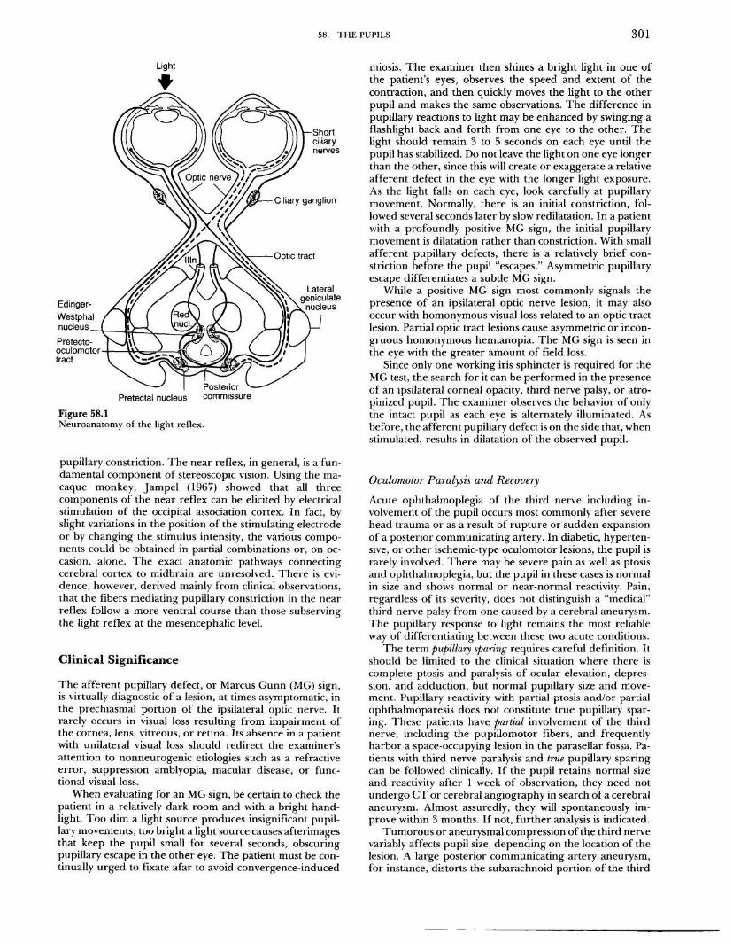
Figure 58.1Neuroanatomy of the light reflex .
pupillary constriction . The near reflex, in general, is a fun-damental component of stereoscopic vision . Using the ma-caque monkey, Jampel (1967) showed that all threecomponents of the near reflex can be elicited by electricalstimulation of the occipital association cortex . In fact, byslight variations in the position of the stimulating electrodeor by changing the stimulus intensity, the various compo-nents could be obtained in partial combinations or, on oc-casion, alone . The exact anatomic pathways connectingcerebral cortex to midbrain are unresolved . There is evi-dence, however, derived mainly from clinical observations,that the fibers mediating pupillary constriction in the nearreflex follow a more ventral course than those subservingthe light reflex at the mesencephalic level .
Clinical Significance
The afferent pupillary defect, or Marcus Gunn (MG) sign,is virtually diagnostic of a lesion, at times asymptomatic, inthe prechiasmal portion of the ipsilateral optic nerve . Itrarely occurs in visual loss resulting from impairment ofthe cornea, lens, vitreous, or retina. Its absence in a patientwith unilateral visual loss should redirect the examiner'sattention to nonneurogenic etiologies such as a refractiveerror, suppression amblyopia, macular disease, or func-tional visual loss .
When evaluating for an MG sign, be certain to check thepatient in a relatively dark room and with a bright hand-light. Too dim a light source produces insignificant pupil-lary movements ; too bright a light source causes afterimagesthat keep the pupil small for several seconds, obscuringpupillary escape in the other eye . The patient must be con-tinually urged to fixate afar to avoid convergence-induced
58 . THE PUPILS
301
miosis. The examiner then shines a bright light in one ofthe patient's eyes, observes the speed and extent of thecontraction, and then quickly moves the light to the otherpupil and makes the same observations . The difference inpupillary reactions to light may be enhanced by swinging aflashlight back and forth from one eye to the other . Thelight should remain 3 to 5 seconds on each eye until thepupil has stabilized . Do not leave the light on one eye longerthan the other, since this will create or exaggerate a relativeafferent defect in the eye with the longer light exposure .As the light falls on each eye, look carefully at pupillarymovement. Normally, there is an initial constriction, fol-lowed several seconds later by slow redilatation . In a patientwith a profoundly positive MG sign, the initial pupillarymovement is dilatation rather than constriction . With smallafferent pupillary defects, there is a relatively brief con-striction before the pupil "escapes ." Asymmetric pupillaryescape differentiates a subtle MG sign .
While a positive MG sign most commonly signals thepresence of an ipsilateral optic nerve lesion, it may alsooccur with homonymous visual loss related to an optic tractlesion. Partial optic tract lesions cause asymmetric or incon-gruous homonymous hemianopia . The MG sign is seen inthe eye with the greater amount of field loss .
Since only one working iris sphincter is required for theMG test, the search for it can be performed in the presenceof an ipsilateral corneal opacity, third nerve palsy, or atrop inized pupil. The examiner observes the behavior of only
the intact pupil as each eye is alternately illuminated . Asbefore, the afferent pupillary defect is on the side that, whenstimulated, results in dilatation of the observed pupil .
Oculomotor Paralysis and RecoveryAcute ophthalmoplegia of the third nerve including in-volvement of the pupil occurs most commonly after severehead trauma or as a result of rupture or sudden expansionof a posterior communicating artery . In diabetic, hyperten-sive, or other ischemic-type oculomotor lesions, the pupil israrely involved . There may be severe pain as well as ptosisand ophthalmoplegia, but the pupil in these cases is normalin size and shows normal or near-normal reactivity . Pain,regardless of its severity, does not distinguish a "medical"third nerve palsy from one caused by a cerebral aneurysm .The pupillary response to light remains the most reliableway of differentiating between these two acute conditions .
The term pupillary sparing requires careful definition. Itshould be limited to the clinical situation where there iscomplete ptosis and paralysis of ocular elevation, depres-sion, and adduction, but normal pupillary size and move-ment. Pupillary reactivity with partial ptosis and/or partialophthalmoparesis does not constitute true pupillary spar-ing. These patients have partial involvement of the thirdnerve, including the pupillomotor fibers, and frequentlyharbor a space-occupying lesion in the parasellar fossa . Pa-tients with third nerve paralysis and true pupillary sparingcan be followed clinically . If the pupil retains normal sizeand reactivity after 1 week of observation, they need notundergo CT or cerebral angiography in search of a cerebralaneurysm. Almost assuredly, they will spontaneously im-prove within 3 months. If not, further analysis is indicated .
Tumorous or aneurysmal compression of the third nervevariably affects pupil size, depending on the location of thelesion. A large posterior communicating artery aneurysm,for instance, distorts the subarachnoid portion of the third

302
IV. THE NEUROLOGIC SYSTEM
nerve and almost always produces mydriasis. In lesions ofthe cavernous sinus, however, the pupillary reaction to alight and near stimulus may be fully preserved in the an-terior cavernous sinus . The oculomotor nerve separates intoa superior and an inferior division . Relative pupillary spar-ing may in part reflect sparing of the inferior division, whichalso innervates the medial and inferior rectus and inferioroblique muscles . It may also be explained by the fact thatthe pupillomotor fibers, in the process of joining the twigto the inferior oblique muscle, may either assume an in-dependent course or descend from the vulnerable super-ficial position in the subarachnoid portion of the nerve toa presumably more protected position, either within thesubstance of the nerve or on its lateral or inferior aspects .
Aberrant regeneration of the third nerve occurs afteraxonal destruction. It is characterized clinically by synkineticeye muscle activity. For instance, there may be elevation ofthe involved lid on adduction, lowering of the lid duringabduction, or elevation of the lid on depression of the eye .The pupil in these cases is usually larger than its mate andalso shows synkinetic activity. It may not react to a brightlight, but portions of the iris sphincter will contract duringadduction, depression, or elevation of the globe, indicatingthat the pupillosphincter contracts simultaneous with themedial or with the inferior or superior rectus muscles,respectively .
Adie's Tonic Pupil Syndrome
Adie's tonic pupil (ATP), the most common cause of isolatedinternal ophthalmoplegia, results from postganglionic para-sympathetic denervation of the internal ocular muscles (theciliary muscle and iris sphincter) . The neuropathologic find-ings comprise nonspecific necrosis and neuronal loss in theshort ciliary nerves and/or ciliary ganglion .
The patient with ATP may be totally asymptomatic, andis often brought to a physician's office by a friend or relativewho notices that he or she has one large pupil. Of the 122patients with ATP studied retrospectively by Thompson(1977),80% had symptoms, which included anisocoria, pho-tophobia, and difficulty with dark adaptation . Ciliary muscle-related symptoms, present in 35% of affected individuals,included blurred vision, pseudomyopia, and brow-ache withnear work.
If the ocular examination is performed at the onset ofsymptoms, the patient manifests a large, immobile pupil .Neurologically, diminished or absent deep tendon reflexesin the lower extremities are found in one-third to half ofpatients (Holmes-Adie syndrome) . With time, aberrantreinnervation of the pupil and ciliary body occurs . Becausethe overwhelming number of postganglionic parasympa-thetic fibers from the ciliary ganglion control accommo-dation, the iris sphincter becomes reinnervated almostexclusively by accommodative elements, and the near reflexconsequently becomes extensive-in fact it is prolonged .Such a "tonic" near response is best appreciated when thepatient changes fixation from a near to a far stimulus . Thenormal pupil readily redilates, while the Adie's pupil re-dilates at a much slower rate .
Aberrant regeneration of the parasympathetic nervesupply to the intraocular muscles also causes sector palsiesof the pupillary sphincter and ciliary muscle . Asynchronouscontractions of these muscles cause the following signs : in-duced astigmatism, tonicity of accommodation, and cholin-ergic supersensitivity of the ciliary muscle . The examiner
notes pupillary light-near dissociation, vermiform contrac-tions of the pupil, and pharmacologic evidence of dener-vation supersensitivity. A dilute parasympathomimetic agentsuch as 0.125% pilocarpine is used for this purpose. It pro-duces marked constriction of an Adie's pupil, but has noeffect on the diameter of a normal pupil . Demonstratingdenervation supersensitivity by this method differentiatesan acute Adie's pupil from the large, immobile pupil ob-served in early third nerve lesions and from a pharmaco-logically dilated pupil .
Although ATP is most commonly a unilateral disorderit can be bilateral, developing in both eyes either simulta-neously •or consecutively . Symmetric bilateral ATPs havebeen observed with widespread peripheral neuropathies suchas in diabetes or the Charcot-Marie-Tooth syndrome . Theyare frequently found in association with other signs of auto-nomic dysfunction-orthostatic hypotension, progressivesegmental anhidrosis, and as a constituent of the Shy-Dragerand Riley-Day syndromes .
Sylvian Aqueduct Syndrome
With rostral midbrain lesions in the area of the pretectalnuclear complex, interruption of the retinotectal fibers withpreservation of the supranuclear accommodative fibers pro-duces bilateral pupillary light-near dissociation . The asso-ciated damage to the pretectal pupilloconstrictor nucleiresults in pupils that are 4 to 6 mm in diameter ; they donot react to light but constrict during the attempt to con-verge; and these findings occur with other signs, such assupranuclear paralysis of upgaze, lid retraction, and con-vergence-retraction nystagmus .
Pharmacologically Dilated Pupil
As an isolated finding, an extremely large pupil, obliteratingthe iris and unresponsive to a light or near stimulus, isalmost always due to inadvertent or factitious applicationof a parasympathomimetic agent (eye drops, scopolamine,jimsonweed, marijuana, LSD) . Medical personnel, includ-ing nurses, doctors, and pharmacists, are especially liableto accidental instillation of mydriatic agents .
Instillation of 1% pilocarpine helps differentiate phar-macologic mydriasis from other causes of a large, unreactivepupil. With parasympathetic denervation of the pupil fromoculomotor palsy or an ATP, the response is prompt miosis .Failure to note any change on the side of the mydriaticpupil is strong clinical evidence of pharmacologic dilatation,provided the pupillosphincter muscle is anatomically intact.
Argyll Robertson Pupil
"Spinal miosis" has been known for some time when Doug-las Argyll Robertson (1869) described his five patients, allof whom had very small pupils . It was found later thatbilateral pupillary light-near dissociation occurred in pa-tients who did not have central nervous system syphilis .Some had tumors in the midbrain (sylvian aqueduct syn-drome), others developed internal ophthalmoplegia fromunknown cause (?ATP), and some patients with diabetesmellitus developed abnormal pupils along with their diffuseperipheral neuropathy . It is now generally accepted thattrue Argyll Robertson pupils related to syphilis are small in

58 . THE PUPILS
303
diameter, irregular in shape, slightly unequal, and fail todilate in darkness or with traditional mydriatic agents . TheArgyll Robertson sign must include relatively intact visualfunction to exclude nonsyphilitic causes of pupillary light-near dissociation .
Homer's SyndromeThe iris pupillodilator fibers are innervated by the sym-pathetic nervous system (Figure 58 .2) . The first-order neu-ron of this pathway resides in the posterolateralhypothalamus. Exiting axons descend uncrossed throughthe brainstem tegmentum to synapse in the intermedio-lateral cell column of the spinal cord at the C8-T2 level .Second-order preganglionic fibers travel along the C8, T1,and T2 motor nerve roots to join and ascend in the sym-pathetic chain over the pulmonary apex to the superiorcervical ganglion. The third-order neuron supplies sudom-otor axons, which are distributed to the face along branchesof the external carotid artery and to the orbits by theophthalmic artery and ophthalmic division of the trigeminalnerve. The distal portions of the third-order neuron releasenorepinephrine, effecting pupillary dilation . For a moredetailed discussion regarding the intracranial sympatheticpathways, the reader should consult Vijayan's article (1978)on pericarotid syndrome .
The classic signs of a Horner's syndrome include ptosisof the upper lid, slight elevation of the lower lid (upside-down ptosis), miosis, and ipsilateral anhidrosis . The illusoryenophthalmos resulting from a narrow palpebral apertureis not measurable .
Occasionally, the signs are minimal . The miosis especiallyneed not be marked; usually the pupillary diameter is re-duced by only .5 to 1 mm. Lesions of the sympathetic path-way proximal to the external carotid artery make theipsilateral face dry, warm, and hyperemic due to dener-
vation of the facial sweat glands and vasoconstrictor fibers .If the lesion is located distal to the superior cervical gan-glion, the postganglionic sudomotor and vasomotor fibersto the face are likely to be preserved. In this case, facialsweating is normal .
Two pharmacologic tests may be applied to patients withHorner's syndrome. Cocaine 5 to 10% prevents the pre-synaptic reuptake of norepinephrine at the sympatheticneuromuscular junction in the pupillodilator muscle. It willdilate a pupil when the entire sympathetic pathway is intact,that is, when norepinephrine is being tonically released .Sympathetic damage reduces the availability of norepi-nephrine at the myoneural junction, so a Horner's pupilmay dilate but not to the same extent as a normal pupil .
Paredrine, a 1 % solution of hydroxyamphetamine, stim-ulates norepinephrine release at the myoneural junction,inducing pupillary dilation . The third-order neuronsproduce, transport, and store norepinephrine . When thethird-order neurons (the superior cervical ganglion or post-ganglionic fibers) are damaged, paredrine produces littleor no pupillary dilation in the affected eye . However, withlesions of the sympathetic pathway that are proximal to thesuperior cervical ganglion, the pupil dilates in response toparedrine because adequate amounts of norepinephrine areavailable for release . Thus, the cocaine test helps differen-tiate a Horner's syndrome from other causes of anisocoria,and the paredrine test can distinguish a third-order neuronHorner's syndrome from first- and second-neuronsyndromes .
In addition to the pharmacologic tests, the topical di-agnosis of Horner's syndrome depends on accompanyingsigns and symptoms. Pain in the homolateral supraclavic-ular fossa and weakness and wasting of the intrinsic handmuscles, for example, suggest an apical lung tumor . Nys-tagmus, numbness of the ipsilateral face and contralateralextremities and trunk, dysarthria, and dysphagia point toinvolvement of the posterolateral medulla . Ipsilateral iris
Figure 58.2Pupillary sympathetic pathway .

304
IV . THE NEUROLOGIC SYSTEM
heterochromia is a good sign of congenital Horner's syn-drome . Horner's pupil plus ipsilateral palsy of cranial nervesIX, X, XI, and XII may be caused by a glomus jugularetumor arising near the carotid bifurcation . Hemifacial pain,along with pharmacologic evidence of a third-order neuronlesion, may be the salient manifestation of an occlusion ordissection of the ipsilateral internal carotid artery .
Essential AnisocoriaAbout 20% of the healthy population have essential ("func-tional," "congenital") anisocoria . Yet it may be "suddenlydiscovered" by a relative or friend, by an eye doctor, or evenby a patient while shaving or applying makeup . In essentialanisocoria, the difference between the pupil diameter re-mains the same regardless of ambient illumination . Withsympathetic denervation, as in Horner's syndrome, the pupilwill not dilate as quickly or as extensively as a normal pupilin darkness, so the difference in pupillary size observed inambient light will be accentuated in subdued illumination .In parasympathetic defects, conversely, the anisocoria in-creases in bright light .
The examiner should assiduously determine the dura-tion of anisocoria. Inspecting a series of old photographscan frequently prove that the anisocoria is not as "newlyacquired" as thought . Obviously, acquired anisocoria of re-cent onset has more ominous implications than anisocoriathat dates back many years or even a lifetime .
References
Bender MB, Fulton JF . Functional recovery in ocular muscles of achimpanzee after section of oculomotor nerve . J Neurophysiol1938 ;1 :144-51 .
Cox TA, Thompson HS, Corbett JJ . Relative afferent pupillarydefects in optic neuritis . Am J Ophthalmol 1981 ;92:685-90 .
Czarnecki JSC, Thompson HS. The iris sphincter in aberrant re-generation of the third nerve . Arch Ophthalmol 1978 ;96:1606-10 .
Giles CL, Henderson JW. Horner's syndrome: an analysis of 216cases . Am J Ophthalmol 1958 ;46 :289-96 .
Goldberg MF, Payne JW, Brunt PW . Ophthalmologic studies offamilial dysautonomia: the Riley-Day syndrome . Arch Ophthal-mol 1968 ;80 :732-43 .
Harriman DGF, Garland H . The pathology of Adie's syndrome .Brain 1968;91 :401-18 .
Hughes JM, Blumenthal JR, Merson MH, et al . Clinical featuresof types A and B food-born botulism . Ann Intern Med 1981 ;95:442-45 .
Jampel RS. Representation of the near-response on the cerebralcortex of the Macaque . Am J Ophthalmol 1959 ;48:573-82 .
Jampel RS, Mindel J. The nucleus for accommodation in the mid-brain of the Macaque. Invest Ophthalmol 1967 ;6:40-50 .
Kerr FWL, Hollowell OW . Location of pupillomotor and accom-modation fibers in the oculomotor nerve : experimental obser-vations on paralytic mydriasis . J Neurol Neurosurg Psych1964;27 :473-81 .
Kissel JT, Burde RM, Klingele TG, et al. Pupil-sparing oculomotorpalsies with internal carotid-posterior communicating arteryaneurysms. Ann Neurol 1983 ; 13 :149-54 .
Korczyn AD, Rubenstein AE, Yahr MD, Axelrod FB . The pupil infamilial dysautonomia . Neurology 1981 ;31 :628-29 .
Kori SH, Foley KM, Posner JB . Brachial plexus lesions in patientswith cancer: 100 cases . Neurology 1981 ;31:45-50 .
Levatin P . Pupillary escape in disease of the retina or optic nerve .Arch Ophthalmol 1959 ;62:768-99 .
Loewenfeld IE . The Argyll Robertson pupil, 1869-1969, a criticalsurvey of the literature. Surv Ophthalmol 1969;14:199-299 .
Loewenfeld IE, Thompson HS . The tonic pupil : a re-evaluation .Am j Ophthalmol 1967 ;63:46-87 .
Loewenfeld IE, Thompson HS . Mechanism of tonic pupil . AnnNeurol 1981 ;10:275-76 .
Lowenstein 0, Loewenfeld IE . Pupillotonic pseudotabes . SurvOphthalmol 1965 ;10:129-85 .
Mikolich JR, Paulson GW, Cross CJ . Acute anticholinergic syn-drome due to Jimson seed ingestion . Ann Intern Med1975;83 :321-25 .
Miller SD, Thompson HS . Pupil cycle time in optic neuritis . Am JOphthalmol 1978 ;85:635-42 .
Nadeau SE, Trobe JD . Pupil sparing in oculomotor palsy : a briefreview . Ann Neurol 1983;13:143-48 .
Riley FC, Moyer NJ . Oculosympathetic paresis associated with clus-ter headaches. Am J Ophthalmol 197 1 ;72:763-68 .
Robertson AD. Four cases of spinal miosis ; with remarks on theaction of light on the pupil. Edinburg Med j 1869;15 :487 .
Spector RH, Faria MA . Aberrant regeneration of the inferior di-vision of the oculomotor nerve. Arch Neurol 1981 ;38 :460-61 .
Thompson HS. Adie's syndrome : some new observations. TransAm Ophthal Soc 1977 ;125 :587-626 .
Thompson HS, Mensher JH. Adrenergic mydriasis in Horner'ssyndrome. Am J Ophthalmol 1971 ;72 :472-80 .
Thompson HS, Newsome DA, Loewenfeld IE . The fixed dilatedpupil . Arch Ophthalmol 1971 ;86:21-27 .
Thompson HS, Zackon DH, Czarnecki, JSC . Tadpole-shaped pu-pils caused by segmental spasm of the iris dilator muscle . AmJ Ophthalmol 1983 ;96:467-77 .
Trobe JD, Glaser JS, Post JD . Meningiomas and aneurysms of thecavernous sinus. Neuro-ophthalmologic features . ArchOphthalmol 1978;96:457-67 .
Vijayan N, Watson C . Pericarotid syndrome . Headache 1978 ;18 :244-54 .
Wirtschafter JD, Volk CR, Sawchuck RJ. Transaqueous diffusionof acetylcholine to denervated iris sphincter muscle : a mecha-nism for the tonic syndrome (Adie's syndrome) . Ann Neurol1978 ;4:1-5 .