5.1 Overview of the cardiovascular system Chp. 5 The ...
12
1 Chp. 5 The cardiovascular system Includes the heart and blood vessels Brings nutrients to cells and helps get rid of wastes Blood is refreshed in the lung, kidneys, intestine and liver Lymphatic vessels help this system by collecting excess fluid surrounding tissues and return it to the cardiovascular system 5.1 Overview of the cardiovascular system What are the function of the cardiovascular system? 1. Generate blood pressure 2. Transport blood 3. Exchange of nutrients and wastes at the capillaries 4. Regulate blood flow as needed 5.1 Overview of the cardiovascular system Arteries and arterioles: Carry blood away from the heart Their walls have 3 layers: Thin inner epithelium Thick smooth muscle layer Outer connective tissue Arterioles are small arteries that regulate blood pressure 5.2 The types of blood vessels
Transcript of 5.1 Overview of the cardiovascular system Chp. 5 The ...

1
Chp. 5The cardiovascular system
Includes the heart and blood vesselsBrings nutrients to cells and helps get rid of wastesBlood is refreshed in the lung, kidneys, intestine and liverLymphatic vessels help this system by collecting excess fluid surrounding tissues and return it to the cardiovascular system
5.1 Overview of the cardiovascular system
What are the function of the cardiovascular system?
1. Generate blood pressure2. Transport blood 3. Exchange of nutrients and wastes at the
capillaries4. Regulate blood flow as needed
5.1 Overview of the cardiovascular system
Arteries and arterioles:
Carry blood away from the heartTheir walls have 3 layers:
Thin inner epitheliumThick smooth muscle layerOuter connective tissue
Arterioles are small arteries that regulate blood pressure
5.2 The types of blood vessels
2
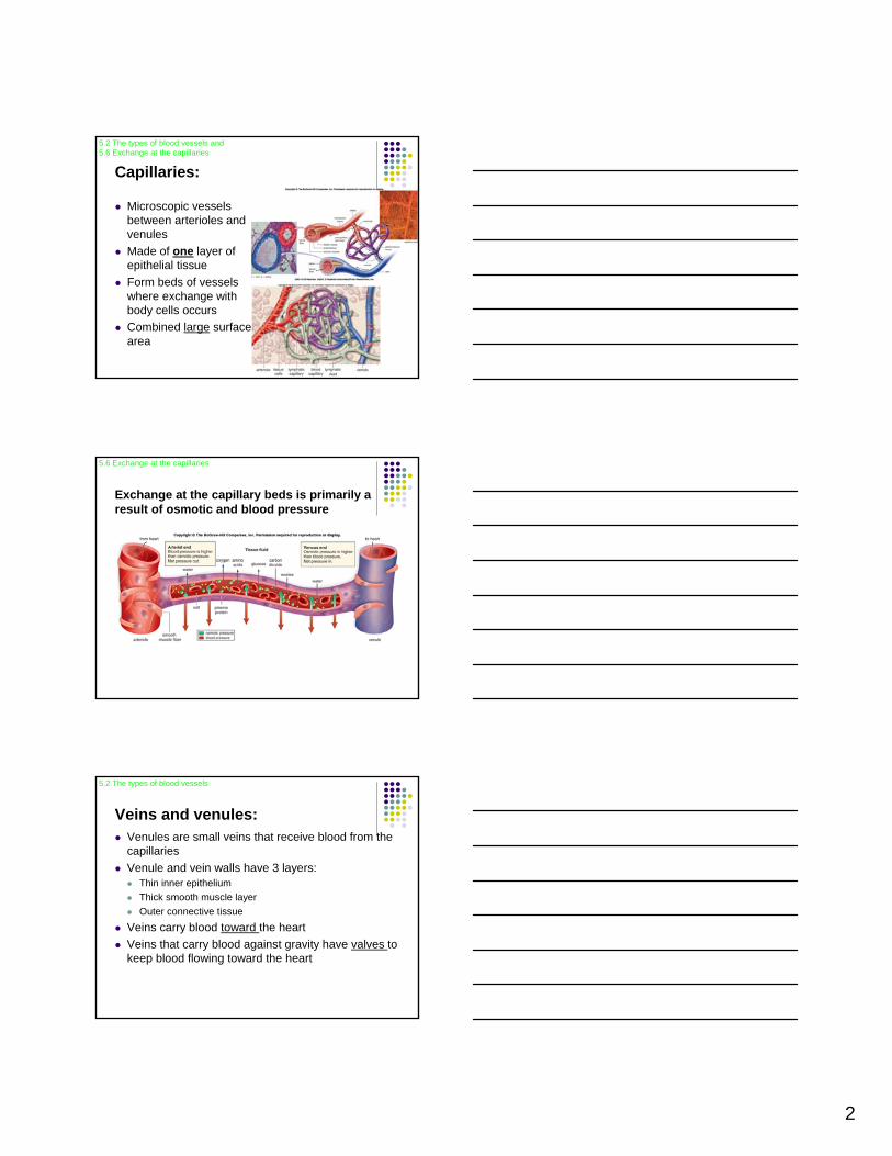
Capillaries:
Microscopic vessels between arterioles and venulesMade of one layer of epithelial tissueForm beds of vessels where exchange with body cells occurs Combined large surface area
5.2 The types of blood vessels and 5.6 Exchange at the capillaries
Exchange at the capillary beds is primarily a result of osmotic and blood pressure
5.6 Exchange at the capillaries
Veins and venules:Venules are small veins that receive blood from the capillaries Venule and vein walls have 3 layers:
Thin inner epitheliumThick smooth muscle layerOuter connective tissue
Veins carry blood toward the heartVeins that carry blood against gravity have valves to keep blood flowing toward the heart
5.2 The types of blood vessels

3
How can you tell the difference between an artery and vein?
5.2 The types of blood vessels
Anatomy of the heart
A large, muscular organ consisting of mostly cardiac tissue called the myocardiumIt is surrounded by a sac called the pericardiumConsists of two sides, right and left, separated by a septumConsists of 4 chambers: 2 atria and 2 ventricles2 sets of valves: semilunar valves and atrioventricular valves (AV valves)The valves give the resulting “lub” and “dup” sound of the heart
5.3 The heart is a double pump
External anatomy of the heart5.3 The heart is a double pump
4
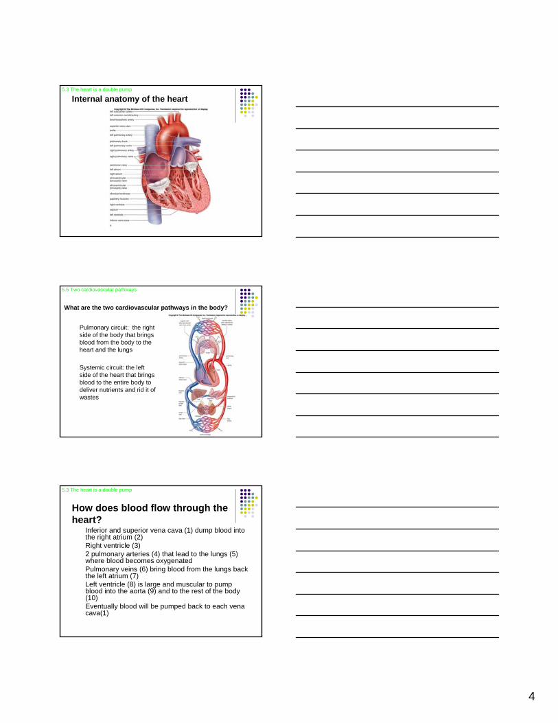
Internal anatomy of the heart5.3 The heart is a double pump
What are the two cardiovascular pathways in the body?
Pulmonary circuit: the right side of the body that brings blood from the body to the heart and the lungs
Systemic circuit: the left side of the heart that brings blood to the entire body to deliver nutrients and rid it of wastes
5.5 Two cardiovascular pathways
How does blood flow through the heart?
Inferior and superior vena cava (1) dump blood into the right atrium (2)Right ventricle (3)2 pulmonary arteries (4) that lead to the lungs (5) where blood becomes oxygenatedPulmonary veins (6) bring blood from the lungs back the left atrium (7)Left ventricle (8) is large and muscular to pump blood into the aorta (9) and to the rest of the body (10)Eventually blood will be pumped back to each vena cava(1)
5.3 The heart is a double pump

5
Visualizing blood flow through the heart5.3 The heart is a double pump
How do the structure of the vessels and heart match their functions?
The left ventricle is much more muscular than the right ventricle because it must pump blood to the entire body
The arteries are more muscular than veins to withstand the higher pressure exerted on them
The veins have a thinner wall and a larger center to store blood
5.3 The heart is a double pump
How does the heartbeat occur?
During systole the atria contract together followed by the ventricles contracting together
This is followed by diastole, a rest phase, when the chambers relax
This cardiac cycle, heartbeat, on average occurs 70 times/minute
5.3 The heart is a double pump

6
What is the cardiac cycle?
5.3 The heart is a double pump
How is the heartbeat controlled?
Internal control:The SA node in the right atrium initiates the heartbeat and causes the atria to contractThis impulse reaches the AV node, also in the right atrium, to send a signal down the AV bundle and Purkinje fibers that causes ventricular contractionThese impulses travel between gap junctions at intercalated disks
External control:heartbeat is also controlled by a cardiac center in the brain and hormones such as epinephrine and norepinephrine
5.3 The heart is a double pump
Visualizing the heartbeat5.3 The heart is a double pump

7
Visualizing the gap junctions at the intercalated disks
5.3 The heart is a double pump
What is an electrocardiogram (ECG)?
A record of the electrical changes in the heart muscle during a cardiac cycleThe atria produce an electrical current when stimulated by the SA node called the P waveThe contraction of the ventricles is the QRS complexThe recovery of the ventricles is called the T waveLooking at these electrical changes allows doctors to detect abnormalities
5.3 The heart is a double pump
How does a “normal” and abnormalECG compare?
5.3 The heart is a double pump

8
What is blood pressure?The pressure against a bloodvessel wall, usually measured in an artery in the armThe highest pressure is during blood ejection from the heart called the systolic pressureThe lowest pressure is the diastolic pressure when the ventricles relaxAverage blood pressure is recorded at about 120/80 mmHg (systolic/diastolic)Reminder: this is controlled by the arterioles
5.4 Features of the cardiovascular system
How is blood pressure categorized?
5.4 Features of the cardiovascular system
What is important about blood flow?Blood flow is under the highest pressure in the arteries but remember the thick, muscular wallsBlood flow is slower in the capillaries which is important to allow time for exchange between cellsBlood pressure is minimal in the veins and venules but blood flow increases
5.4 Features of the cardiovascular system

9
If blood pressure is so low in the veins why does the blood flow increase?
They have help:1. Skeletal muscle
contraction2. Breathing 3. Valves
5.4 Features of the cardiovascular system
The heart’s blood supply: Coronary circulation
There are small coronary arteries that supply the heart that are separate from the systemic and pulmonary pathways of the body
5.5 Two cardiovascular pathways
What is the hepatic portal system?
A system that brings blood from the digestive tract rich in amino acids and glucose to the liver
The liver synthesizes blood proteins and stores the glucose as glycogen
The liver also plays a role in purifying blood from the digestive tract
Finally, the blood will return to the heart via the inferior vena cava
5.5 Two cardiovascular pathways

10
Cardiovasscular Diseases=Most Common causes of death in U.S.A.
Disorders of the blood vessels:Hypertension/high blood pressureAtherosclerosisStrokeHeart attackAneurysm
5.7 Cardiovascular disorders
HypertensionHigh blood pressure results when blood moves through vessels at a rate higher than normal often due to arterial plaque
140/90 mmHg is considered hypertension
A silent killer because there are few symptoms
Can lead to a heart attack, stroke or kidney failure
5.7 Cardiovascular disorders
Atherosclerosis
A build up of plaque in blood vesselsPlaque that is stationary is called athrombus and an embolus when it detaches and can move to distant sitesAssociated with a stroke, heart attack and aneurysm
5.7 Cardiovascular disorders

11
Stroke
Also known as a cerebrovascular accident (CVA)
Usually occurs when a cranial artery is blocked or bursts
Part of the brain dies dues to lack of oxygen
Symptoms may occur including numbness of hands or face, difficulty speaking and inability to see in one eye
5.7 Cardiovascular disorders
Heart attack
Also known as a myocardial infarction (MI)
Part of the heart dies due to lack of oxygen
Can begin with angina pectoris, a pain that radiates down the left arm due to a blockage of a coronary artery
5.7 Cardiovascular disorders
Aneurysm
A ballooning of a blood vessel
Atherosclerosis and hypertension can weaken a vessel and cause ballooning
The most commonly affected is the abdominal artery or the arteries leading to the brain
5.7 Cardiovascular disorders

12
How are disorders of the blood vessels treated?
Dissolving blood clots: t-PA is a drug that dissolves clots
Treating clogged arteries:Bypass surgery: usually a vein from the leg is taken and used to bypass a clogged arteryStents: wire mesh cylinder inserted into a clogged artery to hold it openAngioplasty: a tube with a balloon is inserted into the clogged area and the balloon is then inflated to open the vesselA stent and angioplasty may be used in combination
5.7 Cardiovascular disorders
Disorders of the heart and its treatment
Disorders:Heart failure is when the heart no longer pumps properly
Treatments:Left ventricular assist device(LVAD)Heart transplant either natural or artificial
5.7 Cardiovascular disorders
Health Focus: The do’s and don’ts for prevention of cardiovascular disease?
Do not smokeDo not abuse drugsKeep your weight down to decrease chances of hypertension and Type II diabetesEat a healthy diet
Low in saturated and trans fatsLow in cholesterol
Know your blood cholesterol Exercise
5.7 Cardiovascular disorders