5-Andrews
10
Revista Chilena de Neurocirugía 39: 2013 28 Nanoneurosurgery: some techniques for the future Russell J. Andrews, MD 1 1 Ames Associate (Nanotechnology), National Aeronautics and Space Administration (NASA) Ames Research Center, Moffett Field, CA, USA 94035. Rev. Chil. Neurocirugía 39: 28 - 37, 2013 Abstract Nanotechniques require a rethinking of the surgeon’s tendency to want to cut nervous system tissue more precisely. However, nanotechniques offer the possibility of interacting with the nervous system at the level of the neuron and the glial cell - and even intracellularly - to restore normal nervous system function. After an introduction to the nanorealm as it applies to the nervous system, three nanotechniques are considered: (1) carbon nanotubes, nanofibers, and nanowires; (2) quantum dots and nanoparticles; (3) nanoscaffolds. Carbon nanotubes, nanofibers, and nanowires will greatly enhance the brain-machine interface through orders of magnitude improvement in our ability to record and stimulate nervous system tissue - and they will allow precise focal real-time monitoring of neurotransmitter concentrations throughout the brain. Quantum dots and other nanoparticles will allow us to label various cell types (e.g. neurons, glia, tumors), as well to label intracellularly when necessary. They are already proving to be superior contrast agents for MRI; they will also be valuable tools for identifying and destroying malignant tumors without “collateral damage” to normal brain. Nanoscaffolds possess remarkable properties to improve hemostasis during surgical procedures, but - more importantly for neurosurgery - present the opportunity for true axonal regeneration after nervous system injury. In the treatment of nervous system injury and disorders, size does indeed matter - we, as neurosurgeons, need to think and act at the level of the neuron, the axon, and the synapse. The operating microscope was the “neurosurgeon’s best friend” in the 20th century; nanotechniques will be our best friend in the 21st century! Key words: Deep brain stimulation, nanoparticles, nanoscaffolds, nanotechnology, neuromodulation, neurorepair, neurotransmit- ters, quantum dots. Introduction As neurosurgeons, we are trained to be experts at cutting tissue - especially ner- vous system tissue - increasingly smaller and more precisely. The operating micro- scope was a quantum leap in our efforts to become precise surgeons. Minimally- invasive techniques are the current trend toward reducing “collateral damage” during surgery. But consider an alternative approach: instead of cutting out glioblastomas (or ruptured spinal discs) more precisely - why not attack the tumor cells (or the ruptured disc tissue) one cell at a time? Why have any “collateral damage”; why have any injury of normal tissue? For this, we neurosurgeons need to interact with the nervous system (or any other body organ) on its own level and on its own terms. On its own level means at the level of individual neurons, glial cells, tumor cells, etc. On its own terms means speaking with the nervous system in its own language, i.e. electrically and chemically. In time - perhaps sooner than we think - we neurosurgeons will trade in our scalpels for electrical and chemical messengers. The cellular-level equivalent of the electrical cattle prod and the chemical antibiotic will persuade errant neurons to resume productive firing patterns and depressed (or elated!) chemical brain circuits to adopt a steady course. Gliomas will be targeted by cellular messengers one malignant astrocyte at a time, with virtually no side effects on the surrounding normal tissues. It was scarcely 50 years ago - in late December, 1959 - that the physicist Richard Feynman gave his famous talk entitled, “There’s Plenty of Room at the Bottom”: “I want to talk about the problem of manipulating and controlling things on a small scale… It is a staggeringly small world that is below. In the year 2000, when they look back at this age, they will wonder why it was not until the year 1960 that anyone began seriously to move in this direction.”
-
Upload
rakesh-kumar-mishra -
Category
Documents
-
view
2 -
download
1
description
neuro
Transcript of 5-Andrews
Revista Chilena de Neurocirugía 39: 2013
28
Nanoneurosurgery: some techniques for the future
Russell J. Andrews, MD1
1 Ames Associate (Nanotechnology), National Aeronautics and Space Administration (NASA) Ames Research Center, Moffett Field, CA, USA 94035.
Rev. Chil. Neurocirugía 39: 28 - 37, 2013
Abstract
Nanotechniques require a rethinking of the surgeon’s tendency to want to cut nervous system tissue more precisely. However, nanotechniques offer the possibility of interacting with the nervous system at the level of the neuron and the glial cell - and even intracellularly - to restore normal nervous system function. After an introduction to the nanorealm as it applies to the nervous system, three nanotechniques are considered: (1) carbon nanotubes, nanofibers, and nanowires; (2) quantum dots and nanoparticles; (3) nanoscaffolds. Carbon nanotubes, nanofibers, and nanowires will greatly enhance the brain-machine interface through orders of magnitude improvement in our ability to record and stimulate nervous system tissue - and they will allow precise focal real-time monitoring of neurotransmitter concentrations throughout the brain. Quantum dots and other nanoparticles will allow us to label various cell types (e.g. neurons, glia, tumors), as well to label intracellularly when necessary. They are already proving to be superior contrast agents for MRI; they will also be valuable tools for identifying and destroying malignant tumors without “collateral damage” to normal brain. Nanoscaffolds possess remarkable properties to improve hemostasis during surgical procedures, but - more importantly for neurosurgery - present the opportunity for true axonal regeneration after nervous system injury. In the treatment of nervous system injury and disorders, size does indeed matter - we, as neurosurgeons, need to think and act at the level of the neuron, the axon, and the synapse. The operating microscope was the “neurosurgeon’s best friend” in the 20th century; nanotechniques will be our best friend in the 21st century!
Key words: Deep brain stimulation, nanoparticles, nanoscaffolds, nanotechnology, neuromodulation, neurorepair, neurotransmit-ters, quantum dots.
Introduction
As neurosurgeons, we are trained to be experts at cutting tissue - especially ner-vous system tissue - increasingly smaller and more precisely. The operating micro-scope was a quantum leap in our efforts to become precise surgeons. Minimally-invasive techniques are the current trend toward reducing “collateral damage” during surgery.But consider an alternative approach: instead of cutting out glioblastomas (or ruptured spinal discs) more precisely - why not attack the tumor cells (or the ruptured disc tissue) one cell at a time? Why have any “collateral damage”; why have any injury of normal tissue? For this,
we neurosurgeons need to interact with the nervous system (or any other body organ) on its own level and on its own terms. On its own level means at the level of individual neurons, glial cells, tumor cells, etc. On its own terms means speaking with the nervous system in its own language, i.e. electrically and chemically.In time - perhaps sooner than we think - we neurosurgeons will trade in our scalpels for electrical and chemical messengers. The cellular-level equivalent of the electrical cattle prod and the chemical antibiotic will persuade errant neurons to resume productive firing patterns and depressed (or elated!) chemical brain circuits to adopt a
steady course. Gliomas will be targeted by cellular messengers one malignant astrocyte at a time, with virtually no side effects on the surrounding normal tissues.It was scarcely 50 years ago - in late December, 1959 - that the physicist Richard Feynman gave his famous talk entitled, “There’s Plenty of Room at the Bottom”:“I want to talk about the problem of manipulating and controlling things on a small scale… It is a staggeringly small world that is below. In the year 2000, when they look back at this age, they will wonder why it was not until the year 1960 that anyone began seriously to move in this direction.”
29
Revista Chilena de Neurocirugía 39: 2013
More recently, Richard Smalley (who won the `996 Nobel Prize in Chemistry for C60 “buckeyballs”) said on June 22, 1999:“Twenty years ago, without this crude chemotherapy, I would already be dead. But 20 years from now, nanoscale missiles will target cancer cells in the human body and leave everything else blissfully alone. I may not live to see it. But I am confident it will happen.”Unfortunately Richard Smalley died on October 28, 2005, of non-Hodgkin’s lymphoma. However, his prescient prediction regarding the ability of “nanoscale missiles” to attack cancer cells selectively is likely right on track - nanoparticles are already targeting tumor cells, and by the year 2020 should fulfill Smalley’s goal.Nanotechnology in one sense means working at the nanolevel (10-9 meter, i.e. 1/1,000 of a micron or roughly 1/10,000 the size of a neuron or red blood cell). But as I noted above, construction
one atom or molecule at a time rather than cutting tissue smaller gets at the core of working at the nano level. As Norio Taniguchi (who coined the word, “nanotechnology”) said in 1974:“Nano-technology mainly consists of the processing of, separation, consolidation, and deformation of materials by one atom or one molecule.”In the pages that follow, I will provide a brief overview of three nanotechniques that are almost certainly to have a major impact on neurosurgery in the coming decade. Let’s hope that we as neurosurgeons will envisage, embrace, and create the future - as our pioneering predecessors in neurosurgery have done so often before us.Figure 1 provides a graphic illustration of the macro- to micro- to nano-realm. Most commonly, nanoscale objects are considered to be in the 1 to 100 nanometer (nm) size range. All of the nano materials to be reviewed are fabricated from “building blocks” in this nanoscale
of 1 to 100 nm, although frequently the resulting structures are somewhat larger (but generally within the size of a single cell, i.e. 10 microns or less).
Carbon Nanotubes, Nanofibers, and Nanowires
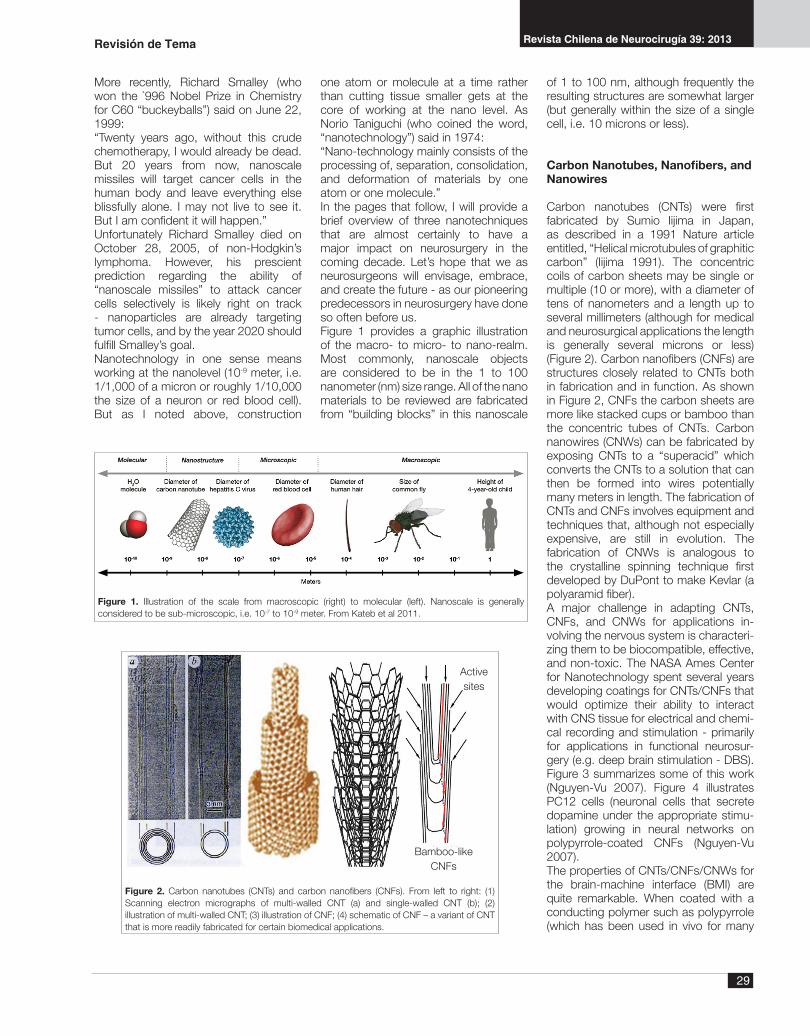
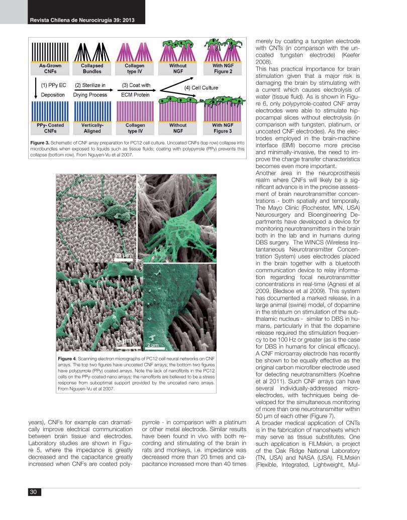
Carbon nanotubes (CNTs) were first fabricated by Sumio Iijima in Japan, as described in a 1991 Nature article entitled, “Helical microtubules of graphitic carbon” (Iijima 1991). The concentric coils of carbon sheets may be single or multiple (10 or more), with a diameter of tens of nanometers and a length up to several millimeters (although for medical and neurosurgical applications the length is generally several microns or less) (Figure 2). Carbon nanofibers (CNFs) are structures closely related to CNTs both in fabrication and in function. As shown in Figure 2, CNFs the carbon sheets are more like stacked cups or bamboo than the concentric tubes of CNTs. Carbon nanowires (CNWs) can be fabricated by exposing CNTs to a “superacid” which converts the CNTs to a solution that can then be formed into wires potentially many meters in length. The fabrication of CNTs and CNFs involves equipment and techniques that, although not especially expensive, are still in evolution. The fabrication of CNWs is analogous to the crystalline spinning technique first developed by DuPont to make Kevlar (a polyaramid fiber).A major challenge in adapting CNTs, CNFs, and CNWs for applications in-volving the nervous system is characteri-zing them to be biocompatible, effective, and non-toxic. The NASA Ames Center for Nanotechnology spent several years developing coatings for CNTs/CNFs that would optimize their ability to interact with CNS tissue for electrical and chemi-cal recording and stimulation - primarily for applications in functional neurosur-gery (e.g. deep brain stimulation - DBS). Figure 3 summarizes some of this work (Nguyen-Vu 2007). Figure 4 illustrates PC12 cells (neuronal cells that secrete dopamine under the appropriate stimu-lation) growing in neural networks on polypyrrole-coated CNFs (Nguyen-Vu 2007).The properties of CNTs/CNFs/CNWs for the brain-machine interface (BMI) are quite remarkable. When coated with a conducting polymer such as polypyrrole (which has been used in vivo for many
Figure 1. Illustration of the scale from macroscopic (right) to molecular (left). Nanoscale is generally considered to be sub-microscopic, i.e. 10-7 to 10-9 meter. From Kateb et al 2011.
Figure 2. Carbon nanotubes (CNTs) and carbon nanofibers (CNFs). From left to right: (1) Scanning electron micrographs of multi-walled CNT (a) and single-walled CNT (b); (2) illustration of multi-walled CNT; (3) illustration of CNF; (4) schematic of CNF – a variant of CNT that is more readily fabricated for certain biomedical applications.
Bamboo-likeCNFs
Activesites
Revisión de Tema
Revista Chilena de Neurocirugía 39: 2013
30
years), CNFs for example can dramati-cally improve electrical communication between brain tissue and electrodes. Laboratory studies are shown in Figu-re 5, where the impedance is greatly decreased and the capacitance greatly increased when CNFs are coated poly-
pyrrole - in comparison with a platinum or other metal electrode. Similar results have been found in vivo with both re-cording and stimulating of the brain in rats and monkeys, i.e. impedance was decreased more than 20 times and ca-pacitance increased more than 40 times
merely by coating a tungsten electrode with CNTs (in comparison with the un-coated tungsten electrode) (Keefer 2008).This has practical importance for brain stimulation given that a major risk is damaging the brain by stimulating with a current which causes electrolysis of water (tissue fluid). As is shown in Figu-re 6, only polypyrrole-coated CNF array electrodes were able to stimulate hip-pocampal slices without electrolysis (in comparison with tungsten, platinum, or uncoated CNF electrodes). As the elec-trodes employed in the brain-machine interface (BMI) become more precise and minimally-invasive, the need to im-prove the charge transfer characteristics becomes even more important.Another area in the neuroprosthesis realm where CNFs will likely be a sig-nificant advance is in the precise assess-ment of brain neurotransmitter concen-trations - both spatially and temporally. The Mayo Clinic (Rochester, MN, USA) Neurosurgery and Bioengineering De-partments have developed a device for monitoring neurotransmitters in the brain both in the lab and in humans during DBS surgery. The WINCS (Wireless Ins-tantaneous Neurotransmitter Concen-tration System) uses electrodes placed in the brain together with a bluetooth communication device to relay informa-tion regarding focal neurotransmitter concentrations in real-time (Agnesi et al 2009, Bledsoe et al 2009). This system has documented a marked release, in a large animal (swine) model, of dopamine in the striatum on stimulation of the sub-thalamic nucleus - similar to DBS in hu-mans, particularly in that the dopamine release required the stimulation frequen-cy to be 100 Hz or greater (as is the case for DBS in humans for clinical efficacy). A CNF microarray electrode has recently be shown to be equally effective as the original carbon microfiber electrode used for detecting neurotransmitters (Koehne et al 2011). Such CNF arrays can have several individually-addressed micro-electrodes, with techniques being de-veloped for the simultaneous monitoring of more than one neurotransmitter within 50 µm of each other (Figure 7). A broader medical application of CNTs is in the fabrication of nanosheets which may serve as tissue substitutes. One such application is FILMskin, a project of the Oak Ridge National Laboratory (TN, USA) and NASA (USA). FILMskin (Flexible, Integrated, Lightweight, Mul-
Figure 3. Schematic of CNF array preparation for PC12 cell culture. Uncoated CNFs (top row) collapse into microbundles when exposed to liquids such as tissue fluids; coating with polypyrrole (PPy) prevents this collapse (bottom row). From Nguyen-Vu et al 2007.
Figure 4. Scanning electron micrographs of PC12 cell neural networks on CNF arrays. The top two figures have uncoated CNF arrays; the bottom two figures have polypyrrole (PPy) coated arrays. Note the lack of nanofibrils in the PC12 cells on the PPy-coated nano arrays; the nanofibrils are believed to be a stress response from suboptimal support provided by the uncoated nano arrays. From Nguyen-Vu et al 2007.

31
Revista Chilena de Neurocirugía 39: 2013
Figure 5. Illustrations of the dramatic increase in capacitance and decrease in impedance provided by PPy-coated CNF arrays in comparison with noble metal (e.g. platinum) electrodes. Courtesy of NASA Ames Nanotechnology.
121086420
60
50
40
30
20
10
0Max
Res
pons
e Vo
ltage
(mV)
(b)
Figure 6. Improved charge transfer with PPy-coated CNF arrays in comparison with other microelectrodes (see text). Filled diamonds: PPy-coated CNF array (short duration field potential). Multiplication signs: PPy-coated CNF array (long duration field potential). Filled circles: tungsten electrodes. Filled squares: uncoated CNF array. Filled triangles: platinum microelectrode array.
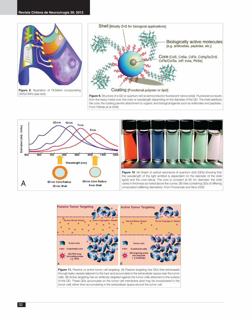
tifunctional skin) is being developed for prosthetic limbs to be able to provide the sensory information of normal skin. The high thermal conductivity of CNTs (as well as other physical properties of CNTs such as remarkable strength and elasticity) will allow artificial "skin" to be manufactured with characteristics simi-lar to normal human skin. A start-up company (Nanocomp Technologies, Inc, Concord, NH, USA) has already begun producing large sheets composed of CNTs; such fabrications may be incorpo-rated into "bionic" artificial skin, with an advantage that CNTs in such bulk forms are not subject to the toxicity concerns of free CNTs/CNFs (Figure 8).
Quantum dots and nanoparticles
Quantum dots (QDs) are particles usually less than 100 nm in diameter which are composed of a heavy metal core (typically cadmium selenium) and an unreactive metallic shell (typically zinc sulfide) (Figure 9). QDs have the interesting and useful property of light emission that depends on the size (diameter) of the QD - the color depends on the size of the QD (Figure 10).The light is particularly bright and stable over time - useful for multiplex labeling (e.g. by attaching different antibodies to different QDs, “color-coding” can be achieved).QDs can be used to track drugs (e.g. cancer therapeutic agents), label cellu-lar and subcellular structures (e.g. neu-rotransmitter receptors), and target tu-mor cells. Tumor (e.g. malignant glioma)
Revisión de Tema
Figure 7. Schematic of needle CNF array currently being fabricated for NASA Ames Nanotechnology and the Mayo Clinic. There are six individually-addressed CNF array “pads” (20 µm diameter) on an microelectrode approximately 0.1 mm in diameter. This electrode can stimulate electrically, record electrically, and record one or more neurotransmitters (e.g. dopamine, serotonin) simultaneously. Courtesy of NASA Ames Nanotechnology.
Revista Chilena de Neurocirugía 39: 2013
32
Figure 8. Illustration of FILMskin incorporating CNTs/CNFs (see text). Figure 9. Structure of a QD or quantum dot (a semiconductor fluorescent nanocrystal). Fluorescence results
from the heavy metal core, the color or wavelength depending on the diameter of the QD. The shell stabilizes the core; the coating permits attachment to organic and biological ligands such as antibodies and peptides. From Pathak et al 2006.
Figure 10. (A) Graph of optical resonance of quantum dots (QDs) showing that the wavelength of the light emitted is dependent on the diameter of the shell (gold) and the core (silica). The core is constant at 60 nm diameter; the shell varies in thickness as noted above the curves. (B) Vials containing QDs of differing composition (differing diameters). From Provenzale and Silva 2009.
Figure 11. Passive vs active tumor cell targeting. (A) Passive targeting has QDs that extravasate through leaky vessels adjacent to the tujor and accumulate in the extracellular space near the tumor cells. (B) Active targeting has an antibody targeted against the tumor cells attached to the surface of the QD. These QDs accumulate on the tumor cell membrane (and may be incorporated in the tumor cell) rather than accumulating in the extracellular space around the tumor cell.

33
Revista Chilena de Neurocirugía 39: 2013
Figure 12. A: Graph of glioblastoma multiforme tumor enhancement comparing ferumoxytol with gadolinium (Gd) and T2-weighted signal abnormalities. B-H: MRI scans - B, Gd T1-weighted; C, T2-weighted; D-H postferumoxytol T1-weighted: D, 4-6 h; E, 6-20 h; F, 24-28 h; G, 48-52 h; H, > 72 h. From Neuwelt et al 2007.
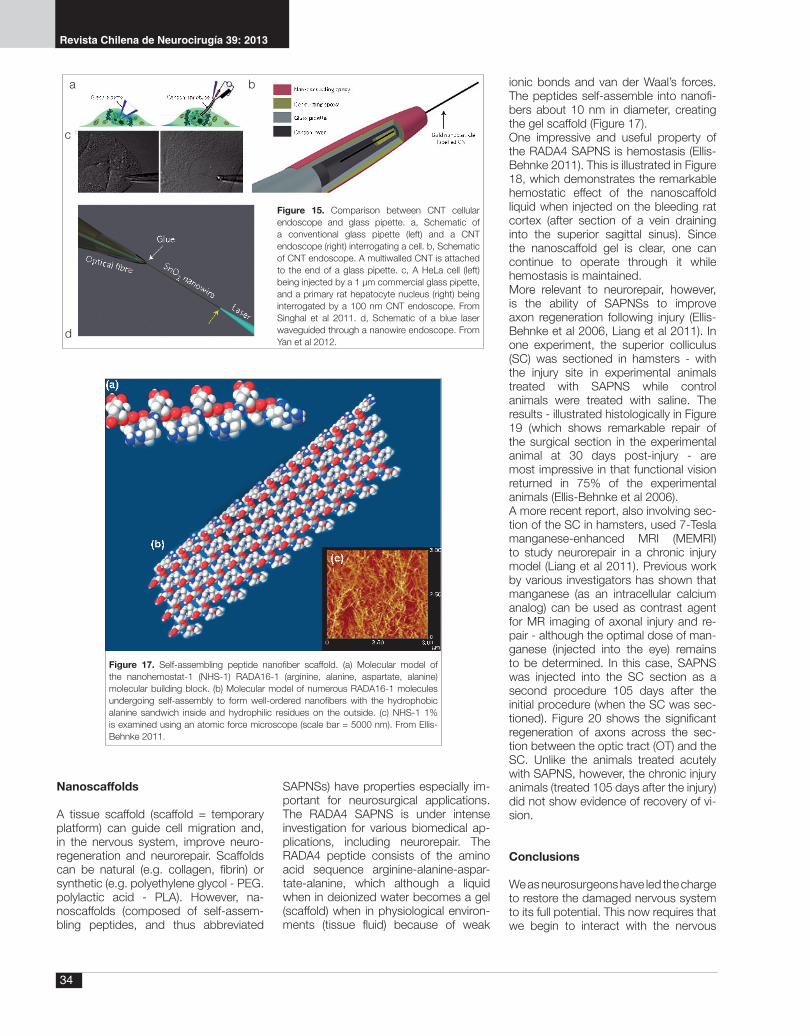
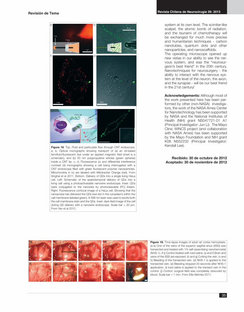
targeting can be accomplished either passively (depending upon the leakiness or lack of the blood-brain barrier in the tumor angiogenic vessels) or actively using antibodies to the tumor cells that are attached to the surface of the QDs (Figure 11).The potential uses of QDs in neuroi-maging - both for diagnostic/therapeutic purposes as well as for improved un-derstanding of cellular/subcellular pro-cesses in vivo - are expanding rapidly.Another nano-sized particle with neu-rosurgical application is the iron oxide nanoparticle. Paramagnetic iron oxide nanoparticles, when used as an MRI contrast agent, can increase signal in-tensity on T1-weighted images for much longer periods than gadolinium (Figure 12) - and with delayed uptake that can be helpful for delineating tumor vas-cularity (Figure 13), as well as the pos-sibility of detecting areas of malignancy not seen on gadolinium-enhanced MRI. Such paramagnetic/superparamagnetic nanoparticles, especially when com-bined with active targeting techniques as illustrated in Figure 11, can enhance tumor identification greatly on MRI (Figu-re 14).Nanowires can be combined with QDs to form a cellular-level endoscope (Singhal et al 2011, Yan et al 2012). In one such endoscope, a CNT is bonded to a fine glass pipette (Figure 15 A), allowing QDs or other nano-sized particles to be injected into or aspirated out of the cell. In another such endoscope, a SnO2 (tin dioxide) nanowire is bonded to a fine optical fiber through which a laser light can be passed (Figure 15 B).The CNT or nanowire is more uniform and also more flexible than the glass pipette or optical fiber, allowing either the CNT or the nanowire to be passed through the cell membrane without injury to the cell. Figure 16 A shows a CNT endoscope injecting 50 nm polypropylene spheres (green) into a cell; Figure 16 B shows a QD which has been delivered into the cell cytoplasm by a nanowire endoscope. Interestingly, the QDs can be detached accurately within the cell much as a neurointerventionalist can detach a stent or balloon in a cerebral artery - in the nanoendoscope situation, a photocleavable (PC) linker detached the QD when light of a certain wavelength is passed through the endoscope. The laser endoscope can also be used to interrogate the intracellular contents by using light of differing frequencies.
Revisión de Tema
Figure 13. Time-of-flight angiography in a patient with a left frontal glioblastoma multiforme. A, without contrast; B, 15 min after Gd infusion; C, 15 min after ferumoxytol infusion. Note the improved visualization of vessels in C compared with B, but the tumor has not yet begun to enhance.
Figure 14. Axial MRI scans of a 9L glioma xenograft grown in the flank of a mouse. Left: modest uptake of nontargeted nanoparticles within the tumor. Right: marked uptake of tumor-targeted (chlorotxin-conjugated) nanoparticles with the tumor.
Revista Chilena de Neurocirugía 39: 2013
34
Figure 17. Self-assembling peptide nanofiber scaffold. (a) Molecular model of the nanohemostat-1 (NHS-1) RADA16-1 (arginine, alanine, aspartate, alanine) molecular building block. (b) Molecular model of numerous RADA16-1 molecules undergoing self-assembly to form well-ordered nanofibers with the hydrophobic alanine sandwich inside and hydrophilic residues on the outside. (c) NHS-1 1% is examined using an atomic force microscope (scale bar = 5000 nm). From Ellis-Behnke 2011.
Nanoscaffolds
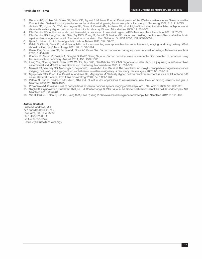
A tissue scaffold (scaffold = temporary platform) can guide cell migration and, in the nervous system, improve neuro-regeneration and neurorepair. Scaffolds can be natural (e.g. collagen, fibrin) or synthetic (e.g. polyethylene glycol - PEG. polylactic acid - PLA). However, na-noscaffolds (composed of self-assem-bling peptides, and thus abbreviated
SAPNSs) have properties especially im-portant for neurosurgical applications. The RADA4 SAPNS is under intense investigation for various biomedical ap-plications, including neurorepair. The RADA4 peptide consists of the amino acid sequence arginine-alanine-aspar-tate-alanine, which although a liquid when in deionized water becomes a gel (scaffold) when in physiological environ-ments (tissue fluid) because of weak
ionic bonds and van der Waal’s forces. The peptides self-assemble into nanofi-bers about 10 nm in diameter, creating the gel scaffold (Figure 17).One impressive and useful property of the RADA4 SAPNS is hemostasis (Ellis-Behnke 2011). This is illustrated in Figure 18, which demonstrates the remarkable hemostatic effect of the nanoscaffold liquid when injected on the bleeding rat cortex (after section of a vein draining into the superior sagittal sinus). Since the nanoscaffold gel is clear, one can continue to operate through it while hemostasis is maintained.More relevant to neurorepair, however, is the ability of SAPNSs to improve axon regeneration following injury (Ellis-Behnke et al 2006, Liang et al 2011). In one experiment, the superior colliculus (SC) was sectioned in hamsters - with the injury site in experimental animals treated with SAPNS while control animals were treated with saline. The results - illustrated histologically in Figure 19 (which shows remarkable repair of the surgical section in the experimental animal at 30 days post-injury - are most impressive in that functional vision returned in 75% of the experimental animals (Ellis-Behnke et al 2006).A more recent report, also involving sec-tion of the SC in hamsters, used 7-Tesla manganese-enhanced MRI (MEMRI) to study neurorepair in a chronic injury model (Liang et al 2011). Previous work by various investigators has shown that manganese (as an intracellular calcium analog) can be used as contrast agent for MR imaging of axonal injury and re-pair - although the optimal dose of man-ganese (injected into the eye) remains to be determined. In this case, SAPNS was injected into the SC section as a second procedure 105 days after the initial procedure (when the SC was sec-tioned). Figure 20 shows the significant regeneration of axons across the sec-tion between the optic tract (OT) and the SC. Unlike the animals treated acutely with SAPNS, however, the chronic injury animals (treated 105 days after the injury) did not show evidence of recovery of vi-sion.
Conclusions
We as neurosurgeons have led the charge to restore the damaged nervous system to its full potential. This now requires that we begin to interact with the nervous
Figure 15. Comparison between CNT cellular endoscope and glass pipette. a, Schematic of a conventional glass pipette (left) and a CNT endoscope (right) interrogating a cell. b, Schematic of CNT endoscope. A multiwalled CNT is attached to the end of a glass pipette. c, A HeLa cell (left) being injected by a 1 µm commercial glass pipette, and a primary rat hepatocyte nucleus (right) being interrogated by a 100 nm CNT endoscope. From Singhal et al 2011. d, Schematic of a blue laser waveguided through a nanowire endoscope. From Yan et al 2012.
a b
c
d
35
Revista Chilena de Neurocirugía 39: 2013
system at its own level. The scimitar-like scalpel, the atomic bomb of radiation, and the tsunami of chemotherapy will be exchanged for much more precise and humanitarian techniques - carbon nanotubes, quantum dots and other nanoparticles, and nanoscaffolds.The operating microscope opened up new vistas in our ability to see the ner-vous system, and was the “neurosur-geon’s best friend” in the 20th century. Nanotechniques for neurosurgery - the ability to interact with the nervous sys-tem at the level of the neuron, the axon, and the synapse - will be our best friend in the 21st century!
Acknowledgements: Although most of the work presented here has been per-formed by other (non-NASA) investiga-tors, the work of the NASA Ames Center for Nanotechnology has been supported by NASA and the National Institutes of Health (NIH) grant NS047721-01 A1 (Principal Investigator: Jun Li). The Mayo Clinic WINCS project (and collaboration with NASA Ames) has been supported by the Mayo Foundation and NIH grant K08 NS52232 (Principal Investigator: Kendall Lee).
Recibido: 30 de octubre de 2012Aceptado: 30 de noviembre de 2012
Revisión de Tema
Figure 16. Top. Fluid and particulate flow through CNT endoscope. a, b, Optical micrographs showing transport of (a) an oil-based ferrofluid-fluorescent dye under an applied magnetic field (inset is a schematic), and (b) 50 nm polypropylene articles (green spheres) inside a CNT tip. c, d, Fluorescence (c) and differential interference contrast (d) micrographs showing a cell being interrogated with a CNT endoscope filled with green fluorescent polymer nanoparticles. Mitochondria in (c) are labeled with Mitotracker Orange (red). From Singhal et al 2011. Bottom. Delivery of QDs into a single living HeLa cell. Left: Schematic of the spatiotemporal delivery of QDs into a living cell using a photoactivatable nanowire endoscope. Inset: QDs were conjugated to the nanowire by photocleavable (PC) linkers. Right: Fluorescence confocal image of a HeLa cell. Showing that the nanoprobe has delivered the QDs (red dot in the cytoplasm) within the cell membrane (labeled green). A 488 nm laser was used to excite both the cell membrane stain and the QDs. Inset: dark-field image of the cell during QD delivery with a nanowire endoscope. Scale bar = 20 µm. From Yan et al 2012.
a b
c d
Figure 18. Time-lapse images of adult rat cortex hemostasis. (a-e) One of the veins of the superior sagittal sinus (SSS) was transected and treated with 1% self-assembling nanohemostat (NHS-1). (f-j) Control treated with iced saline. (a and f) Brain and veins of the SSS are exposed. (b and g) Cutting the vein. (c and h) Bleeding of the transected vein. (d) NHS-1 is applied to the transected vein. (e) Bleeding stopped 20 seconds after NHS-1 application. (i) Iced saline is applied to the transect vein in the control. (j) Control: surgical field was completely obscured by blood. Scale bar = 1 mm. From Ellis-Behnke 2011.

Revista Chilena de Neurocirugía 39: 2013
36
Figure 20. MRI scans of hamsters (A) Axial T1W image 48 hours after injection of MnCl2 in the right eye. The OT is visible along with the projection into the SC (arrowhead). (B) 90 days after BSC cut. Clear MnCl2 accumulation proximal to the transection site (arrow). (C) After SAPNS treatment, the OT shows reconnection to the SC (arrow). (D) Enlarged area of A with the OT and the SC labeled (arrows). (E) Enlarged area of B where the contrast of the Mn signal is clearly visible. The black arrows show the gap between the OT and the SC. (F) Enlarged area of C where the small white arrows show the areas of reconnection of the OT and the SC. BSC brachium of the superior colliculus, OT optic tract, SC superior colliculus, SAPNS self-assembling peptide nanofiber scaffold. From Liang et al 2011.
References
1. Agnesi F, Tye SJ, Bledsoe JM, Griessnauer CJ, Kimble CJ, Sieck GC, et al. Wireless Instantaneous Neurotransmitter Concentration System-based amperometric detection of dopamine, adenosine, and glutamate for intraoperative neurochemical monitoring. J Neurosurg 2009; 111: 701-711.
Figure 19. Self-assembling peptide nanofiber scaffold (SAPNS) in repair of optic tract transection in young hamsters. (a) Dorsel view reconstruction of the hamster brain with cortex removed. Rostral - left; caudal - right. Blue line: location of optic tract transection made at P2 in the superior colliculus (SC) of the midbrain. Pretectal area (PT), lateral posterior nucleus (LP), medial geniculate body (MGB), lateral geniculate body (LGB), inferior colliculus (IC). (b) Schematic of a parasagittal section of the midbrain, with the position and depth of the scalpel cut indicated in the SC. Animals received an injection into the cut of 10 µl of saline in the controls or 10 µl of 1% SAPNS. The dark-field composite photos are parasagittal sections from animals 1, 30, and 60 days after lesion and treatment. Arrows indicate the path and extent of knife cut. Animals killed at one day after lesion and treatment: saline contral animal (c) and SAPNS treatment animal (d). The treated animal (d) has a very small gap, and the surface of the tissue has already started to reconnect both sides. (e and f) 30 day post-lesion animals; (e) is a saline control, and (f) a treated animal. Note the large gap in (e). In the SAPNS treated animal (f), the gap is completely gone, and tissue has reconnected across the injury site. (g) 60 day post-SAPNS-treated animal. Scale bars: (b) = 500 µm; (c-g) = 100 µm. From Ellis-Behnke et al 2006.
37
Revista Chilena de Neurocirugía 39: 2013
2. Bledsoe JM, Kimble CJ, Covey DP, Blaha CD, Agnesi F, Mohseni P, et al. Development of the Wireless Instantaneous Neurotransmitter Concentration System for intraoperative neurochemical monitoring using fast-scan cyclic voltammetry. J Neurosurg 2009; 111: 712-723.
3. de Asis ED, Nguyen-Vu TDB, Arumugam PU, Chen H, Cassell AM, Andrews RJ, et al. High efficient electrical stimulation of hippocampal slices with vertically aligned carbon nanofiber microbrush array. Biomed Microdevices 2009; 11: 801-808.
4. Ellis-Behnke RG. At the nanoscale: nanohemostat, a new class of hemostatic agent. WIREs Nanomed Nanobiotechnol 2011; 3: 70-79.5. Ellis-Behnke RG, Liang Y-X, You S-W, Tay DKC, Zhang S, So K-F, Schneider GE. Nano neuro knitting: peptide nanofiber scaffold for brain
repair and axon regeneration with functional return of vision. Proc Natl Acad Sci USA 2006; 103: 5054-5059.6. Iijima S. Helical microtubules of graphitic carbon. Nature 1991; 354: 56-57.7. Kateb B, Chiu K, Black KL, et al. Nanoplatforms for constructing new approaches to cancer treatment, imaging, and drug delivery: What
should be the policy? NeuroImage 2011; 54: S106-S124.8. Keefer EW, Botterman BR, Romero MI, Rossi AF, Gross GW. Carbon nanotube coating improves neuronal recordings. Nature Nanotechnol
2008; 3: 434-439.9. Koehne JE, Marsh M, Boakye A, Douglas B, Kin IY, Chang SY, et al. Carbon nanofiber array for electrochemical detection of dopamine using
fast scan cyclic voltammetry. Analyst 2011; 136: 1802-1805.10. Liang Y-X, Cheung SWH, Chan KCW, Wu EX, Tay DKC, Ellis-Behnke RG. CNS Regeneration after chronic injury using a self-assembled
nanomaterial and MEMRI for real-time in vivo monitoring. Nanomedicine 2011; 7: 351-359.11. Neuwelt EA, Varallyay CG, Manninger S, Solymosi D, Haluska M, Hunt MA, et al. The potential of ferumoxytol nanoparticle magnetic resonance
imaging, perfusion, and angiography in central nervous system malignancy: a pilot study. Neurosurgery 2007; 60: 601-612.12. Nguyen-Vu TDB, Chen Hua, Cassell A, Andrews RJ, Meyyappan M. Vertically aligned carbon nanofiber architecture as a multifunctional 3-D
neural electrical interface. IEEE Trans Biomed Engr 2007; 54: 1121-1128.13. Pathak S, Cao E, Davidson MC, Jin S, Silva GA. Quantum dot applications to neuroscience: new tools for probing neurons and glia. J
Neurosci 2006; 26: 1893-1895.14. Provenzale JM, Silva GA. Uses of nanoparticles for central nervous system imaging and therapy. Am J Neuroradiol 2009; 30: 1293-301.15. Singhal R, Orynbayeva Z, Sundaram RVK, Niu JJ, Bhattacharyya S, Vitol EA, et al. Multifunctional carbon-nanotube cellular endoscopes. Nat
Nanotech 2011; 6: 57-64.16. Yan R, Park J-H, Choi Y, Heo C-J, Yang S-M, Lee LP, Yang P. Nanowire-based single-cell endoscopy. Nat Nanotech 2012; 7: 191-196.
Author Contact:Russell J. Andrews, MD777 Knowles Drive, Suite 9Los Gatos, CA, USA 95032Ph: 1-408-871-0811Fx: 1-408-353-0275E-mail: <[email protected]>
Revisión de Tema