5. amyloidosis dr. sinhasan, mdzah
20
-
Upload
kciapm -
Category
Health & Medicine
-
view
106 -
download
2
Transcript of 5. amyloidosis dr. sinhasan, mdzah


Amyloid= ‘starch-like’ (Amylum—Latin)
Virchow in mid 19th century
Amyloids: Aggregations of un-degraded, pathologic
protein fibrils because of misfolding mechanisms.
Amyloidosis: a group of diseases.
Background:
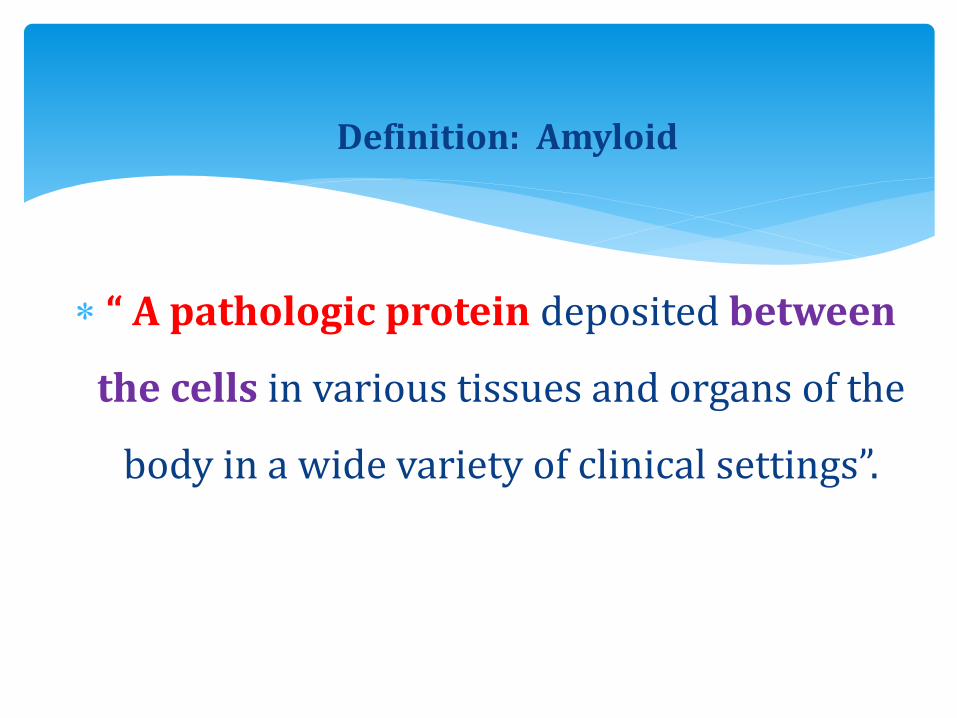
Definition: Amyloid
“ A pathologic protein deposited between
the cells in various tissues and organs of the
body in a wide variety of clinical settings”.

Non-branching fibres
Indefinite length
Diameter of 7.5 –10 nm.
Cross-Beta-pleated sheet conformation:
X-ray crystallography
Infra-red spectroscopy
Physical nature of Amyloid:

95%--consists of fibril proteins.
5%--other glycoproteins
Three most important forms:
1. AL (amyloid light chain): from plasma cells,
2. AA (amyloid associated): from liver,
3. A-Beta amyloid (seen in cerebral lesions of Alzheimer's
disease)
Chemical nature:

SYSTEMIC:
Multiple myeloma—AL
Reactive systemic am—AA
Hemodialysis associated am—Ab2 microglobulins
Hereditary am—AA
Familial Mediterranean fever—AA
Familial amyloidotrophic neuropathy—AATR
Systemic Senile am—ATTR
Classification:

LOCALIZED AMYLOIDOSIS:
Alzheimer's disease –A-beta
Medullary carcinoma –A cal
Islet of Langerhans: AIAPP
Isolated atrial amyloidosis—AANF
Prion disease –PrPsc
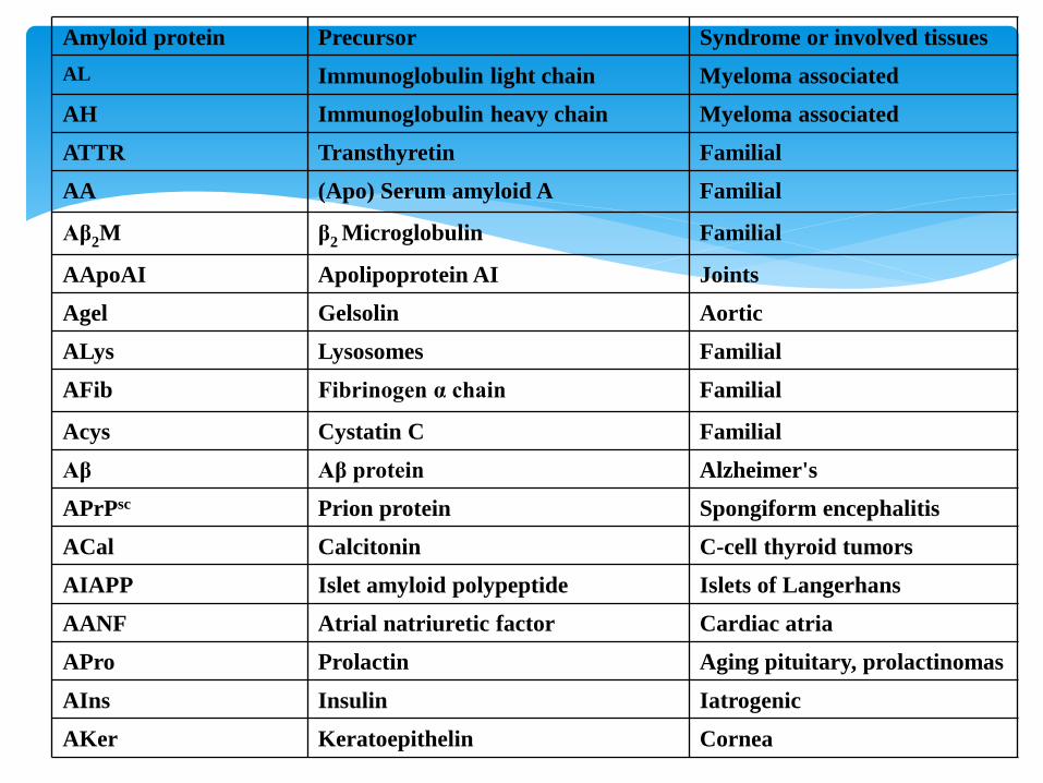
Amyloid protein Precursor Syndrome or involved tissues
AL Immunoglobulin light chain Myeloma associated
AH Immunoglobulin heavy chain Myeloma associated
ATTR Transthyretin Familial
AA (Apo) Serum amyloid A Familial
Aβ2M β2 Microglobulin Familial
AApoAI Apolipoprotein AI Joints
Agel Gelsolin Aortic
ALys Lysosomes Familial
AFib Fibrinogen α chain Familial
Acys Cystatin C Familial
Aβ Aβ protein Alzheimer's
APrPsc Prion protein Spongiform encephalitis
ACal Calcitonin C-cell thyroid tumors
AIAPP Islet amyloid polypeptide Islets of Langerhans
AANF Atrial natriuretic factor Cardiac atria
APro Prolactin Aging pituitary, prolactinomas
AIns Insulin Iatrogenic
AKer Keratoepithelin Cornea


Normal proteins—when produced in excess; have inherent
tendency to fold improperly and form fibrils.
Mutant proteins—that are structurally unstable, prone for
misfolding and aggregation.
Two categories of proteins
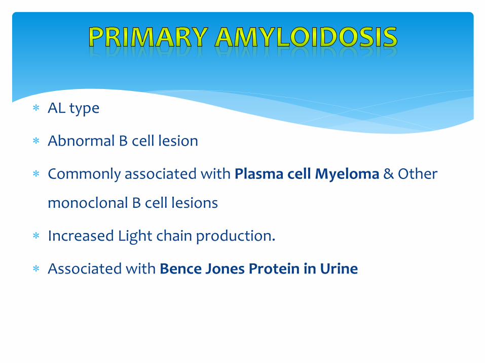
AL type
Abnormal B cell lesion
Commonly associated with Plasma cell Myeloma & Other
monoclonal B cell lesions
Increased Light chain production.
Associated with Bence Jones Protein in Urine

AA type
Secondary to Chronic Inflammatory lesion:
Tuberculosis, Bronchiectasis, Osteomyelitis, Rheumatoid
arthritis, Inflammatory bowel disease, Ankylosing
Spondylitis, IV drug abusers.

Affected organ gets enlarged, firm and waxy appearance.
Painting the cut surface with iodine imparts a yellow color,
and that is transformed to blue-violet after application of
sulphuric acid.
Diagnosis: Congo-red dye: ordinary light gives pink/red color;
polarized light gives apple-green birefringence.
Morphology: Gross

1. Congo Red—View under Polarizing Microscopy—Apple
Green birefringence: Gold standard test.
2. Thioflavin T
3. Van Giessen (Khaki color)
4. PAS (Periodic Acid Schiff )Stain.

Amorphous, eosinophilic, hyaline extra-cellular
protein.
With progressive accumulation, produces pressure
atrophy of adjacent cells.
Microscopically:

Kidney: Focal to diffuse mesangial deposits, Nodular
Basement membrane deposits, Capillary loops, Interstitial
space, Wall of blood vessels
Liver: Hepatomegaly, Waxy surface
Heart: Dew drop like subendocardial nodules, In between
Myocardial fibres
Tongue: Macroglossia, Gingival swellings
Carpel tunnel syndrome.
Morphology:

SPLEEN:
Splenomegaly +
Splenic follicles, white pulp involved—SAGO spleen:
tapioca like granules on gross examination.
Splenic sinuses, red pulp involved– LARDACEOUS spleen
(map like-large deposits).
Qn

Depend on the site involved
Can be unsuspected finding in Autopsy
Can lead to serious organ dysfunction
Renal disease: Nephrotic syndrome
Cardiac: Conduction defects, Restrictive
cardiomyopathy
Hepatosplenomegaly

1. Abdominal fat
2. Tongue
3. Rectal mucosa
4. Renal biopsy
5. Bone marrow biopsy
Biopsy site: