
38_Blood Cell & Urine Sediment Desktop Reference
34
DESKTOP READY RECKONER FOR THE PRACTICING PAEDIATRICIAN TO IDENTIFY CERTAIN BASIC CELLS IN A PERIPHERAL SMEAR & URINE SEDIMENTS Prepared by Dr.S.R.Srinivasa Kannan,MD.Path, Vivek Laboratories, 253,K-11,K.P.Road, Nagercoil-3. 629003 . e mail: [email protected]
-
Upload
john-nirmal -
Category
Documents
-
view
226 -
download
2
Transcript of 38_Blood Cell & Urine Sediment Desktop Reference
7/31/2019 38_Blood Cell & Urine Sediment Desktop Reference
http://slidepdf.com/reader/full/38blood-cell-urine-sediment-desktop-reference 1/34
DESKTOP READY RECKONER FOR THE PRACTICING
PAEDIATRICIAN TO IDENTIFY CERTAIN BASIC CELLS IN A
PERIPHERAL SMEAR & URINE SEDIMENTS
Prepared by
Dr.S.R.Srinivasa Kannan,MD.Path,
Vivek Laboratories, 253,K-11,K.P.Road,Nagercoil-3. 629003 .
e mail: [email protected]

7/31/2019 38_Blood Cell & Urine Sediment Desktop Reference
http://slidepdf.com/reader/full/38blood-cell-urine-sediment-desktop-reference 2/34
• This is not an exhaustive reference.
• This is only a short introduction to the
exciting field of Clinical Microscopy

7/31/2019 38_Blood Cell & Urine Sediment Desktop Reference
http://slidepdf.com/reader/full/38blood-cell-urine-sediment-desktop-reference 3/34
Note
• Areas for morphological assessment should be largeenough that at least 100 WBCs can be identified insamples with a WBC count within the reference intervalfor a specific individual.
• In a wedge blood film preparation, the best area is wherered blood cells barely touch each other.
• Cells should not be damaged by the preparation or staining procedure or by excessive shear forces for microscopy
• In a large blood film preparations; large cells such asmonocytes may be pushed to the periphery and thefeathered edge of the blood film.

7/31/2019 38_Blood Cell & Urine Sediment Desktop Reference
http://slidepdf.com/reader/full/38blood-cell-urine-sediment-desktop-reference 4/34
Good & Bad smear
Smooth & Uniform
staining
Irregular smear, Oil
droplets
7/31/2019 38_Blood Cell & Urine Sediment Desktop Reference
http://slidepdf.com/reader/full/38blood-cell-urine-sediment-desktop-reference 5/34
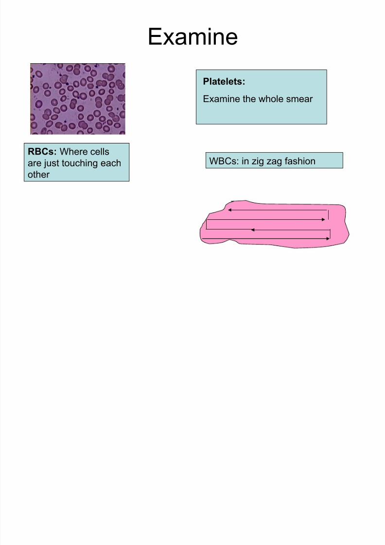
Examine
RBCs: Where cells
are just touching each
other
WBCs: in zig zag fashion
Platelets:
Examine the whole smear
7/31/2019 38_Blood Cell & Urine Sediment Desktop Reference
http://slidepdf.com/reader/full/38blood-cell-urine-sediment-desktop-reference 6/34
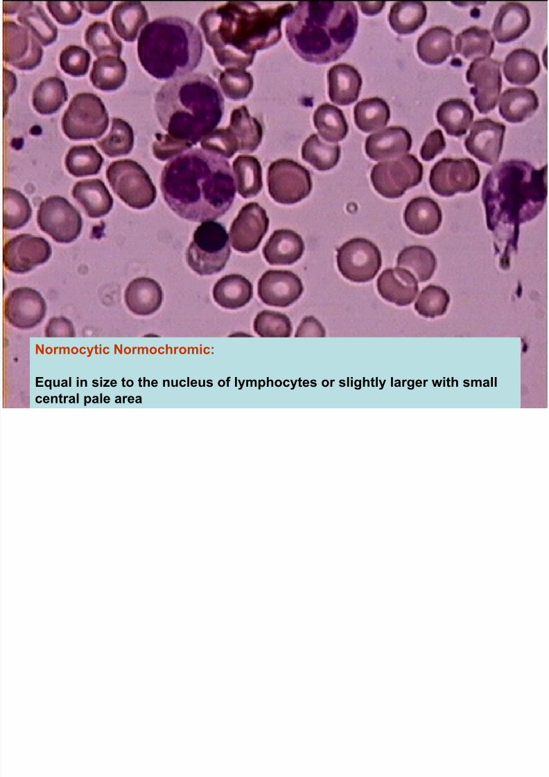
Normocytic Normochromic:
Equal in size to the nucleus of lymphocytes or slightly larger with smallcentral pale area

7/31/2019 38_Blood Cell & Urine Sediment Desktop Reference
http://slidepdf.com/reader/full/38blood-cell-urine-sediment-desktop-reference 7/34
Macrocytic: Larger than the normoytes. Always normochromic

7/31/2019 38_Blood Cell & Urine Sediment Desktop Reference
http://slidepdf.com/reader/full/38blood-cell-urine-sediment-desktop-reference 8/34
• Microcyte: Smaller than the nucleus of the small lymphocyte.
• Always hypochromic-marked central pallor

7/31/2019 38_Blood Cell & Urine Sediment Desktop Reference
http://slidepdf.com/reader/full/38blood-cell-urine-sediment-desktop-reference 9/34
Spherocytes:Small cells with no central pallor

7/31/2019 38_Blood Cell & Urine Sediment Desktop Reference
http://slidepdf.com/reader/full/38blood-cell-urine-sediment-desktop-reference 10/34
Elliptocyte/Ovalocyte : Abnormal congenital oval forms

7/31/2019 38_Blood Cell & Urine Sediment Desktop Reference
http://slidepdf.com/reader/full/38blood-cell-urine-sediment-desktop-reference 11/34
Stomatocytes : Abnormal congenital forms with a central mouth like slit.

7/31/2019 38_Blood Cell & Urine Sediment Desktop Reference
http://slidepdf.com/reader/full/38blood-cell-urine-sediment-desktop-reference 12/34
• Target Cells: Seen in Liver disorders and thalassemias

7/31/2019 38_Blood Cell & Urine Sediment Desktop Reference
http://slidepdf.com/reader/full/38blood-cell-urine-sediment-desktop-reference 13/34
Sickle cells: Sickling at hypoxic conditions. HbS. Drying of slide can pull the plasma and produce sickle shaped artifacts which is of variable size and the pulling effect will be obviousby the clearing in the surrounding areas

7/31/2019 38_Blood Cell & Urine Sediment Desktop Reference
http://slidepdf.com/reader/full/38blood-cell-urine-sediment-desktop-reference 14/34
• Acanthocyte: Irregular cytoplasmicspicules of variable length
• Irregularly disposed over the surface.(red cell Membrane abnormality
• Echinocyte: Many short regular
projections. Usually EDTA effect.

7/31/2019 38_Blood Cell & Urine Sediment Desktop Reference
http://slidepdf.com/reader/full/38blood-cell-urine-sediment-desktop-reference 15/34
MP: P.falciparum
• Basophilic stipplings: Numerous basophilic granules.
Occurs in Thalassemias, lead
poisoning & liver disease
Platelet Mimicking MP

7/31/2019 38_Blood Cell & Urine Sediment Desktop Reference
http://slidepdf.com/reader/full/38blood-cell-urine-sediment-desktop-reference 16/34
Water Artifact: Cells swollen and partly dehemoglobinised

7/31/2019 38_Blood Cell & Urine Sediment Desktop Reference
http://slidepdf.com/reader/full/38blood-cell-urine-sediment-desktop-reference 17/34
Reticulocytes:
seen with New methylene blue
Normoblasts: Normoblasts-Mimics lymphocytes.Note abundance of cytoplasm, irregular borders,
Centrally placed nuclei
7/31/2019 38_Blood Cell & Urine Sediment Desktop Reference
http://slidepdf.com/reader/full/38blood-cell-urine-sediment-desktop-reference 18/34
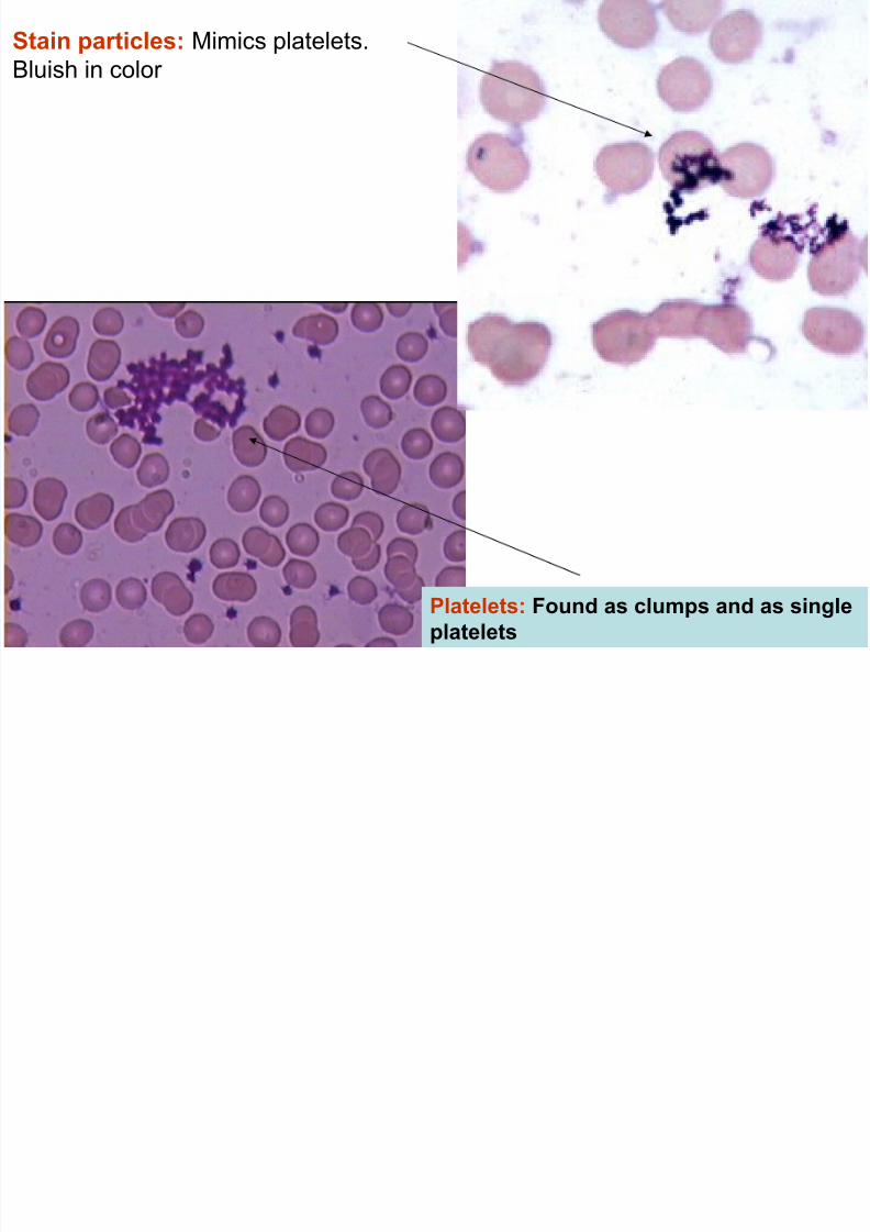
Platelets: Found as clumps and as singleplatelets
Stain particles: Mimics platelets.
Bluish in color
P l h T t l b C t l h hili ( )

7/31/2019 38_Blood Cell & Urine Sediment Desktop Reference
http://slidepdf.com/reader/full/38blood-cell-urine-sediment-desktop-reference 19/34
Polymorph: Two to many lobes. Cytoplasm shows azurophilic (grey)
Granules.
Hypersegmented Neutrophils:
Seen in Megaloblastic Anemia
Toxic granules: Seen in Septicaemia
Eosinophil has
eosin granules

7/31/2019 38_Blood Cell & Urine Sediment Desktop Reference
http://slidepdf.com/reader/full/38blood-cell-urine-sediment-desktop-reference 20/34
Monocytes
Eosinophil
Basophil
Polymorph
7/31/2019 38_Blood Cell & Urine Sediment Desktop Reference
http://slidepdf.com/reader/full/38blood-cell-urine-sediment-desktop-reference 21/34
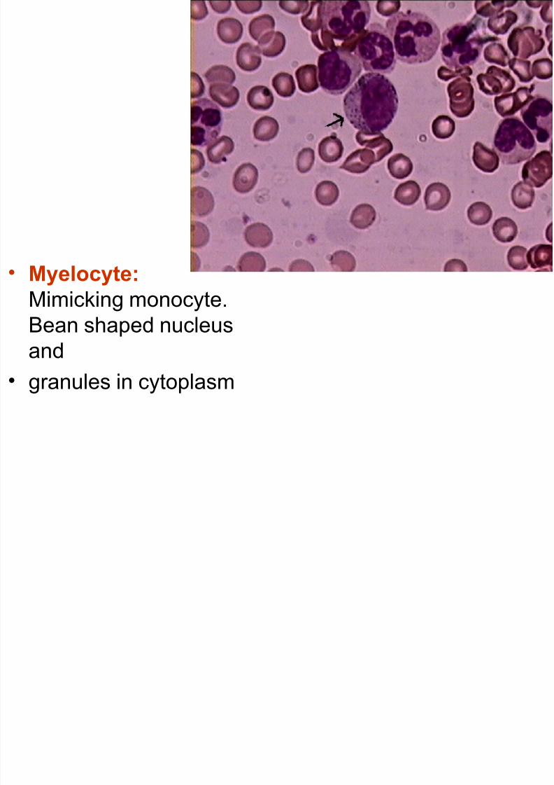
• Myelocyte:
Mimicking monocyte.Bean shaped nucleus
and
• granules in cytoplasm

7/31/2019 38_Blood Cell & Urine Sediment Desktop Reference
http://slidepdf.com/reader/full/38blood-cell-urine-sediment-desktop-reference 22/34
• Promyelocyte:
Comparable to
myelocyte but in
addition has nucleoli.
7/31/2019 38_Blood Cell & Urine Sediment Desktop Reference
http://slidepdf.com/reader/full/38blood-cell-urine-sediment-desktop-reference 23/34
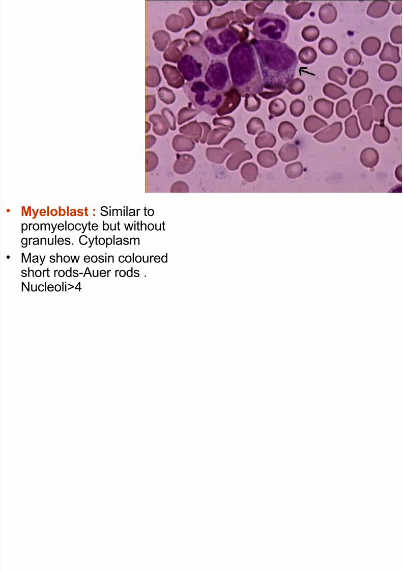
• Myeloblast : Similar topromyelocyte but withoutgranules. Cytoplasm
• May show eosin colouredshort rods-Auer rods .
Nucleoli>4

7/31/2019 38_Blood Cell & Urine Sediment Desktop Reference
http://slidepdf.com/reader/full/38blood-cell-urine-sediment-desktop-reference 24/34
Lymphoblast: Large cell. Cytoplasm is scanty. 1-2 nucleoli. No
granules

7/31/2019 38_Blood Cell & Urine Sediment Desktop Reference
http://slidepdf.com/reader/full/38blood-cell-urine-sediment-desktop-reference 25/34
Storage artifacts:
WBCs show fragmentation of nuclei and can mimic normoblats.

7/31/2019 38_Blood Cell & Urine Sediment Desktop Reference
http://slidepdf.com/reader/full/38blood-cell-urine-sediment-desktop-reference 26/34
• Urine Sediments
iQ®200 Automated Urine Microscopy Analyzer

7/31/2019 38_Blood Cell & Urine Sediment Desktop Reference
http://slidepdf.com/reader/full/38blood-cell-urine-sediment-desktop-reference 27/34
Hyaline cast
Smooth
borders,almost
transparent

7/31/2019 38_Blood Cell & Urine Sediment Desktop Reference
http://slidepdf.com/reader/full/38blood-cell-urine-sediment-desktop-reference 28/34
Cellular cast + yeast cells
Degenerated cellular cast is
granular cast
Border not regular. Cells
and fragments seen

7/31/2019 38_Blood Cell & Urine Sediment Desktop Reference
http://slidepdf.com/reader/full/38blood-cell-urine-sediment-desktop-reference 29/34
Refractile bodies with cut
edges
7/31/2019 38_Blood Cell & Urine Sediment Desktop Reference
http://slidepdf.com/reader/full/38blood-cell-urine-sediment-desktop-reference 30/34
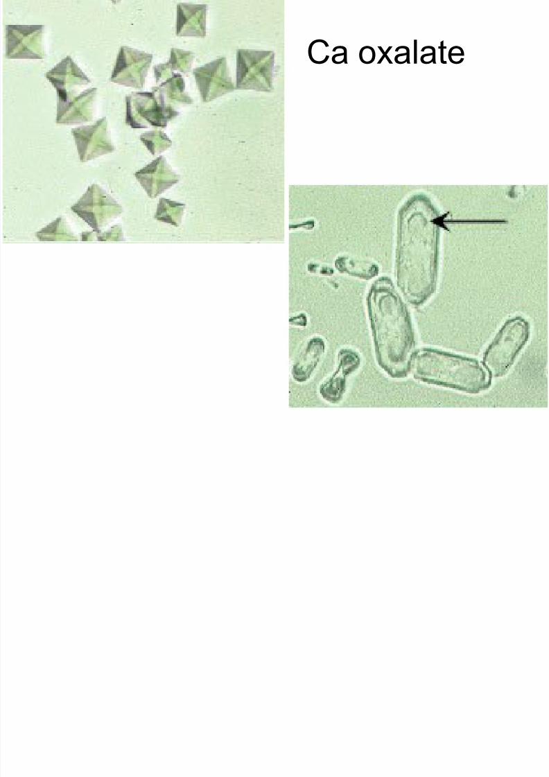
Ca oxalate

7/31/2019 38_Blood Cell & Urine Sediment Desktop Reference
http://slidepdf.com/reader/full/38blood-cell-urine-sediment-desktop-reference 31/34
Triple phosphate
tyrosine
7/31/2019 38_Blood Cell & Urine Sediment Desktop Reference
http://slidepdf.com/reader/full/38blood-cell-urine-sediment-desktop-reference 32/34
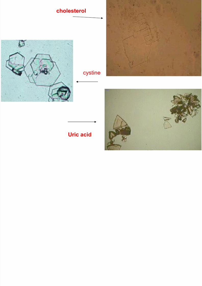
cystine
cholesterol
Uric acid

7/31/2019 38_Blood Cell & Urine Sediment Desktop Reference
http://slidepdf.com/reader/full/38blood-cell-urine-sediment-desktop-reference 33/34
• Each soul is potentially divine.
• The goal is to manifest this divinity within by
controlling nature,external and internal .
• Do this either by work,or worship, or psychic control, or philosophy:by one, or more, or all of these — and be free.

7/31/2019 38_Blood Cell & Urine Sediment Desktop Reference
http://slidepdf.com/reader/full/38blood-cell-urine-sediment-desktop-reference 34/34


![Urine analysis analysis[3359].pdfUrine sediment (Microscopic examination of urine sediment) •Should be performed by trained lab staff •Crystals –uric acid, Ca P or oxalate, Cysteine,](https://static.fdocuments.in/doc/165x107/5ec80a2cfe46c315f91a2ba4/urine-analysis-analysis3359pdf-urine-sediment-microscopic-examination-of-urine.jpg)


