2.cell structure
12
ANATOMY Cell Structure of cell M Humayun jamil 7/4/2014 It includes general review of cell. Structure of cell includes its three major parts and then each organelle is explained separately
-
Upload
m-humayun-jamil -
Category
Education
-
view
24 -
download
1
Transcript of 2.cell structure
ANATOMY
Cell
Structure of cell
M Humayun jamil
7/4/2014
It includes general review of cell. Structure of cell includes its three major parts and then each organelle is explained separately
Cell
Cell is a structural and functional unit of body. Cells were first observed more than 300 years
ago by the English scientist Robert Hooke. cell theory in 1838 and 1839 by two German
biologists, Matthias Schleiden and Theodor Schwann. This theory states that all living organ-
isms are composed of one or more cells and that the cell is the basic unit of structure for all
organisms.
Structure of cell
Cell is basically devided into three parts
1. Cell memberane
2. Cytoplasm and organelles
3. Nucleus
Cell memberane
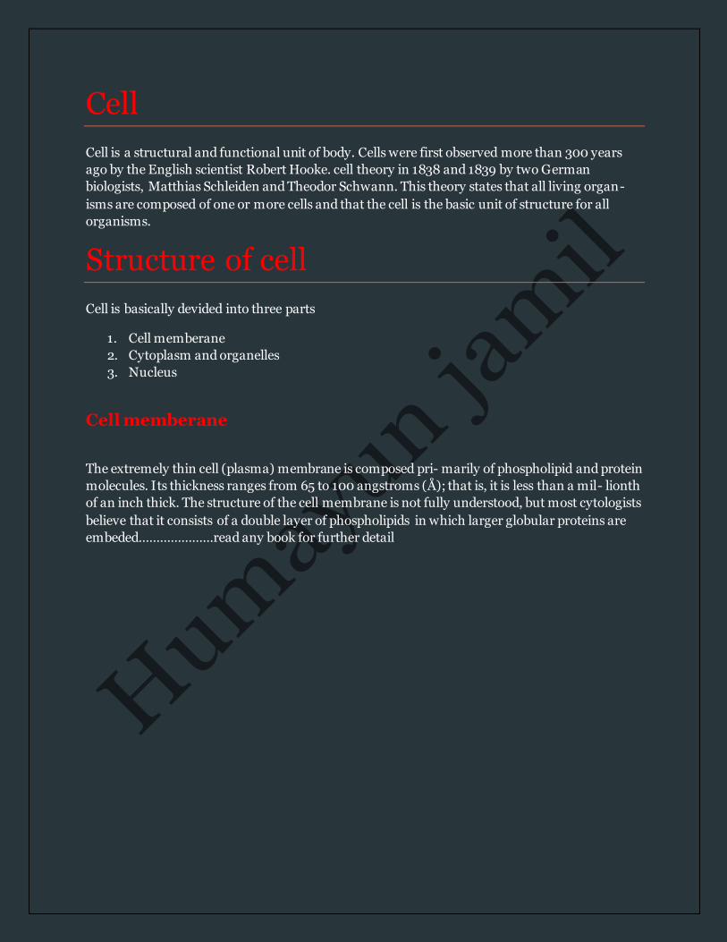
The extremely thin cell (plasma) membrane is composed pri- marily of phospholipid and protein
molecules. Its thickness ranges from 65 to 100 angstroms (Å); that is, it is less than a mil- lionth
of an inch thick. The structure of the cell membrane is not fully understood, but most cytologists
believe that it consists of a double layer of phospholipids in which larger globular proteins are
embeded…………………read any book for further detail
Cytoplasm and Organelles
Cytoplasm refers to the material located within the cell mem- brane but outside the nucleus. The
material within the nucleus is frequently called the nucleoplasm. The term protoplasm is some-
times used to refer to the cytoplasm and nucleoplasm collectively. When observed through an
electron microscope , distinct cellular components called organelles can be seen in the highly
structured cytoplasm. The matrix of the cytoplasm is a jel-lylike substance that is 80% to 90%
water. The organelles and inorganic colloid substances (suspended particles) are dispersed
throughout the cytoplasm. Colloid substances have similar ionic charges that space them
uniformly. Metabolic activity occurs within the organelles of the cyto- plasm. Specific roles such
as heat production, cellular mainte- nance, repair, storage, and protein synthesis are carried out
within the organelles.
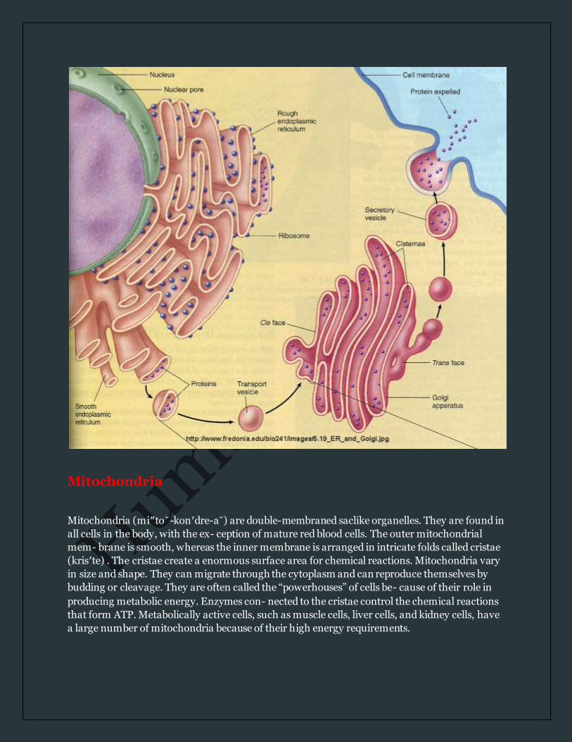
Endoplasmic reticulum Often abbreviated ER, the endoplasmic reticulum (en″do- plaz′mik r˘e-tik′yu˘-lum) is widely
distributed throughout the cyto- plasm as a complex network of interconnected membranes.
Although the name sounds complicated, endoplasmic simply means “within the plasm”
(cytoplasm of the cell) and reticulum means “network.” Between the interconnected mem -
branes are minute spaces, or cisterna, that are connected at one end to the cell membranes. The
tubules may also be connected to other organelles or to the outer nuclear envelope. The ER
provides a pathway for transportation of substances within the cell and a storage area for
synthesized molecules. There are two distinct varieties, either of which may predomi- nate in a
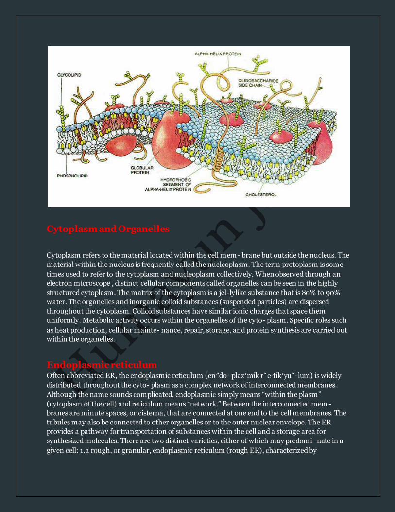
given cell: 1.a rough, or granular, endoplasmic reticulum (rough ER), characterized by

numerous small granules called ribosomes that are attached to the outer surface of the
membranous wall; and 2.a smooth endoplasmic reticulum (smooth ER) that lacks ribosomes.
The membranous wall of rough ER provides a site for protein synthesis within ribosomes.
Smooth ER manufactures certain lipid molecules. Also, enzymes within the smooth ER of liver
cells inactivate or detoxify a variety of chemicals.
Ribosomes
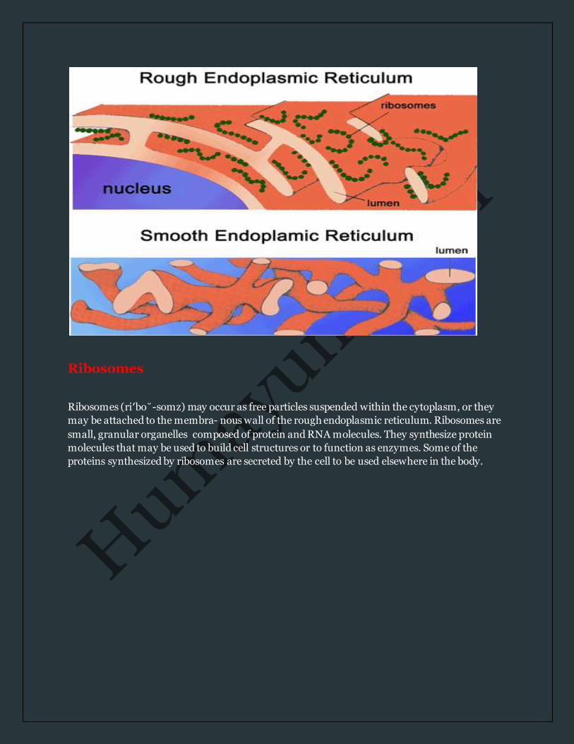
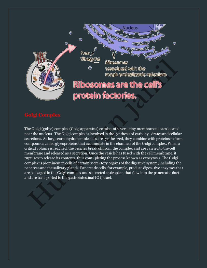
Ribosomes (ri′bo˘-somz) may occur as free particles suspended within the cytoplasm, or they
may be attached to the membra- nous wall of the rough endoplasmic reticulum. Ribosomes are
small, granular organelles composed of protein and RNA molecules. They synthesize protein
molecules that may be used to build cell structures or to function as enzymes. Some of the
proteins synthesized by ribosomes are secreted by the cell to be used elsewhere in the body.
Golgi Complex
The Golgi (gol′je) complex (Golgi apparatus) consists of several tiny membranous sacs located
near the nucleus . The Golgi complex is involved in the synthesis of carbohy- drates and cellular
secretions. As large carbohydrate molecules are synthesized, they combine with proteins to form
compounds called glycoproteins that accumulate in the channels of the Golgi complex. When a
critical volume is reached, the vesicles break off from the complex and are carried to the cell
membrane and released as a secretion. Once the vesicle has fused with the cell membrane, it
ruptures to release its contents, thus com- pleting the process known as exocytosis. The Golgi
complex is prominent in cells of certain secre- tory organs of the digestive system, including the
pancreas and the salivary glands. Pancreatic cells, for example, produce diges- tive enzymes that
are packaged in the Golgi complex and se- creted as droplets that flow into the pancreatic duct
and are transported to the gastrointestinal (GI) tract.
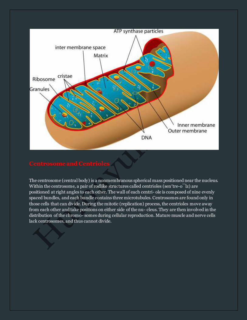
Mitochondria
Mitochondria (mi″to˘-kon′dre-a˘) are double-membraned saclike organelles. They are found in
all cells in the body, with the ex- ception of mature red blood cells. The outer mitochondrial
mem- brane is smooth, whereas the inner membrane is arranged in intricate folds called cristae
(kris′te) . The cristae create a enormous surface area for chemical reactions. Mitochondria vary
in size and shape. They can migrate through the cytoplasm and can reproduce themselves by
budding or cleavage. They are often called the “powerhouses” of cells be- cause of their role in
producing metabolic energy. Enzymes con- nected to the cristae control the chemical reactions
that form ATP. Metabolically active cells, such as muscle cells, liver cells, and kidney cells, have
a large number of mitochondria because of their high energy requirements.
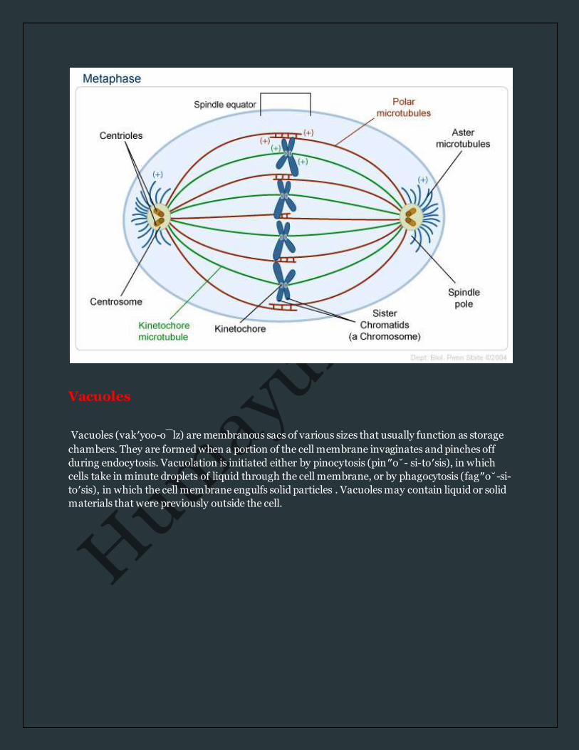
Centrosome and Centrioles
The centrosome (central body) is a nonmembranous spherical mass positioned near the nucleus.
Within the centrosome, a pair of rodlike structures called centrioles (sen′tre-o¯lz) are
positioned at right angles to each other. The wall of each centri- ole is composed of nine evenly
spaced bundles, and each bundle contains three microtubules. Centrosomes are found only in
those cells that can divide. During the mitotic (replication) process, the centrioles move away
from each other and take positions on either side of the nu- cleus. They are then involved in the
distribution of the chromo- somes during cellular reproduction. Mature muscle and nerve cells
lack centrosomes, and thus cannot divide.
Vacuoles
Vacuoles (vak′yoo-o¯lz) are membranous sacs of various sizes that usually function as storage
chambers. They are formed when a portion of the cell membrane invaginates and pinches off
during endocytosis. Vacuolation is initiated either by pinocytosis (pin″o˘- si-to′sis), in which
cells take in minute droplets of liquid through the cell membrane, or by phagocytosis (fag″o˘-si-
to′sis), in which the cell membrane engulfs solid particles . Vacuoles may contain liquid or solid
materials that were previously outside the cell.
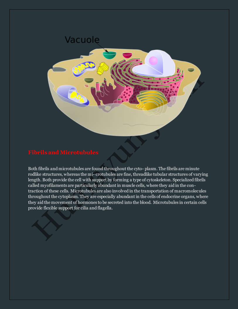
Fibrils and Microtubules
Both fibrils and microtubules are found throughout the cyto- plasm. The fibrils are minute
rodlike structures, whereas the mi- crotubules are fine, threadlike tubular structures of varying
length. Both provide the cell with support by forming a type of cytoskeleton. Specialized fibrils
called myofilaments are particularly abundant in muscle cells, where they aid in the con-
traction of these cells. Microtubules are also involved in the transportation of macromolecules
throughout the cytoplasm. They are especially abundant in the cells of endocrine organs, where
they aid the movement of hormones to be secreted into the blood. Microtubules in certain cells
provide flexible support for cilia and flagella.

Cell Nucleus
The spherical nucleus is usually located near the center of the cell . It is the largest structure of
the cell and contains the genetic material that determines cellular structure and con- trols
cellular activity. Most cells contain a single nucleus. Certain cells, however, such as skeletal
muscle cells, are multinucleated. The long skele- tal muscle fibers contain so much cytoplasm
that several govern- ing centers are necessary. Other cells, such as mature red blood cells, lack
nuclei. These cells are limited to certain types of chemical activities and are not capable of cell
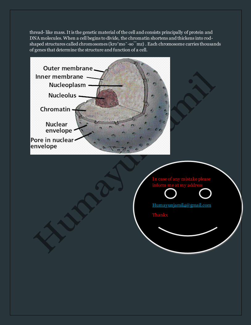
division. The nucleus is enclosed by a bilayered nuclear membrane (nuclear envelope) . The
narrow space between the inner and outer layers of the nuclear membrane is called the nu-
cleolemma cisterna (sis-ter′na). Minute nuclear pores are lo- cated along the nuclear membrane.
These openings are lined with proteins that act as selective gates, allowing certain mole- cules,
such as proteins, RNA, and protein-RNA complexes, to move between the nucleoplasm and the
cytoplasm.
Two important structures within the nucleoplasm of the nucleus determine what a cell will look
like and what functions it will perform: 1. Nucleoli. Nucleoli (noo-kle′o˘-li) are small,
nonmembra- nous spherical bodies composed largely of protein and RNA. It is thought that they
function in the production of ribosomes. As ribosomes are formed, they migrate through the
nuclear membrane into the cytoplasm. 2. Chromatin. Chromatin (kro′ma˘-tin) is a coiled,
In case of any mistake please
inform me at my address
Thankx
thread- like mass. It is the genetic material of the cell and consists principally of protein and
DNA molecules. When a cell begins to divide, the chromatin shortens and thickens into rod-
shaped structures called chromosomes (kro′mo˘-so¯mz) . Each chromosome carries thousands
of genes that determine the structure and function of a cell.