2.4.09 Inf Ostreid Herpes
14
NB: This disease is not listed by the OIE. Manual of Diagnostic Tests for Aquatic Animals 2012 533 CHAPTER 2.4.9 INFECTION WITH OSTREID HERPESVIRUS 1 1. Scope For the purpose of this chapter, infection with ostreid herpesvirus 1 (OsHV-1) is considered to be a viral infection caused by OsHV-1 affecting mainly Pacific cupped oysters, Crassostrea gigas. The term OsHV-1 is used throughout the present chapter to define a virus species. The virus reported by Davison et al. (2005) corresponds to a particular ‘isolate’ or strain collected from infected Pacific oyster larvae in France in 1995. As this ‘isolate’ was the first to be described (through complete genome sequencing), it can be assumed to be a reference type. Other isolates, including OsHV-1 var and OsHV-1 μVar, have also been identified and studied. In this context, OsHV-1 means all virus isolates or strains and the virus reported by Davison et al. (2005) defined as the reference type. Differences in virulence may exist among OsHV-1 strains. 2. Disease information 2.1. Agent factors OsHV-1 is the aetiological agent of a contagious viral disease of Pacific cupped oysters, Crassostrea gigas, affecting also other bivalve species. 2.1.1. Aetiological agent, agent strains OsHV-1 particles have been purified from French C. gigas larvae (Le Deuff & Renault, 1999) and were observed by transmission electron microscopy to be enveloped icosahedral with electron dense cores and a diameter around 120 nm. The intranuclear location of the virus particles, their size and ultrastructure are characteristic of members of the Herpesvirales. The genome structure and sequence, and the capsid morphology (Davison et al., 2005) have been further studied in order to assess OsHV-1 phylogenetic status in relation to vertebrate herpes viruses. The entire virus DNA was sequenced (GenBank accession number AY509253) and OsHV-1 capsids appear structurally similar to those of other herpes viruses that have been studied (Davison et al., 2005). The virus was classified under the name Ostreid herpesvirus 1 (OsHV-1) as the first known species in the family Malacoherpesviridae (Davison et al., 2009). A variant of OsHV-1 (OsHV-1var) has been identified in France (Arzul et al., 2001b) in C. gigas, Ruditapes philippinarum and Pecten maximus. Friedman et al. (2005) and Moss et al. (2007) also described differences in the sequences of OsHV-1 from California and Asia, respectively. Moss et al. (2007) suggested that there are at least two strains in Japan, one in South Korea and two in China (the People’s Rep. of). One of the strains that occurred in China and South Korea was similar in sequence to the OsHV-1 strain from California described by Friedman et al. (2005), and the other strain from China was similar to OsHV-1 from France. More recently, polymerase chain reactions (PCRs) using primer sets IA1-IA2 and C2-C6 enabled the detection of a variant (OsHV-1 μvar) in association with the high mortality events reported in Europe since 2008 (Segarra et al., 2010). The aetiological agent is represented by all strains of ostreid herpesvirus 1 (Arzul et al., 2001b; Davison et al., 2005, Moss et al., 2007, Segarra et al., 2010). 2.1.2. Survival outside the host Maximum survival time outside the host is unknown. Schikorski et al. (2011a; 2011b) presented data on detection by real time PCR of OsHV-1μvar DNA in seawater following cohabitation experiments. The copy numbers of virus DNA in the water in the first 48 hours after injecting spat with virus reached 1 × 10 5 ml –1 , and reached a maximum of 1 × 10 6 ml –1 following infection of cohabiting oysters. The amount of infectious virus is unknown.
-
Upload
adi-darmawan -
Category
Documents
-
view
2 -
download
0
description
jaxz
Transcript of 2.4.09 Inf Ostreid Herpes
NB: This d i sease i s not l i s ted by the OIE.
Manual of Diagnostic Tests for Aquatic Animals 2012 533
C H A P T E R 2 . 4 . 9
INFECTION WITH OSTREID HERPESVIRUS 1
1. Scope
For the purpose of this chapter, infection with ostreid herpesvirus 1 (OsHV-1) is considered to be a viral infection caused by OsHV-1 affecting mainly Pacific cupped oysters, Crassostrea gigas.
The term OsHV-1 is used throughout the present chapter to define a virus species. The virus reported by Davison et al. (2005) corresponds to a particular ‘isolate’ or strain collected from infected Pacific oyster larvae in France in 1995. As this ‘isolate’ was the first to be described (through complete genome sequencing), it can be assumed to be a reference type. Other isolates, including OsHV-1 var and OsHV-1 µVar, have also been identified and studied. In this context, OsHV-1 means all virus isolates or strains and the virus reported by Davison et al. (2005) defined as the reference type. Differences in virulence may exist among OsHV-1 strains.
2. Disease information
2.1. Agent factors
OsHV-1 is the aetiological agent of a contagious viral disease of Pacific cupped oysters, Crassostrea gigas, affecting also other bivalve species.
2.1.1. Aetiological agent, agent strains
OsHV-1 particles have been purified from French C. gigas larvae (Le Deuff & Renault, 1999) and were observed by transmission electron microscopy to be enveloped icosahedral with electron dense cores and a diameter around 120 nm. The intranuclear location of the virus particles, their size and ultrastructure are characteristic of members of the Herpesvirales.
The genome structure and sequence, and the capsid morphology (Davison et al., 2005) have been further studied in order to assess OsHV-1 phylogenetic status in relation to vertebrate herpes viruses. The entire virus DNA was sequenced (GenBank accession number AY509253) and OsHV-1 capsids appear structurally similar to those of other herpes viruses that have been studied (Davison et al., 2005). The virus was classified under the name Ostreid herpesvirus 1 (OsHV-1) as the first known species in the family Malacoherpesviridae (Davison et al., 2009).
A variant of OsHV-1 (OsHV-1var) has been identified in France (Arzul et al., 2001b) in C. gigas, Ruditapes philippinarum and Pecten maximus. Friedman et al. (2005) and Moss et al. (2007) also described differences in the sequences of OsHV-1 from California and Asia, respectively. Moss et al. (2007) suggested that there are at least two strains in Japan, one in South Korea and two in China (the People’s Rep. of). One of the strains that occurred in China and South Korea was similar in sequence to the OsHV-1 strain from California described by Friedman et al. (2005), and the other strain from China was similar to OsHV-1 from France.
More recently, polymerase chain reactions (PCRs) using primer sets IA1-IA2 and C2-C6 enabled the detection of a variant (OsHV-1 µvar) in association with the high mortality events reported in Europe since 2008 (Segarra et al., 2010).
The aetiological agent is represented by all strains of ostreid herpesvirus 1 (Arzul et al., 2001b; Davison et al., 2005, Moss et al., 2007, Segarra et al., 2010).
2.1.2. Survival outside the host
Maximum survival time outside the host is unknown.
Schikorski et al. (2011a; 2011b) presented data on detection by real time PCR of OsHV-1μvar DNA in seawater following cohabitation experiments. The copy numbers of virus DNA in the water in the first 48 hours after injecting spat with virus reached 1 × 10⁵ ml–1, and reached a maximum of 1 × 10⁶ ml–1 following infection of cohabiting oysters. The amount of infectious virus is unknown.
Chapter 2.4.9. — Infection with ostreid herpesvirus 1
534 Manual of Diagnostic Tests for Aquatic Animals 2012
2.1.3. Stability of the agent (effective inactivation methods)
The lack of cell cultures for OsHV-1 has meant that in-vitro studies on the stability of the virus with regard to infectivity have not been done. As an alternative, extracted viral DNA was seeded into seawater and 10 pg μl–1 was detected for 16, 9 and 1 day at 4, 11 and 20°C respectively, and in a second experiment, 100 pg μl–1 was detected after 51 days at each temperature.
The longest time for DNA detection in OsHV-1 released from macerated larvae and seeded into seawater was 22 days at 4°C and 12 days at 20°C (Vigneron et al., 2004). However, the relationship between detection of DNA in the PCR and infectivity of the virus is unknown. As a general rule, the survival of many viruses of aquatic animals outside the host is greatest at lower temperatures.
2.1.4. Life cycle
The life cycle is direct from host to host (Le Deuff et al., 1994; Schikorski et al., 2011a; 2011b).
2.2. Host factors
2.2.1. Susceptible host species
Pacific oyster, C. gigas, Portuguese oyster, C. angulata, suminoe oyster, C. ariakensis, European flat oyster, O. edulis, Manila clam, R. philippinarum, carpet shell clam, R. decussates, scallops, P. maximus (Arzul et al., 2001a; 2001b; Renault et al., 2000).
2.2.2. Susceptible stages of the host
OsHV-1 infection causes mortality in larvae and juveniles of several bivalve species. The virus can be found in adult bivalves most often in absence of mortality.
2.2.3. Species or sub-population predilection (probability of detection)
Crassostrea gigas, O. edulis, R. philippinarum, R. decussates and P. maximus are naturally infected. Young stages including larvae, spat and juveniles seem to be more susceptible to the infection. The virus is easier to detect in moribund animals than in healthy ones.
2.2.4. Target organs and infected tissue
The infection-associated lesions in juveniles are mainly observed in connective tissues of all organs in which fibroblastic-like cells exhibit enlarged nuclei with perinuclear chromatin (Arzul et al., 2002; Lipart & Renault, 2002; Renault et al.; 1995; Schikorsky et al., 2011a).
2.2.5. Persistent infection with lifelong carriers
Apparently healthy oysters, including adults, have been shown to be PCR-positive for OsHV-1 (Arzul et al., 2002; Moss et al., 2007; Sauvage et al., 2009). Pépin et al. (2008) showed that DNA copy numbers mg–1 were high (up to 107) in oysters from populations with abnormal mortalities and low (lowest number detected 101) in populations with no abnormal mortalities. Determining the levels of viral DNA in oysters by quantitative PCR (qPCR) might be a means to differentiate between mechanical carriage of virus and low level of infection.
As the virus (DNA, protein or particles) has been detected in tissues of adult oysters, including the gonad (Arzul et al., 2002; Lipart & Renault, 2002), adults may be a source of infection for larvae or spat, particularly under stressful conditions, e.g. from high temperature (Le Deuff et al., 1996). However, what is not certain is whether true vertical transmission (transmission within the gametes) occurs or whether transmission is horizontal (Barbosa-Solomieu et al., 2005).
2.2.6. Vectors
No vectors are required: the life cycle is direct.
2.2.7. Known or suspected wild aquatic animal carriers
Several bivalve species may act as healthy carriers (see Section 2.2.3).
OsHV-1 µvar DNA has been recently detected in France in blue mussel, Mytilus edulis, and in Donax trunculus (Renault, comm. pers.). However, in these cases, it remains unknown if these bivalve species are susceptible, resistant or may act as vector species.

Chapter 2.4.9. — Infection with ostreid herpesvirus 1
Manual of Diagnostic Tests for Aquatic Animals 2012 535
2.3. Disease pattern
2.3.1. Transmission mechanisms
OsHV-1 DNA has been detected by qPCR in the water around diseased Pacific oysters (Sauvage et al., 2009) and the disease can be experimentally transmitted horizontally via the water (Schikorski et al., 2011a), which presumably is the main natural mode of OsHV-1 transmission.
The first published report (Le Deuff et al., 1994) described rapid transmission of the virus from an extract of diseased larvae to axenic larvae of C. gigas. Inter-species transmission from infected axenic larvae of C. gigas to axenic larvae of C. rivularis and Ostrea edulis was demonstrated experimentally (Arzul et al., 2001b). A suspension of OsHV-1 from R. philippinarum was shown to infect axenic larvae of C. gigas, and a virus suspension from C. gigas was shown to infect axenic larvae of C. angulata (Arzul et al., 2001b).
Experimental transmission of OsHV-1μvar has been described by Schikorski et al. (2011a; 2011b). The disease can be transmitted to spat at 22°C following intramuscular injection of an extract of naturally infected oysters, and also by cohabiting injected oysters with healthy oysters. Using qPCR, the virus was shown to enter the digestive gland and haemolymphatic system, following which the virus was disseminated to other organs.
2.3.2. Prevalence
Reported mortality rates vary considerably between sites and countries and depend on the age of affected stocks. To better understand the implication of OsHV-1 in C. gigas spat mortality outbreaks regularly reported both in the field and in nurseries in France, samples were collected yearly through the French National Network for Surveillance of Mollusc Health between 1997 and 2006 (Garcia et al., 2011). Analyses were carried out by PCR for OsHV-1 detection. Virus DNA was frequently detected in samples collected during mortality events with OsHV-1 detection frequency varying from 9 to 65% depending on the year. Data also demonstrated a particular seasonality and topography of spat oyster mortalities associated with OsHV-1 detection. In the field, mortality outbreaks appeared in summer, preferentially in sheltered environments.
More recently, increased mortality notifications (from 40 to 100%) were reported in 2008–2011 in Europe affecting Pacific oysters. These increased mortalities were associated with the emergence OsHV-1 µvar. OsHV-1 µvar was detected in most of the investigated samples.
2.3.3. Geographical distribution
Europe (France Ireland, Italy, Netherlands, Spain, United Kingdom), Australia, China (People’s Rep. of), Korea, Japan, Morocco, Mexico, New Zealand and United States of America.
2.3.4. Mortality and morbidity
Infection by all strains is often lethal for C. gigas spat and juveniles. Death usually occurs 1 week after infection, during or shortly after the warmest annual water temperatures (Friedman et al., 2005; Garcia et al., 2011; Renault et al., 1994b).
Infected larvae show a reduction in feeding and swimming activities, and mortality can reach 100% in a few days.
2.3.5. Environmental factors
Mortalities associated with the detection of OsHV-1 are more frequent during summer, which might suggest a link between seawater temperature and OsHV-1 infection. The temperature influence on OsHV-1 detection and virus expression was demonstrated for C. gigas larvae (Le Deuff et al., 1996) and strongly suspected for C. gigas spat (Burge et al., 2007; Friedman et al., 2005; Renault et al., 1995; Sauvage et al., 2009). A temperature threshold related to enhanced OsHV-1 expression or mortality appears difficult to define precisely. In the literature, according to the site, the temperature threshold was variable: 22°C to 25°C on the west coast of the USA (Friedman et al., 2005; Burge et al., 2007) and 18 to 20°C in France (Samain et al., 2007; Soletchnik et al., 1999). High seawater temperatures appear to be one of the potential factors influencing OsHV-1 infection.
Moreover, stressful conditions particularly rearing techniques seem to favour OsHV-1 infection. In France, during summer, many oyster transfers occur and might also amplify OsHV-1 transmission.
Chapter 2.4.9. — Infection with ostreid herpesvirus 1
536 Manual of Diagnostic Tests for Aquatic Animals 2012
Spat mortality outbreaks associated with OsHV-1 detection generally presented a patchy distribution in the field. This particular pattern could be partly explained by the nature of the virus. Herpes viruses are enveloped and are assumed to be relatively labile in their environment. Thus, their transmission relies generally on direct contact. These data suggest that when OsHV-1 is excreted by oysters, it would mainly infect nearby oysters. The probable limited dissemination of OsVH-1 in seawater could partly explain the observation of the patchy mortality distribution rather than a uniform distribution as observed in nurseries. In nurseries, oysters are reared at high densities, are very close together and the seawater is often sequentially renewed.
2.4. Control and prevention
2.4.1. Vaccination
None
2.4.2. Chemotherapy
None
2.4.3. Immunostimulation
None
2.4.4. Resistance breeding
Based on recent data, it has been demonstrated that Pacific cupped oyster families resistant or tolerant to OsHV-1 can be obtained (Sauvage et al., 2009).
2.4.5. Restocking with resistant species
None
2.4.6. Blocking agents
None
2.4.7. Disinfection of eggs and larvae
None
2.4.8. General husbandry practices
Bio-security may be successfully applied in confined and controlled facilities such as hatcheries and nurseries in order to protect the facility and the surrounding environment from the introduction of the virus.
As a herpesvirus, OsHV-1 may be assumed to be fragile outside its hosts. High temperature, chemicals or sunlight (UV) may destroy its lipid-containing envelope. However, it has been demonstrated that individual herpes virus species may have different levels of stability to inactivation treatment and that inorganic salts such as Na2SO4 present in seawater may stabilise herpes viruses
(Wallis & Melnick, 1965).
In controlled rearing conditions (mollusc hatchery/nursery), OsHV-1 outbreaks may therefore be controlled through quarantine and hygienic measures including virus inactivation through adapted treatments such as ultraviolet irradiation of the recirculating water and water filtration technologies. However, it is necessary to keep in mind that reduction of virus load depends on the initial titre and the virus reduction capacity of the techniques used for inactivation. If there was an initial concentration of 1 million viruses per litre and the inactivation method used allowed inactivation of 100,000 viruses per litre, there would still numerous infective particles in the treated product.
Moribund and dead oysters should be removed and destroyed whenever feasible. Equipment used in an infected zone should not be sent and used in a non-affected zone without adequate cleaning and disinfection.
Chapter 2.4.9. — Infection with ostreid herpesvirus 1
Manual of Diagnostic Tests for Aquatic Animals 2012 537
3. Sampling
3.1. Selection of individual specimens
Live or moribund individuals should be sampled.
3.2. Preservation of samples for submission
For histology, the best preservative is Davidson’s AFA, but 10% buffered formalin or other standard histology fixatives are also acceptable. For PCR assays, samples must be preserved in 95–100% ethanol or kept frozen (–80°C).
3.3. Pooling of samples
Pooling of small spat is acceptable for PCR/qPCR analyses. However, the effect of pooling samples on PCR/qPCR sensitivity has not been evaluated
3.4. Best organs or tissues
For histology, a 5-µm thick section through the visceral mass that includes digestive gland, gill and mantle is used. For PCR, mantle tissue is best.
3.5. Samples/tissues that are not suitable
Gonad tissues may be not reliable for PCR assays because of the presence of inhibitors.
4. Diagnostic methods
4.1. Field diagnostic methods
4.1.1. Clinical signs
Infection by OsHV-1 causes an acute disease. Animals are likely to die within a few days of demonstrating clinical signs of the disease. Clinical signs may be dead or gaping bivalves but these clinical signs are not specific to infection with OsHV-1.
4.1.2. Behavioural changes
Infected hosts may be slow to close their valves when disturbed but these behavioural changes are not specific to infection with OsHV-1.
4.2. Clinical methods
4.2.1. Gross pathology
Clinical signs may be dead or gaping bivalves but these clinical signs are not specific to infection with OsHV-1.
4.2.2. Clinical chemistry
None
4.2.3. Microscopic pathology
See Section 4.2.6. Fixed sections
4.2.4. Wet mounts
Not applicable
4.2.5. Smears
Not applicable

Chapter 2.4.9. — Infection with ostreid herpesvirus 1
538 Manual of Diagnostic Tests for Aquatic Animals 2012
4.2.6. Fixed sections
The most consistent features of infection with OsHV-1 are nuclear changes including hypertrophy, nuclear margination and pycnosis. The infection-associated lesions in spat are mainly observed in connective tissues in which fibroblastic-like cells exhibit enlarged nuclei with perinuclear chromatin. Highly condensed nuclei (apoptosis features) were also reported in other cells interpreted as haemocytes. These cellular abnormalities are not associated with massive haemocyte infiltration.
Histological examination of the animal is not sufficient to identify infection with herpes virus. Whilst Cowdry type A inclusions (eosinophilic intranuclear inclusions with perinuclear chromatin) are typical of many herpes virus infections they are not a diagnostic feature of herpes virus infections of oysters (Arzul et al., 2002). Cowdry type A inclusions have never been reported following histological examination of infected Pacific cupped oysters in France (Renault et al., 1994a; 1994b). Moreover, intranuclear inclusion bodies were not observed, although there was other cellular/nuclear pathology, in association with OsHV-1 infections in oysters in Mexico (Vásquez-Yeomans et al., 2010) or USA (California) (Friedman et al., 2005).
4.2.7. Electron microscopy/cytopathology
See Section 4.3.1.1.4.
4.3. Agent detection and identification methods
4.3.1. Direct detection methods
4.3.1.1. Microscopic methods
4.3.1.1.1. Wet mounts
Not applicable
4.3.1.1.2. Smears
Not applicable
4.3.1.1.3. Fixed sections
Samples to be taken: live or moribund oysters.
Technical procedure: Sections of tissue that include mantle, digestive gland, gills and adductor muscle should be fixed for 24 hours in 10% formaldehyde fixatives such as Davidson’s AFA or other suitable fixative followed by normal processing for paraffin histology and staining with haematoxylin and eosin. Observations are made at increasing magnifications up to ×400.
Positive controls: These are recommended and are available from the Genetics and Pathology Laboratory, Ifremer, La Tremblade, France. Positive controls are tissue sections from any OsHV-1 infected mollusc.
Levels of validation:
Specificity and sensitivity: Specificity is very low, and sensitivity is good for moderate- to high-intensity infections, but low for low-intensity infections.
Gold standard: None
Interpretation of results:
A positive result is the occurrence of cell abnormalities in tissue sections: Fibroblastic-like cells exhibiting enlarged nuclei with perinuclear chromatin. Highly condensed nuclei are also reported in other cells interpreted as haemocytes. These cellular abnormalities are not associated with massive haemocyte infiltration.
In susceptible host species, within the known range for OsHV-1, a positive result is presumptive evidence of OsHV-1 infection only and should be confirmed by species-specific PCR, in-situ hybridisation (ISH) and/or DNA sequencing.
Availability of commercial tests: No commercially available tests.

Chapter 2.4.9. — Infection with ostreid herpesvirus 1
Manual of Diagnostic Tests for Aquatic Animals 2012 539
4.3.1.1.4. Electron microscopy/cytopathology
Transmission electron microscopy can be used to confirm the presence of viral particles in infected animals.
Tissue samples (containing connective tissue such as mantle) for examination by electron microscopy should be fixed using 2.5% (v/v) glutaraldehyde in 0.1 M cacodylate buffer and post-fixed in 1% (w/v) osmium tetroxide, washed in 0.1 M cacodylate buffer (3 × 10 minutes), dehydrated in a graded series of ethanol (70%, 1 × 10 minutes; 95%, 2 × 15 minutes; 100%, 3 × 20 minutes), washed in propylene oxide (2 × 15 minutes), pre-infiltrated in 50% propylene oxide/50% Epon resin (1 hour), infiltrated in 100% Epon resin (1 hour) and then embedded in Epon resin.
OsHV-1 replication mainly takes place in fibroblastic-like cells throughout connective tissues especially in mantle, labial palps, gills and digestive gland (Renault et al., 1994b; 1995). Virogenesis begins in the nucleus of infected cells where capsids and nucleocapsids are observed. Viral particles then pass through the nuclear membrane into the cytoplasm and enveloped particles are released at the cell surface. Intranuclear and cytoplasmic capsids present a variety of morphological types including electron lucent capsids, toroidal core-containing capsids, and brick-shaped core-containing capsids.
4.3.1.2. Agent isolation and identification
4.3.1.2.1. Cell culture/artificial media
To date, attempts to culture the virus in both vertebrate and invertebrate cell lines and in primary bivalve cell cultures have been unsuccessful.
4.3.1.2.2. Antibody-based antigen detection methods
Specific antibodies have been developed (Arzul et al., 2002). However, they are not currently available for diagnostic purposes.
4.3.1.2.3. Molecular techniques
At present there are a number of different PCR methods available for the detection of OsHV-1. These include both conventional and real-time PCRs (Martenot et al., 2010; Pépin et al., 2008).
A protocol for quantifying OsHV-1 in Pacific oysters based on a Sybr® Green real-time PCR was first developed (Pepin et al., 2008). Martenot et al. (2010) developed an alternative protocol based on TaqMan® chemistry. The quantitation limits were 1000 and 18 UG mg–1 of tissues for the Sybr®
Green-based method and the TaqMan® method, respectively, and the latter protocol has a detection limit of 6 UG mg–1 of tissues. Comparing the two protocols using DNA samples obtained from 210 spat, the kappa index (0.41) indicated a moderate concordance between the protocols, according to the measures of Landis and Koch. All samples that were positive by the reference protocol were also positive by the alternative protocol. Of the 76 samples that were negative by the reference protocol, 49 were positives by the alternative protocol. Although these results may suggest that the alternative protocol can be more sensitive than the reference protocol, formal validation is needed.
A loop-mediated isothermal amplification (LAMP) assay was also developed for OsHV-1 DNA detection (Ren et al., 2010). A set of four primers was designed, based on the sequence of the ATPase subunit of the OsHV-1 DNA-packaging terminase gene. This LAMP technique can be used both in the laboratory and on farms.
Samples to be taken: Live or moribund molluscs. Larvae (100–200 mg), spat (100–200 mg) or 2–3 mm2 tissue pieces are excised aseptically from mantle, placed into 1.5 ml tubes, preserved in 95° alcohol or kept frozen (–80°C). Dissecting utensils should be flamed between samples to prevent cross-contamination.
4.3.1.2.3.1. Conventional PCR assays
Conventional PCR assays have been used successfully to detect OsHV-1 DNA in bivalves and different primer pairs have been designed (see Batista et al., 2007 for a review).
Two pairs of primers (A3/A4 and A5/A6) were designed and used to detect virus DNA in Pacific oyster larvae and spat via nested PCR (Renault et al., 2000a). The specificity of these primer pairs was evaluated using DNA from C. gigas as well as DNA from vertebrate herpesviruses; 500 fg of virus DNA extracted from purified particles was routinely detected.

Chapter 2.4.9. — Infection with ostreid herpesvirus 1
540 Manual of Diagnostic Tests for Aquatic Animals 2012
The one-step PCR assay with the A3/A4 primer pair not only allowed amplification of OsHV-1 DNA but also the detection of a variant of this virus (OsHV-1var) in C. gigas and R. philippinarum larvae (Arzul et al., 2001c).
Other primers were then designed including C2/C6. The combination of primer pairs A3/A4 and A5/A6 allowed less PCR amplification than C2/C6 (21.4% vs 32.4%) when the same larval samples were analysed (Renault & Arzul, 2001). C2/C6 primer pair systematically allowed the detection of 1 fg of purified viral DNA (Renault et al., 2004). A detection limit of 10 fg of purified viral DNA for both primer pairs C13/C5 and Gp3/Gp4 has been reported (Vigneron et al., 2004). As little as 1 pg and 10 pg allowed the C9/C10 and the OsHVDPFor/OsHVDPRev primer pairs, respectively, to detectably amplify a specific product (Webb et al., 2007).
Although PCR specificity has been assessed for some of the primer pairs used to detect virus DNA (see above), this has not been done for all designed primer pairs. Moreover, the amplification conditions that have been used in PCR assays using different primer pairs were based on the conditions optimised for A3/A4 and A5/A6 (Renault et al., 2000a). An experimental procedure scheme used for the detection of OsHV-1 DNA by conventional PCR has been proposed by Bastista et al. (2007).
4.3.1.2.3.2. OsHV-1 specific Sybr® Green PCR assay (Pepin et al., 2008)
Fifty mg of larvae/spat/mantle tissue are ground in 50 µl double-distilled water using a disposable piston. The crushed tissues are diluted six-fold and clarified at 10,000 g for 5 minutes. One hundred µl recovered supernatant are treated using a commercial DNA tissue kit (QIAgen – Qiamp tissue mini kit®) according to the manufacturer’s protocol. Final elution of the DNA is performed with 100 µl TE buffer. The DNA is stored at –20°C. Prior to PCR, DNA concentrations can be measured by absorbance at 260 nm. According to total DNA concentration measured in samples, they are diluted in order to obtain 20 ng total DNA per PCR reaction.
Three sets of primers can be used targeting three regions of viral DNA: (ORF4, ORF88 and ORF99). Primer pairs B4/B3 (Arzul et al., 2001a; ORF99 encoding a BIR protein) and C9/C10 (Barbosa-Solomieu et al., 2004; ORF4) were previously designed for single PCR, whereas the Gp4/Gp7 primer pair (ORF88 encoding a class I membrane protein) was assessed for qPCR. The primer pairs B4/B3, C9/C10 and Gp4/Gp7 yield PCR products of 207, 197 and 85 bp, respectively.
B4: 5’-ACT-GGG-ATC-CGA-CTG-ACA-AC-3’
B3: 5’-GTG-GAG-GTG-GCT-GTT-GAA-AT-3’
C9: 5’-GAG-GGA-AAT-TTG-CGA-GAG-AA-3’
C10: 5’-ATC-ACC-GGC-AGA-CGT-AGG-3’
Gp4: 5’-GGC-GTC-CAA-ACT-CGA-TTA-AA-3’
Gp7: 5’-TTA-CAC-CTT-TGC-CGG-TGA-AT-3’
The C9/C10 primer pair yield reliable parameters for qPCR with OsHV-1 DNA, as well as the B3/B4 primer pair, which show closely similar parameters with a slightly lower E value (96.3%). The Gp4/Gp7 primer pair is less efficient (E = 91.3%) and less sensitive (≥50 copies µl–1). The primer pair C9/C10 appears to be the most sensitive and efficient.
An additional primer pair DPFor/DPRev can be also used producing a197 bp product (ORF100, DNA polymerase).
DPFor: 5’-ATT-GAT-GAT-GTG-GAT-AAT-CTG-TG-3’
DPRev: 5’-GGT-AAA-TAC-CAT-TGG-TCT-TGT-TCC-3’
Targeting different OsHV-1 DNA is important in order to define more precisely viral strains and isolates. Although ORF4 is an interesting candidate to describe diversity because virus polymorphism has been already reported in this area, ORF100 (DNA polymerase) appears to be less polymorphic.
All amplification reactions are performed in a total volume of 25 µl with 96-microwell plates. Each well (25 µl) contains 5 µl extracted DNA dilution (sample) or OsHV-1 genomic DNA (positive control), 12.5 µl Brilliant® SYBR® Green I PCR Master Mix or FullVelocity® Master Mix (Stratagene), 2.5 µl each diluted primer (final concentration 200 nM) and 2.5 µl distilled water. Thermal cycle conditions are: 1 cycle of pre-incubation at 95°C for 10 minutes; 40 cycles of amplification at 95°C for 30 seconds (15 seconds with FullVelocity® Master Mix), 60°C for 45 seconds (30 seconds with FullVelocity® Master Mix) and 72°C for 45 seconds
Chapter 2.4.9. — Infection with ostreid herpesvirus 1
Manual of Diagnostic Tests for Aquatic Animals 2012 541
with Brilliant® Master Mix; and melting temperature curve analysis at 95°C for 60 seconds, 60°C for 30 seconds and 95°C for 30 seconds. Real time PCR analysis should be performed in triplicate with 5 µl sample dilutions as DNA template or a viral DNA control.
Absolute quantitation of copies of OsHV-1 DNA (copies µl–1) is carried out by comparing CT values obtained with the standard curve, using the Thermocycler software. Each experiment includes a positive DNA control (OsHV-1 genomic DNA for absolute quantitation) and blank controls (NTC, no template control consisting of deionised sterile water). PCR efficiency (E) is calculated from standard curves as the percentage of template molecules that is doubled during each cycle ([10 (−1/slope) −1] ×100), with requirements that it fell into the range 95–105% and that the coefficient of determination (R2) is >0.98. In order to allow detection of non-specific products, a dissociation protocol (melt curve) takes place after the amplification cycles. The temperature (Tm) at which SYBR®Green fluorescence is generated by the double-stranded amplicon dissociation is recorded.
The sensitivity was considered to detect systematically 4 DNA copies µl–1. The dynamic range for the qPCR was estimated from several standard curve assays, and a linear relationship was obtained between input copy number of the viral DNA template and CT value for over 5 log 10 dilutions. It was possible to quantitate OsHV-1 DNA copy numbers at least from 10 to 5 × 10⁶ copies µl–1.
4.3.1.2.3.3. OsHV-1 specific TaqMan(®) PCR assay (Martenot et al., 2010)
The target was the B region of the OsHV-1 genome, which encodes a putative apoptosis inhibitor (Arzul et al., 2001b). Primer pairs and two TaqMan® probes were designed to detect simultaneously the target gene and an internal control (IC). The IC was a synthesised sequence containing at each end the forward OsHV1BF (5’-GTC-GCA-TCT-TTG-GAT-TTA-ACA-A-3’) and reverse B4 (5’-ACT-GGG-ATC-CGA-CTG-ACA-AC-3’) primers. The B4 primer used for the TaqMan PCR was the same as that published by Pepin et al. (2008).
The amplification of the targeted region and IC was performed by using the OsHV1BF and B4 primers. The B (5’-TGC-CCC-TGT-CAT-CTT-GAG-GTA-TAG-ACA-ATC-3’) and the IC (5’-ATC-GGG-GGG-GGG-GGT-TTT-TTT-TTT-ATC-G-3’) probes were labelled at the 5’ end with the fluorescent reporter dyes TxR and FAM, respectively, and at the 3’ end with an appropriate quencher (BHQI or BHQII).
The reaction mixture contained 12.5 µl premix ExTaq® 2× Takara® (Lonza, Verviers, Belgium), 0.5 µl each primer (20 µM), 0.5 µl TaqMan® probes (10 µM) and 9 µl water. Two µl DNA sample was added to 23 µl reaction mixture. The amplification was performed in two stages under the following conditions: 1 cycle of 95°C for 10 seconds, followed by 40 cycles of amplification at 95°C for 5 seconds, 60°C for 20 seconds. The virus quantitation was carried out by comparison with standard curve values.
4.3.1.2.3.4. OsHV-1 specific in-situ hybridisation
The in-situ hybridisation (ISH) procedure described here uses a digoxigenin (DIG)-labelled DNA probe to detect OsHV-1 in formalin-fixed, paraffin-embedded tissue (Arzul et al., 2002; Lipart & Renault 2002). This assay can detect the generic and emergent strains.
Sections of tissue that include mantle, digestive gland, gills and adductor muscle should be fixed for 24 hours in Davidson’s AFA or other suitable fixative and processed using standard procedures for histological examination.
Seven µm thick tissue sections on silane-prepTM slides are dewaxed in xylene (2 × 5 minutes), treated in absolute ethanol (2 × 5 minutes) and air dried at room temperature (15 minutes). Sections are then permeabilised with proteinase K (100 µg ml–1 in distilled water) for 30 minutes at 37°C in a humid chamber. Proteolysis is stopped by one 3-minute wash in 0.1 M Tris, 0.1 M NaCl buffer (pH 7.5) at room temperature. Sections are dehydrated in 95° ethanol for 1 minute, absolute ethanol for 1 minute and air dried (15 minutes).
A prehybridisation step is carried out with pre-hybridisation buffer (50% formamide, 10% dextran sulfate, 4 × SSC [0.06 M Na3citrate, 0.6 NaCl, pH 7], 250 µg ml–1 yeast tRNA and
10% Denhart) for 30 minutes at 42°C in a humid chamber. The prehybridisation buffer solution is replaced with 100 µl hybridisation buffer solution containing 50 µl digoxigenin-labelled probe (5 ng µl–1) and 50 µl hybridisation buffer (50% formamide, 10% dextran sulfate, 4× SSC, 250 µg ml–1 yeast tRNA and 10% Denhart). Slides are covered with plastic coverslips (Polylabo, France). DIG-labelled probes are synthesised from OsHV-1 genomic DNA (100 pg per reaction) by incorporation of digoxigenin-11-dUTP (Boehringer Mannheim, Germany) during conventional PCR. The primer pair C1/C6 is used:
Chapter 2.4.9. — Infection with ostreid herpesvirus 1
542 Manual of Diagnostic Tests for Aquatic Animals 2012
C1: 5’- TTC-CCC-TCG-AGG-TAG-CTT-TT -3’
C6: 5’- GTG-CAC-GGC-TTA-CCA-TTT-TT -3’
Target DNA and digoxigenin-labelled probe are denatured at 95°C for 5 minutes and the hybridisation is carried out overnight at 42°C in a humid chamber.
After hybridisation, coverslips were removed carefully and slides were washed for 10 minutes in 1 × SSC (0.2% BSA) at 42°C. Specifically bound probe was detected using a peroxidase-conjugated mouse IgG antibody again digoxigenin (Boehringer Mannheim, Germany) diluted 1:250 in 1 × PBS (1 hour at room temperature). Unbound peroxidase-conjugated antibody was removed by six washes in 1 × PBS (5 minutes). Diaminobenzidine (DAB) tetrahydrochloride was diluted in 1 × PBS (0.7 mg ml–1). The colour solution was added to tissue sections (500 µl) and incubated at room temperature in the dark for 20 minutes. The reaction was stopped with two 1 × PBS washes. Slides were stained for 20 seconds in Unna Blue (RAL, France) followed by ethanol dehydration and mounted in Eukitt via xylene.
Specific dark brown intra-cellular staining is indicative of the presence of viral DNA.
Thirty Pacific oyster adults have been analysed using three different techniques: PCR, ISH and immunochemistry, in order to detect OsHV-1 in subclinical individuals (Arzul et al., 2002). PCR and ISH allowed detection of oyster herpes virus DNA in 93.3% and 86.6%, respectively, of analysed oysters while polyclonal antibodies allowed detection of viral proteins in 76.6% of analysed adult oysters.
4.3.1.2.4. Agent purification
OsHV-1 can be purified from infected animals using a previously developed technique (Le Deuff & Renault, 1999)
4.3.2. Serological methods
None applicable.
5. Rating of tests against purpose of use
Should perinuclear chromatin be observed by histology, electron microscopy at least should be undertaken to identify any virus-like particles present and demonstrate their location within cells. Viruses observed by EM should be described as e.g. herpes virus-like until further investigations are done to provide further evidence of the identity of the virus. As different herpes viruses are morphologically similar, a virus should only be described as OsHV-1 if it had been shown to have identity with the latter virus using OsHV-1 specific primers or probes.
For OsHV- 1, the presence of intracellular viral proteins, specific OsHV-1 messenger RNA, non-structural proteins and TEM demonstrating virions within cells constitute evidence for replication, but detection of viral presence by PCR alone does not. As many moribund/dead oysters from populations with abnormal mortalities had high copy numbers of viral DNA, it may be possible in some cases to extrapolate those data to infer that OsHV-1 has replicated in animals (from known or new host species) with such high levels of viral DNA. However, rigorous evaluation and validation is required before those data could be used in that way.
It may be possible to demonstrate viral infectivity by passage to a susceptible host with appropriate control animals (bioassay). Detection of mortality or characteristic changes associated with detection of the virus is an important consideration in the assessment but not conclusive evidence of host susceptibility. The anatomical location of the pathogen is important also to exclude potential passive contamination of the host. This information can be obtained by techniques such as TEM, immuno-histochemistry or in situ hybridisation.
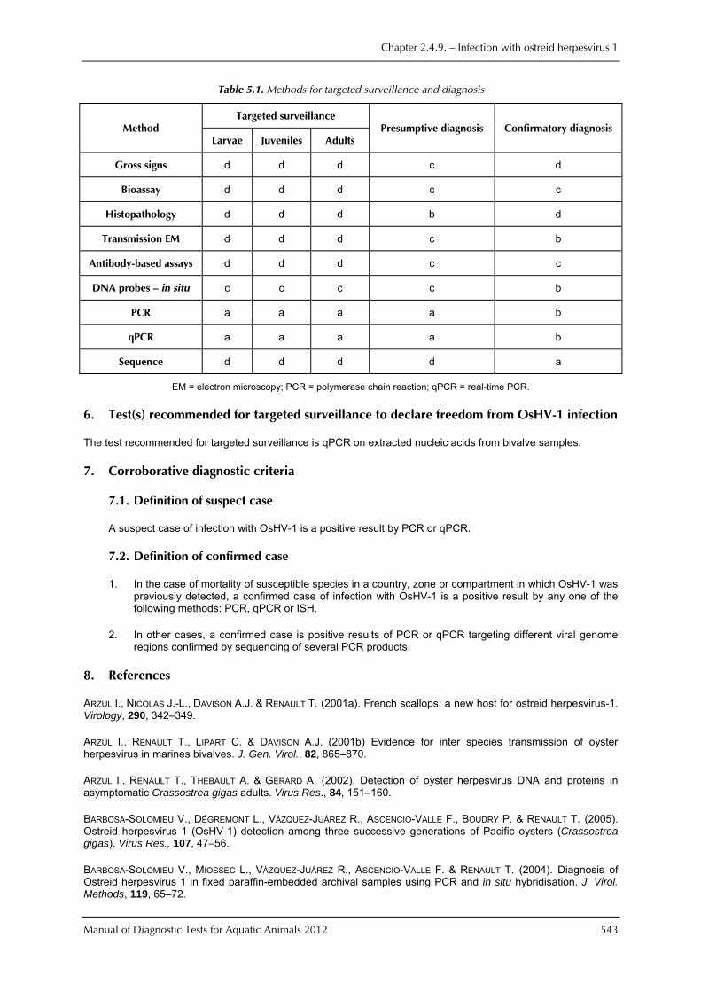
As an example, the methods currently available for targeted surveillance and diagnosis are listed in Table 5.1. The designations used in the Table indicate: a = the method is the recommended method for reasons of availability, utility, and diagnostic specificity and sensitivity; b = the method is a standard method with good diagnostic sensitivity and specificity; c = the method has application in some situations, but cost, accuracy, or other factors severely limits its application; and d = the method is presently not recommended for this purpose.
Chapter 2.4.9. — Infection with ostreid herpesvirus 1
Manual of Diagnostic Tests for Aquatic Animals 2012 543
Table 5.1. Methods for targeted surveillance and diagnosis
Method Targeted surveillance
Presumptive diagnosis Confirmatory diagnosis Larvae Juveniles Adults
Gross signs d d d c d
Bioassay d d d c c
Histopathology d d d b d
Transmission EM d d d c b
Antibody-based assays d d d c c
DNA probes — in situ c c c c b
PCR a a a a b
qPCR a a a a b
Sequence d d d d a
EM = electron microscopy; PCR = polymerase chain reaction; qPCR = real-time PCR.
6. Test(s) recommended for targeted surveillance to declare freedom from OsHV-1 infection
The test recommended for targeted surveillance is qPCR on extracted nucleic acids from bivalve samples.
7. Corroborative diagnostic criteria
7.1. Definition of suspect case
A suspect case of infection with OsHV-1 is a positive result by PCR or qPCR.
7.2. Definition of confirmed case
1. In the case of mortality of susceptible species in a country, zone or compartment in which OsHV-1 was previously detected, a confirmed case of infection with OsHV-1 is a positive result by any one of the following methods: PCR, qPCR or ISH.
2. In other cases, a confirmed case is positive results of PCR or qPCR targeting different viral genome regions confirmed by sequencing of several PCR products.
8. References
ARZUL I., NICOLAS J.-L., DAVISON A.J. & RENAULT T. (2001a). French scallops: a new host for ostreid herpesvirus-1. Virology, 290, 342–349.
ARZUL I., RENAULT T., LIPART C. & DAVISON A.J. (2001b) Evidence for inter species transmission of oyster herpesvirus in marines bivalves. J. Gen. Virol., 82, 865–870.
ARZUL I., RENAULT T., THEBAULT A. & GERARD A. (2002). Detection of oyster herpesvirus DNA and proteins in asymptomatic Crassostrea gigas adults. Virus Res., 84, 151–160.
BARBOSA-SOLOMIEU V., DÉGREMONT L., VÁZQUEZ-JUÁREZ R., ASCENCIO-VALLE F., BOUDRY P. & RENAULT T. (2005). Ostreid herpesvirus 1 (OsHV-1) detection among three successive generations of Pacific oysters (Crassostrea gigas). Virus Res., 107, 47–56.
BARBOSA-SOLOMIEU V., MIOSSEC L., VÁZQUEZ-JUÁREZ R., ASCENCIO-VALLE F. & RENAULT T. (2004). Diagnosis of Ostreid herpesvirus 1 in fixed paraffin-embedded archival samples using PCR and in situ hybridisation. J. Virol. Methods, 119, 65–72.
Chapter 2.4.9. — Infection with ostreid herpesvirus 1
544 Manual of Diagnostic Tests for Aquatic Animals 2012
BATISTA F.M., ARZUL I., PEPIN J.F., RUANO F., FREIDMAN C., BOUDRY P. & RENAULT T. (2007) Detection of ostreid herpesvirus-1 DNA in bivalve molluscs: a critical review. J. Virol. Methods, 139 (1), 1–11.
BURGE C.A., JUDAH L.R., CONQUEST L.L., GRIFFIN F.J., CHENEY D.P., SUHRBIER A., VADOPALAS B., OLIN P.G., RENAULT
T. & FRIEDMAN C.S. (2007). Summer seed mortality of the Pacific oyster, Crassostrea gigas Thunberg grown in Tomales Bay, California, USA: The influence of oyster stock, planting time, pathogens, and environmental stressors. J. Shellfish Res., 26, 163–172.
DAVISON A.J., EBERLE R., EHLERS B., HAYWARD G.S., MCGEOCH D.J., MINSON A.C., PELLETT P.E., ROIZMAN B., STUDDERT M.J. & THIRY E. (2009). The order Herpesvirales. Arch. Virol., 154, 171–177.
DAVISON A.J., TRUS B.L., CHENG N., STEVEN A.C., WATSON M.S., CUNNINGHAM C., LE DEUFF R.M. & RENAULT T. (2005). A novel class of herpesvirus with bivalve hosts. J. Gen. Virol., 86, 41–53.
FRIEDMAN C.S., ESTES R.M., STOKES N.A., BURGE C.A., HARGOVE J.S., BARBER B.J., ELSTON R.A., BURRESON E.M. &
REECE K.S. (2005). Herpes virus in juvenile Pacific oysters Crassostrea gigas from Tomales Bay, California, coincides with summer mortality episodes. Dis. Aquat. Org., 63 (1), 33–41.
GARCIA C., THÉBAULT A., DÉGREMONT L., ARZUL I., MIOSSEC L., ROBERT M., CHOLLET B., FRANÇOIS C., JOLY J.-P., FERRAND S., KERDUDOU N. & RENAULT T. (2011). OsHV-1 detection and relationship with C. gigas spat mortality in France between 1998 and 2006. Vet. Res, 42, 73–84.
LE DEUFF R.M., NICOLAS J.L., RENAULT T. & COCHENNEC N. (1994) Experimental transmission of a herpes-like virus to axenic larvae of Pacific oyster, Crassostrea gigas. Bull. Eur. Assoc. Fish Pathol., 14, 69–72.
LE DEUFF R.-M. & RENAULT T. (1999). Purification and partial genome characterization of a herpes-like virus infecting the Japanese oyster, Crassostrea gigas. J. Gen. Virol., 80, 1317–1322.
LE DEUFF R.-M., RENAULT T. & GERARD A. (1996). Effects of temperature on herpes-like virus detection among hatchery-reared larval Pacific oyster Crassostrea gigas, Dis. Aquat. Org., 24, 149–157.
LIPART C. & RENAULT T. (2002). Herpes-like virus detection in Crassostrea gigas spat using DIG-labelled probes. J. Virol. Methods, 101, 1–10.
MARTENOT C., ODEN E., TRAVAILLE E., MALAS J.P. & HOUSSIN M. (2010). Comparison of two real-time PCR methods for detection of ostreid herpesvirus 1 in the Pacific oyster Crassostrea gigas. J. Virol. Methods, 70 (1–2), 86–99.
MOSS J.A., BURRESON E.M., CORDES J.F., DUNGAN C.F., BROWN G.D., WANG A., WU X. & REECE K.S. (2007). Pathogens in Crassostrea ariakensis and other Asian oyster species: implications for non-native oyster introduction to Chesapeake Bay. Dis. Aquat. Org., 77, 207–223.
PEPIN J. F., RIOU A. & RENAULT T. (2008). Rapid and sensitive detection of ostreid herpesvirus 1 in oyster samples by real-time PCR. J. Virol. Methods, 149, 269–276.
REN W., RENAULT T., CAI Y. & WANG C. (2010) Development of a loop-mediated isothermal amplification assay for rapid and sensitive detection of ostreid herpesvirus 1 DNA. J. Virol. Methods, 170, 30–36.
RENAULT T., COCHENNEC N., LE DEUFF R.M. & CHOLLET B. (1994a) Herpes-like virus infecting Japanese oyster (Crassostrea gigas) spat. Bull. Eur. Assoc. Fish Pathol., 14, 64–66.
RENAULT T., LE DEUFF R.M., COCHENNEC N., CHOLLET B. & MAFFART P. (1995). Herpes-like viruses associated with high mortality levels in larvae and spat of Pacific oysters, Crassostrea gigas: A comparative study, the thermal effects on virus detection in hatchery-reared larvae, reproduction of the disease in axenic larvae. Vet. Res., 26, 539–543.
RENAULT T., LE DEUFF R.-M., COCHENNEC N. & MAFFART P. (1994b). Herpesviruses associated with mortalities among Pacific oyster, Crassostrea gigas, in France – comparative study. Revue de Médecine Vétérinaire, 145, 735–742.
RENAULT T., LE DEUFF R.-M., LIPART C. & DELSERT C. (2000). Development of a PCR procedure for the detection of a herpes-like virus infecting oysters in France. J. Virol. Methods, 88, 41–50.
SAMAIN J.F., DÉGREMONT L., SOLETCHNIK P., HAURE J., BÉDIER E., ROPERT M., MOAL J., HUVET A., BACCA H., VAN
WORMHOUDT A., DELAPORTE M., COSTIL K., POUVREAU S., LAMBERT C., BOULO V., SOUDANT P., NICOLAS J.L., LE ROUX
Chapter 2.4.9. — Infection with ostreid herpesvirus 1
Manual of Diagnostic Tests for Aquatic Animals 2012 545
F., RENAULT T., GAGNAIRE B., GERTET F.,BOUTET I., BURGEOT T. & BOUDRY P. (2007). Genetically based resistance to summer mortality in the Pacific oyster (Crassostrea gigas) and its relationship with physiological, immunological characteristics and infection processes. Aquaculture, 268 (1–4), 227–243.
SAUVAGE C., PEPIN J.F., LAPEGUE S., BOUDRY P. & RENAULT T. (2009). Ostreid herpes virus 1 infection in families of the Pacific oyster, Crassostrea gigas, during a summer mortality outbreak: difference in viral DNA detection and quantification using real-time PCR. Virus Res., 142, 181–187.
SCHIKORSKI D., FAURY N., PEPIN J.F., SAULNIER D., TOURBIEZ D. & RENAULT T. (2011a). Experimental ostreid herpesvirus 1 infection of the Pacific oyster Crassostrea gigas: kinetics of virus DNA detection by q-PCR in seawater and in oyster samples. Virus Res., 155 (1), 28–34.
SCHIKORSKI D., RENAULT T., SAULNIER D., FAURY N., MOREAU P. & PEPIN J.F. (2011b). Experimental infection of Pacific oyster Crassostrea gigas spat by ostreid herpesvirus 1: demonstration of oyster spat susceptibility. Vet. Res., 42, 1–13.
SEGARRA A., PEPIN J.F., ARZUL I., MORGA B., FAURY N. & RENAULT T. (2010). Detection and description of a particular Ostreid herpesvirus 1 genotype associated with massive mortality outbreaks of Pacific oysters, Crassostrea gigas, in France in 2008. Virus Res., 153, 92–95.
SOLETCHNIK P., LE MOINE O., FAURY N., RAZET D., GEAIRON P. & GOULLETQUER P. (1999). Mortalité de l’huître Crassostrea gigas dans le bassin de Marennes-Oléon : étude de la variabilité spatiale de son environnement et de sa biologie par un système d’informations géographiques (SIG), Aquatic Living Resources,. 12, 131–143.
VÁSQUE YEOMANS R., GARCÍA-ORTEGA M. & CÁCERES-MARTÍNEZ J. (2010). Gill erosion and herpesvirus in Crassostrea gigas cultured in Baja California, Mexico. Dis. Aquat. Org., 89, 137–144.
VIGNERON V., SOLLIEC G., MONTANIE H. & RENAULT T. (2004). Detection of ostreid herpes virus 1 (OsHV-1) DNA in seawater by PCR: influence of water parameters in bioassays. Dis. Aquat. Org., 62, 35–44.
WALLIS C. & MELNICK J. (1965). Thermostabilization and thermosensitization of herpesvirus. J. Bacteriol., 90, 1632–1637.
WEBB S.C., FIDLER A. & RENAULT T. (2007). Primers for PCR-based detection of ostreid herpes virus-1 (OsHV-1): Application in a survey of New Zealand molluscs. Aquaculture, 272, 126–139.
* * *