2404 Targeting the Cancer-Stroma Interaction: A Potential
13
See discussions, stats, and author profiles for this publication at: https://www.researchgate.net/publication/221868626 Targeting the Cancer-Stroma Interaction: A Potential Approach for Pancreatic Cancer Treatment Article in Current Pharmaceutical Design · February 2012 DOI: 10.2174/13816128112092404 · Source: PubMed CITATIONS 55 READS 653 6 authors, including: Some of the authors of this publication are also working on these related projects: Arginine metabolism and pancreatic cancer View project CSC and salinomycin View project Xuqi Li Xi'an Jiaotong University 100 PUBLICATIONS 2,241 CITATIONS SEE PROFILE Qingyong Ma Xi'an Jiaotong University 279 PUBLICATIONS 6,468 CITATIONS SEE PROFILE Qinhong Xu Xi'an Jiaotong University 83 PUBLICATIONS 2,410 CITATIONS SEE PROFILE Wanxing Duan Xi'an Jiaotong University 95 PUBLICATIONS 2,448 CITATIONS SEE PROFILE All content following this page was uploaded by Xuqi Li on 04 June 2014. The user has requested enhancement of the downloaded file.
Transcript of 2404 Targeting the Cancer-Stroma Interaction: A Potential

See discussions, stats, and author profiles for this publication at: https://www.researchgate.net/publication/221868626
Targeting the Cancer-Stroma Interaction: A Potential Approach for Pancreatic
Cancer Treatment
Article in Current Pharmaceutical Design · February 2012
DOI: 10.2174/13816128112092404 · Source: PubMed
CITATIONS
55READS
653
6 authors, including:
Some of the authors of this publication are also working on these related projects:
Arginine metabolism and pancreatic cancer View project
CSC and salinomycin View project
Xuqi Li
Xi'an Jiaotong University
100 PUBLICATIONS 2,241 CITATIONS
SEE PROFILE
Qingyong Ma
Xi'an Jiaotong University
279 PUBLICATIONS 6,468 CITATIONS
SEE PROFILE
Qinhong Xu
Xi'an Jiaotong University
83 PUBLICATIONS 2,410 CITATIONS
SEE PROFILE
Wanxing Duan
Xi'an Jiaotong University
95 PUBLICATIONS 2,448 CITATIONS
SEE PROFILE
All content following this page was uploaded by Xuqi Li on 04 June 2014.
The user has requested enhancement of the downloaded file.
2404 Current Pharmaceutical Design, 2012, 18, 2404-2415
1873-4286/12 $58.00+.00 © 2012 Bentham Science Publishers
Targeting the Cancer-Stroma Interaction: A Potential Approach for Pancreatic Cancer Treatment
Xuqi Li1, Qingyong Ma
1*, Qinhong Xu
1, Wanxing Duan
1, Jianjun Lei
1 and Erxi Wu
2
1Department of Hepatobiliary Surgery, First Affiliated Hospital of Medical College, Xi’an Jiaotong University, Xi’an 710061,
Shaanxi, China; 2Department of Pharmaceutical Sciences, North Dakota State University, Fargo, ND 58105, USA
Abstract: Recent studies have demonstrated that the interaction between the cancer and the stroma, play a key role in the development of pancreatic cancer. The desmoplasia, which consists of fibroblasts, pancreatic stellate cells, lymphatic and vascular endothelial cells, im-
mune cells, pathologic increased nerves, and the extracellular matrix (ECM), creates a complex tumor microenvironment that promotes pancreatic cancer development, invasion, metastasis, and resistance to chemotherapy. Thus, the potential approach for targeting the com-
ponents of this desmoplastic reaction or the pancreatic tumor microenvironment might represent a novel therapeutic approach to ad-vanced pancreatic carcinoma. Novel therapies that target on the pancreatic tumor microenvironment should become one of the more ef-
fective treatments for pancreatic cancer.
Keywords: Pancreas carcinoma, stroma, tumor desmoplasia, mechanism, anticancer treatment, therapeutic targets.
INTRODUCTION
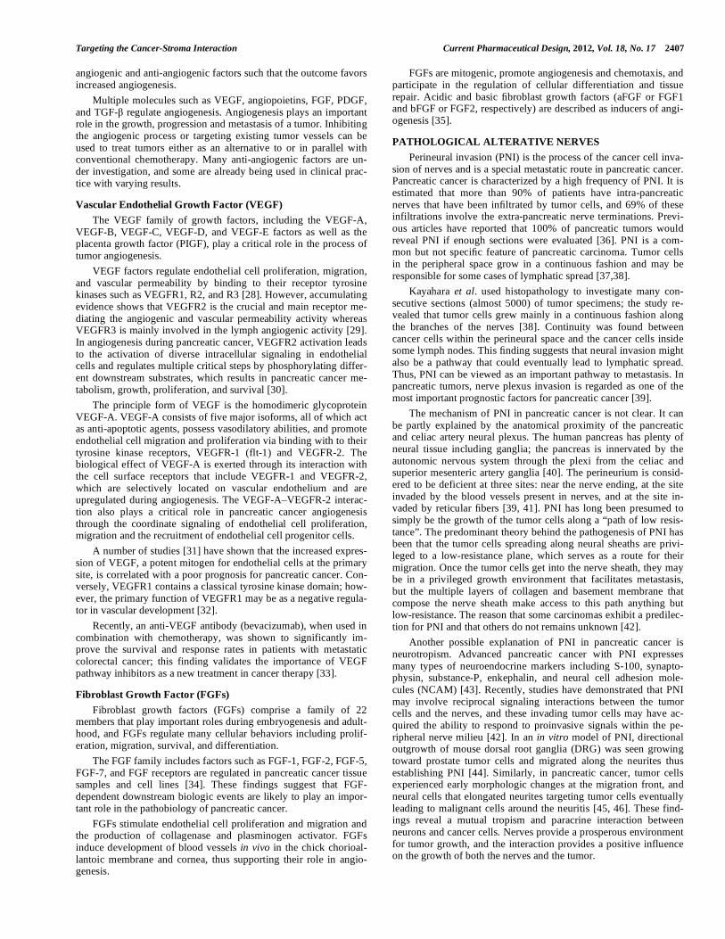
Pancreatic cancer is the most lethal human malignancy and has a ratio of death to incidence up to 96% [1]. Pancreatic adenocarci-noma is locally invasive and is surrounded by a dense desmoplastic reaction, which can involve adjacent vital structures. The desmo-plasia, which consists of fibroblasts, pancreatic stellate cells, lym-phatic and vascular endothelial cells, immune cells, pathologic increased nerves and the ECM, creates a complex tumor microenvi-ronment that promotes pancreatic cancer development, invasion, metastasis and resistance to chemotherapy (Fig. (1)). The molecular mechanisms of the tumor-stroma interaction are very complex. This review focuses on the mechanisms by which the interaction be-tween the cancer cell and the stroma influence pancreatic cancer progression. Studies on the pancreatic tumor microenvironment will bring new concepts that will ultimately contribute to new diagnoses and treatments for this disease.
PANCREATIC STELLATE CELLS
From a histological point of view, pancreatic cancers may have the most prominent stromal reaction of all of the epithelial tumors; there is a remarkable increase in the connective tissue that infil-trates and envelopes the neoplasm [2]. Studies of human pancreatic cancer have shown that the mesenchymal cells secrete many of cytokines such as insulin-like growth factor-I (IGF-I) and fibroblast growth factor (FGF), which have an impact on disease prognosis (Fig. (2)). Because it is possible to isolate and culture pancreatic stellate cells (PSC) in vitro [3, 4] and these cells are responsible for producing the stromal reaction in pancreatic cancer [5], determining which mechanisms mediate the epithelial-stromal interactions in pancreatic cancer is important.
In healthy tissue, PSCs are quiescent; in diseased states, under the influence of growth factors, cytokines, and oxidative stress, PSCs are activated and adopt a myofibroblast-like phenotype and then secret excess amounts of extracellular matrix (ECM) and ma-trix degrading enzymes [3]. Growth factors that are known to in-duce PSC activation such as transforming growth factor-h1 (TGF-h1), platelet-derived growth factor (PDGF) and vascular endothelial growth factor (VEGF) are secreted by pancreatic cancer cells
*Address correspondence to this author at the Department of Hepatobiliary
Surgery, The First Affiliated Hospital of Medical College, Xi’an Jiaotong University, 277 West Yanta Road, Xi’an 710061, Shaanxi, China; Tel: +86
29 8532 3899; Fax: +86 29 8532 3899; E-mail: [email protected]
[5, 6]. Additionally, it has been shown that cancer cells can also secrete the ECM metalloproteinase inducer (EMMPRIN) [4]. This secretion leads to increased matrix metalloproteinase 2 secretions by PSCs; matrix metalloproteinase 2 has been associated with the invasive phenotype of pancreatic cancer cell lines [7].
PSCs can also act on pancreatic cancer cells, which affect their biological behavior. How PSCs and the desmoplasia promote the growth of tumor cells in adenocarcinomas is only partially under-stood [8].
Proliferation and Apoptosis
The growth rate of a tumor that forms when both cancer cells and PSCs are injected subcutaneously into the flanks of nude mice is significantly increased compared to the tumors that form when only the cancer cells are injected [6]. As opposed to the tumors that are initiated by injection of only the cancer cells, the tumors initi-ated with the co-injection of cancer cells and PSCs have a desmo-plasia similar to that observed in human pancreatic adenocarcinoma [6]. The growth advantage of pancreatic cancer cells in the presence of PSCs may be mediated by two mechanisms: increased mitosis and decreased programmed cell death (apoptosis). As observed in the co-injection model [9], PSC secretions displayed a marked in-fluence on the promotion of cancer cell proliferation, and this effect was partially abolished by neutralizing antibodies against the mito-genic factor PDGF. Other factors secreted by PSCs such as stromal-derived factor-1, EGF, IGF-1, or FGF are also likely to exert mito-genic effects on cancer cells, and studies examining the role of these and other factors are currently underway [2].
Resistance to apoptosis is a common trait of many tumors. It has been shown that PSCs reduce basal level of apoptosis in various pancreatic cancer cell lines in vitro [10, 11]. Importantly, when mice were injected with PSCs and cancer cells, this led to reduced apoptosis in vivo; this result is in contrast to animals that were only injected with cancer cells. The resistance to apoptosis could be related to an increased expression of anti-apoptotic proteins such as Bcl-2 and Bcl-xL. Additionally, growth factors such as IGF-1 and basic fibroblast growth factor (bFGF) may mediate the anti-apoptotic effects of pancreatic cancer cells. PSCs are myofibro-blast-like cells that are found in the areas of the pancreas with an exocrine function [8], and abundant producers of ECM compo-nents, such as various types of collagen. A conceptually novel study proved the importance of ECM stiffening, which is an abundance of collagen cross-linking, in the process of tumor growth and invasion

Targeting the Cancer-Stroma Interaction Current Pharmaceutical Design, 2012, Vol. 18, No. 17 2405
[12]. During the malignant progression of the oncogene-expressing breast epithelium in transgenic mice, an increase in ECM stiffening and activation of the prototypical PI3 kinase and Akt survival sig-naling pathway synergestically caused the downstream ligation of integrin by collagen fibers [13].
Invasion and Metastasis
As cancers progress into the malignant state, they acquire the characteristic of invading the surrounding tissue and seeding metas-tases in the lymphatic system or blood vessels. For invasion to oc-cur, tumor cells need to attain a migratory phenotype, which leads to extensive remodeling of the surrounding ECM. Compared with mice injected with pancreatic cancer cells alone, mice co-injected with human PSCs and cancer cells showed regional and distant metastasis and fibrotic bands (desmoplasia) containing activated PSCs within the tumors [9, 14]. PSCs contribute to the invasive and metastatic process by inducing the epithelial to mesenchymal tran-sition of pancreatic cancer cells [15]. Pancreatic cancer cells co-cultured with PSCs exhibited loose cell contacts and a scattered, fibroblast-like appearance. The expression of E-cadherin, cy-tokeratin 19, and membrane-associated -catenin decreased in can-cer cells co-cultured with PSCs relative to cells cultured without PSCs whereas vimentin and Snail (Snail-1) expression increased. The migration of pancreatic cancer cells was increased when they were co-cultured with PSCs. The decrease of E-cadherin expression induced by PSCs was not altered by treatment with an anti-TGF- -neutralizing antibody, which excludes a central role for TGF- in this process [15]. Moreover, tumor-derived pancreatic stellate cells
stimulate pancreatic cancer cell invasion possibly through release of thrombospondin-2 [16]. Additionally, 1 integrins play an essential role in the adhesion and invasion of pancreatic carcinoma cells [11].
Chemotherapy Resistance
The stromal compartment not only provides an abundance of stroma-derived factors that facilitate cancer initiation, growth and progression but also has an effect on the therapeutic outcome and provides ample opportunities for drug targeting. Pancreatic stellate cells could inhibit the effects of chemotherapy and radiation on tumor cells [14]. Indeed, a stroma-derived gene expression pattern can predict the clinical outcome in breast cancer [17]. Further evi-dence supporting the theory that cancer associated fibroblasts (CAFs) can determine therapeutic outcome in breast cancer patients came from the demonstration that a CAF gene signature is predic-tive of the response to neoadjuvant chemotherapy [18]. Moreover, preclinical studies suggest that CAFs may also regulate the resis-tance to antiangiogenic therapy and targeted therapy that uses epi-dermal growth factor receptor tyrosine kinase inhibitors [19, 20]. In addition to directly modulating the sensitivity of tumor cells to anticancer agents, PDGF receptors of CAFs increase interstitial hypertension and reduce transcapillary transport in tumors to influ-ence the transcapillary transport of drugs [21, 22].
Targeting Therapy
The cell surface serine protease fibroblast activation protein (FAP) is selectively expressed on tumor-associated fibroblasts and pericytes in epithelial tumors; a study showed that genetic deletion
Fig. (1). Desmoplasia. The desmoplasia consisting of fibroblasts, pancreatic stellate cells, lymphatic and vascular endothelial cells, immune cells, pathologic
increased nerves, and ECM creates a complex tumor microenvironment that promotes pancreatic cancer development, invasion, metastasis and resistance to
chemotherapy. Targeting the interaction between cancer and stroma in the pancreatic tumor microenvironment may contribute to the design of new diagnosis
techniques and treatments for this disease.

2406 Current Pharmaceutical Design, 2012, Vol. 18, No. 17 Li et al.
and pharmacologic inhibition of FAP inhibited tumor growth in mouse models of epithelial-derived solid tumors. The results indi-cated that FAP depletion inhibits the tumor cell proliferation indi-rectly, increases the accumulation of collagen, decreases the myofi-broblast content, and decreases the blood vessel density in tumors [23]. The inhibition of the stromal PDGF receptors reduced the proliferation and angiogenesis in cervical lesions by suppressing the expression of the angiogenic factor FGF-2 and the epithelial cell growth factor FGF-7, which are secreted by CAFs. These effects were recapitulated using neutralizing antibodies against the PDGF receptors. When treated with a ligand trap for the FGFs, the angio-genic phenotype was impaired, and this effect was similar to treat-ment with imatinib [24]. A study showed that the ligand-dependent activation of the hedgehog (Hh) pathway in the stromal microenvi-ronment is a paracrine requirement for Hh signaling in cancer. Spe-cific inhibition of Hh signaling using small molecule inhibitors, a neutralizing anti-Hh antibody or genetic deletion of smoothened (Smo) in the mouse stroma leads to growth inhibition in xenograft tumor models [25]. Moreover, in a mouse model of pancreatic can-cer, a co-administration of gemcitabine and IPI-926, a drug that depletes tumor-associated stromal tissue via inhibition of Hedgehog signaling pathway, transiently increase the intratumoral vascular density and intratumoral concentration of gemcitabine and thus lead to a transient stabilization of the disease [26].
VASCULAR MICROENVIRONMENT
The tumor microenvironment is extremely complex and de-pends on the interaction between the tumor cells and responding host cells. Angiogenesis, new blood vessel growth from the preex-isting vasculature, is a preeminent feature of the successful growth of all solid tumors. As one of the hallmarks of cancer of the exo-crine pancreas, angiogenesis is an essential event that is involved in pancreatic cancer progression and metastasis.
The proliferative index of tumors decreases as the distance from the nearest capillary blood vessel increases, and the rapid exponen-tial growth of tumors is dependent on the vascularization of the
tumor mass. Without angiogenesis, pancreatic tumors are limited in size by the distance that oxygen can diffuse, namely, 1–2 mm. Hy-poxia results when the rate of new blood vessel growth is exceeded by the growth of the tumor. Hypoxia in pancreatic cancer results in changes at a transcriptional level, which alter cellular metabolism and stimulates angiogenesis [27].
Angiogenesis is not necessarily linked to invasive pancreatic cancer, but it is an early event in pancreatic cancer genesis; the process of angiogenesis consists of multiple, sequential, and inter-dependent steps with the involvement of myriad positive and nega-tive regulators of angiogenesis. The survival of pancreatic cancers and their metastases are dependent on the balance of endogenous
Fig. (2). The interaction between pancreatic cancer cells and PSC. Selected mediators and their effects on biological behavior changes are depicted. Trans-
forming growth factor- (TGF- ), platelet-derived growth factor (PDGF), and VEGF, which are secreted by pancreatic cancer cells, are known to induce PSC
activation. Under the influence of these growth factors, PSCs are activated and transformed into a myofibroblast-like phenotype; the PSCs then secret excess
amounts of extracellular matrix (ECM) and matrix degrading enzymes to remodel the cancer microenvironment. Simultaneously, the transformation promotes
their proliferation and migration capacity. PSCs can also act on pancreatic cancer cells, which can affect their biological behavior. PSCs have a marked influ-
ence on promoting cancer cell proliferation through PDGF. Stromal-derived factor-1(SDF-1), EGF, IGF-1, or FGF, when secreted by PSCs, are also likely to
display mitogenic effects on cancer cells. EMMPRIN, which is secreted by cancer cells, can increase the MMP-2 secretion by PSCs; MMP-2 has been associ-
ated with the invasive phenotype of pancreatic cancer cell lines. Resistance to apoptosis might be related to the increased expression of the anti-apoptotic pro-
teins Bcl-2 and Bcl-xL and of the growth factors IGF-1 and FGF. Tumor-derived pancreatic stellate cells stimulate pancreatic cancer cell invasion likely
through the release of thrombospondin-2 and 1 integrins. PDGF receptors of PSCs increase interstitial hypertension and reduce transcapillary transport in
tumors and thus influence the transcapillary transport of drugs. SDF-1 can also influence the therapeutic outcome.
Targeting the Cancer-Stroma Interaction Current Pharmaceutical Design, 2012, Vol. 18, No. 17 2407
angiogenic and anti-angiogenic factors such that the outcome favors increased angiogenesis.
Multiple molecules such as VEGF, angiopoietins, FGF, PDGF, and TGF- regulate angiogenesis. Angiogenesis plays an important role in the growth, progression and metastasis of a tumor. Inhibiting the angiogenic process or targeting existing tumor vessels can be used to treat tumors either as an alternative to or in parallel with conventional chemotherapy. Many anti-angiogenic factors are un-der investigation, and some are already being used in clinical prac-tice with varying results.
Vascular Endothelial Growth Factor (VEGF)
The VEGF family of growth factors, including the VEGF-A, VEGF-B, VEGF-C, VEGF-D, and VEGF-E factors as well as the placenta growth factor (PIGF), play a critical role in the process of tumor angiogenesis.
VEGF factors regulate endothelial cell proliferation, migration, and vascular permeability by binding to their receptor tyrosine kinases such as VEGFR1, R2, and R3 [28]. However, accumulating evidence shows that VEGFR2 is the crucial and main receptor me-diating the angiogenic and vascular permeability activity whereas VEGFR3 is mainly involved in the lymph angiogenic activity [29]. In angiogenesis during pancreatic cancer, VEGFR2 activation leads to the activation of diverse intracellular signaling in endothelial cells and regulates multiple critical steps by phosphorylating differ-ent downstream substrates, which results in pancreatic cancer me-tabolism, growth, proliferation, and survival [30].
The principle form of VEGF is the homodimeric glycoprotein VEGF-A. VEGF-A consists of five major isoforms, all of which act as anti-apoptotic agents, possess vasodilatory abilities, and promote endothelial cell migration and proliferation via binding with to their tyrosine kinase receptors, VEGFR-1 (flt-1) and VEGFR-2. The biological effect of VEGF-A is exerted through its interaction with the cell surface receptors that include VEGFR-1 and VEGFR-2, which are selectively located on vascular endothelium and are upregulated during angiogenesis. The VEGF-A–VEGFR-2 interac-tion also plays a critical role in pancreatic cancer angiogenesis through the coordinate signaling of endothelial cell proliferation, migration and the recruitment of endothelial cell progenitor cells.
A number of studies [31] have shown that the increased expres-sion of VEGF, a potent mitogen for endothelial cells at the primary site, is correlated with a poor prognosis for pancreatic cancer. Con-versely, VEGFR1 contains a classical tyrosine kinase domain; how-ever, the primary function of VEGFR1 may be as a negative regula-tor in vascular development [32].
Recently, an anti-VEGF antibody (bevacizumab), when used in combination with chemotherapy, was shown to significantly im-prove the survival and response rates in patients with metastatic colorectal cancer; this finding validates the importance of VEGF pathway inhibitors as a new treatment in cancer therapy [33].
Fibroblast Growth Factor (FGFs)
Fibroblast growth factors (FGFs) comprise a family of 22 members that play important roles during embryogenesis and adult-hood, and FGFs regulate many cellular behaviors including prolif-eration, migration, survival, and differentiation.
The FGF family includes factors such as FGF-1, FGF-2, FGF-5, FGF-7, and FGF receptors are regulated in pancreatic cancer tissue samples and cell lines [34]. These findings suggest that FGF-dependent downstream biologic events are likely to play an impor-tant role in the pathobiology of pancreatic cancer.
FGFs stimulate endothelial cell proliferation and migration and the production of collagenase and plasminogen activator. FGFs induce development of blood vessels in vivo in the chick chorioal-lantoic membrane and cornea, thus supporting their role in angio-genesis.
FGFs are mitogenic, promote angiogenesis and chemotaxis, and participate in the regulation of cellular differentiation and tissue repair. Acidic and basic fibroblast growth factors (aFGF or FGF1 and bFGF or FGF2, respectively) are described as inducers of angi-ogenesis [35].
PATHOLOGICAL ALTERATIVE NERVES
Perineural invasion (PNI) is the process of the cancer cell inva-sion of nerves and is a special metastatic route in pancreatic cancer. Pancreatic cancer is characterized by a high frequency of PNI. It is estimated that more than 90% of patients have intra-pancreatic nerves that have been infiltrated by tumor cells, and 69% of these infiltrations involve the extra-pancreatic nerve terminations. Previ-ous articles have reported that 100% of pancreatic tumors would reveal PNI if enough sections were evaluated [36]. PNI is a com-mon but not specific feature of pancreatic carcinoma. Tumor cells in the peripheral space grow in a continuous fashion and may be responsible for some cases of lymphatic spread [37,38].
Kayahara et al. used histopathology to investigate many con-secutive sections (almost 5000) of tumor specimens; the study re-vealed that tumor cells grew mainly in a continuous fashion along the branches of the nerves [38]. Continuity was found between cancer cells within the perineural space and the cancer cells inside some lymph nodes. This finding suggests that neural invasion might also be a pathway that could eventually lead to lymphatic spread. Thus, PNI can be viewed as an important pathway to metastasis. In pancreatic tumors, nerve plexus invasion is regarded as one of the most important prognostic factors for pancreatic cancer [39].
The mechanism of PNI in pancreatic cancer is not clear. It can be partly explained by the anatomical proximity of the pancreatic and celiac artery neural plexus. The human pancreas has plenty of neural tissue including ganglia; the pancreas is innervated by the autonomic nervous system through the plexi from the celiac and superior mesenteric artery ganglia [40]. The perineurium is consid-ered to be deficient at three sites: near the nerve ending, at the site invaded by the blood vessels present in nerves, and at the site in-vaded by reticular fibers [39, 41]. PNI has long been presumed to simply be the growth of the tumor cells along a “path of low resis-tance”. The predominant theory behind the pathogenesis of PNI has been that the tumor cells spreading along neural sheaths are privi-leged to a low-resistance plane, which serves as a route for their migration. Once the tumor cells get into the nerve sheath, they may be in a privileged growth environment that facilitates metastasis, but the multiple layers of collagen and basement membrane that compose the nerve sheath make access to this path anything but low-resistance. The reason that some carcinomas exhibit a predilec-tion for PNI and that others do not remains unknown [42].
Another possible explanation of PNI in pancreatic cancer is neurotropism. Advanced pancreatic cancer with PNI expresses many types of neuroendocrine markers including S-100, synapto-physin, substance-P, enkephalin, and neural cell adhesion mole-cules (NCAM) [43]. Recently, studies have demonstrated that PNI may involve reciprocal signaling interactions between the tumor cells and the nerves, and these invading tumor cells may have ac-quired the ability to respond to proinvasive signals within the pe-ripheral nerve milieu [42]. In an in vitro model of PNI, directional outgrowth of mouse dorsal root ganglia (DRG) was seen growing toward prostate tumor cells and migrated along the neurites thus establishing PNI [44]. Similarly, in pancreatic cancer, tumor cells experienced early morphologic changes at the migration front, and neural cells that elongated neurites targeting tumor cells eventually leading to malignant cells around the neuritis [45, 46]. These find-ings reveal a mutual tropism and paracrine interaction between neurons and cancer cells. Nerves provide a prosperous environment for tumor growth, and the interaction provides a positive influence on the growth of both the nerves and the tumor.
2408 Current Pharmaceutical Design, 2012, Vol. 18, No. 17 Li et al.
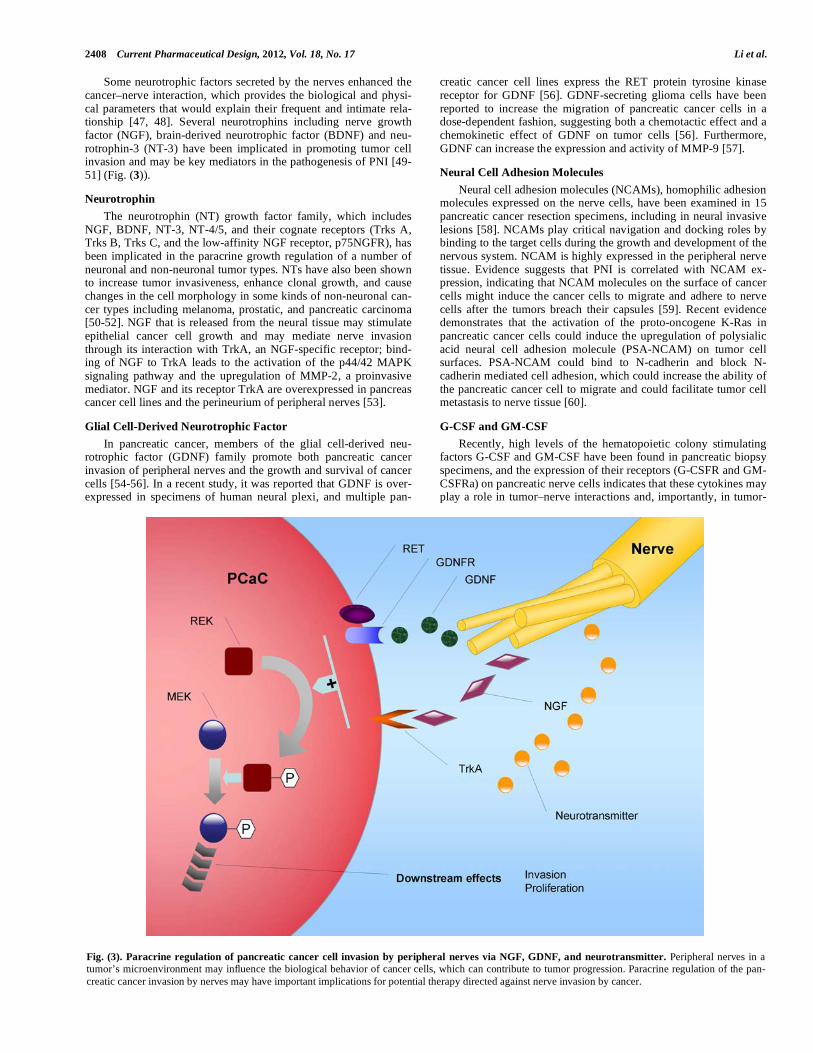
Some neurotrophic factors secreted by the nerves enhanced the cancer–nerve interaction, which provides the biological and physi-cal parameters that would explain their frequent and intimate rela-tionship [47, 48]. Several neurotrophins including nerve growth factor (NGF), brain-derived neurotrophic factor (BDNF) and neu-rotrophin-3 (NT-3) have been implicated in promoting tumor cell invasion and may be key mediators in the pathogenesis of PNI [49-51] (Fig. (3)).
Neurotrophin
The neurotrophin (NT) growth factor family, which includes NGF, BDNF, NT-3, NT-4/5, and their cognate receptors (Trks A, Trks B, Trks C, and the low-affinity NGF receptor, p75NGFR), has been implicated in the paracrine growth regulation of a number of neuronal and non-neuronal tumor types. NTs have also been shown to increase tumor invasiveness, enhance clonal growth, and cause changes in the cell morphology in some kinds of non-neuronal can-cer types including melanoma, prostatic, and pancreatic carcinoma [50-52]. NGF that is released from the neural tissue may stimulate epithelial cancer cell growth and may mediate nerve invasion through its interaction with TrkA, an NGF-specific receptor; bind-ing of NGF to TrkA leads to the activation of the p44/42 MAPK signaling pathway and the upregulation of MMP-2, a proinvasive mediator. NGF and its receptor TrkA are overexpressed in pancreas cancer cell lines and the perineurium of peripheral nerves [53].
Glial Cell-Derived Neurotrophic Factor
In pancreatic cancer, members of the glial cell-derived neu-rotrophic factor (GDNF) family promote both pancreatic cancer invasion of peripheral nerves and the growth and survival of cancer cells [54-56]. In a recent study, it was reported that GDNF is over-expressed in specimens of human neural plexi, and multiple pan-
creatic cancer cell lines express the RET protein tyrosine kinase receptor for GDNF [56]. GDNF-secreting glioma cells have been reported to increase the migration of pancreatic cancer cells in a dose-dependent fashion, suggesting both a chemotactic effect and a chemokinetic effect of GDNF on tumor cells [56]. Furthermore, GDNF can increase the expression and activity of MMP-9 [57].
Neural Cell Adhesion Molecules
Neural cell adhesion molecules (NCAMs), homophilic adhesion molecules expressed on the nerve cells, have been examined in 15 pancreatic cancer resection specimens, including in neural invasive lesions [58]. NCAMs play critical navigation and docking roles by binding to the target cells during the growth and development of the nervous system. NCAM is highly expressed in the peripheral nerve tissue. Evidence suggests that PNI is correlated with NCAM ex-pression, indicating that NCAM molecules on the surface of cancer cells might induce the cancer cells to migrate and adhere to nerve cells after the tumors breach their capsules [59]. Recent evidence demonstrates that the activation of the proto-oncogene K-Ras in pancreatic cancer cells could induce the upregulation of polysialic acid neural cell adhesion molecule (PSA-NCAM) on tumor cell surfaces. PSA-NCAM could bind to N-cadherin and block N-cadherin mediated cell adhesion, which could increase the ability of the pancreatic cancer cell to migrate and could facilitate tumor cell metastasis to nerve tissue [60].
G-CSF and GM-CSF
Recently, high levels of the hematopoietic colony stimulating factors G-CSF and GM-CSF have been found in pancreatic biopsy specimens, and the expression of their receptors (G-CSFR and GM-CSFRa) on pancreatic nerve cells indicates that these cytokines may play a role in tumor–nerve interactions and, importantly, in tumor-
Fig. (3). Paracrine regulation of pancreatic cancer cell invasion by peripheral nerves via NGF, GDNF, and neurotransmitter. Peripheral nerves in a
tumor’s microenvironment may influence the biological behavior of cancer cells, which can contribute to tumor progression. Paracrine regulation of the pan-
creatic cancer invasion by nerves may have important implications for potential therapy directed against nerve invasion by cancer.

Targeting the Cancer-Stroma Interaction Current Pharmaceutical Design, 2012, Vol. 18, No. 17 2409
induced pain [61]. The presence of hematopoietic factors in the tumor microenvironment is a very common finding, and hema-topoietic factors are known to act on both myeloid and tumor cells, which stimulates the proliferation of the cells [62].
Myelin-Associated Glycoprotein
Myelin-associated glycoprotein (MAG) is a membrane-bound protein expressed by myelinating Schwann cells in the periaxonal membrane. On Schwann cells, MAG binds both to gangliosides on the axons and to the mucin, MUC1, expressed by pancreatic tumor cells [63]. Laminin-5 released from the cancer cells has been re-ported to correlate with PNI in head and neck squamous carcino-mas, which supports the hypothesis that the deposition of basement membrane components may be required in the process of nerve invasion [64].
Chemokines
A recent study indicated that tumor cells from human pancre-atic cancers strongly upregulate the chemokine receptor CX3CR1, which is not expressed in the normal pancreatic epithelium, and that the CX3CL1/CX3CR1 axis mediates PNI in pancreatic cancer [65]. CX3CR1 exclusively binds the transmembrane chemokine CX3CL1 (also known as fractalkine or neurotactin) expressed by neurons, nerve fibers and activated endothelial cells [66-69]. CX3CR1+ pancreatic ductal adenocarcinoma (PDAC) tumor cells migrate in response to the ligand CX3CL1 and specifically adhere to neural cells. To determine the relationship between the high
CX3CR1 expression by tumor cells and neural tropism, a system-atic evaluation of perineural invasion was performed. High expres-sion of the receptor was significantly associated with pathologically detected prominent PNI. The biological significance of PNI and the expression of CX3CR1 in pancreatic cancer were investigated in the clinical outcome of the patients. Higher CX3CR1 expression and perineural invasion were strongly associated with local and earlier tumor recurrence. Thus, CX3CR1 expression by cancer cells is an independent factor predicting local tumor recurrence in re-sected pancreatic carcinoma patients [65].
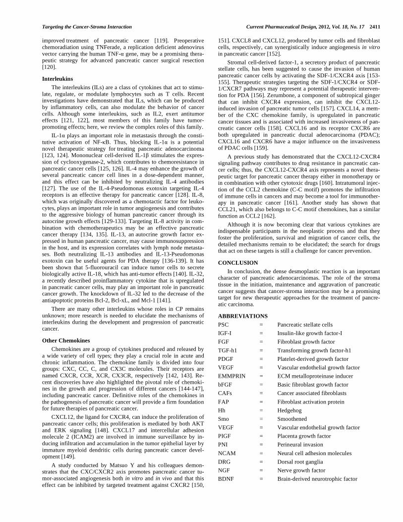
ECM
One of the most important features of PDAC is the develop-ment of the desmoplastic reaction around tumor cells; this phe-nomenon is mainly due to excess ECM production, which is mainly produced by the activated pancreatic stellate cells [70-72]. The ECM, which is composed of collagens, noncollagenous glycopro-teins, glycosaminoglycans, proteoglycans, proteinases, growth fac-tors, and matricellular proteins, is an important component of the tumor microenvironment. Koninger et al. has described that malig-nant cells can alter the composition of the ECM and transform their microenvironment in a tumor-favorable way. Although some stud-ies focus on the tumor's impact on its microenvironment [73], much research focuses on the reverse effect: the ability of the surrounding microenvironment to influence the tumor. The ECM is one of the main constituents of the tumor microenvironment; however, it has been thought that the dense ECM around pancreatic cancer cells
Fig. (4). Excess extracellular matrix (ECM) production in pancreatic tumor microenvironment. From a histologic standpoint, the prominent stromal
reaction in the pancreatic tumor local microenvironment is one of the most remarkable characteristics of pancreatic cancers, and the stroma is thought to be an
active participant in the process of pancreatic cancer initiation, progression, metastasis and chemoresistance. Increased deposition of the ECM, which is mainly
composed of collagens, glycoproteins, proteoglycans, proteinases, and growth factors, is the culprit of this desmoplastic reaction. Most members of the ECM,
including collagens, fibronectins, and laminins, can bind to integrins, a large family of type I transmembrane heterodimeric receptors that play key roles in
downstream cell processes. When several members of the ECM bind to integrins, they then cause the activation of multiple downstream signaling cascades,
which include the Src–FAK, Ras–MEK–MAPK, and PI3K signaling pathways that control biological and cellular functions such as cell adhesion, migration,
proliferation, cell differentiation, and apoptosis. A more complete understanding of the interactions between the altered ECM surrounding tumor cells and
pancreatic ductal adenocarcinoma will help to elucidate the mechanisms of tumor biological behaviors and to develop new targets for pancreatic cancer pre-
vention.

2410 Current Pharmaceutical Design, 2012, Vol. 18, No. 17 Li et al.
may represent a host barrier against malignant invasion [74]. Ac-cumulating evidence shows that the ECM is an important compo-nent that regulates the biological behaviors of pancreatic cancer such as tumor growth, differentiation, survival and motility [75, 76] (Fig. (4)).
Collagens
Collagens are the main ingredients of the ECM. Abnormal ex-pression of type I collagen, the predominant component of the des-moplastic reaction on pancreatic cancer cells, facilitates the malig-nant phenotype of pancreatic cancer cells and promotes gemcit-abine resistance [77, 78]. The activation of 2 1 integrin, which is mediated by type I collagen, can promote pancreatic cancer cell proliferation and migration and contributes to the generation of the malignant phenotype [79]. Work done by Yasushi S and his col-leagues revealed that type I collagen can activate the c-Jun-NH-2–terminal-kinase-1 and upregulate N-cadherin expression to promote pancreatic cancer metastasis [80]. Studies also show that type I collagen can modulate the expression of Snail and E-cadherin; both of these proteins are important transcription factors of the epithe-lial-mesenchymal transition (EMT) process, which results in the increased invasion of pancreatic cancer cells [81,82]. Type IV col-lagen may be a useful biomarker to evaluate pancreatic cancer prognosis and to predict the effect of pancreatic cancer treatments [83].
Glycoproteins
Pancreatic cancer cells with high levels of laminin 2 expres-sion that are associated with long nerve invasion reveal an impor-tant role for laminin 2 in nerve invasion[84]. Laminin-1 adheres to
6 1 integrin, and it can induce CXCR4 and IL-8 expression, which may play a mechanistic role in pancreatic cancer metastasis [85, 86].
Proteoglycans
Proteoglycans, which are abundantly present in normal and neoplastic tissues, can modulate paracrine growth factor signaling events both directly and indirectly [87]. Versican, a member of the proteoglycan family, can regulate the cell adhesion, survival, prolif-eration, and migration of cancer cells in pancreatic carcinoma [88, 89].
Proteinases
Before the invasion and distant metastasis can occur, cancer cells must degrade the surrounding basement membrane; this proc-ess largely relies on the role of proteinases, especially the matrix metalloproteinases (MMPs). MMPs are zinc-dependent proteinases that are frequently expressed in cancer; MMPs play important roles in tumor growth and invasion, and they are positively correlated with a poorer prognosis and shorter patient survival time in pancre-atic ductal adenocarcinoma.
Some data suggest that the activation of MMP-2 and MMP-9 plays an key role in cancer invasion and metastasis by digesting the ECM and potentially aiding in the development of the desmoplastic reaction in pancreatic cancer [90-93]. RNA interference targeting MMP-2 may be an effective therapeutic strategy for cancer [94]. MMP-7 is a member of the MMP family, and overexpression of MMP-7 may be involved in oncogenesis and degradation of ex-tracellular matrix and is thought to promote the subsequent invasion of pancreatic cancer cells [95, 96]. MMP-21, which may be upregu-lated by EGF, is a marker of differentiation, and MMP-26 expres-sion is associated with metastases in pancreatic cancer [97]. John-son et al. demonstrated the role of human kallikrein 7 (hK7), a ser-ine protease with aberrant expression in pancreatic cancer, in cell invasion by showing hK7’s ability to cleave E-cadherin [98].
Matricellular Proteins
Thrombospondin-1 (TSP-1) and TGF- 1 can upregulate the urokinase plasminogen activator (uPA) and its receptor (uPAR) and promote pancreatic tumor cell invasion [99]. In a study by Tobita et al., TSP-1 was found to play an important role in cancer cell growth and metastasis, and its immunoreactivity was found to act as a good prognostic predictor of human pancreatic cancer [100]. Throm-bospondin-2 (TSP-2), a secreted extracellular matrix glycoprotein released by tumor-derived pancreatic stellate cells, stimulated the spread of pancreatic cancer [101]. Increased levels of MMP-2 found in TSP-2-null fibroblasts revealed a different role for TSP-2 in cancer; this conflicting result may be due to the different sources of cancer cells [102].
Tenascin-C (TNC), synthesized by pancreatic stellate cells, is upregulated in pancreatic cancer and potentially promotes pancre-atic cancer progression [103, 104]. It has also been reported that TNC expression correlates with cell differentiation [105]. Fi-bronectin and laminin can stimulate reactive oxygen species (ROS) production and thus increase pancreatic cancer cell survival [106]. Recent data by Wu have demonstrated that laminin-induced FAK phosphorylation led to the increased chemoresistance to gemcit-abine in pancreatic cancer cell lines [107]. The overexpression of fibrinogen (FBG), a central protein in the hemostasis pathway, in-duced cytokine and collagen production, which contributed to the desmoplastic reaction in pancreatic cancer [108-110].
Clearly, the articles mentioned above do not cover all the de-
tails of the ECM; these papers studied the extensively researched components of the ECM that are altered in pancreatic carcinoma microenvironments and produce various effects on tumor biological behaviors during cancer development. Hence, the ECM components may be effective targets for pancreatic cancer therapeutic strategies.
INFLAMMATORY CELLS
The relationship between inflammatory cells and tumors has been highlighted in recent years. Recent data have shown that in-flammation is a critical constituent of the tumor microenvironment and that it plays an important role in carcinogenesis, tumor prolif-eration, angiogenesis, metastasis and resistance to chemotherapy [111, 112]. Evidence suggests that pancreatic inflammation may be involved in the progression of pancreatic malignancy [113, 114]. Inflammation is mainly mediated by cytokines, most of which are produced by the cancer cells and inflammatory cells. Here, we will review the cytokines involved in pancreatic cancer that may play key roles in the progression of the tumor.
Tumor Necrosis Factor (TNF)
Tumor necrosis factor, a cytokine involved in systemic inflam-mation, possesses a wide range of proinflammatory actions. Al-though its anticancer and cancer-promoting effects have be studied in the past [115], it is still used as anticancer agent. Here, we dis-cuss its effects on pancreatic cancer. Human pancreatic tumor cells are highly resistant to TNF; however, the delivery of TNF to HER-2/neu expressing pancreatic tumor cells may be an effective therapy for pancreatic cancer especially when utilized in combination with 5-fluorouracil (5-FU) [116]. Tumor necrosis factor-alpha (TNF- ) is the most frequently mentioned member of TNF family; its anti-tumor activity is undermined by the activation of I-kappa-B-alpha kinase (IKK), which in turn activates nuclear factor- B (NF- B) to help cancer cells survive; a specific inhibitor or small interfering RNA against IKK improves the anticancer efficacy of TNF- [117].
Another report showed that TNF- induces claudin-1 expres-sion and is thus responsible for the proliferation of pancreatic can-cer cells [118]. The combination of human TNF-alpha and gemcit-abine may be a potentially useful therapeutic approach for the
Targeting the Cancer-Stroma Interaction Current Pharmaceutical Design, 2012, Vol. 18, No. 17 2411
improved treatment of pancreatic cancer [119]. Preoperative chemoradiation using TNFerade, a replication deficient adenovirus vector carrying the human TNF- gene, may be a promising thera-peutic strategy for advanced pancreatic cancer surgical resection [120].
Interleukins
The interleukins (ILs) are a class of cytokines that act to stimu-late, regulate, or modulate lymphocytes such as T cells. Recent investigations have demonstrated that ILs, which can be produced by inflammatory cells, can also modulate the behavior of cancer cells. Although some interleukins, such as IL2, exert antitumor effects [121, 122], most members of this family have tumor-promoting effects; here, we review the complex roles of this family.
IL-1 plays an important role in metastasis through the consti-tutive activation of NF- B. Thus, blocking IL-1 is a potential novel therapeutic strategy for treating pancreatic adenocarcinoma [123, 124]. Mononuclear cell-derived IL-1 stimulates the expres-sion of cyclooxygenase-2, which contributes to chemoresistance in pancreatic cancer cells [125, 126]. IL-4 may enhance the growth of several pancreatic cancer cell lines in a dose-dependent manner, and this effect can be inhibited by neutralizing IL-4 antibodies [127]. The use of the IL-4-Pseudomonas exotoxin targeting IL-4 receptors is an effective therapy for pancreatic cancer [128]. IL-8, which was originally discovered as a chemotactic factor for leuko-cytes, plays an important role in tumor angiogenesis and contributes to the aggressive biology of human pancreatic cancer through its autocrine growth effects [129-133]. Targeting IL-8 activity in com-bination with chemotherapeutics may be an effective pancreatic cancer therapy [134, 135]. IL-13, an autocrine growth factor ex-pressed in human pancreatic cancer, may cause immunosuppression in the host, and its expression correlates with lymph node metasta-ses. Both neutralizing IL-13 antibodies and IL-13-Pseudomonas exotoxin can be useful agents for PDA therapy [136-139]. It has been shown that 5-fluorouracil can induce tumor cells to secrete biologically active IL-18, which has anti-tumor effects [140]. IL-32, a recently described proinflammatory cytokine that is upregulated in pancreatic cancer cells, may play an important role in pancreatic cancer growth. The knockdown of IL-32 led to the decrease of the antiapoptotic proteins Bcl-2, Bcl-xL, and Mcl-1 [141].
There are many other interleukins whose roles in CP remains
unknown; more research is needed to elucidate the mechanisms of interleukins during the development and progression of pancreatic cancer.
Other Chemokines
Chemokines are a group of cytokines produced and released by a wide variety of cell types; they play a crucial role in acute and chronic inflammation. The chemokine family is divided into four groups: CXC, CC, C, and CX3C molecules. Their receptors are named CXCR, CCR, XCR, CX3CR, respectively [142, 143]. Re-cent discoveries have also highlighted the pivotal role of chemoki-nes in the growth and progression of different cancers [144-147], including pancreatic cancer. Definitive roles of the chemokines in the pathogenesis of pancreatic cancer will provide a firm foundation for future therapies of pancreatic cancer.
CXCL12, the ligand for CXCR4, can induce the proliferation of pancreatic cancer cells; this proliferation is mediated by both AKT and ERK signaling [148]. CXCL17 and intercellular adhesion molecule 2 (ICAM2) are involved in immune surveillance by in-ducing infiltration and accumulation in the tumor epithelial layer by immature myeloid dendritic cells during pancreatic cancer devel-opment [149].
A study conducted by Matsuo Y and his colleagues demon-strates that the CXC/CXCR2 axis promotes pancreatic cancer tu-mor-associated angiogenesis both in vitro and in vivo and that this effect can be inhibited by targeted treatment against CXCR2 [150,
151]. CXCL8 and CXCL12, produced by tumor cells and fibroblast cells, respectively, can synergistically induce angiogenesis in vitro in pancreatic cancer [152].
Stromal cell-derived factor-1, a secretory product of pancreatic stellate cells, has been suggested to cause the invasion of human pancreatic cancer cells by activating the SDF-1/CXCR4 axis [153-155]. Therapeutic strategies targeting the SDF-1/CXCR4 or SDF-1/CXCR7 pathways may represent a potential therapeutic interven-tion for PDA [156]. Zerumbone, a component of subtropical ginger that can inhibit CXCR4 expression, can inhibit the CXCL12-induced invasion of pancreatic tumor cells [157]. CXCL14, a mem-ber of the CXC chemokine family, is upregulated in pancreatic cancer tissues and is associated with increased invasiveness of pan-creatic cancer cells [158]. CXCL16 and its receptor CXCR6 are both upregulated in pancreatic ductal adenocarcinoma (PDAC); CXCL16 and CXCR6 have a major influence on the invasiveness of PDAC cells [159].
A previous study has demonstrated that the CXCL12-CXCR4 signaling pathway contributes to drug resistance in pancreatic can-cer cells; thus, the CXCL12-CXCR4 axis represents a novel thera-peutic target for pancreatic cancer therapy either in monotherapy or in combination with other cytotoxic drugs [160]. Intratumoral injec-tion of the CCL2 chemokine (C-C motif) promotes the infiltration of immune cells in cancers and may become a tool for immunother-apy in pancreatic cancer [161]. Another study has shown that CCL21, which also belongs to C-C motif chemokines, has a similar function as CCL2 [162].
Although it is now becoming clear that various cytokines are indispensable participants in the neoplastic process and that they foster the proliferation, survival and migration of cancer cells, the detailed mechanisms remain to be elucidated; the search for drugs that act on these targets is still a challenge for cancer prevention.
CONCLUSION
In conclusion, the dense desmoplastic reaction is an important character of pancreatic adenocarcinomas. The role of the stroma tissue in the initiation, maintenance and aggravation of pancreatic cancer suggests that cancer-stroma interaction may be a promising target for new therapeutic approaches for the treatment of pancre-atic carcinoma.
ABBREVIATIONS
PSC = Pancreatic stellate cells
IGF-I = Insulin-like growth factor-I
FGF = Fibroblast growth factor
TGF-h1 = Transforming growth factor-h1
PDGF = Platelet-derived growth factor
VEGF = Vascular endothelial growth factor
EMMPRIN = ECM metalloproteinase inducer
bFGF = Basic fibroblast growth factor
CAFs = Cancer associated fibroblasts
FAP = Fibroblast activation protein
Hh = Hedgehog
Smo = Smoothened
VEGF = Vascular endothelial growth factor
PIGF = Placenta growth factor
PNI = Perineural invasion
NCAM = Neural cell adhesion molecules
DRG = Dorsal root ganglia
NGF = Nerve growth factor
BDNF = Brain-derived neurotrophic factor

2412 Current Pharmaceutical Design, 2012, Vol. 18, No. 17 Li et al.
NT = Neurotrophin
GDNF = Glial cell-derived neurotrophic factor
PSA = Polysialic acid
MAG = Myelin-associated glycoprotein
ECM = Eextracellular matrix
EMT = Epithelial-mesenchymal transition
MMPs = Matrix metalloproteinases
hK7 = Human kallikrein 7
TSP = Thrombospondin
uPA = Urokinase plasminogen activator
TNC = Tenascin-C
ROS = reactive oxygen species
FBG = fibrinogen
TNF = Tumor necrosis factor
5-FU = 5-fluorouracil
TNF- = Tumor necrosis factor-alpha
NF- B = Nuclear factor- B
IKK = I-kappa-B-alpha kinase
ILs = Interleukins
ICAM2 = Intercellular adhesion molecule 2
PDAC = Pancreatic ductal adenocarcinoma
ACKNOWLEDGMENTS
This study was supported by grants from the National Natural Science Foundation (Grant serial No.81172360 and No.30900705), 13115 Major Project (2010ZDKG-49), Scientific Grant of Xi'an City (2009 No.SF09027), and Pilot Project Grant from NIH P20 RR020151.
REFERENCES
[1] Jemal A, Siegel R, Xu J, Ward E. Cancer statistics, 2010. CA Can-cer J Clin 2010; 60: 277-300.
[2] Vonlaufen A, Phillips PA, Xu Z, et al. Pancreatic stellate cells and pancreatic cancer cells: an unholy alliance. Cancer Res 2008; 68:
7707-10. [3] Apte MV, Wilson JS. Stellate cell activation in alcoholic pancreati-
tis. Pancreas 2003; 27: 316-20. [4] Bachem MG, Zhou S, Buck K, Schneiderhan W, Siech M. Pancre-
atic stellate cells--role in pancreas cancer. Langenbecks Arch Surg 2008; 393: 891-900.
[5] Apte MV, Park S, Phillips PA, et al. Desmoplastic reaction in pancreatic cancer: role of pancreatic stellate cells. Pancreas 2004;
29: 179-87. [6] Bachem MG, Schunemann M, Ramadani M, et al. Pancreatic car-
cinoma cells induce fibrosis by stimulating proliferation and matrix synthesis of stellate cells. Gastroenterology 2005; 128: 907-21.
[7] Ellenrieder V, Alber B, Lacher U, et al. Role of MT-MMPs and MMP-2 in pancreatic cancer progression. Int J Cancer 2000; 85:
14-20. [8] Omary MB, Lugea A, Lowe AW, Pandol SJ. The pancreatic stel-
late cell: a star on the rise in pancreatic diseases. J Clin Invest 2007; 117: 50-9.
[9] Vonlaufen A, Joshi S, Qu C, et al. Pancreatic stellate cells: partners in crime with pancreatic cancer cells. Cancer Res 2008; 68: 2085-
93. [10] Logsdon CD, Simeone DM, Binkley C, et al. Molecular profiling
of pancreatic adenocarcinoma and chronic pancreatitis identifies multiple genes differentially regulated in pancreatic cancer. Cancer
Res 2003; 63: 2649-57. [11] Arao S, Masumoto A, Otsuki M. Beta1 integrins play an essential
role in adhesion and invasion of pancreatic carcinoma cells. Pan-creas 2000; 20: 129-37.
[12] Levental KR, Yu H, Kass L, et al. Matrix crosslinking forces tumor progression by enhancing integrin signaling. Cell 2009; 139: 891-
906.
[13] Pietras K, Ostman A. Hallmarks of cancer: interactions with the
tumor stroma. Exp Cell Res 2010; 316: 1324-31. [14] Hwang RF, Moore T, Arumugam T, et al. Cancer-associated stro-
mal fibroblasts promote pancreatic tumor progression. Cancer Res 2008; 68: 918-26.
[15] Kikuta K, Masamune A, Watanabe T, et al. Pancreatic stellate cells promote epithelial-mesenchymal transition in pancreatic cancer
cells. Biochem Biophys Res Commun 2010; 403: 380-4. [16] Farrow B, Berger DH, Rowley D. Tumor-derived pancreatic stel-
late cells promote pancreatic cancer cell invasion through release of thrombospondin-2. J Surg Res 2009; 156: 155-60.
[17] Finak G, Bertos N, Pepin F, et al. Stromal gene expression predicts clinical outcome in breast cancer. Nat Med 2008; 14: 518-27.
[18] Farmer P, Bonnefoi H, Anderle P, et al. A stroma-related gene signature predicts resistance to neoadjuvant chemotherapy in breast
cancer. Nat Med 2009; 15: 68-74. [19] Crawford Y, Kasman I, Yu L, et al. PDGF-C mediates the angio-
genic and tumorigenic properties of fibroblasts associated with tu-mors refractory to anti-VEGF treatment. Cancer Cell 2009; 15: 21-
34. [20] Wang W, Li Q, Yamada T, et al. Crosstalk to stromal fibroblasts
induces resistance of lung cancer to epidermal growth factor recep-tor tyrosine kinase inhibitors. Clin Cancer Res 2009; 15: 6630-8.
[21] Pietras K, Ostman A, Sjoquist M, et al. Inhibition of platelet-derived growth factor receptors reduces interstitial hypertension
and increases transcapillary transport in tumors. Cancer Res 2001; 61: 2929-34.
[22] Pietras K, Rubin K, Sjoblom T, et al. Inhibition of PDGF receptor signaling in tumor stroma enhances antitumor effect of chemother-
apy. Cancer Res 2002; 62: 5476-84. [23] Santos AM, Jung J, Aziz N, Kissil JL, Pure E. Targeting fibroblast
activation protein inhibits tumor stromagenesis and growth in mice. J Clin Invest 2009; 119: 3613-25.
[24] Pietras K, Pahler J, Bergers G, Hanahan D. Functions of paracrine PDGF signaling in the proangiogenic tumor stroma revealed by
pharmacological targeting. PLoS Med 2008; 5: e19. [25] Yauch RL, Gould SE, Scales SJ, et al. A paracrine requirement for
hedgehog signalling in cancer. Nature 2008; 455: 406-10. [26] Olive KP, Jacobetz MA, Davidson CJ, et al. Inhibition of Hedge-
hog signaling enhances delivery of chemotherapy in a mouse model of pancreatic cancer. Science 2009; 324: 1457-61.
[27] Garcea G, Doucas H, Steward WP, Dennison AR, Berry DP. Hy-poxia and angiogenesis in pancreatic cancer. ANZ J Surg 2006; 76:
830-42. [28] Hicklin DJ, Ellis LM. Role of the vascular endothelial growth
factor pathway in tumor growth and angiogenesis. J Clin Oncol 2005; 23: 1011-27.
[29] Kowanetz M, Ferrara N. Vascular endothelial growth factor signal-ing pathways: therapeutic perspective. Clin Cancer Res 2006; 12:
5018-22. [30] He L, Wu Y, Lin L, et al. Hispidulin, a small flavonoid molecule,
suppresses the angiogenesis and growth of human pancreatic can-cer by targeting vascular endothelial growth factor receptor 2-
mediated PI3K/Akt/mTOR signaling pathway. Cancer Sci 2011; 102: 219-25.
[31] Seo Y, Baba H, Fukuda T, Takashima M, Sugimachi K. High ex-pression of vascular endothelial growth factor is associated with
liver metastasis and a poor prognosis for patients with ductal pan-creatic adenocarcinoma. Cancer 2000; 88: 2239-45.
[32] Cao Y. Positive and negative modulation of angiogenesis by VEGFR1 ligands. Sci Signal 2009; 2: re1.
[33] Hicklin DJ, Ellis LM. Role of the vascular endothelial growth factor pathway in tumor growth and angiogenesis. J Clin Oncol
2005; 23: 1011-27. [34] Tassi E, Wellstein A. The angiogenic switch molecule, secreted
FGF-binding protein, an indicator of early stages of pancreatic and colorectal adenocarcinoma. Semin Oncol 2006; 33: S50-6.
[35] Makrilia N, Lappa T, Xyla V, Nikolaidis I, Syrigos K. The role of angiogenesis in solid tumours: an overview. Eur J Intern Med
2009; 20: 663-71. [36] Pour PM, Bell RH, Batra SK. Neural invasion in the staging of
pancreatic cancer. Pancreas 2003; 26: 322-5. [37] Kayahara M, Nakagawara H, Kitagawa H, Ohta T. The nature of
neural invasion by pancreatic cancer. Pancreas 2007; 35: 218-23. [38] Li J, Ma Q. Hyperglycemia promotes the perineural invasion in
pancreatic cancer 2008; 71:386-9.

Targeting the Cancer-Stroma Interaction Current Pharmaceutical Design, 2012, Vol. 18, No. 17 2413
[39] Liu B, Lu KY. Neural invasion in pancreatic carcinoma. Hepatobil-
iary Pancreat Dis Int 2002; 1: 469-76. [40] Pour PM, Bell RH, Batra SK. Neural invasion in the staging of
pancreatic cancer. Pancreas 2003; 26: 322-5. [41] Nagakawa T, Kayahara M, Ueno K, Ohta T, Konishi I, Miyazaki I.
Clinicopathological study on neural invasion to the extrapancreatic nerve plexus in pancreatic cancer. Hepatogastroenterology 1992;
39: 51-5. [42] Liebig C, Ayala G, Wilks JA, Berger DH, Albo D. Perineural inva-
sion in cancer: a review of the literature. Cancer 2009; 115: 3379-91.
[43] Kenmotsu M, Gochi A, Ishii H, et al. [Relationship between per-ineural invasion and local recurrence of rectal carcinoma: a pre-
liminary study with immunohistochemical staining with anti-NCAM: preliminary report]. Nippon Geka Gakkai Zasshi 1990; 91:
1759. [44] Ayala GEW, T. M. Shine HDS, M. Frolov AC, S. Rowley D. In
vitro dorsal root ganglia and human prostate cell line interaction: redefining perineural invasion in prostate cancer. Prostate 2001; 49:
213-23. [45] Ceyhan GOD, I. E. Altintas BR, U. Thiel GM, M. W. Giese NAF,
H. Schafer KH. Neural invasion in pancreatic cancer: a mutual tro-pism between neurons and cancer cells. Biochemical and Biophysi-
cal Research Communications 2008; 374: 442-7. [46] Dai H, Li R, Wheeler T, et al. Enhanced survival in perineural
invasion of pancreatic cancer: an in vitro approach. Hum Pathol 2007; 38: 299-307.
[47] Zhu ZW, Friess H, Wang L, et al. Nerve growth factor exerts dif-ferential effects on the growth of human pancreatic cancer cells.
Clin Cancer Res 2001; 7: 105-12. [48] Zhu Z, Kleeff J, Kayed H, et al. Nerve growth factor and enhance-
ment of proliferation, invasion, and tumorigenicity of pancreatic cancer cells. Mol Carcinog 2002; 35: 138-47.
[49] Geldof AA, De Kleijn MA, Rao BR, Newling DW. Nerve growth factor stimulates in vitro invasive capacity of DU145 human
prostatic cancer cells. J Cancer Res Clin Oncol 1997; 123: 107-12. [50] Miknyoczki SJ, Lang D, Huang L, Klein-Szanto AJ, Dionne CA,
Ruggeri BA. Neurotrophins and Trk receptors in human pancreatic ductal adenocarcinoma: expression patterns and effects on in vitro
invasive behavior. Int J Cancer 1999; 81: 417-27. [51] Djakiew D, Pflug BR, Delsite R, et al. Chemotaxis and chemokine-
sis of human prostate tumor cell lines in response to human pros-tate stromal cell secretory proteins containing a nerve growth fac-
tor-like protein. Cancer Res 1993; 53: 1416-20. [52] Marchetti D, McQuillan DJ, Spohn WC, Carson DD, Nicolson GL.
Neurotrophin stimulation of human melanoma cell invasion: se-lected enhancement of heparanase activity and heparanase degrada-
tion of specific heparan sulfate subpopulations. Cancer Res 1996; 56: 2856-63.
[53] Okada Y, Eibl G, Guha S, Duffy JP, Reber HA, Hines OJ. Nerve growth factor stimulates MMP-2 expression and activity and in-
creases invasion by human pancreatic cancer cells. Clin Exp Metastasis 2004; 21: 285-92.
[54] Ayala GE, Dai H, Tahir SA, et al. Stromal antiapoptotic paracrine loop in perineural invasion of prostatic carcinoma. Cancer Res
2006; 66: 5159-64. [55] Ceyhan GO, Giese NA, Erkan M, et al. The neurotrophic factor
artemin promotes pancreatic cancer invasion. Ann Surg 2006; 244: 274-81.
[56] Okada Y, Takeyama H, Sato M, et al. Experimental implication of celiac ganglionotropic invasion of pancreatic-cancer cells bearing
c-ret proto-oncogene with reference to glial-cell-line-derived neu-rotrophic factor (GDNF). Int J Cancer 1999; 81: 67-73.
[57] Okada Y, Eibl G, Duffy JP, Reber HA, Hines OJ. Glial cell-derived neurotrophic factor upregulates the expression and activation of
matrix metalloproteinase-9 in human pancreatic cancer. Surgery 2003; 134: 293-9.
[58] Kameda K, Shimada H, Ishikawa T, et al. Expression of highly polysialylated neural cell adhesion molecule in pancreatic cancer
neural invasive lesion. Cancer Lett 1999; 137: 201-7. [59] Seki HT, J. Sato YK, Y. Umezawa AK, K. Neural cell adhesion
molecule (NCAM) and perineural invasion in bile duct cancer. J Surg Oncol 1993; 53: 78-83.
[60] Schreiber SC, Giehl K, Kastilan C, et al. Polysialylated NCAM represses E-cadherin-mediated cell-cell adhesion in pancreatic tu-
mor cells. Gastroenterology 2008; 134: 1555-66.
[61] Schweizerhof M, Stosser S, Kurejova M, et al. Hematopoietic
colony-stimulating factors mediate tumor-nerve interactions and bone cancer pain. Nat Med 2009; 15: 802-7.
[62] Marchesi F, Piemonti L, Mantovani A, Allavena P. Molecular mechanisms of perineural invasion, a forgotten pathway of dis-
semination and metastasis. Cytokine Growth Factor Rev 2010; 21: 77-82.
[63] Swanson BJ, McDermott KM, Singh PK, Eggers JP, Crocker PR, Hollingsworth MA. MUC1 is a counter-receptor for myelin-
associated glycoprotein (Siglec-4a) and their interaction contributes to adhesion in pancreatic cancer perineural invasion. Cancer Res
2007; 67: 10222-9. [64] Anderson TD, Feldman M, Weber RS, Ziober AF, Ziober BL.
Tumor deposition of laminin-5 and the relationship with perineural invasion. Laryngoscope 2001; 111: 2140-3.
[65] Marchesi F, Piemonti L, Fedele G, et al. The chemokine receptor CX3CR1 is involved in the neural tropism and malignant behavior
of pancreatic ductal adenocarcinoma. Cancer Res 2008; 68: 9060-9. [66] Verge GM, Milligan ED, Maier SF, Watkins LR, Naeve GS, Foster
AC. Fractalkine (CX3CL1) and fractalkine receptor (CX3CR1) dis-tribution in spinal cord and dorsal root ganglia under basal and neu-
ropathic pain conditions. Eur J Neurosci 2004; 20: 1150-60. [67] Pan Y, Lloyd C, Zhou H, et al. Neurotactin, a membrane-anchored
chemokine upregulated in brain inflammation. Nature 1997; 387: 611-7.
[68] Bazan JF, Bacon KB, Hardiman G, et al. A new class of mem-brane-bound chemokine with a CX3C motif. Nature 1997; 385:
640-4. [69] Cardona AE, Pioro EP, Sasse ME, et al. Control of microglial
neurotoxicity by the fractalkine receptor. Nat Neurosci 2006; 9: 917-24.
[70] Masamune A, Watanabe T, Kikuta K, Shimosegawa T. Roles of pancreatic stellate cells in pancreatic inflammation and fibrosis.
Clin Gastroenterol Hepatol 2009; 7: S48-54. [71] Masamune A, Shimosegawa T. Signal transduction in pancreatic
stellate cells. J Gastroenterol 2009; 44: 249-60. [72] Pandol S, Edderkaoui M, Gukovsky I, Lugea A, Gukovskaya A.
Desmoplasia of pancreatic ductal adenocarcinoma. Clin Gastroen-terol Hepatol 2009; 7: S44-7.
[73] Koninger J, Giese T, di MFF, et al. Pancreatic tumor cells influ-ence the composition of the extracellular matrix. Biochem Biophys
Res Commun 2004; 322: 943-9. [74] Erkan M, Michalski CW, Rieder S, et al. The activated stroma
index is a novel and independent prognostic marker in pancreatic ductal adenocarcinoma. Clin Gastroenterol Hepatol 2008; 6: 1155-
61. [75] Brakebusch C, Bouvard D, Stanchi F, Sakai T, Fassler R. Integrins
in invasive growth. J Clin Invest 2002; 109: 999-1006. [76] Stupack DG, Cheresh DA. Get a ligand, get a life: integrins, signal-
ing and cell survival. J Cell Sci 2002; 115: 3729-38. [77] Armstrong T, Packham G, Murphy LB, et al. Type I collagen pro-
motes the malignant phenotype of pancreatic ductal adenocarci-noma. Clin Cancer Res 2004; 10: 7427-37.
[78] Dangi-Garimella S, Krantz SB, Barron MR, et al. Three-dimensional collagen I promotes gemcitabine resistance in pancre-
atic cancer through MT1-MMP-mediated expression of HMGA2. Cancer Res 2011; 71: 1019-28.
[79] Grzesiak JJ, Bouvet M. The alpha2beta1 integrin mediates the malignant phenotype on type I collagen in pancreatic cancer cell
lines. Br J Cancer 2006; 94: 1311-9. [80] Shintani Y, Hollingsworth MA, Wheelock MJ, Johnson KR. Colla-
gen I promotes metastasis in pancreatic cancer by activating c-Jun NH(2)-terminal kinase 1 and up-regulating N-cadherin expression.
Cancer Res 2006; 66: 11745-53. [81] Imamichi Y, Konig A, Gress T, Menke A. Collagen type I-induced
Smad-interacting protein 1 expression downregulates E-cadherin in pancreatic cancer. Oncogene 2007; 26: 2381-5.
[82] Shields MA, Dangi-Garimella S, Krantz SB, Bentrem DJ, Munshi HG. Pancreatic Cancer Cells Respond to Type I Collagen by Induc-
ing Snail Expression to Promote Membrane Type 1 Matrix Metal-loproteinase-dependent Collagen Invasion. J Biol Chem 2011; 286:
10495-504. [83] Ohlund D, Lundin C, Ardnor B, Oman M, Naredi P, Sund M. Type
IV collagen is a tumour stroma-derived biomarker for pancreas cancer. Br J Cancer 2009; 101: 91-7.

2414 Current Pharmaceutical Design, 2012, Vol. 18, No. 17 Li et al.
[84] Mitsunaga S, Fujii S, Ishii G, et al. Nerve invasion distance is
dependent on laminin gamma2 in tumors of pancreatic cancer. Int J Cancer 2010; 127: 805-19.
[85] Patrick CW Jr, Wu X. Integrin-mediated preadipocyte adhesion and migration on laminin-1. Ann Biomed Eng 2003; 31: 505-14.
[86] Grzesiak JJ, Smith KC, Burton DW, Deftos LJ, Bouvet M. In-tegrin-mediated laminin-1 adhesion upregulates CXCR4 and IL-8
expression in pancreatic cancer cells. Surgery 2007; 141: 804-14. [87] Friedl A. Proteoglycans: master modulators of paracrine fibroblast-
carcinoma cell interactions. Semin Cell Dev Biol 2010; 21: 66-71. [88] Wight TN. Versican: a versatile extracellular matrix proteoglycan
in cell biology. Curr Opin Cell Biol 2002; 14: 617-23. [89] Skandalis SS, Kletsas D, Kyriakopoulou D, Stavropoulos M, Theo-
charis DA. The greatly increased amounts of accumulated versican and decorin with specific post-translational modifications may be
closely associated with the malignant phenotype of pancreatic can-cer. Biochim Biophys Acta 2006; 1760: 1217-25.
[90] Ellenrieder V, Alber B, Lacher U, et al. Role of MT-MMPs and MMP-2 in pancreatic cancer progression. Int J Cancer 2000; 85:
14-20. [91] Yang X, Staren ED, Howard JM, Iwamura T, Bartsch JE, Appert
HE. Invasiveness and MMP expression in pancreatic carcinoma. J Surg Res 2001; 98: 33-9.
[92] Schneiderhan W, Diaz F, Fundel M, et al. Pancreatic stellate cells are an important source of MMP-2 in human pancreatic cancer and
accelerate tumor progression in a murine xenograft model and CAM assay. J Cell Sci 2007; 120: 512-9.
[93] Nagai S, Nakamura M, Yanai K, et al. Gli1 contributes to the inva-siveness of pancreatic cancer through matrix metalloproteinase-9
activation. Cancer Sci 2008; 99: 1377-84. [94] Zhi YH, Song MM, Wang PL, Zhang T, Yin ZY. Suppression of
matrix metalloproteinase-2 via RNA interference inhibits pancre-atic carcinoma cell invasiveness and adhesion. World J Gastroen-
terol 2009; 15: 1072-8. [95] Li YJ, Wei ZM, Meng YX, Ji XR. Beta-catenin up-regulates the
expression of cyclinD1, c-myc and MMP-7 in human pancreatic cancer: relationships with carcinogenesis and metastasis. World J
Gastroenterol 2005; 11: 2117-23. [96] Tan X, Egami H, Abe M, Nozawa F, Hirota M, Ogawa M. In-
volvement of MMP-7 in invasion of pancreatic cancer cells through activation of the EGFR mediated MEK-ERK signal transduction
pathway. J Clin Pathol 2005; 58: 1242-8. [97] Bister V, Skoog T, Virolainen S, Kiviluoto T, Puolakkainen P,
Saarialho-Kere U. Increased expression of matrix metalloprotein-ases-21 and -26 and TIMP-4 in pancreatic adenocarcinoma. Mod
Pathol 2007; 20: 1128-40. [98] Johnson SK, Ramani VC, Hennings L, Haun RS. Kallikrein 7 en-
hances pancreatic cancer cell invasion by shedding E-cadherin. Cancer 2007; 109: 1811-20.
[99] Albo D, Berger DH, Vogel J, Tuszynski GP. Thrombospondin-1 and transforming growth factor beta-1 upregulate plasminogen ac-
tivator inhibitor type 1 in pancreatic cancer. J Gastrointest Surg 1999; 3: 411-7.
[100] Tobita K, Kijima H, Dowaki S, et al. Thrombospondin-1 expres-sion as a prognostic predictor of pancreatic ductal carcinoma. Int J
Oncol 2002; 21: 1189-95. [101] Farrow B, Berger DH, Rowley D. Tumor-derived pancreatic stel-
late cells promote pancreatic cancer cell invasion through release of thrombospondin-2. J Surg Res 2009; 156: 155-60.
[102] Yang Z, Kyriakides TR, Bornstein P. Matricellular proteins as modulators of cell-matrix interactions: adhesive defect in throm-
bospondin 2-null fibroblasts is a consequence of increased levels of matrix metalloproteinase-2. Mol Biol Cell 2000; 11: 3353-64.
[103] Esposito I, Penzel R, Chaib-Harrireche M, et al. Tenascin C and annexin II expression in the process of pancreatic carcinogenesis. J
Pathol 2006; 208: 673-85. [104] Chen J, Chen Z, Chen M, et al. Role of fibrillar Tenascin-C in
metastatic pancreatic cancer. Int J Oncol 2009; 34: 1029-36. [105] Juuti A, Nordling S, Louhimo J, Lundin J, Haglund C. Tenascin C
expression is upregulated in pancreatic cancer and correlates with differentiation. J Clin Pathol 2004; 57: 1151-5.
[106] Edderkaoui M, Hong P, Vaquero EC, et al. Extracellular matrix stimulates reactive oxygen species production and increases pan-
creatic cancer cell survival through 5-lipoxygenase and NADPH oxidase. Am J Physiol Gastrointest Liver Physiol 2005; 289:
G1137-47.
[107] Wu W, Zhiyong L, Xiaohua S, Xinyu R, Kai W, Tonghua L. Intrin-
sic chemoresistance to gemcitabine is associated with constitutive and laminin-induced phosphorylation of FAK in pancreatic cancer
cell lines. Mol Cancer 2009; 8: 125. [108] Simpson-Haidaris PJ, Rybarczyk B. Tumors and fibrinogen. The
role of fibrinogen as an extracellular matrix protein. Ann N Y Acad Sci 2001; 936: 406-25.
[109] Bloomston M, Zhou JX, Rosemurgy AS, Frankel W, Muro-Cacho CA, Yeatman TJ. Fibrinogen gamma overexpression in pancreatic
cancer identified by large-scale proteomic analysis of serum sam-ples. Cancer Res 2006; 66: 2592-9.
[110] Masamune A, Kikuta K, Watanabe T, et al. Fibrinogen induces cytokine and collagen production in pancreatic stellate cells. Gut
2009; 58: 550-9. [111] Coussens LM, Werb Z. Inflammation and cancer. Nature 2002;
420: 860-7. [112] Mantovani A, Allavena P, Sica A, Balkwill F. Cancer-related in-
flammation. Nature 2008; 454: 436-44. [113] Farrow B, Evers BM. Inflammation and the development of pan-
creatic cancer. Surg Oncol 2002; 10: 153-69. [114] Whitcomb DC. Inflammation and Cancer V. Chronic pancreatitis
and pancreatic cancer. Am J Physiol Gastrointest Liver Physiol 2004; 287: G315-9.
[115] Mocellin S, Nitti D. TNF and cancer: the two sides of the coin. Front Biosci 2008; 13: 2774-83.
[116] Lyu MA, Kurzrock R, Rosenblum MG. The immunocytokine scFv23/TNF targeting HER-2/neu induces synergistic cytotoxic ef-
fects with 5-fluorouracil in TNF-resistant pancreatic cancer cell lines. Biochem Pharmacol 2008; 75: 836-46.
[117] Zhang Y, Gavriil M, Lucas J, et al. IkappaBalpha kinase inhibitor IKI-1 conferred tumor necrosis factor alpha sensitivity to pancre-
atic cancer cells and a xenograft tumor model. Cancer Res 2008; 68: 9519-24.
[118] Kondo J, Sato F, Kusumi T, et al. Claudin-1 expression is induced by tumor necrosis factor-alpha in human pancreatic cancer cells. Int
J Mol Med 2008; 22: 645-9. [119] Murugesan SR, King CR, Osborn R, et al. Combination of human
tumor necrosis factor-alpha (hTNF-alpha) gene delivery with gem-citabine is effective in models of pancreatic cancer. Cancer Gene
Ther 2009; 16: 841-7. [120] Chadha MK, Litwin A, Levea C, et al. Surgical resection after
TNFerade therapy for locally advanced pancreatic cancer. JOP 2009; 10: 535-8.
[121] Degrate L, Nobili C, Franciosi C, et al. Interleukin-2 immunother-apy action on innate immunity cells in peripheral blood and tu-
moral tissue of pancreatic adenocarcinoma patients. Langenbecks Arch Surg 2009; 394: 115-21.
[122] Grande C, Firvida JL, Navas V, Casal J. Interleukin-2 for the treatment of solid tumors other than melanoma and renal cell carci-
noma. Anticancer Drugs 2006; 17: 1-12. [123] Matsuo Y, Sawai H, Ochi N, et al. Interleukin-1alpha secreted by
pancreatic cancer cells promotes angiogenesis and its therapeutic implications. J Surg Res 2009; 153: 274-81.
[124] Melisi D, Niu J, Chang Z, et al. Secreted interleukin-1alpha in-duces a metastatic phenotype in pancreatic cancer by sustaining a
constitutive activation of nuclear factor-kappaB. Mol Cancer Res 2009; 7: 624-33.
[125] Angst E, Reber HA, Hines OJ, Eibl G. Mononuclear cell-derived interleukin-1 beta confers chemoresistance in pancreatic cancer
cells by upregulation of cyclooxygenase-2. Surgery 2008; 144: 57-65.
[126] Ramachandran C, Resek AP, Escalon E, Aviram A, Melnick SJ. Potentiation of gemcitabine by Turmeric Force in pancreatic cancer
cell lines. Oncol Rep 2010; 23: 1529-35. [127] Prokopchuk O, Liu Y, Henne-Bruns D, Kornmann M. Interleukin-4
enhances proliferation of human pancreatic cancer cells: evidence for autocrine and paracrine actions. Br J Cancer 2005; 92: 921-8.
[128] Kawakami K, Kawakami M, Husain SR, Puri RK. Targeting inter-leukin-4 receptors for effective pancreatic cancer therapy. Cancer
Res 2002; 62: 3575-80. [129] Wigmore SJ, Fearon KC, Sangster K, Maingay JP, Garden OJ,
Ross JA. Cytokine regulation of constitutive production of inter-leukin-8 and -6 by human pancreatic cancer cell lines and serum
cytokine concentrations in patients with pancreatic cancer. Int J Oncol 2002; 21: 881-6.

Targeting the Cancer-Stroma Interaction Current Pharmaceutical Design, 2012, Vol. 18, No. 17 2415
[130] Shi Q, Abbruzzese JL, Huang S, Fidler IJ, Xiong Q, Xie K. Consti-
tutive and inducible interleukin 8 expression by hypoxia and acido-sis renders human pancreatic cancer cells more tumorigenic and
metastatic. Clin Cancer Res 1999; 5: 3711-21. [131] Xie K. Interleukin-8 and human cancer biology. Cytokine Growth
Factor Rev 2001; 12: 375-91. [132] Takamori H, Oades ZG, Hoch OC, Burger M, Schraufstatter IU.
Autocrine growth effect of IL-8 and GROalpha on a human pan-creatic cancer cell line, Capan-1. Pancreas 2000; 21: 52-6.
[133] Li M, Zhang Y, Feurino LW, et al. Interleukin-8 increases vascular endothelial growth factor and neuropilin expression and stimulates
ERK activation in human pancreatic cancer. Cancer Sci 2008; 99: 733-7.
[134] Zhu YM, Woll PJ. Mitogenic effects of interleukin-8/CXCL8 on cancer cells. Future Oncol 2005; 1: 699-704.
[135] Kamohara H, Takahashi M, Ishiko T, Ogawa M, Baba H. Induction of interleukin-8 (CXCL-8) by tumor necrosis factor-alpha and leu-
kemia inhibitory factor in pancreatic carcinoma cells: Impact of CXCL-8 as an autocrine growth factor. Int J Oncol 2007; 31: 627-
32. [136] Formentini A, Prokopchuk O, Strater J, et al. Interleukin-13 exerts
autocrine growth-promoting effects on human pancreatic cancer, and its expression correlates with a propensity for lymph node me-
tastases. Int J Colorectal Dis 2009; 24: 57-67. [137] Shimamura T, Fujisawa T, Husain SR, Joshi B, Puri RK. Interleu-
kin 13 mediates signal transduction through interleukin 13 receptor alpha2 in pancreatic ductal adenocarcinoma: role of IL-13 Pseudo-
monas exotoxin in pancreatic cancer therapy. Clin Cancer Res 2010; 16: 577-86.
[138] Fujisawa T, Nakashima H, Nakajima A, Joshi BH, Puri RK. Tar-geting IL-13Ralpha2 in human pancreatic ductal adenocarcinoma
with combination therapy of IL-13-PE and gemcitabine. Int J Can-cer 2011; 128: 1221-31.
[139] Fujisawa T, Joshi B, Nakajima A, Puri RK. A novel role of inter-leukin-13 receptor alpha2 in pancreatic cancer invasion and metas-
tasis. Cancer Res 2009; 69: 8678-85. [140] Carbone A, Rodeck U, Mauri FA, et al. Human pancreatic carci-
noma cells secrete bioactive interleukin-18 after treatment with 5-fluorouracil: implications for anti-tumor immune response. Cancer
Biol Ther 2005; 4: 231-41. [141] Nishida A, Andoh A, Inatomi O, Fujiyama Y. Interleukin-32 ex-
pression in the pancreas. J Biol Chem 2009; 284: 17868-76. [142] Pellegrino A, Vacca A, Scavelli C, Dammacco F. [Chemokines and
tumors]. Recenti Prog Med 2002; 93: 642-54. [143] Trakatelli C, Frydas S, Hatzistilianou M, Papadopoulos E, Simeon-
idou I, Founta A, et al. Chemokines as markers for parasite-induced inflammation and tumors. Int J Biol Markers 2005; 20:
197-203. [144] Yoong KF, Afford SC, Jones R, et al. Expression and function of
CXC and CC chemokines in human malignant liver tumors: a role for human monokine induced by gamma-interferon in lymphocyte
recruitment to hepatocellular carcinoma. Hepatology 1999; 30: 100-11.
[145] Huang F, Geng XP. Chemokines and hepatocellular carcinoma. World J Gastroenterol 2010; 16: 1832-6.
[146] Barbieri F, Bajetto A, Florio T. Role of chemokine network in the development and progression of ovarian cancer: a potential novel
pharmacological target. J Oncol 2010; 2010: 426956.
[147] Strieter RM, Belperio JA, Burdick MD, Sharma S, Dubinett SM,
Keane MP. CXC chemokines: angiogenesis, immunoangiostasis, and metastases in lung cancer. Ann N Y Acad Sci 2004; 1028: 351-
60. [148] Shen X, Artinyan A, Jackson D, Thomas RM, Lowy AM, Kim J.
Chemokine receptor CXCR4 enhances proliferation in pancreatic cancer cells through AKT and ERK dependent pathways. Pancreas
2010; 39: 81-7. [149] Hiraoka N, Yamazaki-Itoh R, Ino Y, et al. CXCL17 and ICAM2
are associated with a potential anti-tumor immune response in early intraepithelial stages of human pancreatic carcinogenesis. Gastro-
enterology 2011; 140: 310-21. [150] Matsuo Y, Raimondo M, Woodward TA, et al. CXC-
chemokine/CXCR2 biological axis promotes angiogenesis in vitro and in vivo in pancreatic cancer. Int J Cancer 2009; 125: 1027-37.
[151] Wente MN, Keane MP, Burdick MD, et al. Blockade of the chemokine receptor CXCR2 inhibits pancreatic cancer cell-induced
angiogenesis. Cancer Lett 2006; 241: 221-7. [152] Matsuo Y, Ochi N, Sawai H, et al. CXCL8/IL-8 and
CXCL12/SDF-1alpha co-operatively promote invasiveness and an-giogenesis in pancreatic cancer. Int J Cancer 2009; 124: 853-61.
[153] Gao Z, Wang X, Wu K, Zhao Y, Hu G. Pancreatic stellate cells increase the invasion of human pancreatic cancer cells through the
stromal cell-derived factor-1/CXCR4 axis. Pancreatology 2010; 10: 186-93.
[154] Wang Z, Ma Q, Liu Q, et al. Blockade of SDF-1/CXCR4 signalling inhibits pancreatic cancer progression in vitro via inactivation of
canonical Wnt pathway. Br J Cancer 2008; 99: 1695-703. [155] Wehler T, Wolfert F, Schimanski CC, et al. Strong expression of
chemokine receptor CXCR4 by pancreatic cancer correlates with advanced disease. Oncol Rep 2006; 16: 1159-64.
[156] Liang JJ, Zhu S, Bruggeman R, et al. High levels of expression of human stromal cell-derived factor-1 are associated with worse
prognosis in patients with stage II pancreatic ductal adenocarci-noma. Cancer Epidemiol Biomarkers Prev 2010; 19: 2598-604.
[157] Sung B, Jhurani S, Ahn KS, et al. Zerumbone down-regulates chemokine receptor CXCR4 expression leading to inhibition of
CXCL12-induced invasion of breast and pancreatic tumor cells. Cancer Res 2008; 68: 8938-44.
[158] Wente MN, Mayer C, Gaida MM, et al. CXCL14 expression and potential function in pancreatic cancer. Cancer Lett 2008; 259: 209-
17. [159] Wente MN, Gaida MM, Mayer C, et al. Expression and potential
function of the CXC chemokine CXCL16 in pancreatic ductal ade-nocarcinoma. Int J Oncol 2008; 33: 297-308.
[160] Singh S, Srivastava SK, Bhardwaj A, Owen LB, Singh AP. CXCL12-CXCR4 signalling axis confers gemcitabine resistance to
pancreatic cancer cells: a novel target for therapy. Br J Cancer 2010; 103: 1671-9.
[161] Okudaira K, Tsuzuki Y, Hokari R, et al. Effects of intratumoral injection of CCL2 on monocyte-endothelial cell interactions in
mouse pancreatic cancer. Microcirculation 2007; 14: 241-51. [162] Turnquist HR, Lin X, Ashour AE, et al. CCL21 induces extensive
intratumoral immune cell infiltration and specific anti-tumor cellu-lar immunity. Int J Oncol 2007; 30: 631-9.
Received: December 15, 2011 Accepted: January 18, 2012
View publication statsView publication stats