INFLAMMATION PLASMA PROTEIN–DERIVED MEDIATORS Of Inflammation.
Upload
kciapmCategory
view
44download
2

Vasodilatation induced by Chemical mediators: Histamine &
Nitric oxide
Initially starts in the Arterioles later in the Capillaries.
Followed by Increased vascular permeability.
Stasis of blood, Loss of Plasma, Proteins and later the
WBCs.

Immediate Transient Response: 30 min. Mediated by
Histamine & leukotrienes.
Delayed response: 2 Hrs. Mediated by Kinins, complements.
Prolonged response: Mediated by direct endothelial Injury.

Regulated by the binding of complimentary Adhesion
molecules on WBC & Endothelial surface.
Surface expression is modified by Chemical mediators like
cytokines which causes Chemoattraction.
There are Four classes of Cell Adhesion Molecules

i. Selectins:: L, E, P types
ii. Immunoglobulin family:: ICAM, VCAM
iii. Integrins:: Beta-1 & Beta- 2 types.
iv. Mucin like Glycoproteins:: CD 44

1. Rolling of WBCs in Lumen
2. Adhesion to Endothelium
3. Arrest of motion
4. Activation
5. WBC migration
6. Leukocyte Homing.

Is Migration of WBCs in tissue towards the site of Injury
LOCOMOTION oriented along the CHEMICAL GRADIENT.
Chemoattractants can be Exogenous or Endogenous.
Bacterial products, Complements, leukotriens, Cytokines
are Chemoattractants.

Chemotaxis is the directional and purposeful
movement of cells by amoeboid movement
toward an area of injury in response to a
chemical mediator.

WBCs move by extending the filopodia.
Rapid assembly of Actin monomers into Actin polymers
occurs
Actin regulating proteins are ::
-Filamin
-Gelsolin
-Profilin
-Calmodulin.

Activated by Microbes, Necrotic cells, Ag-Ab complexes,
Chemotactic factors, Cytokines.
Activation leads to::
-Production of Arachidonic acid metabolites
-Secretion of Lysosomal enzymes
-Degranulation
-Secretion of cytokines
-Modulation of CAM

Movement of neutrophils toward the endothelial lining
Causes of margination
electrical charge on the endothelial cells changes
blood viscosity increases
chemical mediators

Diapedesis means “cell-walking”
When activated, neutrophils squeeze through the
endothelial gaps into the tissues by a process known as
Diapedesis

1. Recognition & Attachment of the particles to be
ingested by the leukocytes.
2. Engulfment, with subsequent formation of a phagocytic
vacuole.
3. Killing & Degradation

Mannose Receptors & Scavenger Receptors
Best attachment if the Microbe is Opsonised
by the Immunoglobulin (Ig G) or by
Complements C3b.

Extensions of the cytoplasm –pseudopods –flow around the
particle to be engulfed
Formation of phagosome: Created by plasma membrane of cell
Phagolysosome formation: fusion of phagosome membrane
with Lysosomal membrane.
Release of lysosomal granules.

Oxygen dependent Killing mechanisms
Burst in O2 consumption, Reactive O2 species.
Increased production of Hydrogen peroxide, Hydroxyl
ions, Superoxide ions, Halide ions.
“H2O2 + MPO + Halide” is the most efficient Bactericidal
mechanism in us.
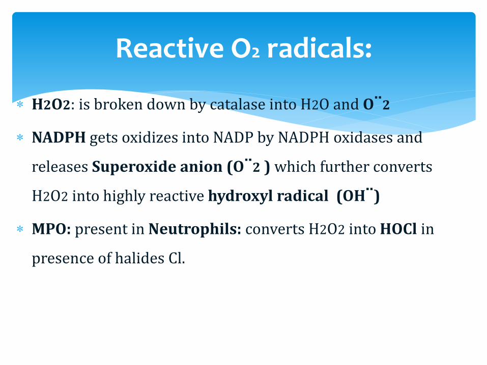
H2O2: is broken down by catalase into H2O and O¨2
NADPH gets oxidizes into NADP by NADPH oxidases and
releases Superoxide anion (O¨2 ) which further converts
H2O2 into highly reactive hydroxyl radical (OH¨)
MPO: present in Neutrophils: converts H2O2 into HOCl in
presence of halides Cl.
Reactive O2 radicals:

O2 independent Mechanism of killing:
Lysozymes
Lactoferrin
Major Basic protein
Defensins
Elastase

Substance released during phagocytosis:
1. Lysosomal enzymes
2. Reactive oxygen intermediates
3. Products of arachidonic acid metabolites (prostaglandins and
leukotrienes)
These products causes endothelial damage and tissue damage.
So---Persistent/ unchecked leukocyte infiltrate itself can become an
offender.

Leads to Increased susceptibility to Infections
Defects have been identified in all levels of WBC function
Defects can be genetically inherited or Acquired.
Defects in WBC Adhesion, Phagolysosome function,
microbicidal activity
Defects in phagolysosome formation
Neutropenia, defective degranulation, delayed microbial
killing
Neutrophils –with giant granules
Increased susceptibility to infections, Albinism (due to
defect in melanocytes), nerve defects.
Also platelet dysfunction—leading to bleeding disorders.
Due to defect in microbicidal activity
Inherited defects in the genes encoding the NADPH oxidase—
which generates Superoxide.
Patients are susceptible for recurrent bacterial infections.

Acute
Inflammation
Resolution/Healing
& Repair
Chronic
Inflammation
Abscess
SinusFistula
Ulcer
Injury
Definition: Dynamic, vascularized, injury
Calor, Rubor, Dolor, Tumor, Loss of function
Vasodilatation, exudation, chemotaxis, phagocytosis.
Chemical mediators – Prostaglandins, Kinins, etc
Types Acute/Chronic, Suppurative, serous, fibrinous.
Acute / Chronic differences.

Acute & Chronic inflammation.
Suppurative
Fibrinous
Serous
Granulomatous
Eosinophilic

Exudative Inflammation:
Suppuration – Purulent - Bacterial
Fibrinous – pneumonia – fibrosis
Serous – excess clear fluid – lung
Haemorrhagic – >Damage - anthrax.
Chronic inflammation: with healing.
Granulomatous – clusters of epitheloid* cells eg. TB, Fungus,
Foreign body.