
1.Summary - Publikationen der UdS: Zur Startseite · 2 dysfunction. It goes deeper insight in the...
120
1 1. Summary Hypertension represents a significant global public health problem and economic burden. The progressive nature of the disease from asymptomatic status to end-organ failure contributes to cardiovascular morbidity and mortality as it is responsible about systolic and diastolic heart failure and renal impairment. Despite achievements in pharmacological therapies to provide effective blood pressure control, still considerable percentage of patients are lacking of blood pressure control due to multiple factors. The novel approach of selective renal denervation may be considered as an important tool for treatment of resistant hypertension. In our study, we evaluated, by using transthoracic two- dimensional, doppler and strain rate echocardiography, whether renal denervation has ability to reverse cardiac remodeling processes followed hypertension in patients with refractory hypertension and hence a positive effect on systolic and diastolic dysfunctions. Beside lowering peripheral blood pressure and resting heart rate, we found that renal sympathectomy significantly reduces left ventricular mass and volumes. It facilitates the reverse remodeling that accompanied by improvement of left ventricular diastolic and systolic functions markedly evidenced in the responder’s population and particularly those who had evidence of diastolic dysfunction at baseline. Left atrial volume, which was evidenced to be a surrogate marker for the risk of atrial fibrillation development, (Wachtell et al., 2005 b) also significantly reduced after renal denervation independable from blood pressure response. However; left ventricular filling pressure did not exhibit the expected significant changes in our study which recommends coincidental assessment with invasive manoeuvre and long period of follow-up. Results derived from strain rate imaging supports that left ventricular systolic impairment is in part related to an increased left ventricular mass in patients with hypertension. It is proved that the improvement in echocadiographic parameters as well as left ventricular mass, in parallel with blood pressure reduction after renal denervation, contributed to the improvement of longitudinal left ventricular systolic function and relaxation and stiffness of the left ventricule. These results highlight the function of subendocardium which might play a role in the pathology of diastolic dysfunction. Accordingly, our data support strain rate imaging as a sensitive tool for detection of subclinical phases of left ventricular
Transcript of 1.Summary - Publikationen der UdS: Zur Startseite · 2 dysfunction. It goes deeper insight in the...

1
1. Summary
Hypertension represents a significant global public health problem and economic burden.
The progressive nature of the disease from asymptomatic status to end-organ failure
contributes to cardiovascular morbidity and mortality as it is responsible about systolic
and diastolic heart failure and renal impairment. Despite achievements in
pharmacological therapies to provide effective blood pressure control, still considerable
percentage of patients are lacking of blood pressure control due to multiple factors. The
novel approach of selective renal denervation may be considered as an important tool for
treatment of resistant hypertension. In our study, we evaluated, by using transthoracic
two- dimensional, doppler and strain rate echocardiography, whether renal denervation
has ability to reverse cardiac remodeling processes followed hypertension in patients with
refractory hypertension and hence a positive effect on systolic and diastolic dysfunctions.
Beside lowering peripheral blood pressure and resting heart rate, we found that renal
sympathectomy significantly reduces left ventricular mass and volumes. It facilitates the
reverse remodeling that accompanied by improvement of left ventricular diastolic and
systolic functions markedly evidenced in the responder’s population and particularly
those who had evidence of diastolic dysfunction at baseline. Left atrial volume, which
was evidenced to be a surrogate marker for the risk of atrial fibrillation development,
(Wachtell et al., 2005 b) also significantly reduced after renal denervation independable
from blood pressure response. However; left ventricular filling pressure did not exhibit
the expected significant changes in our study which recommends coincidental assessment
with invasive manoeuvre and long period of follow-up.
Results derived from strain rate imaging supports that left ventricular systolic impairment
is in part related to an increased left ventricular mass in patients with hypertension. It is
proved that the improvement in echocadiographic parameters as well as left ventricular
mass, in parallel with blood pressure reduction after renal denervation, contributed to the
improvement of longitudinal left ventricular systolic function and relaxation and stiffness
of the left ventricule. These results highlight the function of subendocardium which
might play a role in the pathology of diastolic dysfunction. Accordingly, our data support
strain rate imaging as a sensitive tool for detection of subclinical phases of left ventricular

2
dysfunction. It goes deeper insight in the analysis of left ventricular systolic and diastolic
functions than do conventional echocardiography and tissue doppler imaging. It might
therefore improve the clinical management of patients with heart failure with preserved
ejection fraction. The further role of strain rate imaging in clinical practice has to be
studied in more details in larger trials.
We conclude that in addition to blood pressure lowering effect, the selective denervation
of the renal sympathetic nerves reverses cardiac remodeling and improves both systolic
and diastolic functions in patients with refractory hypertension. These data suggest a
cardiovascular prognostic benefit of renal denervation in patients with refractory
hypertension. After reviewing drug trials, (Devereux et al., 2004 and Pierdomenico et al.,
2010) we recommend investigation of the cardiovascular prognostic benefit of renal
denervation in further big trials.

3
2. Zusammenfassung
Wir konnten durch Verwendung transthorakaler Echokardiographie inclusive strain rate
imaging zeigen, dass die renale sympathische Denervation in Patienten mit
therapierefraktärer arterieller Hypertonie neben Blutdruck und Herzfrequenz auch die
linksventrikuläre Masse und das linksventrikuläre Volumen reduziert sowie das
linksventrikuläre Remodelling verbessert.
Weiter verbessert die renale Denervation die systolische und diastolische
linksventrikuläre Funktion in denjenigen Patienten, die mir einer Blutdrucksenkung
reagierten und im Besonderen in denjenign mit diastolischer Dysfunktion bei
Studieneinschluss. Wie erwartet war die Verbesserung stärker in den Patienten, die die
Zielblutdruckwerte erreichten.
Linksventrikuläre Füllungsdrücke zeigten keine signifikante Veränderung in unserer
Studie was auf die Überlegenheit invasiver Messungen und langer Nachbeobachtung
hindeutet.
Der in unserer Studie dargestellte Effekt der renalen Denervation auf die myokardiale
Struktur und Funktion suggeriert einen kardioprotektiven Effekt der renalen Denervation
in Patienten mit therapierefraktärer arterieller Hypertonie.
Wir fanden das strain rate imaging eine sensitive Technik zur Identifizierung
subklinischer Phasen der linksventrikulären Dysfunktion. In unserer Studie wurde es
angewandt um die frühen Veränderungen der linksventrikulären systolischen und
diastolischen Funktion kurz nach renaler Denervation zu verstehen. strain rate imaging
und mitral annular plane systolic excursion waren in der Frühphase nach Therapie
sensitiver als konventionelle Echoparameter zur Bestimmung der linksventrikulären
systolischen Funktion. Die strain rate imaging Bestimmung deutete darauf hin, dass die
systolische Funktionseinschränkung ist mit einer gesteigerten linkventrikulären Masse in
Patienten mit arterieller Hypertonie assoziiert und zeigte dass die Verbesserung
echokardiographischer funktioneller Parameter wie auch des Massenindices parallel zur
Blutdruckreduktion zur Verbesserung der systolischen Funktion sowie der
linksventrikulären Relaxation und Steifigkeit beiträgt, die in Patienten mit adäquater
Blutdruckreduktion und diastolischer Dysfunktion vor Studienbeginn beobachtet wurde.

4
strain rate imaging unterstrich zudem die Funktion des Subendokards, das ebenfalls eine
Rolle in der Pathophysiology der diastolischen Dysfunktion spielen könnte.
strain rate imaging als neuartige echokardiographische Methode, gibt einen fundierten
Einblick in die Analyse der linksventrikulären systolischen und diastolischen
Dysfunktion als die konventionelle Echokardiographie und Gewebedoppler-
Echokardiographie. Es könnte daher die klinische Betreuung von Patienten mit
diastolischer Herzinsuffizienz verbessern. Die weitere Rolle von strain rate imaging in
der klinischen Praxis muss im Detail in größeren Studien untersucht werden.

5
3. Introduction
3.1. Essential hypertension
3.1.1. Size of the problem and cardiovascular risk
Essential hypertension (HTN), or primary high blood pressure, is a common disorder
mostly asymptomatic where systemic blood pressure (BP) remains abnormally elevated
for a sustained period of time (Chobanian et al., 2003). HTN is a significant growing
global health problem affecting approximately 1.2 billion people worldwide (WHO
2002). One in three adults is affected by high BP and the number of patients with newly
discovered hypertension is projected to rise even more (Kearney et al., 2005). Projections
show that by 2030, an additional 27 million people could have hypertension, a 9.9%
increase in prevalence from 2010 (Heidenreich et al., 2011). HTN remains a leading
cause of premature death, responsible for 12.8% of total deaths worldwide, (WHO 2009)
being a main cause of stroke, congestive heart failure and kidney disease. About 62% of
cerebrovascular and 49% of ischemic heart disease patients are attributed to suboptimal
blood pressure control. Following diabetes, HTN is the second most common cause of
end-stage renal failure and 80% of chronic kidney disease patients develop HTN at some
point in the course of their disease (WHO 2002). From 1998 to 2008, the death rate
caused by HTN increased 20.2%, and the actual number of deaths rose 49.7% (Centers
for Disease Control and Prevention. 2011). The risk of cardiovascular mortality is related
linearly with both systolic and diastolic pressures, doubling for every 20-mm Hg and 10-
mm Hg increase in the systolic and diastolic blood pressure, respectively, above 115/75
mm Hg (Roger et al., 2012). The prevalence of hypertension increases with age, obesity
and sedentary lifestyles (Whelton et al., 2002). Since all three factors are growing
worldwide, HTN treatment represents a huge and growing clinical challenge as well as a
major public health cost burden. The estimated annual global healthcare expenditure
directly attributable to hypertension is estimated by $500 billion (Lawes et al., 2008). On
the other hand, World Health Organization report has classified HTN as a risk factor for
cardiovascular diseases and one of the most important preventable causes of premature

6
morbidity and mortality in developed as well as developing countries (WHO 2002 and
Ezzati et al., 2002).
For actual detection of the size of the problem, European Society of Cardiology,
European Society of Hypertension and the United States Joint National Committee on
Prevention, Detection, Evaluation and Treatment of High Blood Pressure, have
established guidelines for blood pressure classification (see Table 1). (Chobanian et al.,
2003).
Classification Systolic (mmHg) Diastolic (mmHg)
Normal
Prehypertension
HTN:
Stage 1
Stage 2
90–119
120–139
140−159
≥160
60–79
80–89
90–99
≥100
Table 1: Blood pressure classification (Chobanian et al., 2003)
(Note that target blood pressure for diabetics is <130/80)
3.1.2. Blood pressure regulating mechanisms
BP is controlled by a complex processes in the body through the interaction of electrical,
mechanical and hormonal influences. The sympathetic nervous system (SNS) is the main
component of blood pressure control as it connects the brain, heart, blood vessels and
kidneys, each of which plays a vital role in the regulation of the body’s BP. The brain
mainly plays an important role, processing and sending signals to the rest of the SNS.
The heart plays an important mechanical role by adjusting the heart rate and the force of
cardiac contractility to regulate BP. The blood vessels themselves also play a mechanical

7
role, influencing BP by either dilating to lower blood pressure or constricting to raise BP.
The final and perhaps the most central contributors to the regulation of BP are the
kidneys. They play electrical, mechanical and hormonal roles to regulate systemic blood
pressure. The kidneys regulate BP by signaling the need for increased or lowered
pressure through the SNS (electrical), by controlling the amount of fluid in the body
(mechanical) and by releasing hormones that influence the response of the heart and
blood vessels (hormonal) (Kearney et al., 2005).
3.1.3. Essential hypertension as a consequence of chronic elevated central and renal
sympathetic tone
A network of afferent and efferent sensory, chemo- and baroreceptor nerve fibers, lie in
the adventitia of the renal artery allover the kidney (Vonend et al., 2003). This net work
terminates in the blood vessels, the juxtaglomerular apparatus and the renal tubules
(Barajas et al., 1992). It connects with hypothalamus providing an integration of the renal
artery with the brain stem (DiBona 2005). This signaling pathway is responsible for
altering the peripheral arterial resistance, the venous capacitance vasculature, peripheral
and central chemoreceptors, and sympathetic activity of the kidney (Schlaich et al.,
2004). Activation of renal sensory afferent nerve is caused by various stimuli such as
renal ischemia, hypoxia, and oxidative stress as well as intrinsic renal diseases (DiBona
2005). Following these stimuli, the sensory afferent signaling reaches the posterior
hypothalamus, directly influences central sympathetic outflow, throughout the entirety of
the sympathetic system including kidneys (Esler 2010). Accordingly, activation of the
sympathetic nerve fibers that innervate all organs involved in the direct control of
peripheral vascular resistance, management of central and peripheral chemo-receptors,
directly affect cardic contractility, vascular tone, heart rate and rhythm, management of
total body salt and water through both renal mechanisms and control of intravascular
circulating blood volume in the splanchnic vessels and of course the kidney itself
(DiBona 2005). The consequences of incease central sympathetic drive responsible for
many diseases linked to hypertension and systolic heart failure, such as insulin resistance,
8
sleep disorders, diuretic resistance and congestion (Fitzgerald 2009). Therefore, it is well
established that the renal blood flow and renal sympathetic nerve activity are very
important in the regulation of renal function and fundamental to the control of blood
pressure (Barrett et al., 2001) and there is considerable evidence that activation of the
SNS plays an important role in the pathogenesis of several cardiovascular diseases,
including HTN (Lohmeier 2001).
It has been investigated that essential hypertension is largely neurogenic, both initiated
and sustained by SNS over-activity proved using radiotracer dilution methodology
measuring spillover of noradrenaline from the kidney (Esler 2000).
Thus, reduction of excessive central sympathetic activity, following the selective removal
of renal signals to the hypothalamus, is therapeutically attractive in the treatment of
disorders commonly linked by sympathetic over-activity (Doumas et al., 2010).
3.2. Hypertensive heart disease
Chronically elevated systemic blood pressure and sustained increased workload on the
heart leads to a series of pathological changes (remodeling processes) and begin as a
compensatory mechanism to minimize wall stress. They mainly take place in the left
ventricle (LV) in the form of hypertrophy and remodeling (Bauml et al., 2010).
Pathological changes involve myocardial fibrosis, ischemia, cardiomyocyte impairment,
apoptosis, endothelial dysfunction and increased arterial stiffness. All these abnormalities
together often lead into a vicious circle which determine the phenotype form of
hypertensive heart disease (Hellenic 2008).
One of the key components in the development of hypertensive heart disease is the
myocardial fibrosis (Yamamoto et al., 2002). It is mainly accused to promote diastolic
dysfunction (DD) resulting in the increase of LV filling pressure. These changes
compromise cardiac function after a series of poorly characterized events (transition to
failure), in which the left ventricle dilates and the LV ejection fraction (EF) (systolic
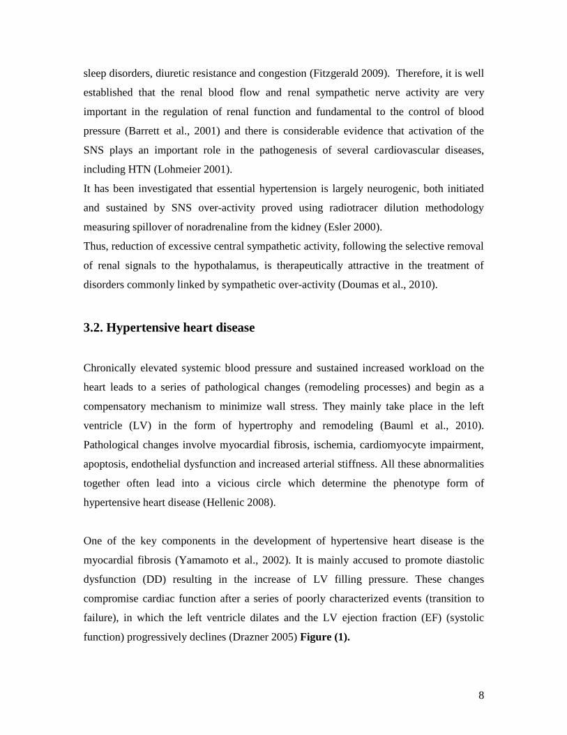
function) progressively declines (Drazner 2005) Figure (1).
9
Figure (1): Hypertension and transition to failure. MI; myocardial infarction, cLVH;
concentric left ventricular hypertrophy, EF; ejection fraction. (Drazner 2005).
Clinically hypertensive heart disease covers a broad spectrum ranging from
asymptomatic LV remodeling (either a concentric or an eccentric pattern) to overt heart
failure (with either a preserved or a reduced left ventricular ejection fraction) (Drazner
2011).
3.2.1. LV remodeling and myocardial hypertrophy
The combination of volume and pressure overloads causes LV geometric adaptations.
This LV adaptation ranges from concentric remodeling and concentric left ventricular
hypertrophy (LVH) up to heart failure (transition to failure, see before) (Hellenic 2008).
The influences of neurohormones beside response to the mechanical stress from elevated
BP are considered the mechanisms responsible about progression from LV hypertrophy
to failure (Hill et al., 2008, Diez et al., 2010). The most commonly response of the
myocardium from the longstanding pumping against an elevated after load is LVH. LVH
is defined as an abnormal increase in the mass of the left ventricular myocardium caused
by a chronically increased workload on the heart (Lorell et al., 2000). The development
of LVH is highly correlated with systolic hypertension. In the Framingham Heart Study,
10
even borderline isolated systolic hypertension at an elderly age was associated with
increased left ventricular wall thickness and impaired diastolic filling (Sagie et al., 1993).
There is considerable inter individual variation in the progression from HTN to the
increase in LV mass and the changes in geometric pattern (ventricular dilatation or wall
thickening) (Drazner 2011).
3.2.1.1. Echocardiographic diagnosis of LVH
The diagnosis of LVH is important because it is an indicator of end-organ damage in
arterial hypertension. The risk of cardiovascular morbidity and mortality is two-to-four-
fold increased in LVH patients compared to patients with normal left ventricular mass as
LVH may facilitate many cardiac complications of HTN, including congestive heart
failure, ventricular arrhythmias, myocardial ischemia and sudden death (Casale et al.,
1986). On the other hand, treatment-induced regression of LVH decreases adverse
cardiovascular events and improves overall survival; therefore treatment of LVH should
be considered as well as BP control in hypertensive patients (Okin et al., 2004).
It is well established that echocardiography is the test of choice to assess LVH. It is much
more sensitive than electrocardiography (Devereux et al., 1986). Left ventricular mass
index (LVMI) is considered the most valid parameter to define LVH. The left ventricular
wall thickness alone is not a good indicator of LVH and does not accurately assess the
presence of LVH (Leibowitz et al., 2007). Accordingly, wall thickness should not be used
alone to define this pathology, as often happens in clinical practice. Echocardiography
can measure end-diastolic diameter (EDD), posterior wall thickness (PW), and
interventricular septum thickness (IVS) Figure (2). From these measurements and the
patient’s height and weight, LVMI can be calculated (Lang et al., 2005). By
echocardiography, we can also detect other associated structural and functional
abnormalities.

11
Figure (2): Left ventricular hypertrophy. It is diagnosed by an elevated left ventricularmass index, which is calculated from the intraventricular septal thickness (IVSd),posterior wall thickness (PWTd), and left ventricular end-diastolic internal diameter(LVIDd) (Devereux et al., 1986).
3.2.2. Left atrial remodeling in HTN
A time-dependent adaptive regulation processes against external stressors is established
to maintain homeostasis, these adaptive processes are referred to as LA remodeling
(Nattel 1999). In HTN, volume/pressure overload and diastolic dysfunction represents the
main stressors (Colucci et al., 2005). The LA remodeling mechanisms depend on the
strength and the duration of exposure to these “stressors” and they are reversible in the
earlier and midterm stages of LA structural and functional disturbances (Everett et al.,
2000 and Kumagai et al., 2003) and usually irreversible over the longer term (Li et al.,
1999). These include myocyte growth, hypertrophy, necrosis, and apoptosis; alterations in
the composition of extracellular matrix; recalibration of energy production and
expenditure; changes in the expression of cellular ionic channels and atrial hormones
(Colucci et al., 2005). These maladaptive processes which lead to LA remodeling

12
resulted in structural, functional, electrical, metabolic, and neurohormonal consequences
(Casaclang-Verzosa G et al., 2008).
3.2.2.1. Consequences of LA remodeling in HTN
Ultrastructural changes in hypertension- induced remodeling are marked by extensive
interstitial fibrosis and myocyte hypertrophy (Khan et al., 2004) which provides circuits
for re-entry (Shi et al., 2001) making the LA more vulnerable to atrial fibrillation (AF)
development. Whereas atrial dilatation is the hallmark of structural remodeling, atrial
arrhythmias, especially AF, are the most common manifestations of LA electrical
remodeling (Schoonderwoerd et al., 2005). In hypertensive patients, who present with an
enlarged left atrium, have a 42% increased risk of developing atrial fibrillation (Krahn et
al., 1995), cardiovascular morbidity e.g. heart failure (Bayes-Genis et al., 2007) , stroke
(Douglas et al., 2003) and sudden cardiac death (Wachtell et al., 2005 a and Alsaileek et
al., 2006).
3.2.2.2. Echocardiographic assessment of LA structural remodeling
In sinus rhythm, left atrial volume has a high sensitivity and specificity as a measurement
of diastolic dysfunction and LV filling pressures (Tsang et al., 2002). Echocardiographic
assessment of left atrial volume indexed to body surface area (LAVI) was proposed as a
marker of both diastolic LV dysfunction and cardiovascular risk and may improve the
process of cardiovascular risk stratification (Alsaileek et al., 2006). It is required also to
support the diagnosis of heart failure with normal ejection fraction which can complicate
uncontrolled longstanding HTN (Yoshida et al., 2009).
3.2.3. Neurohormonal disturbances associate LA and LV remodeling with HTN
Increases in atrial natriuretic peptide (ANP) (Dietz et al., 2005), brain natriuretic peptide
(BNP) (Tsioufis et al., 2006), angiotensin II (Ang-II), aldosterone, transforming growth
factor beta-1(Hanna et al., 2004), and sympathetic hyperinnervation (Tsioufis et al.,
13
2006) have been described in association with the remodeling process. Atrial natriuretic
peptide is a direct vasodilator, which lowers systemic blood pressure and inhibits renin
and endothelin secretion, myocyte hypertrophy, and fibroblast collagen synthesis
(Miyauchi et al., 2003). Mechanical stretching of the LA is the strongest stimulus for
ANP secretion, which is augmented by endothelin and inhibited by nitric oxide (Franco et
al., 2004). Cardiac BNP is another marker for LA and LV remodeling in response to an
increase of atrial or ventricular diastolic stretch and their secretion results in natriuresis,
vasodilation, and improved LV relaxation (Lim et al., 2006 and Barclay et al., 2006). In
the case of LA remodeling, BNP is significantly correlated with indexed LA volume in
patients with diastolic heart failure (Lim et al., 2006), stable chronic heart failure (Barclay
et al., 2006), hypertension (Inoue et al., 2000), In contrast to its usefulness in
symptomatic isolated diastolic LV dysfunction, natriuretic peptides were a suboptimal
screening test for preclinical diastolic LV dysfunction (Boldt et al., 2004).
3.2.4. Diastolic dysfunction in hypertensive heart
The function of the heart can be classified into (1) pumping function during the systolic
phase, impairment of which can lead to low cardiac output, which has usually been
evaluated by the presence of low EF and (2) filling function, during diastolic phase, and
it’s impairment results from decrease the LV compliance and relaxation with increase of
the LV stiffness and that can lead to congestion (Mandinov et al., 2000). Ventricular
relaxation is a dynamic active process leading to flow of blood from the left atrium (LA)
into the LV across a pressure gradient (Ommen et al., 2000). Changes in diastolic
compliance and relaxation are direct consequences of exceeded tissue stiffness
(Mandinov et al., 2000). There is growing evidences that myocardial stiffening depends
mainly upon myocardial fibrosis rather than LV wall thickening and myocyte
hypertrophy in hypertensive heart (Yamamoto et al., 2002). Therefore, diastolic
dysfunction (functional impairment) can be also presented in hypertensive patients with
normal LV mass. (Aeschbacher et al., 2001).

14
EF may be normal in the presence of diastolic dysfunction in a phenomena referred as
‘heart failure with normal EF (HFNEF)’ or ‘heart failure with preserved EF (HFPEF)’
(Yu et al., 2002 and Paulus et al., 2007).
3.2.4.1. Problem of heart failure with preserved ejection fraction (HFPEF)
The prevalence of cases presented HFPEF increased from 38 to 54% of all heart failure
cases over the last two decades. The prevalence of HFPEF in the heart failure population
rises by about 1% a year especially among elderly, female gender, diabetics obesity
population, arterial hypertension, and LVH (Owan et al., 2006). Symptoms due to
HFPEF, as in systolic heart failure, are attributed to low cardiac output and pulmonary
congestion. As the increase in LV filling pressure is the direct cause of pulmonary
congestion, assessment of LV filling pressure has been becomes a main clinical concern,
not only in systolic heart failure but also in DHF (Yu et al., 2002 and Baicu et al., 2005).
Morbidity, hospitalization rates, and healthcare costs per patient are very similar between
systolic and diastolic heart failure (Bursi et al., 2006). Moreover, no great difference in
mortality between both forms of heart failure was found (Bhatia et al., 2006 and Bursi et
al., 2006).
3.3. Echocardiographic assessment of diastolic function in hypertensive
patients
Echocardiography can indirectly assess myocardial relaxation, LV stiffness, and filling
pressures to predict the hemodynamic performance of LV during diastole. Doppler study
is primarily applied to evaluate hemodynamic and the diastolic function. Study of
morphologic and functional properties of the heart which correlates with diastolic
dysfunction is recommended during the Doppler echocardiographic assessment and
important to establish the diagnosis of diastolic dysfunction in certain difficult situations
and as well as for the definition of HFPEF. These properties involve LVH, LA volume
and function and pulmonary artery systolic and diastolic pressures (Nagueh et al., 2009).

15
3.3.1. Blood flow Doppler assessment of LV diastolic function
Pulsed wave Doppler is mainly used to identify Isovolumetric LV relaxation time
(IVRT), ratio of peak early (E) to peak atrial (A) Doppler mitral valve flow velocity,
deceleration time (DT) of early Doppler mitral valve flow velocity, (see methods) and
ratio of pulmonary vein systolic (S) and diastolic (D) flow velocities were originally
considered to be indicative of diastolic LV dysfunction and allow classification of the
grade of DD if they exceeded specific cut-off values indexed for age groups (Nagueh et
al., 2009). The combined use of these variables provided a semi quantitative estimate of
LV end-diastolic pressure is also supported by observations in hypertensive patients
(Rusconi et al., 2001).
3.3.2. Assessment of LV diastolic function using Tissue Doppler Imaging (TDI)
TDI measures myocardial motion (measured as tissue velocity) relative to the transducer
with high spatial (mm) and temporal resolution. Tissue velocity indicates the rate at
which a particular point in the myocardium moves toward or away from the transducer.
In this setting TD derived velocity is obtained via pulsed Doppler (PW) (by placing a
sample volume at a particular location). Measurements can be obtained at the septal and
at the lateral side of the mitral annulus (Ommen et al., 2000). The peak systolic (S)
shortening velocity and the early diastolic (E’) lengthening velocities are considered to be
sensitive measures of LV systolic or diastolic function. E depends on LA pressures, LV
relaxation kinetics, and age but E’ depends mostly on LV relaxation kinetics and age.
Hence, in the ratio E/E’, effects of LV relaxation kinetics and age are eliminated and the
ratio becomes a measure of left atrial driving pressure or LV filling pressure. E’ can also
reflect the amount of blood entering the LV during early filling, whereas E represents the
gradient necessary to make this blood enter the LV. A high E/E’ thus represents a high
gradient for a low shift in volume (Rivas-Gotz et al., 2003 and Diwan et al., 2005). Cut
off values for the E/E’ is established in which when the ratio exceeds 15, LV filling
pressures are elevated and when the ratio is lower than 8, LV filling pressures is
considered to be low i.e normal. An E/E’ ratio ranging from 8 to 15 is suggestive but non

16
conclusive evidence of diastolic LV dysfunction and needs to be supported with other
non-invasive investigations to confirm the diagnosis (Ommen et al., 2000). It is known
now that E/E’ is superior as predictor of prognosis than clinical or other
echocardiographic variables (Hillis et al., 2004). See Table 2 and Figure (3).
A pulm–A mitral = time difference between pulmonary vein flow A-wave duration and mitralflow A-wave duration; E/A = ratio of early to late diastolic mitral inflow waves; e’ early diastolicvelocity of mitral annulus; E/e’ = ratio of the mitral inflow E wave to the tissue Doppler e’ wave;HF heart failure; LV left ventricular. Different cut-off points exist in different consensusdocuments. (Paulus et al., 2007 and Nagueh et al., 2009). For the cut-off points mentioned in thistable both septal and average e’ may be used.bHighly variable and unsuitable for diagnosis on itsown; largely depending on loading conditions; age-corrected normal values exist (Nagueh et al,2009).
Table 2: Common echocardiographic measures of left ventricular diastolic dysfunction in
patients with heart failure (McMurray et al., 2012).

17
Figure (3): Doppler criteria for classification of diastolic function. E, early mitralvalve flow velocity; E´, early TD lengthening velocity; E/A, ratio of early (E) to late (A)mitral valve flow velocity; DT, deceleration time; S, peak systolic shortening velocity;Vp colour M-Mode flow propagation (Redfield et al., 2003).
Doppler echocardiographic technique is used frequently in the daily practice as a tool to
provide functional informative data. It is widely available, safe non invasive and
inexpensive imaging modality (Nagueh et al., 1997).
3.3.3. Need for newly applied techniques in the assessment of diastolic function
Although hemodynamic data with echocardiography often simply established, it may be
valid in a given patient but are not necessarily applicable to all patients. Doppler
echocardiographic measurements of diastolic function can show individual variability
18
and can even vary from day to day in the same patient. the mitral inflow velocity profile
is affected by several factors including age, heart rate, volume status, left atrial pressure,
and rate of myocardial relaxation, sometime it is restricted in accurately evaluating the
diastolic function (Alarm et al., 1992 and Sohn et al., 1997). Also, the diagnosis of
diastolic heart failure using the available guidelines is difficult to apply in some cases.
Thus the optimum way of treatment is still controversial in such cases as long as the
mechanisms of the disease are not completely defined. Simple informative diagnostic
tests are needed especially for better identification of the early stages of diastolic
dysfunction (Vinereanu et al., 2005). To apply echocardiographic methods for
investigating of such problems, it is possible to choose either a general simple approach
with high feasibility and reproducibility or a more sophisticated one. Although the former
is suited for clinically trials, the latter may be superior for answering mechanistic
questions. Recently, deformation measurements by strain analysis appear to have good
reproducibility and can be applied to investigate regional deformation and to approach
mechanistic problems missed by conventional parameters (Nagueh et al., 2009).
3.4. Strain and strain rate imaging (SRI)
Strain is defined as a quantitative measurement of the amount of movement of an object
in response to applied force and it is expressed by %, whereas SR is the rate at which this
change or movement occurs and expressed by s-1 (Abraham et al., 2007).
Echocardiographic strain and is a tool for the evaluation of myocardial systolic function
and has been found to be superior to velocity analysis by TDI, or conventional ultrasound
techniques, for quantification of regional myocardial function. The spectrum of clinical
applications is very wide due its ability to differentiate between active and passive
movement of myocardial segments and to evaluate components of myocardial function
such as longitudinal myocardial shortening that are not visually assessable. The high
sensitivity of both TDI-derived and 2D speckle tracking derived strain and strain rate data
for the early detection of myocardial dysfunction recommend this new non-invasive
diagnostic method for routine clinical use (D’hooge et al., 2002 and Dandel et al., 2009).
Systolic strain represents percentage shortening when measurements are done in the long
19
axis and percentage radial thickening in the short axis. Accordingly, lengthening and
thickening strains are assigned positive values and shortening and thinning strains
negative values (Dandel et al., 2009). SRI may also provide important quantitative data in
the evaluation of diastolic function. The high temporal resolution of strain imaging
provides the ability to analyze short-lived mechanical events during diastole (Takemoto
et al., 2005). In which regional diastolic strain ratios are related to regional stiffness and
can differentiate between stunned and infarcted myocardium (Pellikka et al., 2004). The
evidence exists that changes in early diastolic strain rate can predict angiographic disease
(Perk et al., 2007).
3.4.1. Speckle-tracking derived two-dimensional (2D)-strain
It is newly emerging technology that measures myocardial deformation (strain) by
tracking speckles (acoustic markers) in grayscale echocardiographic images. The motion
pattern of myocardial tissue is reflected by the motion pattern of speckles. The speckles
function as natural acoustic markers that can be tracked from frame to frame. Later on
velocity and strain are obtained by automated measurement of distance between speckles.
Special software allows spatial and temporal image processing with the ability to
recognize and select these elements on ultrasound images and then automatically
calculates strain and strain rate values (Abraham et al., 2007 and Dandel et al., 2009).
The method is angle independent; therefore, measurements can be obtained
simultaneously from multiple regions within an image plane. The 2D echocardiographic
loops obtained from parasternal and apical views are processed offline. This requires only
one cardiac cycle to be acquired but SRI data can be obtained only with high resolution
image quality at high frame rate (about 50-70 frames per second (FPS)) (Marwick et al.,
2006 and Dandel et al., 2007).

20
3.4.2. Emerging role of SRI in the evaluation of hypertensive heart disease
3.4.2.1. SRI in the assessment of diastolic function
As mentioned before, Doppler and TDI methods are widely available and easy applied
method to identify DD. Although that, the comprehensive understanding of diastolic
pathophysiology is still considered as a clinical challenge due to the drawbacks and some
times difficulties in applying these parameters (Nagueh et al., 2009). Garcia-Fernandez et
al., 1999 proved that despite of normal mitral flow Doppler, still up to 40% of the
myocardial segments may have measurable regional diastolic motion abnormalities. In
hypertensive patients, SRI technique permits the evaluation of changes of LV diastolic
function even if they are free from clinical manifestations of HF (Bruch C, et al., 2002).
However, the role of strain and strain rate imaging in defining diastolic dysfunction is
still under investigation (Nagueh et al., 2009, Oxborough et al., 2009 and Amundsen et
al., 2009).
3.4.2.2. SRI in the assessment of systolic function
TDI was used before to study LV systolic function. The advantage of pulsed-wave TDI is
that it does not require high-end equipment, specific software, or offline analysis
(Anderson et al., 2008). But it has drawbacks hinder the accuracy of evaluation
myocardium mechanics during systole. TDI interrogates motion at a single point in the
myocardium with reference to a point outside the chest (the transducer). It is found that it
is influenced by translational motion of the chest wall and tethering effects (normal apical
segments pull an abnormal basal segment toward the apex). Moreover, single point
interrogation (depicting tissue displacement) does not fully capture true myocardial
mechanics (D’hooge et al., 2002). Accordingly, it requires sampling of multiple regions
from different cardiac cycles, which is time consuming and renders tissue velocity peaks
more difficult to identify (Anderson et al., 2008). In contrast, strain rate imaging
technology now enables quantitative measurement of regional LV function independent
of cardiac rotational motion, chest wall movements and tethering artefacts and thus may

21
be superior to tissue velocity in depicting regional or global myocardial function
(D’hooge et al., 2002). Speckle tracking derived from 2D SRI can also provide an angle-
independent imaging for myocardial deformation (Langeland et al., 2004). Impairment in
longitudinal myocardial function was reported in different groups of patients despite
normal EF and FS (Koyama et al., 2003). This impairment was not observed with TDV
(Ballo et al., 2007) but was determined by SRI (Koyama et al., 2003). Systemic Arterial
Hypertension leads to tiny vascular abnormalities beside myocardial fibrosis which
mainly harms the endocardium (Martinez et al., 2003). Affection of subendocardial
function could be early detected by examining the longitudinal function abnormalities
using global longitudinal strain measurement (Wang et al., 2008). Application of SRI
using speckle tracking has been used in detecting subclinical myocardial abnormalities
resulted from hypertrophy and hypoperfusion in LV hypertrophy, as well as in
distinguishing the different causes of LV hypertrophy (Pavlopoulos et al., 2008 and
Imbalzano et al., 2011). Accordingly, the application of a 2D speckle tracking is
promising and can be widely applied, as a simple clear diagnostic test, in the study of
subclinical or overt LV systolic dysfunction and for better understanding of the early
stages of diastolic dysfunction (Nagueh et al., 2009) with improved both interobserver
and intraobserver variability compared with TDI derived strain. Therefore, more studies
are needed to define its clinical value in defining LVH associated subclinical systolic
dysfunction (Afonso et al., 2008).
3.5. Mitral annular plane systolic excursion (MAPSE)
3.5.1. Background
The LV wall is composed of both circular (radial) and longitudinal myocardial fibers, in
which the later fibers contracting earlier and dominate in subepicardial and
subendocardial layers and in the papillary muscles (Timek et al., 2001). It has a complex
three-dimensional motion pattern composed of: longitudinal apical motion along the
ventricular long axis (base to apex), rotation and sphincter-like motion (Zaky et al.,

22
1967). The apical displacement represents shortening of the left ventricle along its long
axis. Mitral annulus is considered tightly coupled component of the mitral valve/left
atrial/left ventricular complex that aids in effective efficient valve closure as well as
uncomplicated left ventricular filling (Timek et al., 2001).
3.5.2. Functional importance and clinical application
Cardiac disorders can impair both longitudinal and radial contractile function. However,
it has been suggested that in pathologic conditions (e.g., myocardial ischemia or
hypertrophy, dilated cardiomyopathy, and hypertrophic cardiomyopathy), long-axis
myocardial function is the first to be impaired (Bolognesi et al., 2001). Impairment of
Long axis left ventricular contraction and relaxation has attracted interest in recent years
as it could help in early diagnosis of cardiac dysfunction. It is easy to be evaluated
through measuring of mitral annular plane systolic excursion (MAPSE) by M-mode
echocardiography. MAPSE was found to be correlated to LV function calculated by two-
dimensional echocardiography, radionuclide ventriculography, as well as contrast
cineangiography (Höglund et al., 1989 and Alam et al., 1992 a) which makes it a good
indicator of LV systolic function and very sensitive parameter in many different cardiac
pathologies (Höglund et al., 1989, Pai et al., 1991 and Florian et al., 2012). Left
atrioventricular plane displacement was found to be reduced in AF, most likely due to the
absence of atrial contration which contributes to left ventricular diastolic filling (Alam et
al., 1992 b). Thus, it is possible that left atrioventricular plane displacement is also
affected by left ventricular diastolic performance (Willenheimer et al., 1999). Therefore,
MAPSE has been established as a method of measuring left ventricular systolic and
diastolic function and as an index of left atrial function (Jones et al., 1991, Pai et al.,
1991, Alam et al., 1992 c and Simonson et al., 1998).

23
3.6. Poor treatment reality
3.6.1. Therapeutic approaches in the management of HTN
Pharmaceutical therapies used in the management of HTN involve multiple agents acting
with different mechanisms of action. Centrally acting sympatholytic drugs aim to disrupt
the electrical signals involved in the activation of the SNS. Agents designed to lower the
mechanical load in the circulatory system, such as diuretics, inhibit sodium and water
retention to lower fluid volume. Perhaps most effective, several agents target electrical,
mechanical and endocrine activities of the kidneys (see before). These drugs include beta
blockers (to reduce enzymes release and heart rate), angiotensin-converting enzyme
(ACE) inhibitors, angiotensin receptor blockers (ARB) and aldosterone blockers (to
counteract rennin angiotensin aldosteron system RAAS) (Chobanian et al., 2003). The
aim of antihypertensive therapy should be to reduce the blood pressure as far as possible
(Heagerty et al., 2004). A reduction in SBP by 10 mmHg is associated with a reduction in
cardiovascular events of up to 25% and these effects are more prominent in stroke than in
coronary artery disease (Perkovic et al., 2007). Another potential therapeutic target for
the treatment of hypertension is to reverse structural and functional changes result from
maladaptive regulatory mechanisms (remodeling process) that is activated to overcome
the increase in pressure-volume work load and to minimize wall stress that associate the
increase in arterial blood pressure.
It is proved that patients who had a lower left ventricular mass index during treatment
with antihypertensive drugs had lower rates of cardiovascular morbidity and all-cause
mortality (Okin et al., 2004). Regression of electrocardiographic LVH in hypertensive
patients has also been shown to be associated with decrease the incidence of atrial
fibrillation (Okin et al., 2006) and hospitalizations for heart failure (Okin et al., 2007).
To treat HTN induce DD, we should look for the treatment of the underlying HTN
adequately as a preventable factor and that will contribute to counteract the remodeling
process accompanied HTN being the main mechanism for the development of DD (Diez
et al., 2002 Yusuf et al., 2003 and Bursi et al., 2006).

24
The direct impact of reversing LA remodeling on cardiovascular outcomes remains to be
seen, but the evidence indirectly suggests significant reduction of risk of AF development
as an outcome (Wachtell et al., 2005 b). Still the drugs that modify the renin angiotensin-
aldosterone system appear to have particularly powerful effects on LV and LA
remodeling processes and their consequence e.g AF development and risk of stroke,
beyond their beneficial effects on blood pressure regulation (Tsang et al., 2002, Tsang et
al., 2006 and Tanaba et al.; 2009).
3.6.2. Problem of resistant HTN
Despite the availability of numerous effective pharmacologic agents, adequate BP
reduction is not achieved in a large number of patients. It is not unusual, as failure to
achieve BP control even occurred with proper use of multiple antihypertensive drugs.
Several patient and physician related aspects contribute to this problem (Sarafidis et al.,
2008). About 50% of patients with HTN remain uncontrolled and approximately 15–20%
percent of those are resistant (Lloyd-Jones et al, 2010). Resistant hypertension is defined
as blood pressure that remains above goal in spite of the concurrent use of 3
antihypertensive agents of different classes including diuretics (Pantelis et al., 2008).
This leaves patients at high risk for major cardiovascular events as long standing
uncontrolled HTN is strongly associated with increasing the risk of stroke, heart failure,
coronary heart disease, chronic kidney disease diabetes, obstructive sleep apnea and LVH
(David et al., 2008 and WHO 2009).
Approximately 45% of ischemic heart disease deaths and 51% of stroke deaths are
directly linked to systolic BP (SBP) (WHO 2009). Efforts to overcome resistance to
pharmacotherapy have attracted the attention into the role of the sympathetic nervous
system in such problems. It plays a critical role in the pathophysiology of HTN and its
adverse sequences mainly LVH making it an attractive therapeutic target (Schlaich et al.
2011 a).

25
3.7. Renal sympathetic denervation (RD)
Certain disease conditions were linked to sympathetic nervous system hyperactivity. this
cluster of diseases includes hypertension (Esler et al., 1988), heart failure (Triposkiadis et
al., 2009), sleep disturbances (Narkiewicz et al., 1998), metabolic syndrome and
glycemic control (Mancia et al., 2007), and chronic kidney disease (Hausberg et al.,
2002). the risk of deaths among heart failure patients is associated with sympathetic
nervous system hyperactivity (Triposkiadis et al., 2009). Salt and water retention in some
forms of heart failure may be mediated by renal sympathetic activity and selective renal
denervation may in part used for the treatment or prevention of heart failure and the
cardiorenal syndrome (Sobotka et al., 2011). Elevated sympathetic activity of the renal
efferent nerves in hypertension mediated by promoting sodium retention, decreasing
renal blood flow and glomerular filtration, and increasing renin release, which known as
activation of the renin-angiotensin-aldosterone neurohormonal cascade (DiBona et al.,
2010). Similarly, hypothalamic signaling activates renal somatic afferent nerve activity,
which may mediate effects indirectly related to systemic hypertension (Kandzari et al.,
2012). The mechanistic relationship between renal nerve activation and high blood
pressure was recognized and investigated since long time to find therapeutic
opportunities related to selective renal sympathectomy in patients with refractory
hypertension (DiBona et al., 2010). Surgical attempts to attenuate sympathetic drive in
attempt to reduce BP have been applied as early as the 1920s in severely hypertensive
patients. Although severe side effects developed after the procedure, improvements in BP
control and survival were demonstrated in treated patients, highlighting the effectiveness
of this old concept (Smithwick et al., 1953). Recently, efforts have led to the emergence
of a novel catheter-based approach using radiofrequency (RF) energy for selectively
ablation and disruption of the renal nerves. This new technique provides safe and durable
blood pressure reduction and that offers hope of successful therapy for the group of
hypertensive patients unresponding, unwilling or unable to take maximal doses of
multiple anti hypertensive therapy (Kandzari et al., 2012).
Analysis of initial pilot studies (SYMPLICITY HTN-1 First-in-Human and additional
phase I studies), showed sustained reduction in office-based systolic and diastolic blood
26
pressures from baseline measurements through 2 years. A reduction in systolic blood
pressure of at least 10 mmHg was achieved in 92% of patients (Krum et al., 2009).
Preliminary results of patients with uncontrolled hypertension of the open-label
randomized SYMPLICITY HTN-2 trial in Europe and Australia represented a 84% of
patients treated with sympathetic denervation experienced a systolic blood pressure
reduction exceeding 10 mm Hg, and more than 80% had an office blood pressure <160
mm Hg after 6 months. No recorded procedural complications were observed, and renal
imaging at 6 months did not identify any renal abnormalities directly caused by
denervation or requiring therapy which consider RD as a safe and efficient for the
treatment of resistant HTN (Symplicity HTN-2 Investigators 2010).
SYMPLICITY HTN-3 takes place now in 90 research sites allover the United States. It is
a prospective, randomized single-blind trial in which 530 patients will be randomized
(316 treatment and 158 control). It is designed to evaluate and define the safety and
effectiveness of catheter-based bilateral renal denervation for the treatment of
uncontrolled hypertension despite taking at least 3 antihypertensive medications of
different classes involving diuretice. The primary end point is detected by the change in
office-based systolic blood pressure (SBP) from baseline to 6 months and a major
secondary effectiveness analysis is the change in average 24-hour SBP by ambulatory
blood pressure monitoring from baseline to 6 months (Kandzari et al., 2012). The results
of the SYMPLICITY HTN-3 pivotal trial will be submitted to the US Food and Drug
Administration for approval of catheter-based renal denervation as a treatment option for
resistant hypertension (Kandzari et al., 2012). In addition, it is found that renal
sympathetic denervation may have a multiple benifits beyond those directly related to
high blood pressure. Recent reports detected that patients with insulin resistance or type
II diabetes mellitus, polycystic ovary syndrome, and hypertension have also improved
glucose metabolism insulin sensitivity and good glycemic control with renal
sympathectomy (Mahfoud et al., 2011 and Schlaich et al., 2011 b) which may therefore
provide protection in patients with resistant HTN and metabolic disorders at high
cardiovascular risk (Mahfoud et al., 2011).

27
As discussed, (see before), HTN results in structural alterations which are associated with
functional impairment of the LV, i.e., abnormal diastolic function promoting an increased
in diastolic filling pressures. Although it was proved that RD is effective and safe therapy
for resistant HTN, still the impact of RD on structural modulations and functional
impairment is unclear. Theoretically, as long as increased sympathetic drive is believed to
contribute to LVH, the reduction of sympathetic outflow following RD is supposed to
facilitate the regression of LV remodeling and its consequences (Tsang et al., 2002). If
proved true in prospective studies, this therapy may have unique value suggests a
prognostic cardioprotective benefit of RD in patients with refractory hypertension.
4. Aim of the study
Using transthoracic 2 D echocardiography, Doppler and SRI, we studied the effect of the
RD, as a newly applied therapeutic tool for treatment of resistant HTN, on structural
modulations of the heart and functional impairment of LV secondary to HTN. We also
studied SRI parameters as new quantitative indices of intrinsic cardiac deformation to
answer the question whether or not SRI provides more information from analysing
systolic and diastolic functions of the heart than do the conventional echocardiographic
parameters.

28
5. Patients and Methods
5.1. Patient population
All patients were given a written informed consent for invasive procedures. The research
protocol was approved by the local institutional Ethics Committee (Nr. 67/11).
68 patients received RD were enrolled based on having an elevated office systolic BP
(160 ≥ mm Hg) despite taking ≥3 types of antihypertensive drugs, including a diuretics,
at target or maximal tolerated dose. Patients were excluded if they had an estimated
glomerular filtration rate (eGFR) of ≤45 mL/min per 1.73 m2 (according to MDRD
formula), a known secondary cause of HTN or chronic kidney disease. Patients with
significant renovascular abnormalities and hemodynamically significant renal artery
stenosis were also excluded. Renovascluar pathology was assessed by angiography,
magnetic resonance angiography, computed tomography angiography, or duplex
ultrasound. Patients had to be ≥18 years of age. Patients were investigated before
(baseline) and 6 months following renal sympathetic denervation. All the patients
underwent complete history and physical examination, assessment of vital signs, review
of medications and blood and renal chemistry. The high, weight, heart rate, systolic and
diastolic blood pressures were measured at the baseline and at follow-up visits. Body
surface area (BSA) and body mass index (BMI) were calculated. For assessment of BP,
24-h BP recordings in addition to office BP measurements at the hospital before
enrolment and at the follow-up visits were performed.
5.2. Renal sympathetic denervation procedure
After confi rmation of eligibility, we introduced the treatment catheter (Symplicity by
Ardian Inc, Palo Alto, CA, USA and Medtronic, Inc., Mountain View, CA) into each
renal artery via femoral access. We applied discrete, RF ablations lasting up to 2 min
each and of 8 watts or less to obtain up to six ablations separated both longitudinally and
rotationally within each renal artery. During ablation, the catheter system monitored tip
temperature and impedance, altering RF energy delivery in response to a predetermined
algorithm.

29
5.3. Echocardiography
Conventional echocardiographic examinations were performed using a Vivid E9 digital
ultrasound system (General Electric VingMed Ultrasound, Horten, Norway) with a 3.0
MHz transducer. All echocardiographic studies were performed by experienced
echocardiographers. The patients were studied in the left lateral recumbent position. The
observer obtained images, together with a simultaneous ECG signal, along the parasternal
long and short axes and from the apical 4-, 2-chamber and long-axis views. All
recordings included at least 3 cardiac cycles and were digitally stored for off-line
analysis. Echocardiographic techniques and calculations of different cardiac dimensions
and diastolic function evaluation were performed in accordance with the
recommendations of The American Society of Echocardiography Committee (Nagueh et
al., 2009). One examining physician performed >80% of the procedures, ensuring high
reproducibility. Speckle-tracking derived two-dimensional (2D)-strain was done offline
by one investigator. The patients received echocardiographic investigation before and 6
months after RD.
5.3.1. Determination of 2D echocardiographic parameters
Examinations included measurements of cardiac dimensions, including the
interventricular septal thickness (IVS), the LV end-diastolic dimension (EDD), the LV
end-systolic dimension (ESD) and the LV posterior wall thickness (PW) as measured by
M-mode echocardiography at the chordae tendineae level the LV (Lang et al., 2005).
EF was estimated by calculating LV ejection fraction (EF) using the biplane method of
discs (modified Simpson’s rule), which is the currently recommended method, as follow:
Ejection fraction = (EDV-ESV)/EDV. In which (EDV): end-diastolic volume and (ESV):
end-systolic volume (Lang et al., 2005). See Figure (4).

30
Figure (4): 2-D measurements for volume calculations of left ventricle using thebiplane method of discs (modified Simpson’s rule), in the apical four-chamber (A4C)and apical two-chamber (A2C) views at end diastole (LV EDD) and at end-systole(LVESD). The papillary muscles should be excluded from the cavity in the tracing (Langet al., 2005).
Left ventricular mass was determined by the following formula from the
recommendations of the American Society of Echocardiography (Lang et al., 2005).
LV mass = 0.8 × {1.04[(LVIDd + PW + IVS)3 – (LVIDd)3]} + 0.6, where LVIDd, PW,
and IVS are LV end-diastolic dimension, posterior wall thickness at end diastole, and
septal wall thickness at end diastole, respectively. Left ventricular mass index (LVMI)
were taken as LV mass divided by body surface area. Cutoff values for the left
ventricular mass index have been proposed; as follows: values of > 95 g/m2 in women
and > 116 g/m2 in men to define LVH (Okin et al., 2004 and Lang et al., 2005).
Similarly, LA volume indexed calculated from the following formula:
LA Vol / (0.007184* Length0.725*Weight0.425) (Lang et al., 2005).

31
LA volume evaluated by biplane method of disks (modified Simpson’s rule) by using
apical 4-champer and apical 2-champer views at ventricular end systole (maximum LA
size) (Lang et al., 2005). See Figure (5).
Figure (5): Measurement of left atrial volume from biplane method of disks(modified Simpson’s rule) usingapical 4-chamber (A4C) and apical 2-chamber (A2C)views at ventricular end systole (maximum LA size) (Lang et al., 2005).

32
5.3.2. Mitral annular plane systolic excursion (MAPSE)
MAPSE was measured by using the apical 4-chamber view focused on the left ventricle.
An M-mode vector was placed through the mitral annulus close to the septal and the
lateral wall, respectively. The vector was adjusted to be as parallel to the walls as
possible by using anatomical M-mode where necessary. MAPSE was measured in
millimetres and values of both walls were averaged (Höglund et al., 1988). See Figure
(6).
Figure (6): Mitral annulus systolic velocity is assessed with M-mode in apical four-chamber view, placing the examination beam on the lateral mitral annulus (Pai et al.,1991).
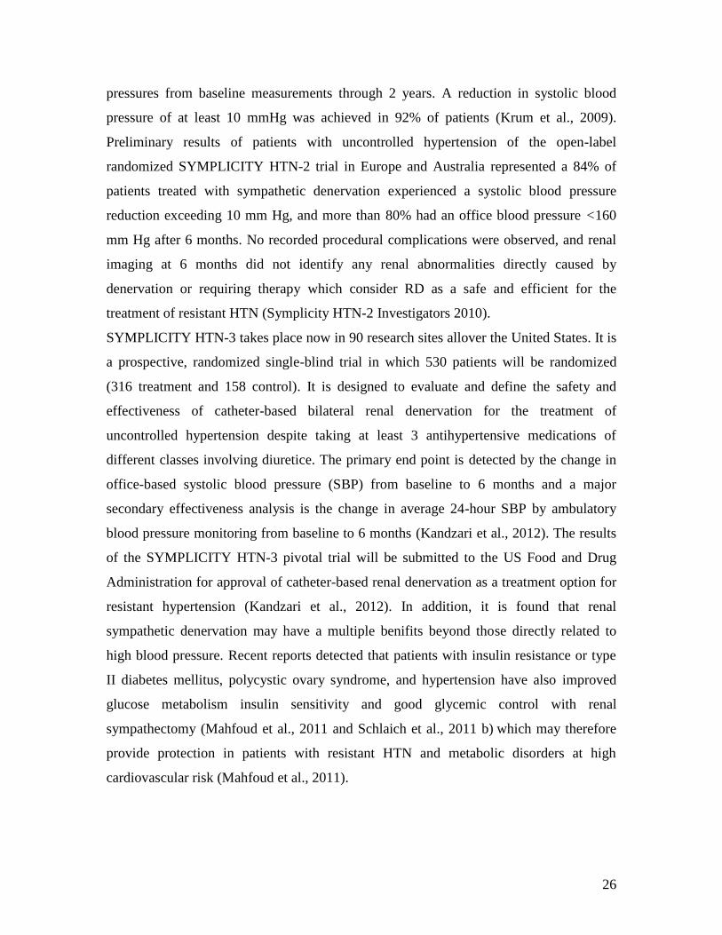
5.3.3. Pulsed wave (PW) Doppler and Tissue Doppler imaging (TDI)
Conventional echocardiographic studies were performed using “standard” diastolic
parameters. Pulsed-wave Doppler measurements of LV inflow velocity (mitral inflow
measurements) in which the sample volume was placed at the tip of the mitral leaflets in
the apical 4-chamber view. The following Doppler indices were measured: peak early
(E), peak late (A) flow velocities, E/A ratio, deceleration time (DT) Figure (7). Mitral
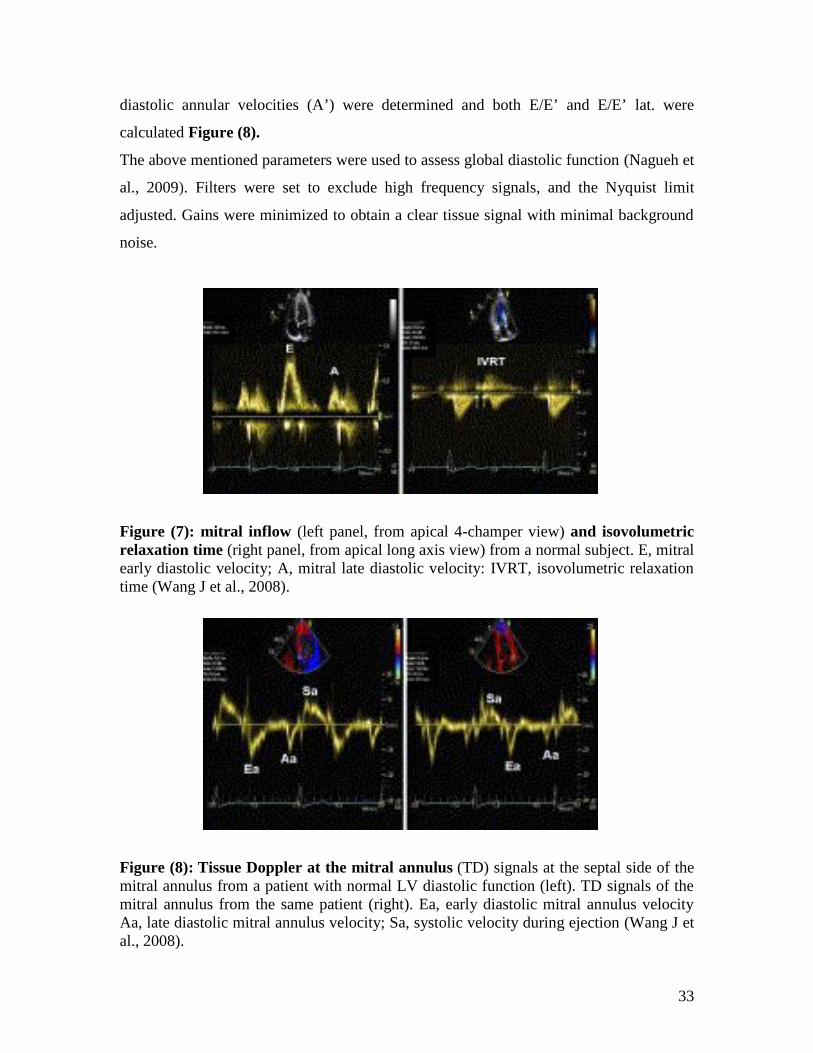
annular velocities were also recorded from the apical 4-chamber view with the pulse-
wave Doppler sample volume, (2 mm in size) placed in the septal and lateral corners of
the mitral annulus under tissue Doppler imaging mode. Peak early diastolic (E’) and late
33
diastolic annular velocities (A’) were determined and both E/E’ and E/E’ lat. were
calculated Figure (8).
The above mentioned parameters were used to assess global diastolic function (Nagueh et
al., 2009). Filters were set to exclude high frequency signals, and the Nyquist limit
adjusted. Gains were minimized to obtain a clear tissue signal with minimal background
noise.
Figure (7): mitral inflow (left panel, from apical 4-champer view) and isovolumetricrelaxation time (right panel, from apical long axis view) from a normal subject. E, mitralearly diastolic velocity; A, mitral late diastolic velocity: IVRT, isovolumetric relaxationtime (Wang J et al., 2008).
Figure (8): Tissue Doppler at the mitral annulus (TD) signals at the septal side of themitral annulus from a patient with normal LV diastolic function (left). TD signals of themitral annulus from the same patient (right). Ea, early diastolic mitral annulus velocityAa, late diastolic mitral annulus velocity; Sa, systolic velocity during ejection (Wang J etal., 2008).

34
5.3.4. SRI data Analysis
Myocardial deformation measurements were performed using tissue speckle tracking.
Three cardiac cycles were recorded in apical four-, two-chamber, and long-axis views
using grey-scale acquisition at a frame rate is between 70 and 90 frames per second.
Cardiac cycles were recorded in a cine loop format and stored digitally for subsequent
off-line analysis at EchoPAC PC (GE Healthcare, Horton, Norway) workstation for two-
dimensional (2D) strain analysis version 110.0.2 (GE Vingmed), averaging three cardiac
cycles, which allowing quantitative evaluation of the myocardial function. Offline
analysis of apical 4, 2-chamber and long axis views images were completed by tracing
the endocardium in end diastole and the thickness of the region of interest adjusted to
include the entire myocardium (Moen et al., 2011). Images with frame rates below 40 Hz
and above 80 Hz were excluded from analysis to ensure adequate temporal and spatial
resolution as well as accurate frame to frame tracking as regards measurement of strain or
SRI (strain rate imaging) (Horton et al., 2009).
Strain and strain rate curves were obtained in each of 3 views (4-, 2-chamber and long-
axis views) in which all LV myocardial segments (6 segments per view) are presented.
Peak strain was defined as the peak negative value on the strain curve during the entire
cardiac cycle (Reisner et al., 2004). The average value of peak systolic longitudinal strain
and peak systolic strain rate from all three views were then calculated as global systolic
longitudinal strain (GS) or (SISYS) Figure (9) and (10) and global systolic strain rate
(GSR) or (SRSYS), respectively Figure (11) (Nagueh et al., 2009).
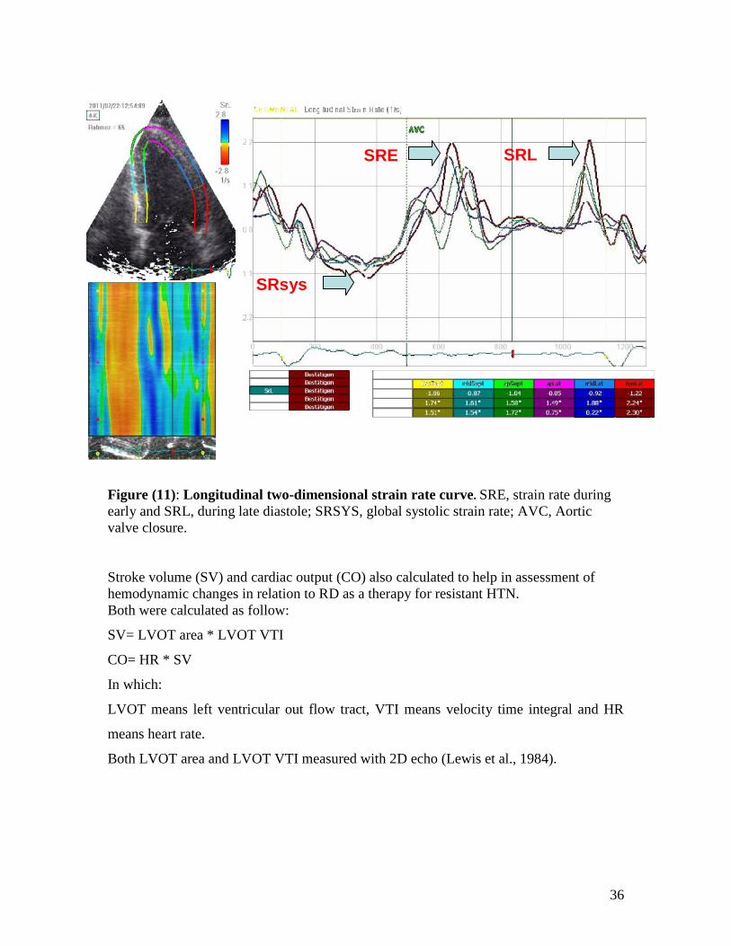
Similarly, peak global strain rate during early (SRE) and late (SRL) diastole were
determined. Diastolic indices E/SRE and SRE/SRL were calculated Figure (11) (Wang et
al., 2007). The off-line analysis was performed by one investigator.

35
Figure (9): Systolic longitudinal two-dimensional strain curve taken from apical 4-champer view. Peak strain (arrow) is the peak negative value on the strain curve duringthe entire cardiac cycle. Segmental strain curves are presented by continuous lines andthe global strain curve by interrupted line.
Figure (10): Average value of peak global systolic longitudinal strain from all threeviews was then calculated as global longitudinal strain (GS).
36
Figure (11): Longitudinal two-dimensional strain rate curve. SRE, strain rate duringearly and SRL, during late diastole; SRSYS, global systolic strain rate; AVC, Aorticvalve closure.
Stroke volume (SV) and cardiac output (CO) also calculated to help in assessment ofhemodynamic changes in relation to RD as a therapy for resistant HTN.Both were calculated as follow:
SV= LVOT area * LVOT VTI
CO= HR * SV
In which:
LVOT means left ventricular out flow tract, VTI means velocity time integral and HR
means heart rate.
Both LVOT area and LVOT VTI measured with 2D echo (Lewis et al., 1984).
SRE SRL
SRsys

37
6. Statistics
SPSS Version 20.0 was used for statistical analysis. Results are displayed as mean±SEM.
Frequencies are expressed as percentages. Statistical significance of the comparison of
measurements at 6 months follow-up and baseline were assessed by a paired Student’s t
test for parametric data and by an unpaired t-test when comparing the differences
between subgroups. For categorical data, X 2 analysis was used. Pearson’s correlation
coefficient (r) was used for analyses of linear correlations.

38
7. Results
7.1. Patient baseline characteristics
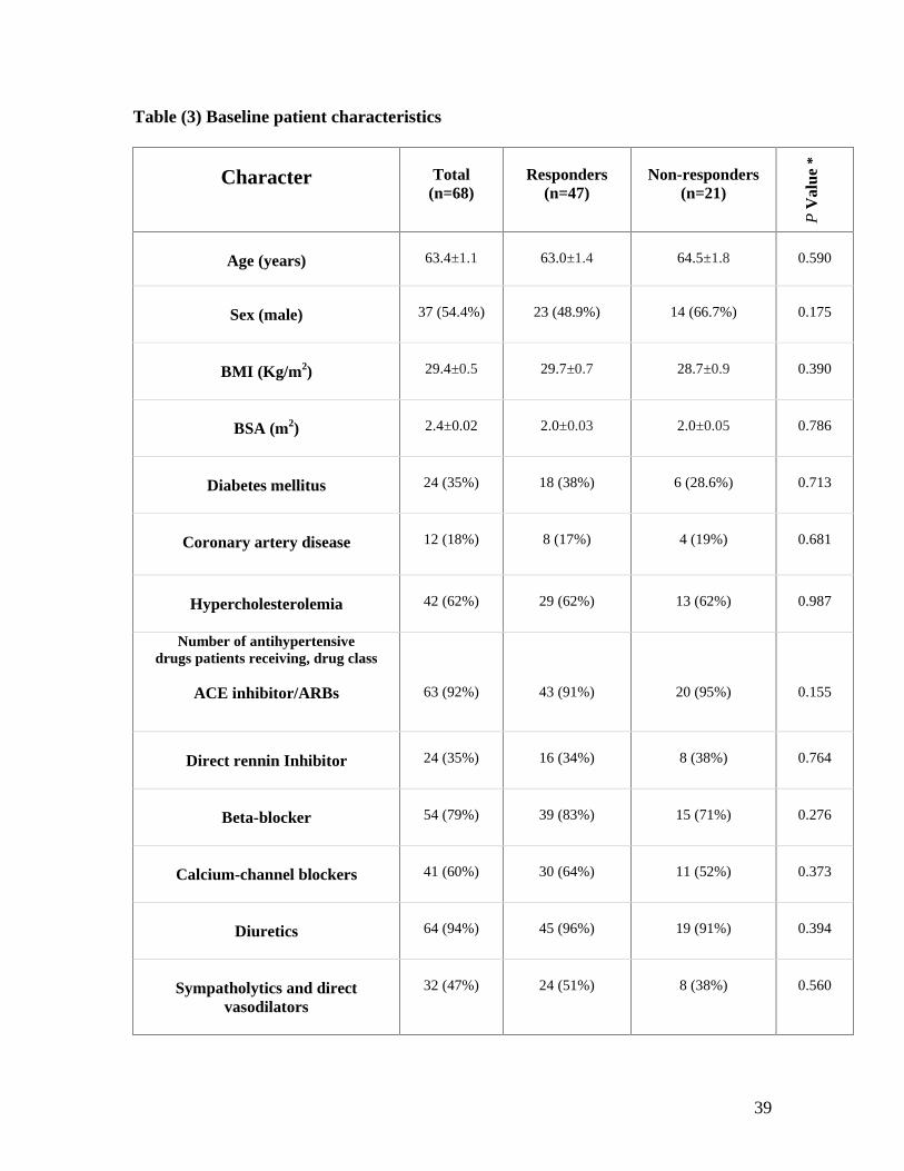
The current study included 68 patients (Table 3), 37 males (54.4 %) and 31 females (45.6
%) with mean age 63.4±1.1 years. The patients group included 12 patients had coronary
artery disease, 24 patients had diabetes mellitus (DM) (19 with type 2 DM) and 42
patients hypercholesterolemia. 64 (94%) patients were receiving diuretics; 54 patients
(79.4%) were taking ß-blocker, 41 (60.3 %) taking calcium channel blocker, 63 (92.6%)
receiving an angiotensin-converting enzyme inhibitor, angiotensin II receptor blocker, or
both and 44 (64.7 %) receiving peripheral direct vasodilator, centrally acting
sympatholytic agents or both. Body mass index (BMI) and body surface area (BSA) were
similar at baseline and at 6 months (BMI 29.4±0.5 kg/m2 and 2.0 m2, respectively).
Baseline and 6 months follow-up were needed for inclusion in the present study. There
were no changes in medication during the study period in any of the patients up to the 6-
month follow-up.
39
Table (3) Baseline patient characteristics
Character Total(n=68)
Responders(n=47)
Non-responders(n=21)
PV
alue
*
Age (years) 63.4±1.1 63.0±1.4 64.5±1.8 0.590
Sex (male) 37 (54.4%) 23 (48.9%) 14 (66.7%) 0.175
BMI (Kg/m2) 29.4±0.5 29.7±0.7 28.7±0.9 0.390
BSA (m2) 2.4±0.02 2.0±0.03 2.0±0.05 0.786
Diabetes mellitus 24 (35%) 18 (38%) 6 (28.6%) 0.713
Coronary artery disease 12 (18%) 8 (17%) 4 (19%) 0.681
Hypercholesterolemia 42 (62%) 29 (62%) 13 (62%) 0.987
Number of antihypertensivedrugs patients receiving, drug class
ACE inhibitor/ARBs 63 (92%) 43 (91%) 20 (95%) 0.155
Direct rennin Inhibitor 24 (35%) 16 (34%) 8 (38%) 0.764
Beta-blocker 54 (79%) 39 (83%) 15 (71%) 0.276
Calcium-channel blockers 41 (60%) 30 (64%) 11 (52%) 0.373
Diuretics 64 (94%) 45 (96%) 19 (91%) 0.394
Sympatholytics and directvasodilators
32 (47%) 24 (51%) 8 (38%) 0.560

40
Values are mean ± SEM or n (%). * For comparison between subgroups, the Pearsonchi-square test was performed.BMI: Body mass index, BSA: Body surface area, ACE =Angiotensin-converting enzyme: ARB = angiotensin receptor blocker.
7.2. Effect of RD on hemodynamic
At baseline, overall mean sitting office systolic blood pressure was 173±3 mmHg, and
mean sitting office diastolic blood pressure was 92±2 mm Hg, with a heart rate of 68±1
bpm. Renal denervation significantly reduced systolic (-22±3 mmHg; P<0.0001, by 12.5
%) Figure (12 and 13) and diastolic (-10±2mmHg; P<0.0001, by 11 %) blood pressures
as well as the mean HR (-12±3 bpm; P<0.0001, by 17.6 %) 6 months after the procedure.
The patients were divided according to their reduction of BP after RD into responders
and non-responders in which the reduction of blood pressure > 10 mmHg will be defined
as response to RD. Accordingly, 47 patients of the treated patients (69 %) were
considered as responders and 21 (31 %) are non-responders. The responders group
showed significant reduction in systolic, diastolic BP (-33±3 mmHg and -14±3 mmHg
respectively; P<0.0001) and also HR (- 8±3; P<0.0001); however the non-responders
sub-group did not show significant reduction in BP after RD but they actually exhibited
increase in SBP. On the other hand, HR reduced significantly in this sub group (-21±6;
P=0.0004).
A significant difference in the reduction of SBP, DBP and HR was reported between 2
subgroups (P< 0.0001, P=0.01 and P=0.03 respectively) (Table 4). The SV showed
increase in all patients but that was statistically non significant. The CO is dependent on
HR, so as long as the later decreased significantly, also CO deceased (Table 4) and
Figure (14).

41
Table (4) Changes in hemodynamic in response to renal denervation
Cha
ract
erTotal
(n=68)Responders
(n=47)Non-responders
(n=21)
PV
alue
*
Baseline 6 month Baseline 6 month Baseline 6 month
SBP(mm Hg)
173.0±3.0 151±3.2** 174.8±3.5 141.9±2.7** 168±5.4 172.3±6.8* <0.0001
DBP(mm Hg)
92.0±2.3 82.3±1.5** 93.3±2.9 79.8±1.8** 89.0±4.1 88.3±2.8 0.009
Pulsepressure(mmHg)
82.4±2.9 68.7±2.7** 83.5±3.6 62.2±3.9** 75.9±6.2 84.0±6.4* <0.0001
HR(bpm)
68.3±1.4 60.7±1.2** 67.2±1.1 60.4±1.4** 70.8±2.7 61.2±2.3* 0.027
SV (ml) 85.6±2.2 89.2±2.2 85.6±2.7 88.9±3.9 86.6±4.4 88.8±4.8 0.976
CO(ml/min)
5.7±0.2 5.4±0.2 5.8±0.0 5.4±0.0* 5.9±0.35 5.4±0.4 0.831
Values are mean ± SEM. **P ≤0.001, *P≤0.05 between 6 months and baseline value.p Value * for difference in ∆ between responders and non-responders subgroups, theunpaired t-test was performed.SBP = Systolic Blood Pressure, DBP = Diastolic Blood Pressure, HR = Heart Rate, SV =stroke volume, CO = cardiac output.

42
Figure (12): Systolic and diastolic blood pressure (A) and changes in blood pressure
(B) 6 months after renal denervation.
A
* p< 0.0001, **p=0.016
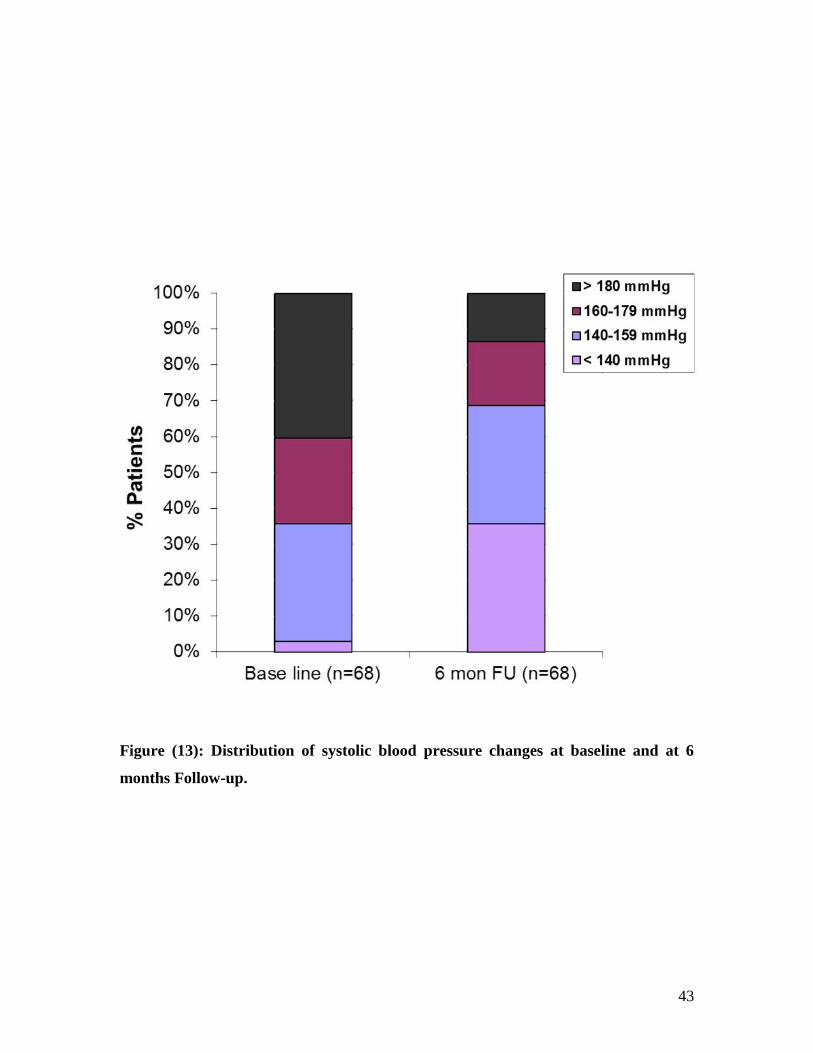
43
Figure (13): Distribution of systolic blood pressure changes at baseline and at 6
months Follow-up.

44
Figure (14): Effect of renal denervation on stroke volume (A), heart rate (B) and
cardiac output (C) at rest after 6 months compared with baseline.

45
7.3. Changes in cardiac structures in response to renal denervation
As shown in (Table 5), the LV mass index reduced significantly in the whole group
(-19.37±2.6 gm/m²; P<0.0001 by 14.7%). The reduction of LV mass was due to
reduction of interventricular septum and posterior wall (-0.65 mm±0.17; P=0.0002 and -
0.66 mm±0.19; P=0.0009 respectively). That is beside the reduction of LV internal
diameter in diastole (-2.40±0.43 mm; P<0.0001). Furthermore, a 21 % reduction in LV
end-diastolic volume (-19.25±3.14 ml; P<0.0001) and an 11% reduction in left atrial
volume index (-3.77±0.71 ml/m²; P<0.0001) was also found. If we look to the subgroups,
responders showed regression in LV mass index which was not stronger than in the
whole group (-17.00±3.00 gm/m²; P<0.0001). That came with a significant reduction in
interventricular septum and posterior wall (-0.76 mm±0.20; P=0.0005 and-0.48mm±0.22;
P=0.04 respectively). LV end diastolic diameter also found to be significantly reduced in
this subgroup (-2.03±0.50 mm; P<0.0001) to contribute to over all reduction in LV mass
index. Responders showed a similar reduction in both left atrial volume index
(-4.01±0.79 ml/m²; P<0.0001) and LV end-diastolic volume (-20.04±4.15 ml; P<0.0001).
Interestingly, among non-responders the change in left atrial and left ventricular structure was as
strong as in the responder group, with significant regression in the mean LV mass index
(-24.89±4.89 gm/m²; P<0.0001). That was mainly because of the significant reduction in
the posterior wall and LV end diastolic parameter (- 0.99 mm±0.37; P=0.015 and -3.36
mm±0.81; P=0.0005 respectively) without observed significant reduction of the
interventricular septum (-0.44 mm±0.30; P=0.161). We found less strongly pronounced but
still significant reduction in both left atrial volume index and LV end-diastolic volume in
this subgroup (-3.35±1.53 ml/m²; P=0.041 and -18.35±6.22 ml; P=0.008) Figure (15
and 16).

46
Table (5): Changes in cardiac structures in response to renal denervation
Cha
ract
erTotal
(n=68)Responders
(n=47)Non-responders
(n=21)
PV
alue
*
Baseline 6 month Baseline 6 month Baseline 6 month
LVMI(g/m2) 134.9±4.1 115.5±3.2** 131.7±4.4 114.9±3.9** 141.9±8.9 117.0±6.0** 0.144
LVEDD(mm)
52.12±0.6 49.7±0.6** 51.2±0.8 49.2±0.7** 54.2±0.9 50.1±0.9** 0.138
LVESD(mm)
31.7±0.6 30.3±0.5 30.9±0.7 29.8±0.2 33.24±1.2 31.2±1.0 0.239
IVS(mm)
12.8±0.2 12.1±0.2** 12.9±0.3 12.1±0.3** 12.5±0.4 12.0±0.4 0.394
PW(mm)
12.2±0.3 11.5±0.2** 12.2±0.3 11.7±0.2* 12.1±0.4 11.1±0.4* 0.251
LVEDvol(ml)
98.7±3.6 79.4±3.6** 98.3±4.4 78.6±3.2** 99.5±6.4 81.1±6.5* 0.860
LVESvol(ml)
32.0±1.2 27.5±1.1* 30.5±1.2 26.7±3.4* 36.04±2.3 29.3±2.5* 0.167
LAVI(ml/ m2)
34.3±1.03 30.5±0.9** 33.6±1.1 29.6±1.1** 35.8±2.2 32.5±1.2* 0.949
MAPSE(mm)
14.7±0.3 15.4±0.2* 14.8±0.4 15.6±0.3* 14.5±0.6 14.9±0.4 0.487
EF(%)
65.2±0.8 65.43±0.6 65.9±1.0 65.6±0.7 63.4±1.2 65.1±1.3 0.396

47
Values are mean ± SEM. **P ≤0.001, *P≤0.05 between 6 months and baseline value. pValue * for difference in ∆ between responders and non-responders subgroups, theunpaired t-test was performed.LVMI = Left Ventricular Mass Index, LVEDD = Left ventricular End DiastolicDimension, LVESD = Left ventricular End Systolic Dimension, IVS = Interventricularseptum Thickness, PW = Posterior Wall Thickness, LVED vol = Left Ventricular EndDiastolic Volume, LVESvol= Left Ventricular End Systolic Volume, LAVI = Left AtrialVolume Index, MAPSE = Mitral annular plane systolic excursion, EF= Ejection Fraction.
Figure (15): Left atrial volume index decreased 6 months after RD compared with
baseline independent from blood pressure response.

48
Figure (16): Effect of renal denervation on interventricular septum (A), posterior
wall (B), left ventricular end diastolic dimension (LVEDD) (C), left ventricular end
systolic dimension (LVESD) (D) and left ventricular mass index (E) after 6 months
compared with baseline.

49
7.4. Changes in systolic function in response to renal denervation
MAPSE, which considered as a longitudinal systolic function parameter, showed a
significant increase in the whole group and responders 6 months after RD (0.71±0.26
mm; P=0.008 and 0.87±0.30 mm; P=0.007 respectively) with no observed significant
change in EF. No significant change in MAPSE was observed (-0.45±0.51 ml/m²;
P=0.388) in non-reponders and also no observed significant change in EF in both
subgroups Figure (17, 24).

50
Figure (17): Effect of renal denervation on left ventricular end systolic volume
(LVESV) (A), left ventricular end diastolic volume (LVEDV) (B) and ejection
fraction (EF) (C) after 6 months compared with baseline. EF: derived from biplane
method.

51
7.5. Changes in diastolic function in response to renal denervation
7.5.1 Blood flow Doppler and TDI assessment of left ventricular dysfunction during
diastole
On analyzing LV diastolic function (Table 6), the whole group showed significantly
increase in E/A ratio after 6 months (0.89±0.04 versus 1.01±0.06; P=0.001) with
significant shortening in deceleration time (DT) and isovolumetric relaxation time
(IVRT) from baseline values (-24.85±8.93 ms; P=0.007 and -6.97±2.57 ms; P=0.012
respectively). Similarly, the responders showed increased E/A ratio significantly
(0.89±0.05 versus 1.00±0.06; P=0.013) with significant shortening in DT and IVRT from
baseline values (-30.91±10.77ms; P=0.006 and -8.55±2.93 ms; P=0.006 respectively)
after 6 months. Studying the non-responders subgroup, only DT was significantly
changed (-30.59±13.57 ms; P=0.035) without any observed changes involved E/A ratio
or IVRT (0.18±0.09 ms; P=0.061 and -1.68±5.14 ms; P=0.747 respectively) Figure
(18and 19). Concerning the mean values of both E/E’ and E/E’ lat. ratios, no significant
changes were observed compared with baseline values in all patients although significant
improvement (increase) in E’sep and E’lat. were reported in whole group as well as
responders Figure (20 and 21).
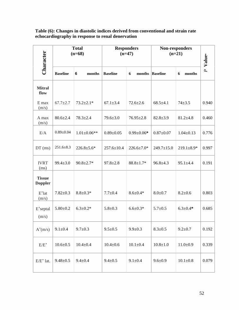
52
Table (6): Changes in diastolic indices derived from conventional and strain rateechocardiography in response to renal denervation
Cha
ract
erTotal
(n=68)Responders
(n=47)Non-responders
(n=21)
PV
alue
*
Baseline 6 months Baseline 6 months Baseline 6 months
Mitralflow
E max(m/s)
67.7±2.7 73.2±2.1* 67.1±3.4 72.6±2.6 68.5±4.1 74±3.5 0.940
A max(m/s)
80.6±2.4 78.3±2.4 79.6±3.0 76.95±2.8 82.8±3.9 81.2±4.8 0.460
E/A 0.89±0.04 1.01±0.06** 0.89±0.05 0.99±0.06* 0.87±0.07 1.04±0.13 0.776
DT (ms) 251.6±8.3 226.8±5.6* 257.6±10.4 226.6±7.0* 249.7±15.0 219.1±8.9* 0.997
IVRT(ms)
99.4±3.0 90.8±2.7* 97.8±2.8 88.8±1.7* 96.8±4.3 95.1±4.4 0.191
TissueDoppler
E’lat(m/s)
7.82±0.3 8.8±0.3* 7.7±0.4 8.6±0.4* 8.0±0.7 8.2±0.6 0.803
E’septal
(m/s)
5.80±0.2 6.3±0.2* 5.8±0.3 6.6±0.3* 5.7±0.5 6.3±0.4* 0.685
A’(m/s) 9.1±0.4 9.7±0.3 9.5±0.5 9.9±0.3 8.3±0.5 9.2±0.7 0.192
E/E’ 10.6±0.5 10.4±0.4 10.4±0.6 10.1±0.4 10.8±1.0 11.0±0.9 0.339
E/E’ lat. 9.48±0.5 9.4±0.4 9.4±0.5 9.1±0.4 9.6±0.9 10.1±0.8 0.079

53
StrainRate
imaging
SISYS(%)
-17.5±0.41 -18.7±0.35* -17.6±0.5 -18.8±0.5* -17.5±0.7 -18.7±0.4 0.912
SRSYS(s−1)
-0.93±0.03 -0.94±0.02 -0.94±0.03 -0.94±0.03 -0.9±0.05 -0.95±0.04 0.482
SRE(s−1)
0.86±0.04 0.90±0.03 0.86±0.04 0.88±0.03 0.87±0.06 0.96±0.06 0.331
SRL(s−1)
0.83±0.03 0.85±0.027 0.84±0.04 0.87±0.03 0.83±0.05 0.83±0.04 0.781
SRE/L 1.1±0.06 1.2±0.05 1.1±0.09 1.07±0.06 1.1±0.08 1.2±0.09 0.154
E/SRE(m)
84.0±3.9 84.9±3.3 82.3±4.4 86.0±4.1 87.7±8.3 80.6±5.1 0.114
Values are mean ± SEM. **P ≤0.001, *P≤0.05 between 6 months and baseline value. PValue * for difference in ∆ between responders and non-responders subgroups, theunpaired t-test test was performed.E/A, the ratio of early (E) to late (A) mitral flow peak velocities; DT = Deceleration time,IVRT = Isovolumetric Relaxation Time. E’ septal and E′ lat. = early diastolic peakvelocities of mitral annulus at septal and lateral site respectively; E/E’ and E/E’ lat. = LVfilling index; SISYS, systolic strain; SRSYS, systolic strain rate; SRE, strain rate duringearly diastole; SRL, during late diastole; SRE/L, strain rate early-to-late ratio; E/SRE = Emax-to-SRE.

54
Figure (18): Impact of renal denervation on mitral inflow velocities andisovolumetric relxation time. Improvement in the mitral flow indices (E max, A max,E/A ratio and deceleration time) 6 months after RD compared with baseline (A).Shortening in Isovolumetric relaxation time (IVRT) 6 months after RD compared withbaseline (B). Note reduction of HR. See (Table 6).

55
Figure (19): Effect of renal denervation on Doppler derived diastolic parameters;
Deceleration time (DT) (A), isovolumetric relaxation time (IVRT) (B) and E/A ratio
(C) after 6 months compared with baseline.

56
Figure (20): Impact of renal denervation on mitral annular velocities. Septal andlateral mitral annular velocities were recorded from the apical 4-chamber view with thepulse-wave Doppler placed in the septal and lateral corners of the mitral annulus undertissue Doppler imaging mode. Peak early diastolic (E’) and late diastolic annularvelocities (A’) were determined and both E/E’ and E/E’ lat. were calculated. (A) beforeRD and (B) after RD. See (Table 6).
A
B

57
Figure (21): Effect of renal denervation on tissue doppler derived diastolic
parameters; E’ septal (A), E’ lateral (B) and E/E’ ratio (C) after 6 months compared
with baseline.

58
7.5.2. Change in Left Ventricular Filling Patterns
E/E’ ratio was used to classify the different patterns of LV filling pressures. It is
considered normal if the E/E’ ratio is lower than 8, LV filling pressures are low and
considered abnormal if E/E’ more than 15, LV filling pressures is high. Then we
considered trans mitral flow parameters to sub classify this abnormal high LV filling
pattern in to 3 stages. If DT ˃ 275 ms, IVRT ˃ 105 ms and E/A ratio ˂ 0.5, the patient
was diagnosed to have abnormal relaxation (functionally stage 1 diastolic dysfunction). If
the previously mentioned trans mitral flow parameters were normal despite the high E/E’
ratio, the patient was diagnosed to have pseudonormalisation pattern (functionally stage 2
diastolic dysfunction). In case of DT ˂ 150 ms, IVRT ˂ 60 ms and E/A ratio ˃ 2, the
patient was considered to have restrictive flow pattern (functionally stage 3 diastolic
dysfunction). An E/E’ ratio ranging from 8 to 15 is considered suggestive but not a
diagnostic evidence of diastolic LV dysfunction and needs to be implemented with trans
mitral blood flow to confirm the diagnosis. At baseline, abnormal relaxation had the most
common prevalence between patients (37); they represented 54.4%, whereas (24)
represented 35.3 % had normal mitral valve flow profile. 7 patients (10.3 %) had
pseudonormal flow profile, and no patient had restrictive flow pattern at baseline. After 6
months of treatment, the prevalence of normal flow pattern increased to 47 (69.2%) and
abnormal relaxation decreased to 19 (27.9%) and the prevalence of pseudonormal pattern
decreased to 2 patients (2.9%), (P=0.027) Figure (22).

59
Figure (22): Improvement in Left Ventricular Filling Patterns 6 months after RD
compared with baseline. Chi square test was used to find the significance of difference
in filling pattern before and after renal denervation.
7.6. Strain and strain rate imaging (SRI)
7.6.1. Systolic SRI parameters
Longitudinal LV systolic function is estimated by mean strain SL and SR. Both represent
systolic parameters of SRI. The whole group as well as responders showed significant
increase in global longitudinal strain SL (-17.53±0.41 versus -18.65±0.35; P=0.003 and -
17.55±0.49 versus -18.83±0.84; P=0.013, respectively) Figures (23 and 24). In contrast,
GSR did not show significant differences between FU and baseline. 6 months after RD,
non-responders showed no observed significant improvement in global longitudinal
systolic SRI parameters (Table 6).

60
Figure (23): Global longitudinal strain (Bull`s Eye) before (A) and 6 months after
RD (B).

61
Figure (24): Effect of renal denervation on Mitral annular systolic excursion
(MAPSE) (A), longitudinal strain (B) and longitudinal strain rate (C) after 6 months
compared with baseline.
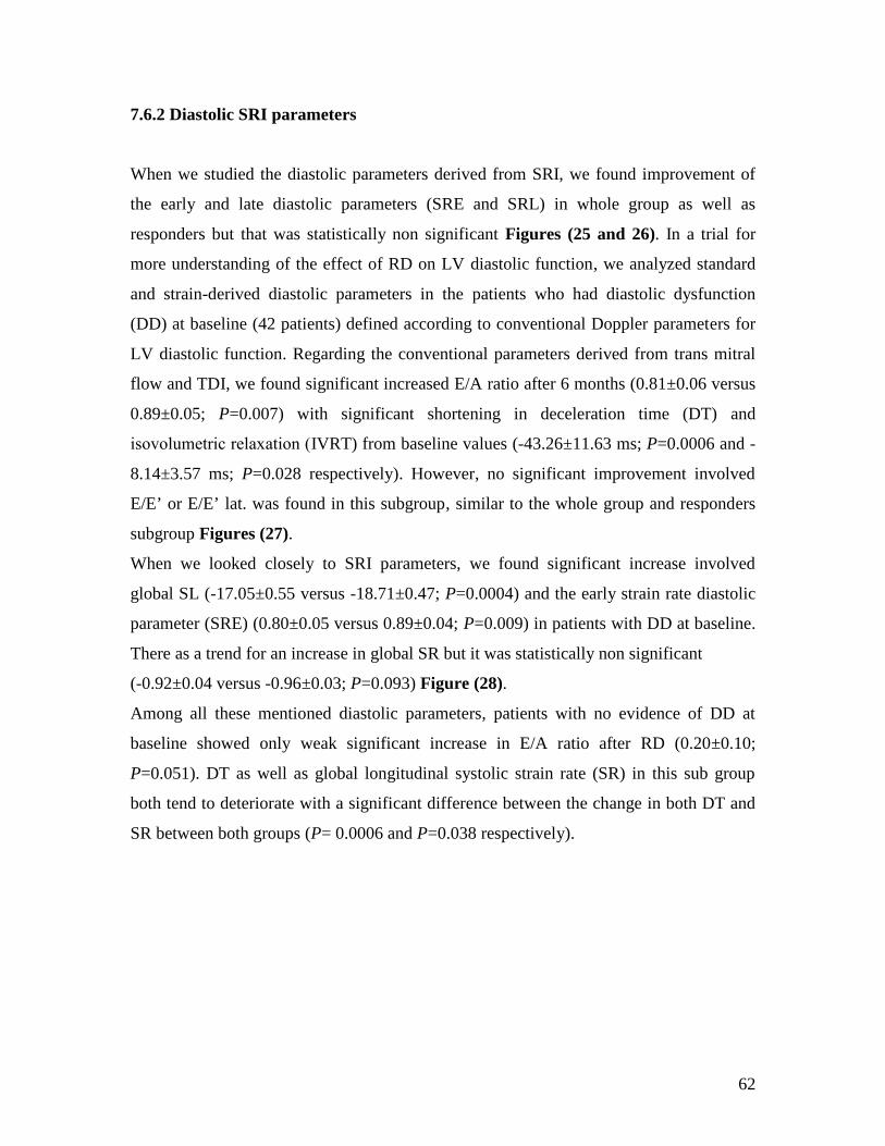
62
7.6.2 Diastolic SRI parameters
When we studied the diastolic parameters derived from SRI, we found improvement of
the early and late diastolic parameters (SRE and SRL) in whole group as well as
responders but that was statistically non significant Figures (25 and 26). In a trial for
more understanding of the effect of RD on LV diastolic function, we analyzed standard
and strain-derived diastolic parameters in the patients who had diastolic dysfunction
(DD) at baseline (42 patients) defined according to conventional Doppler parameters for
LV diastolic function. Regarding the conventional parameters derived from trans mitral
flow and TDI, we found significant increased E/A ratio after 6 months (0.81±0.06 versus
0.89±0.05; P=0.007) with significant shortening in deceleration time (DT) and
isovolumetric relaxation (IVRT) from baseline values (-43.26±11.63 ms; P=0.0006 and -
8.14±3.57 ms; P=0.028 respectively). However, no significant improvement involved
E/E’ or E/E’ lat. was found in this subgroup, similar to the whole group and responders
subgroup Figures (27).
When we looked closely to SRI parameters, we found significant increase involved
global SL (-17.05±0.55 versus -18.71±0.47; P=0.0004) and the early strain rate diastolic
parameter (SRE) (0.80±0.05 versus 0.89±0.04; P=0.009) in patients with DD at baseline.
There as a trend for an increase in global SR but it was statistically non significant
(-0.92±0.04 versus -0.96±0.03; P=0.093) Figure (28).
Among all these mentioned diastolic parameters, patients with no evidence of DD at
baseline showed only weak significant increase in E/A ratio after RD (0.20±0.10;
P=0.051). DT as well as global longitudinal systolic strain rate (SR) in this sub group
both tend to deteriorate with a significant difference between the change in both DT and
SR between both groups (P= 0.0006 and P=0.038 respectively).

63
Figure (25): Systolic and diastolic strain rate before (A) and 6 months after RD (B)involving segments of 3 champers view (apical long axis view) .

64
Figure (26): Effect of renal denervation on early diastolic strain rate SRE (A) and
late diastolic strain rate SRL (B) after 6 months compared with baseline.

65
Figure (27): Impact of renal denervation on deceleration time (A), isovolumetric
relaxation time (B), E/A ratio (C) and E/E’ ratio (D) after 6 months compared with
baseline in patients with resistant hypertension divided according to the existence of
diastolic dysfunction at baseline. DD: diastolic dysfunction.

66
Figure (28): Impact of renal denervation on strain rate imaging derived systolic
parameters; global longitudinal strain (A), global longitudinal strain rate (B) and
diastolic parameters; early diastolic strain rate (C) and late diastolic strain rate (D)
after 6 months compared with baseline in patients with resistant hypertension
divided according to the existence of diastolic dysfunction at baseline. DD: diastolic
dysfunction.
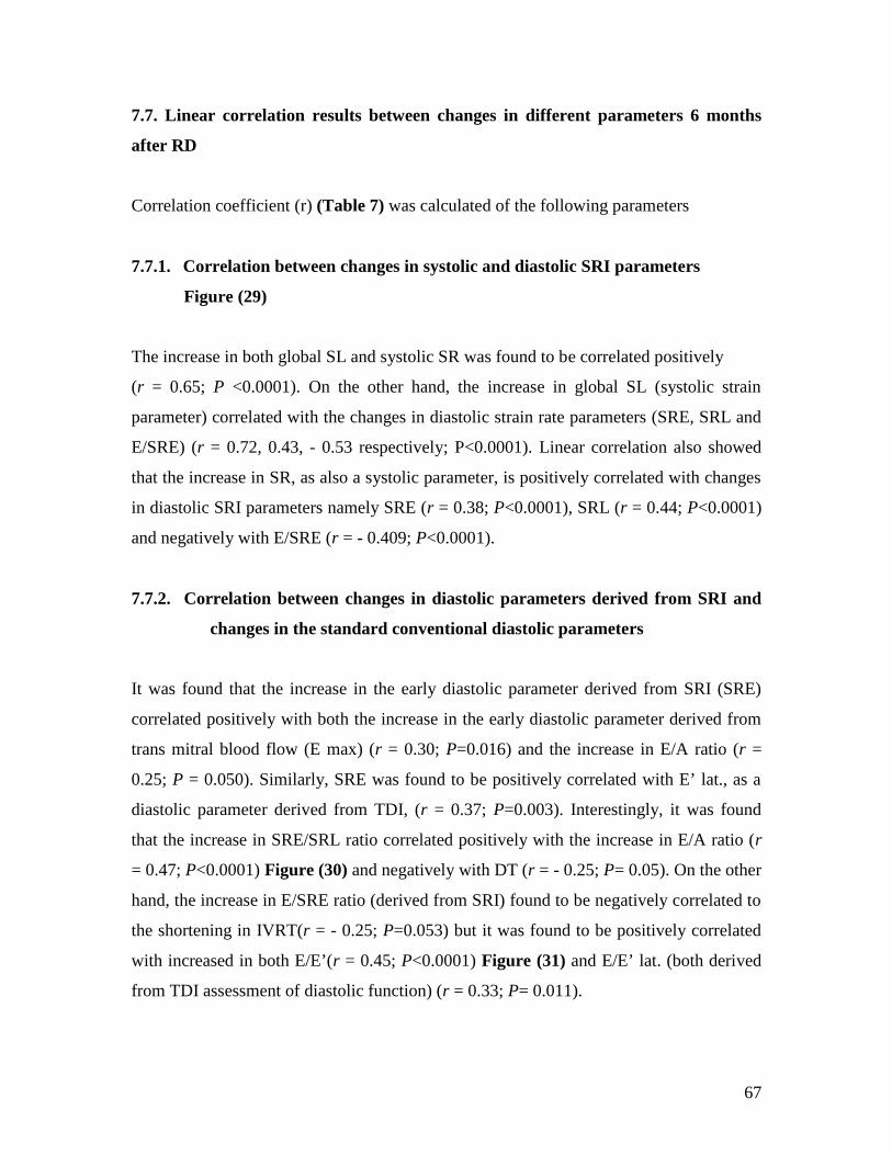
67
7.7. Linear correlation results between changes in different parameters 6 months
after RD
Correlation coefficient (r) (Table 7) was calculated of the following parameters
7.7.1. Correlation between changes in systolic and diastolic SRI parameters
Figure (29)
The increase in both global SL and systolic SR was found to be correlated positively
(r = 0.65; P <0.0001). On the other hand, the increase in global SL (systolic strain
parameter) correlated with the changes in diastolic strain rate parameters (SRE, SRL and
E/SRE) (r = 0.72, 0.43, - 0.53 respectively; P<0.0001). Linear correlation also showed
that the increase in SR, as also a systolic parameter, is positively correlated with changes
in diastolic SRI parameters namely SRE (r = 0.38; P<0.0001), SRL (r = 0.44; P<0.0001)
and negatively with E/SRE (r = - 0.409; P<0.0001).
7.7.2. Correlation between changes in diastolic parameters derived from SRI and
changes in the standard conventional diastolic parameters
It was found that the increase in the early diastolic parameter derived from SRI (SRE)
correlated positively with both the increase in the early diastolic parameter derived from
trans mitral blood flow (E max) (r = 0.30; P=0.016) and the increase in E/A ratio (r =
0.25; P = 0.050). Similarly, SRE was found to be positively correlated with E’ lat., as a
diastolic parameter derived from TDI, (r = 0.37; P=0.003). Interestingly, it was found
that the increase in SRE/SRL ratio correlated positively with the increase in E/A ratio (r
= 0.47; P<0.0001) Figure (30) and negatively with DT (r = - 0.25; P= 0.05). On the other
hand, the increase in E/SRE ratio (derived from SRI) found to be negatively correlated to
the shortening in IVRT(r = - 0.25; P=0.053) but it was found to be positively correlated
with increased in both E/E’(r = 0.45; P<0.0001) Figure (31) and E/E’ lat. (both derived
from TDI assessment of diastolic function) (r = 0.33; P= 0.011).

68
7.7.3. Correlation between the changes in global systolic SRI and LV parameters
We found that the increase in global SL (longitudinal systolic parameter derived from
SRI) correlated positively with the increase in MAPSE which is considered also as
longitudinal systolic function parameter measured by 2D echocardiography (r = 0.27; P =
0.033) and negatively with the regression in LV mass index in response to RD
(r = - 0.26; P = 0.041). The change in LVEF, as assessed by biplane Simpson’s method,
also correlated positively with the increase in MAPSE (r = 0.33; P=0.008) without
observed significant correlation between it and the changes involved SRI systolic
parameters 6 months after RD.
7.7.4. Correlations between different other parameters in response to treatment of
resistant HTN using RD
MAPSE, was found to be positively correlated with late diastolic parameter derived from
trans mitral flow (A max). The same also was found between regression of LVEDV and
increase in E’ lat., diastolic parameter derived from TDI, (r = 0.27; P = 0.031). This
increase in E’ lat. was found to be positively correlated with the increase in the global SL
(r = 0.35; P = 0.005). In contrast, increase in early diastolic SRI parameter (SRE) was
detected to be negatively correlated with reduction in LV mass (r = - 0.27; P = 0.033).
Shortening in DT was observed to be positively correlated with reduction in DBP
(r = 0.36; P = 0.001). Similarly, decrease of LAVI was positively correlated with
reduction of SBP which considered statistically weak positive correlation (r = 0.23; P =
0.051). Except that, no observed correlations were found between the different echo
parameters and the reduction of BP or HR.
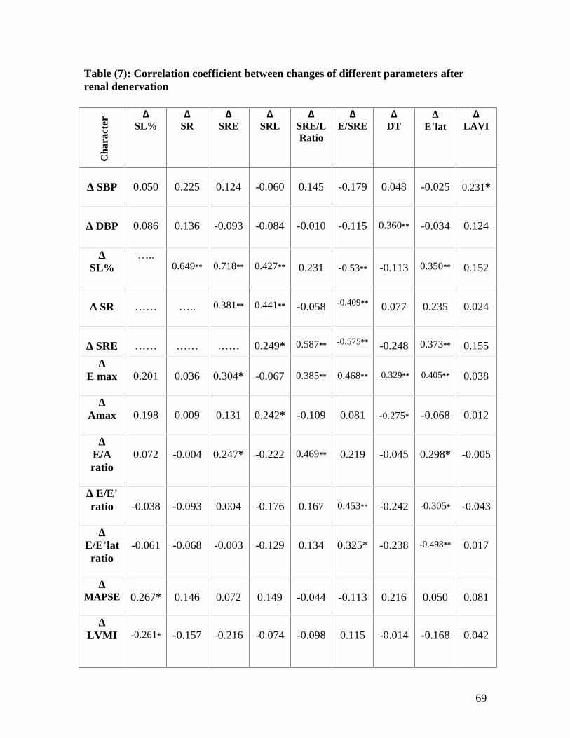
69
Table (7): Correlation coefficient between changes of different parameters afterrenal denervation
Cha
ract
er∆
SL%∆
SR∆
SRE∆
SRL∆
SRE/LRatio
∆E/SRE
∆DT
∆E’lat
∆LAVI
∆ SBP 0.050 0.225 0.124 -0.060 0.145 -0.179 0.048 -0.025 0.231*
∆ DBP 0.086 0.136 -0.093 -0.084 -0.010 -0.115 0.360** -0.034 0.124
∆SL%
…..0.649** 0.718** 0.427** 0.231 -0.53** -0.113 0.350** 0.152
∆ SR …… ….. 0.381** 0.441** -0.058 -0.409** 0.077 0.235 0.024
∆ SRE …… …… …… 0.249* 0.587** -0.575** -0.248 0.373** 0.155
∆E max 0.201 0.036 0.304* -0.067 0.385** 0.468** -0.329** 0.405** 0.038
∆Amax 0.198 0.009 0.131 0.242* -0.109 0.081 -0.275* -0.068 0.012
∆E/Aratio
0.072 -0.004 0.247* -0.222 0.469** 0.219 -0.045 0.298* -0.005
∆ E/E’ratio -0.038 -0.093 0.004 -0.176 0.167 0.453** -0.242 -0.305* -0.043
∆E/E’lat
ratio-0.061 -0.068 -0.003 -0.129 0.134 0.325* -0.238 -0.498** 0.017
∆MAPSE 0.267* 0.146 0.072 0.149 -0.044 -0.113 0.216 0.050 0.081
∆LVMI -0.261* -0.157 -0.216 -0.074 -0.098 0.115 -0.014 -0.168 0.042

70
** Correlation is significant at the 0.01 level (2-tailed).* Correlation is significant at the 0.05 level (2-tailed).SBP = Systolic Blood Pressure, DBP = Diastolic Blood Pressure, LVMI = LeftVentricular Mass Index, LVEDvol = Left Ventricular End Diastolic Volume, LAVI =Left Atrial Volume Index, MAPSE = Mitral annular plane systolic excursion, E/A, theratio of early (E) to late (A) mitral flow peak velocities; DT = Deceleration time, E′ lat. =early diastolic peak velocities of mitral annulus at lateral site; E/E′ and E/E′ lat. ratios =LV filling index; SL, systolic strain; SR, systolic strain rate; SRE, strain rate during early;SRL, during late diastole; SRE/L, strain rate early-to-late ratio; E/SRE = E max-to-SRE.

71
Figure (29): Correlation coefficient between changes in systolic strain and diastolic
strain rate imaging parameters 6 months after renal denervation.
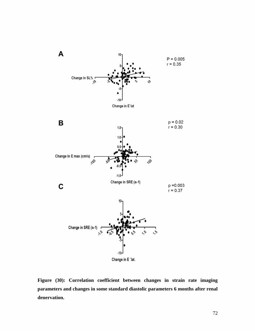
72
Figure (30): Correlation coefficient between changes in strain rate imaging
parameters and changes in some standard diastolic parameters 6 months after renal
denervation.

73
Figure (31): Correlation coefficient between changes in standard diastolic ratios and
changes in diastolic ratios derived from strain rate imaging 6 months following
renal denervation.

74
8. Discussion
We analyzed in the current study the effect of denervation of the renal sympathetic as
treatment of resistant HTN on hypertension-related cardiac remodeling and functional
impairment. In addition to the BP-lowering effect, we were able to observe reduction of
LV mass, LA volume, improvement of systolic and diastolic parameters by using
conventional 2D echocardiography, Doppler and longitudinal strain derived from 2 D
echocardiography speckle tracking. The improvement observed after 6 months follow-up
period. Reduction of peripheral BP was the main target in the treatment of patients with
arterial hypertension for many years. Recently, the evidence is increasing indicates that
the mode of BP control may differentially affect outcome (Lindholm et al., 2005 and
Williams et al., 2006). It has been observed that reduction in cardiovascular morbidity
and mortality does not automatically follow the lowering of peripheral BP as a main
target (De Caterina et al., 2010). It is still affected by other confounders mainly LVH as
intermediate endpoint which is shown to be reliably linked to cardiovascular prognosis
(Devereux et al. 2004 and Pierdomenico et al., 2010).
8.1. Effect of RD on hemodynamics
Chronic stimulation of the sympathetic nervous system is responsible for the
development and maintenance of arterial hypertension (Sobotka et al., 2011) and it is a
key component of the signalling pathways altered in hypertension-related cardiac
remodeling (Mancia et al., 2010). The Simplicity trials (HTN-I and HTN-2) identified a
significant and safe reduction in blood pressure achieved with catheter-based renal
denervation in patients with resistant HTN which provided insights into the critical role
of renal sympathetic and somatic nerves in mediating resistant hypertension (Simplicity
HTN-I by Krum H, et al., 2009 and Symplicity HTN-2 Investigators. 2010).
In our study, we noticed a significant reduction of the blood pressure in the whole group.
We further subdivided the patients according to the pattern of response into two sub
group, in which the response was defined as reduction of SBP>10 mmHg. Until now, we
cannot predict which population will respond satisfactory to RD. Heart rate (HR) is a

75
predictor of cardiovascular mortality and morbidity in the general population, as well as
in patients with hypertension and heart failure (Kolloch et al., 2008, Böhm et al., 2010
and Reil et al., 2011).
Beside BP lowering effect, our findings indicate a reduced resting HR in whole group
after RD. That was independent on BP response as both responders and nonresponders
also showed a significant decrease in resting HR. This finding confirms recent studies
analysing the effect of RD on HR at rest (Ukena et al., 2011 and Brandt et al., 2012).
Reduction of HR in patients who lack a relevant reduction of BP suggests that also in BP
non-responders there is a relevant anti-sympathetic effect of RD.
8.2. Effect of RD on different echocardiographic parameters
Many echocardiographic findings were significantly improved between BP responders
and non-responders, in comparison with baseline. However, there was a trend of BP
nonresponders to have maintained high BP and to show even deterioration in few cardiac
systolic and diastolic parameters.
8.2.1. Regression of the left ventricular hypertrophy
The presence of LVH, as an indicator of end-organ damage, is associated with an
increased rate of cardiovascular events and death independent of other cardiovascular risk
factors and, notably, independent of BP values (Ruilope et al., 2008 and Bombelli et al.,
2009). The sympathetic nervous system mediates hypertension-induced hypertrophy via
direct stimulation of cardiomyocyte beta-adrenergic receptors as prove with experimental
evidence (Perlini et al., 2005). Cardiac fibrosis and inflammation have been provided to
be a result from mast cell stimulation, activation of afferent sympathetic nerves,
angiotensin II production, and norepinephrine release, rather than being mediated via
alphaadrenergic receptors (Levick et al., 2010).
In hypertensive rat models both beta-blockade and sympathectomy attenuated LVH,
whereas accompanying myocardial interstitial fibrosis was abolished by sympathectomy

76
or doxazosin but left unchanged by beta-blockade (Perlini S, et al., 2005). These
experimental results were further supported by clinical observations. Antihypertensive
treatment with angiotensin receptor antagonism (losartan) has superior efficacy for
reversing LVH, a cardinal manifestation of hypertensive target organ damage, than
conventional atenolol-based treatment as demonstrated in The LIFE study (Losartan
Intervention For Endpoint reduction in Hypertension) (Dahlof et al., 2002).
In our study, we obtained a pronounced reduction of the LVMI by 19 gm/ m² (- 15%
from baseline) after RD, indicating a more pronounced effect of RD on LV mass
regression and LVH reduction in this treatment resistant patient group compared with the
effect of different antihypertensive drug classes used for treatment of uncomplicated
hypertension (Klingbeil et al., 2003). This finding was also found in both involved
subgroups in our study which denotes that the regression, following RD, was found
regardless of BP response after the procedure. This supports the hypothesis of BP-
independent effects of RD on LVH which was observed with animal models, in which
LV mass regression occurred also in 5 of 6 sympathectomy “nonresponders” (Perlini et
al., 2005). This was also further approved by a meta-analysis demonstrating that beta-
blockers induced significantly less LVH regression compared with various other
antihypertensive drugs, especially blockers of the rennin-angiotensin system beyond the
effects of BP reduction (Dahlof et al., 1992). That might explain, why there was no
observed significant correlation between changes in LVMI and changes in SBP or DBP
in our study. Recently, Brandt et al 2012 studied the effect of RD on LV mass and
detected the significant reduction of LV mass index in the group received RD but not in
the control group, who did not receive RD as a therapy for resistant HTN.
Catheter-based RD reduces noradrenaline surge by reduction of the renal sympathetic
efferent activity (Krum et al., 2011). Furthermore, by the ablation of afferent renal
nerves, which stimulate sympathetic outflow in the hypothalamus, the whole-body
sympathetic activation is also reduced (Schlaich et al., 2009). Thus, in contrast to beta-
blockers, RD diminishes both beta- and alpha-receptor–mediated sympathetic
hyperactivity. It was not possible to distinguish how these changes resulted from RD, but
the aforementioned signaling pathways, at the end, lead to decrease pressure overload,

77
which probably contributed to diminish LVEDD in both sub-groups. The later contributes
mathematically to the calculated LV mass index.
We conclude that RD promotes LVH regression independent on BP reduction effects.
8.2.2. Effect of RD on diastolic function
The term diastolic dysfunction refers to an abnormality of diastolic compliance, filling or
relaxation of the left ventricle, regardless of whether the patient is symptomatic or not
and whether the EF is normal or abnormal (Greenberg et al., 2002). Increased afterload
increases mechanical stress, which provokes collagen synthesis and LVH through
internal mechanisms and production of some mediators (Komuro et al., 1991). Increased
percentage of myocardial collagen seems to be the main underlying cause of DD of the
LV. That is noticed in hypertensive patients with or without LVH (Gottdiener 1993). Left
ventricular diastolic dysfunction often occurs before systolic dysfunction in patients with
HTN (Poulsen et al., 2003). In addition, asymptomatic DD is reported to be present in
40–60% of those patients with coronary artery disease, hypertension, valvular heart
disease, hypertrophic cardiomyopathy, diabetes, or cardiac amyloidosis, who develop
heart failure with apparently normal global systolic and diastolic functions of LV on
conventional echocardiography (Vasan et al., 2000). LV diastolic dysfunction has been
recognized as an important primary cause of heart failure (Weidemann et al., 2002).
Several studies indicated a close relation between refractory hypertension and LVH, as
well as LVH and DD (Diez et al., 2002 and Salles GF et al., 2010). Unfortunately there is
no clinical way to assess accurately the amount of collagen in the LV. Left ventricular
diastolic function might reflect, in part, the degree of fibrosis in the myocardium and it is
a potential means of measuring myocardial stiffness, relaxation and determination of LV
filling pressures (Nagueh SF et al., 2009). Doppler parameters usually used in our daily
practice to assess the diastolic function of the LV. Besides inducing LVH regression, RD
in our study resulted after 6 months in a 6.7% shortening IVRT, 18 % increases in E/A
ratio and 10% reduction in mitral valve deceleration time in all patients received RD as a
treatment of resistant HTN. Similar response was found concerning the same diastolic

78
parameters in responders. It was also found a significant positive correlation between the
reductions in DT and the reduction in DBP in the whole group after 6 months from RD.
Regarding the non-responders, they showed only significant shortening of DT with out
any observed significant changes involving the other Doppler parameters despite LVH
regression. E/E` ratio, as an LV filling pressure parameter was not significantly changed
after RD in our study.
The results of antihypertensive treatment on LV diastolic function are also conflicting.
Regression of LVH has been associated with preserved, (Zakynthinos et al. 2004)
improved, (Trimarco et al., 1989, White et al., 1989, Gottdiener 1991 and Muiesan et al.,
1991) or even deteriorated (Sen et al. , 1979) diastolic LV filling after antihypertensive
therapy.
Looking to our patients, the responders showed better improvement in Doppler diastolic
parameters, as actually expected, than non responders although the later showed a
significant DBP reduction but that did not corresponding to criteria of response. It is
proved that the decrease of the pressure overload per se affects myocardial collagen
content and probably normalizing connective tissue in response to antihypertensive
treatment. (Little et al., 1995 and Mizushige et al., 2000) and will lead to improvement of
active relaxation and reduction of passive chamber stiffness (Ohno et al., 1994).
Reduction of Doppler diastolic parameters in our patients corresponds well with what is
found in animals (Dussaillant et al., 1996), patients with aortic stenosis and aortic valve
replacement (Villari et al., 1995), and in response to ACE Inhibitors and ARBs as
antihypertensive agents (Klingbeil et al., 2000, Verdecchia et al., 2000 and Zakynthinos
et al., 2004). In these studies, also the LV filling pressures, represented by E/E`, did not
show change after therapy in spite of the reduction in LVMI. They observed that the
changes in diastolic stiffness and LV filling pressure depend more on myocardial
collagen than it does on myocardial mass (Dussaillant et al., 1996) in which the decrease
in hemodynamic overload and the reverse of myocardial remodeling have been
associated with a decrease in muscle mass but associated with a relative increase in
79
fibrous tissue. Fibrous tissue regresses less rapidly than myocardial tissue, necessitating 7
years for normalization of myocardial stiffness in one study (Villari et al., 1995).
A potential impact of RD on myocardial fibrosis remains speculative. Experimentally, a
central inhibition of the SNS rapidly reduces cardiac hypertrophy and improves
relaxation without any observed effect on LV filling pressures in hypertensive rabbits.
(Signolet et al., 2008). On the other hand, Brandt et al 2012 supported that RD
significantly improved tissue-Doppler derived LV filling pressures. In our study, due to
the simultaneous increase in both E and E` values, the overall E/E` ratio remained
eventually unchanged.
We conclude that improvement of diastolic parmeters following RD was mainly
prominent in responders’ population.
8.2.3. Effect of RD on LA volume
Left atrial enlargement is considered as an early sign of hypertensive heart disease in
patients with no other discernible cause (Culpepper et al., 1983). It is also a powerful
independent risk factor for stroke, atrial fibrillation and congestive heart failure
(Verdecchia et al., 2003, Gerdts et al., 2007 and Kim et al., 2009).
LA enlargement in hypertensive patients is a complex and multi factorial phenomenon
(Lind et al., 1995). It could be affected by changes in LV systolic and diastolic function.
It is well established that LA reservoir and pump functions are increased in hypertensive
patients with LVH and impaired LV relaxation (Erol et al., 2001) as impairment in LA
function associating LA remodeling process. The prevention of LA enlargement should
be based on a comprehensive strategy addressed mainly at reducing the development of
LVH or at promoting its regression and at controlling high blood pressure. (Lind et al.,
1995).
LA may not enlarge in a symmetrical fashion, and any or all three of its orthogonal axes
may increase at rates greater than the others. Hence, the use of a single linear dimension
may not accurately reflect either LA volume or its change (Pritchett et al., 2003). In the
present study, LA volumes were measured echocardiographically according to a biplane
80
method combining measures obtained from four chamber and two-chamber apical views
and then LAVI was calculated.
We found that all patients, including both sub groups, showed a significant reduction in
LAVI (all 11%, responders 9.6% and non responders 9.4 % respectively) after renal
artery ablation.
A positive correlation also found between reduction in LAVI and the reduction in SBP in
all our patients. Cuspidi et al, found the high SBP was independently correlated to LA
size (Cuspidi et al., 2005).
In our patient, LA enlargement at the baseline was associated with abnormal
conventional diastolic parameter. BP reduction beside regression of LVMI and reduction
of LVED volume and dimension were reported. Reduction in LA size was found to be
associated all these changes in our study.
Simek et al., 1995 reported that LA volume and function are important determinants of
LV filling particularly when LV compliance is reduced, which is the case with LVH. It
was also observed with telmisartan that a reduction of LV end-diastolic pressure and
regression of LVMI were associated with an increase in diastolic filling and with a
significant reduction of active and passive emptying contribution of left atrium to LV
stroke volume (Mattioli et al., 2005). Beside that, we recorded in our study a regression
of most of diastolic function parameters, which was significantly prominent in
responders. That suggests an improvement in both impaired relaxation and abnormal high
passive stiffness of the LV. All these factors together reflected on LA size and functions
and might also contribute to the reverse of LA remodeling process after RD which has
been recently observed (Brandt et al., 2012). We can conclude that modification of LV
geometry and reduction of LV end-diastolic pressure and volumes after RD, express the
reduction of LA volumes. This also associated with the significant improvement of the
diastolic filling parameters that related to active relaxation and passive chamber stiffness
in responders. Reduction of LA size might also reflect an early improvement of LV
relaxation and stiffness in non responders even if there no significant improvement in

81
Doppler parameters was recorded, in which the utility and accuracy of Doppler
parameters could be limited. (Nagueh SF et al., 2009).
8.2.4. Effect of RD on systolic function
Several studies determined that the systolic function of the heart did not change with
antihypertensive therapy. This is in keeping with data demonstrating that the systolic
function of the heart is well preserved in patients treated with angiotensin receptor
blocker (Losartan), despite the decrease of LV mass (Klingbeil et al., 2000, Verdecchia et
al., 2000 and Omvik et al., 2000). Although the LVED volume improved significantly in
our patients on top of relieving the afterload, but this did not contribute to LVEF which
did not show significant change in our study.
As long as we have not enough available data determining the effect of RD on
myocardial structure and function so we could not understand perfectly the effect of RD
on LV systolic function. However, certain echocardiographic parameters e.g MAPSE,
GSL and GSR are helpful to analyse the effect of RD on LV systolic function. These
parameters reflect the state of longitudinal systolic function of LV, which considered an
element of the global LV systolic function.
Long-axis myocardial systolic function is impaired first (Henein et al., 1998) as seen in
different pathologic conditions (e.g., myocardial ischemia or hypertrophy). Also,
echocardiographic analysis of longitudinal myocardial function could help in early
diagnosis of early cardiac systolic dysfunction, especially in patients with normal global
systolic function and EF. (Vinereanu et al., 2005 a and Elnoamany et al., 2006). MAPSE
found to be useful in the assessment of long-axis myocardial function and a good
indicator of LV systolic function (Emilsson K et al., 2000 and Qin et al., 2004) as it
correlates with EF (Florian et al., 2012). It was also found that MAPSE is a simple and a
sensitive marker for impaired longitudinal function. It may be the earliest marker of
myocardial dysfunction in patients having cardiomyopathies with preserved ejection
fraction (Florian et al., 2012) in whom MAPSE observed to be abnormally low compared

82
with the normal population (Elnoamany et al., 2006). In our study, we found that MAPSE
increased significantly in the whole group as well as responders (4.8% and 5.7%
respectively) 6 months after RD as a therapy for resistant HTN. Improvement of MAPSE
after therapy may denote early recovery of long-axis myocardial function, which is
suspected the first to be lost as part of LVH pathology, and this recovery associated also
the regression of LV mass index and Doppler diastolic parameters in the same studied
population. This improvement may be also preceding the recovery of the whole LV
systolic function represented by EF which did not show significant increase in our study,
compared with Berndt et al 2012. We supposed that, especially this increase in MAPSE
correlated positively with changes in EF as well as the improvement of systolic strain
parameters (see later) in our patients after RD.
On the other hand, the non responders did not show an observed significant improvement
in MAPSE despite the regression of LV mass index. These observations came in
association also with failure of the Doppler diastolic parameters to recover in the
mentioned sub group.
8.2.5. Impact of RD on 2D echocardiography SRI parameters
8.2.5.1. Impact of RD on diastolic SRI parameters
Assessment of diastolic dysfunction complicating systemic hypertension is frequently
encountered in daily clinical practice. There is an emerging role for SRI in the assessment
of DD as it provides new physiologic and pathophysiologic information. Early strain rate
parameter (SRE) is shown to be useful for detecting abnormalities in early diastolic phase
(Støylen et al., 2001). Global SRE reflects LV relaxation, LV stiffness and is influenced
by preload conditions (Park et al, 2004 and Pislaru et al., 2004). Similar to E′, SRE is
influenced by late systolic recoil and arterial load (Borlaug et al., 2007, Wang et al 2007,
Pavlopoulos et al., 2008 and Wang et al 2008 a). Some researchers are convinced that
SRE was the most informative parameter in the assessment of hypertensive hypertrophic
subjects, especially in those having concentric hypertrophy pattern of LVH (Kim et al.,
2008). Reduction of global SRE was associated with global DD (Pislaru et al., 2004 and

83
Park et al, 2006) as both peak strain rate and propagation of stretching are reduced in
diastolic dysfunction (Støylen et al., 2001).
The ratio of E and SRE (E/SRE) was observed to be significantly related to LV filling
pressure and might predict it (Goto et al., 2006, Dokainish et al., 2008. and Kasner et al.,
2010). Recently, E/SRE ratio was introduced as analogous to the LV filling index
parameter, E/E′ (Wang J, et al., 2007).
In our study, SRE did not show significant change in the whole patients after RD as well
as the subgroups. As SRE reflects LV relaxation and stiffness, we subdivided the patients
according to the existence of DD at the baseline, according to the novel standard
parameters. We observed that those who had evidence of DD at baseline showed
improvement in SRE 6 months after the procedure. That came in parallel with the
significant improvement of the conventional diastolic parameters derived from Doppler
and TDI in the same subgroup. This was not observed in the others who had no evidence
of DD at baseline. That confirms that SRE is influenced mainly by LV relaxation. 37
(54.4%) patients from our population had impaired relaxation at base line decreased to be
only 19 (27.9%) patients who had impaired relaxation at the end of follow-up period.
Improvement of SRE was also found to be positively correlated with improvement of
E max, E/A ratio, E’ sep. and E’ lat. On the other hand, it was negatively correlated with
decrease in LVEDD which supports that the improvement of diastolic parameters is
linked to change of LV geometry and LV systolic function (see later). E/SRE ratio did
not show statistically significant improvement in the all the involved subgroups,
however, the changes in E/SRE ratio was found to be positively correlated with both E/E’
and E/E’ lat. ratios and that is corresponding to Wang et al observations (Wang J, et al.,
2008 b).
In contrast to the SRE, strain rate during the late diastolic filling (SRL) was not observed
to show significant improvement, also in the subgroup of patients who had DD at
baseline. This was unexpected since it was demonstrated that the strain rate pattern after
the early lengthening wave depends mainly on LV passive recoil processes in late
diastole. Impairment of early diastolic filling is usually compensated by the augmentation
of LV filling during atrial systole incrementing the late ventricular lengthening (Kasner et
al., 2010). Kasnerl et al 2010 found that late diastolic filling was related neither to LV

84
relaxation nor compliance parameters obtained from pressure-volume loop analysis when
they estimated the diastolic function in HFpEF by SRI compared with pressure–volume
loop analysis. They owed that to mild impairment of left atrial function and thus no
abnormalities were found in the late diastolic lengthening in their population.
In contrast to SRI, the TDI measurements are still quicker and may be more convenient
for daily practice for evaluation of diastolic function of LV, whereas SRI data needs
deeper insight but enables more detailed analysis of diastolic pathophysiology. It was
helpful in better understanding the changes in the early diastolic phase, as well as systolic
phase (see later), of the LV that occurred in concomitant with LV mass regression after
RD therapy. Concomitant invasive manoeuvre for the analysis of pressure-volume loops
during diastole in our population is recommended to support these findings and to
understand myocardial pathomechanisms in late diastole.
8.2.5.2. Impact of RD on systolic SRI parameters
The conventional echocardiography is considered to be reliable for ventricular wall
motion analysis and assessment of regional myocardial function, but the visual estimation
of wall motion is still very subjective and highly operator dependent. It also has high
interobserver and intraobserver variability with limited evaluation of radial displacement
and deformation, without the possibility of assessing myocardial shortening (Sheehan et
al., 2002 and Perk et al., 2007). During recent years, velocity imaging, displacement and
deformation imaging (strain and strain-rate imaging) have emerged as valuable tools for
more detailed and reliable echocardiographic assessment of myocardial function (Perk et
al., 2007 and Yu et al., 2007).
Systemic arterial hypertension leads to LV macro and microvascular abnormalities beside
interstitial myocardial fibrosis. The endocardium is the most vulnerable part to the
harmful effects of interstitial fibrosis and hypoperfusion (Martinez et al., 2003). The
longitudinal function abnormalities can be early detected by examining subendocardial
function, using global longitudinal strain measurement (Wang et al., 2008 a). Application
of SRI using Speckle tracking has been used in detecting subclinical myocardial changes

85
in LV hypertrophy, as well as in distinguishing the different causes of LV hypertrophy
(Pavlopoulos et al., 2008 and Imbalzano et al., 2011). Global longitudinal systolic strain
and strain rate can also reflect diastolic function of LV. It is proved that impaired global
diastolic filling, with a preserved ejection fraction (EF > 50%) is associated with
longitudinal systolic dysfunction (Vinereanu et al 2005 b). It is found also that
longitudinal systolic function estimated by mean strain and SR decreased in the
hypertensive group but further deteriorated in the diastolic dysfunction group, compared
with control (Pavlopoulos et al., 2008). As we know, the myocardial lengthening is
associated with the onset of mitral inflow into the LV (Perry et al., 2008); this may
suggest that changes in global longitudinal strain rate may be early markers for
disturbances of global diastolic function. Accordingly, measuring of global longitudinal
systolic 2D strain appears to be even more important, In agreement with Wang et al
(Wang J, et al., 2007).
We aimed in the present study to investigate the effect of RD on GSL and GSR. We
studied whether the regression of LVMI is associated with improvement of the global
longitudinal strain and hence the myocardial longitudinal systolic function of LV after
treatment of resistant hypertension using RD and if that reflecting on the diastolic
function of LV. We observed that the GSL increased (improved) in the whole group as
well as the responders (6.6% and 7.1% respectively). Changes are the same in non-
responders, albeit not significant probably because of the lower number. On the other
hand, the involved patients did not show significant change concerning GSR after RD.
Interestingly; the improvement in GSL was negatively correlated with LVMI and was
positively correlated with MAPSE, which considered also another parameter to evaluate
longitudinal systolic function of LV. That confirms a relevant improving in
subendocardial function and hence the longitudinal systolic function of LV secondary to
regression of LVMI after RD therapy. There were no available reports, according to our
knowledge, describing the effect of antihypertensive therapy on longitudinal LV systolic
functions in hypertensive patients using SRI. A recent study investigated whether strain,
and strain rate could be useful to detect subtle left ventricular dysfunction in patients with
aortic stenosis (in which there is also a pressure overload on LV) and changes in regional
myocardial function after aortic valve replacement. The authors concluded that strain and

86
strain rate parameters seemed to relate to LV function and aortic stenosis severity. They
seemed to be more sensitive than tissue velocity and conventional echocardiography in
detecting tiny changes in myocardial function after AVR and even before the expected
improvement in LV mass and LV function (Iwahashi et al., 2006).
In another study (Becker et al., 2007), the authors tried to recognize the impact of
changes in LV loading conditions on myocardial deformation parameters. They revealed
that myocardial deformation parameters change significantly immediately after AVR for
aortic stenosis or aortic insufficiency indicating a dependency of determined myocardial
deformation parameters on LV preload and afterload. By studying the correlation
between systolic and diastolic SRI parameters, we found the changes in GSL and GSR
both correlated positively with changes SRE and SRL and both correlated negatively with
E/SRE ratio in the whole group. That helps in better understanding the myocardial
deformation during systolic and diastolic phases and supports the high relation between
longitudinal systolic and diastolic dysfunction. It also explains how the improvement in
systolic parameters in response to RD as a treatment of resistant hypertension could
contribute to the improvement in LV relaxation and stiffness and hence the diastolic
function of the LV. Furthermore, it also suggests that subendocardial myocardium
performs a functioning compartment and contributes to the whole cardiac performance.
This compartment seems to play an important role in development and progression of
diastolic dysfunction and diastolic heart failure which is corresponded to some recent
studies (Brutsaert et al., 2006 and Pavlopoulos et al., 2008).

87
9. Limitations of the Study
No control group in this prospective 6-month study. Control group with placebo, which
would be the ideal control, is probably over the ethical standpoint for such a long period.
The coexistence of coronary artery disease has not been excluded in all the hypertensive
patients; however, there was no evidence for coronary artery disease from ECG or chest
pain on exertion. Independent confirmation of global DD using invasive hemodynamic
measurements was not performed. Still a major limitation using echocardiographic
speckle tracking is its dependence on high-quality images. Adequate speckles in the
echocardiographic image for speckle tracking to track in each frame are important for it
to work optimally. Reduction in image quality (e.g. poor quality parasternal window and
significant lung disease) significantly affecting the ability of the software to process the
data.
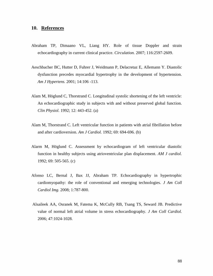
88
10. References
Abraham TP, Dimaano VL, Liang HY. Role of tissue Doppler and strain
echocardiography in current clinical practice. Circulation. 2007; 116:2597-2609.
Aeschbacher BC, Hutter D, Fuhrer J, Weidmann P, Delacretaz E, Allemann Y. Diastolic
dysfunction precedes myocardial hypertrophy in the development of hypertension.
Am J Hypertens. 2001; 14:106 -113.
Alam M, Höglund C, Thorstrand C. Longitudinal systolic shortening of the left ventricle:
An echocardiographic study in subjects with and without preserved global function.
Clin Physiol. 1992; 12: 443-452. (a)
Alam M, Thorstrand C. Left ventricular function in patients with atrial fibrillation before
and after cardioversion. Am J Cardiol. 1992; 69: 694-696. (b)
Alarm M, Höglund C. Assessment by echocardiogram of left ventricular diastolic
function in healthy subjects using atrioventricular plan displacement. AM J cardiol.
1992; 69: 505-565. (c)
Afonso LC, Bernal J, Bax JJ, Abraham TP. Echocardiography in hypertrophic
cardiomyopathy: the role of conventional and emerging technologies. J Am Coll
Cardiol Img. 2008; 1:787-800.
Alsaileek AA, Osranek M, Fatema K, McCully RB, Tsang TS, Seward JB. Predictive
value of normal left atrial volume in stress echocardiography. J Am Coll Cardiol.
2006; 47:1024-1028.

89
Amundsen BH, Crosby J, Steen PA, Torp H, Slordahl SA, Støylen A. Regional
myocardial long-axis strain and strain rate measured by different tissue Doppler and
speckle tracking echocardiography methods: a comparison with tagged magnetic
resonance imaging. Eur J Echocardiogr. 2009; 10:229-237.
Anderson LJ, Miyazaki C, Sutherland GR, Oh JK. Patient selection and
echocardiographic assessment of dyssynchrony in cardiac resynchronization therapy.
Circulation. 2008; 117:2009-2023.
Baicu CF, Zile MR, Aurigemma GP, Gaasch WH. Left ventricular systolic performance,
function, and contractility in patients with diastolic heart failure. Circulation. 2005;
111:2306-2312.
Ballo P, Quatrini I, Giacomin E, Motto A, Mondillo S. Circumferential versus
longitudinal systolic function in patients with hypertension: a nonlinear relation. J Am
Soc Echocardiogr. 2007; 20: 298-306.
Barajas L, Liu L, Powers K. Anatomy of the renal innervation: Intrarenal aspects and
ganglia of origin. Can. J. Physiol. Pharmacol. 1992; 70: 735-749.
Barclay JL, Kruszewski K, Croal BL, Cuthbertson BH, Oh JK, Hillis GS. Relation of left
atrial volume to B-type natriuretic peptide levels in patients with stable chronic heart
failure. Am J Cardiol. 2006; 98: 98-101.
Barrett CJ, Navakatikyan AM, Malpas CS. Long-term control of renal blood flow: what
is the role? Am J Physiol Regul Integr Comp Physiol. 2001; 280: R1534-R1545.
Bauml MA, Underwood AD. Left ventricular hypertrophy: An overlooked cardiovascular
risk factor. Cleve Clin J Med. 2010; 77: 381-387.

90
Bayes-Genis A, Vazquez R, Puig T, Fernandez-Palomeque C, Fabregat J, Bardajı´ A, et
al. Left atrial enlargement and NT-proBNP as predictors of sudden cardiac death in
patients with heart failure. Eur J Heart Fail. 2007; 9:802–807.
Becker M, Kramann R, Dohmen G, Luckhoff A, Autschbach R, Kelm M, et al. Impact of
left ventricular loading conditions on myocardial deformation parameters: analysis of
early and late changes of myocardial deformation parameters after aortic valve
replacement. J Am Soc Echocardiogr. 2007; 20: 681-689.
Bhatia RS, Tu JV, Lee DS, Austin PC, Fang J, Haouzi A, Gong Y, Liu PP. Outcome of
heart failure with preserved ejection fraction in a population-based study. N Engl J
Med. 2006; 355:260–269.
Böhm M, Swedberg K, Komajda M, et al. Heart rate as a risk factor in chronic heart
failure (SHIFT): the association between heart and outcomes in a randomized
placebo-controlled trial. Lancet. 2010; 376:886-894.
Boldt A, Wetzel U, Lauschke J, et al. Fibrosis in left atrial tissue of patients with atrial
fibrillation with and without underlying mitral valve disease. Heart. 2004; 90:400-
405.
Bolognesi R, Tsialtas D, Barilli AL, et al. Detection of early abnormalities of left
ventricular function by hemodynamic, echo-tissue Doppler imaging, and mitral
Doppler flow techniques in patients with coronary artery disease and normal ejection
fraction. J Am Soc Echocardiogr. 2001; 14: 764-772.
Bombelli M, Facchetti R, Carugo S, et al. Left ventricular hypertrophy increases
cardiovascular risk independently of in-office and out-office blood pressure values. J
Hypertens. 2009; 27:2458-2464.

91
Borlaug BA, Melenovsky V, Redfield MM, Kessler K, Chang HJ, Abraham TP et al.
Impact of arterial load and loading sequence on left ventricular tissue velocities in
humans. J Am Coll Cardiol. 2007; 50:1570-1577.
Brandt MC, Mahfoud F, Reda S, Schirmer SH, Erdmann E, Böhm M, Hoppe UC. Renal
sympathetic denervation reduces left ventricular hypertrophy and improves cardiac
function in patients with resistant hypertension. J Am Coll Cardiol. 2012; 59:901-909.
Brutsaert DL. Cardiac dysfunction in heart failure: the cardiologist’s love affair with time
progress in cardiovascular diseases. Prog Cardiovasc Dis J. 2006; 49: 157–81.
Bursi F, Weston SA, Redfield MM, Jacobsen SJ, Pakhomov S, Nkomo VT, Meverden
RA, Roger VL. Systolic and diastolic heart failure in the community. JAMA. 2006;
296:2209-2216.
Casaclang-Verzosa G, Gersh BJ, Tsang TS. Structural and functional remodeling of the
left atrium: clinical and therapeutic implications for atrial fibrillation. J Am Coll
Cardiol. 2008; 51:1-11.
Casale PN, Devereux BR, and Milner M. Value of echocardiographic measurement of
left ventricular mass in predicting cardiovascular morbid events in hypertensive men.
Annals of Internal Medicine. 1986; 105: 173-178.
Centers for Disease Control and Prevention. Vital Statistics Public Use Data Files - 2008
Mortality Multiple Cause Files. Available at
http;//www.cdc.gov/nchs/data_access/Vitalstatsonline.htm #Mortality_ Multiple.
Accessed September 23, 2011.
Centers for Disease Control and Prevention. National Center for Health Statistics. Health
Data Interactive. http://www.cdc.gov/nchs/hdi.htm. Accessed July 19, 2011.

92
Chobanian A, et al. Seventh report of the joint national committee on prevention,
detection, evaluation and treatment of high blood pressure (JNC7). Hypertension.
2003; 42:1206–1252.
Colucci WS, Braunwald E. Pathophysiology of heart failure. In: Zipes D, Libby P,
Bonow RO, Braunwald E, editors. Braunwald’s Heart Disease: A Textbook of
Cardiovascular Medicine. 7th edition. Philadelphia, PA: W.B. Saunders, 2005:509-
538.
Culpepper WS. Cardiac anatomy and function in juvenile hypertension: current
understanding and future concerns. Am J Med.. 1983; 75: 57-61.
Cuspidi C, Meani S, Fusi V, Valerio C, Catini E, Sala C, Sampieri C, Magrini F,
Zanchetti A. Prevalence and correlates of left atrial enlargement in essential
hypertension: role of ventricular geometry and the metabolic syndrome The
Evaluation of Target Organ Damage in Hypertension study. J Hypertens. 2005,
23:875-882.
D’hooge J, Herbots L, Sutherland GR. Quantitative assessment of intrinsic regional
myocardial deformation by Doppler strain rate echocardiography in humans:
validation against three-dimensional tagged magnetic resonance imaging.
Circulation. 2002; 106:50-56.
Dahlof B, Pennert K, Hansson L. Reversal of left ventricular hypertrophy in hypertensive
patients. A metaanalysis of 109 treatment studies. Am J Hypertens. 1992; 5:95-110.
Dahlof B, Devereux RB, Kjeldsen SE, et al. Cardiovascular morbidity and mortality in
the Losartan Intervention For Endpoint reduction in hypertension study (LIFE): a
randomised trial against atenolol. Lancet. 2002; 359:995-1003.

93
Dandel M, Knosalla C, Lehmkuhl H, Hetzer R. Non-Doppler twodimensional strain
imaging – clinical application. J Am Soc Echocardiogr. 2007; 20:234-243.
Dandel M, Lehmkuhl H, Knosalla C, Suramelashvili N and Hetzer R. Strain and strain
rate imaging by echocardiography – basic concepts and clinical applicability Curr
Cardiol Rev. 2009, 5, 133-148.
David A. Calhoun, MD, FAHA, Jones D, et al. Resistant hypertension: diagnosis,
evaluation, and treatment. Hypertension. 2008; 51:1403-1419.
De Caterina AR, Leone AM. Why beta-blockers should not be used as first choice in
uncomplicated hypertension. Am J Cardiol. 2010; 105: 1433-1438.
Devereux RB, Alonso DR, Lutas EM, et al. Echocardiographic assessment of left
ventricular hypertrophy: comparison to necropsy findings. Am J Cardiol. 1986;
57:450-458.
Devereux RB, Wachtell K, Gerdts E, et al. Prognostic significance of left ventricular
mass change during treatment of hypertension. JAMA. 2004; 292:2350-2356.
Di Tullio MR, Sacco RL, Sciacca RR, Homma S. Left atrial size and the risk of ischemic
stroke in an ethnically mixed population. Stroke. 1999; 30:2019-2024.
DiBona GF. Physiology in perspective: the wisdom of the body. Neural control of the
kidney. Am J Physiol Regul Integr Comp Physiol. 2005; 289:R633–R641
DiBona GF, Esler M. Translational medicine: the antihypertensive effect of renal
denervation. Am J Physiol Regul Integr Comp Physiol. 2010;298:R245-R253.

94
Diez J, Querejeta R, Lopez B, Gonzalez A, Larman M, Martinez Ubago JL. Losartan-
dependent regression of myocardial fibrosis is associated with reduction of left
ventricular chamber stiffness in hypertensive patients. Circulation. 2002; 105:2512-
2517.
Dietz JR. Mechanisms of atrial natriuretic peptide secretion from the atrium. Cardiovasc
Res. 2005; 68:8-17.
Diez J, Frohlich ED. A translational approach to hypertensive heart disease.
Hypertension. 2010; 55:1-8.
Diwan A, McCulloch M, Lawrie GM, Reardon MJ, Nagueh SF. Doppler estimation of
left ventricular filling pressures in patients with mitral valve disease. Circulation.
2005; 111:3281-3289.
Dokainish H, Sengupta R, Pillai M, Bobek J, Lakkis N. Usefulness of new diastolic strain
and strain rate indexes for the estimation of left ventricular filling pressure. Am J
Cardiol. 2008; 101:1504-1509.
Doumas M, Douma S. Renal sympathetic denervation: the jury is still out. Lancet. 2010;
376:1878-1880.
Douglas PS: The left atrium: A biomarker of chronic diastolic dysfunction and
cardiovascular disease risk. J Am Coll Cardiol. 2003; 42:1206-1207.
Drazner MH. The transition from hypertrophy to failure: how certain are we?
Circulation. 2005; 112:936-938.
Drazner MH. The progression of hypertensive heart disease, Circulation. 2011; 123:327-
334.
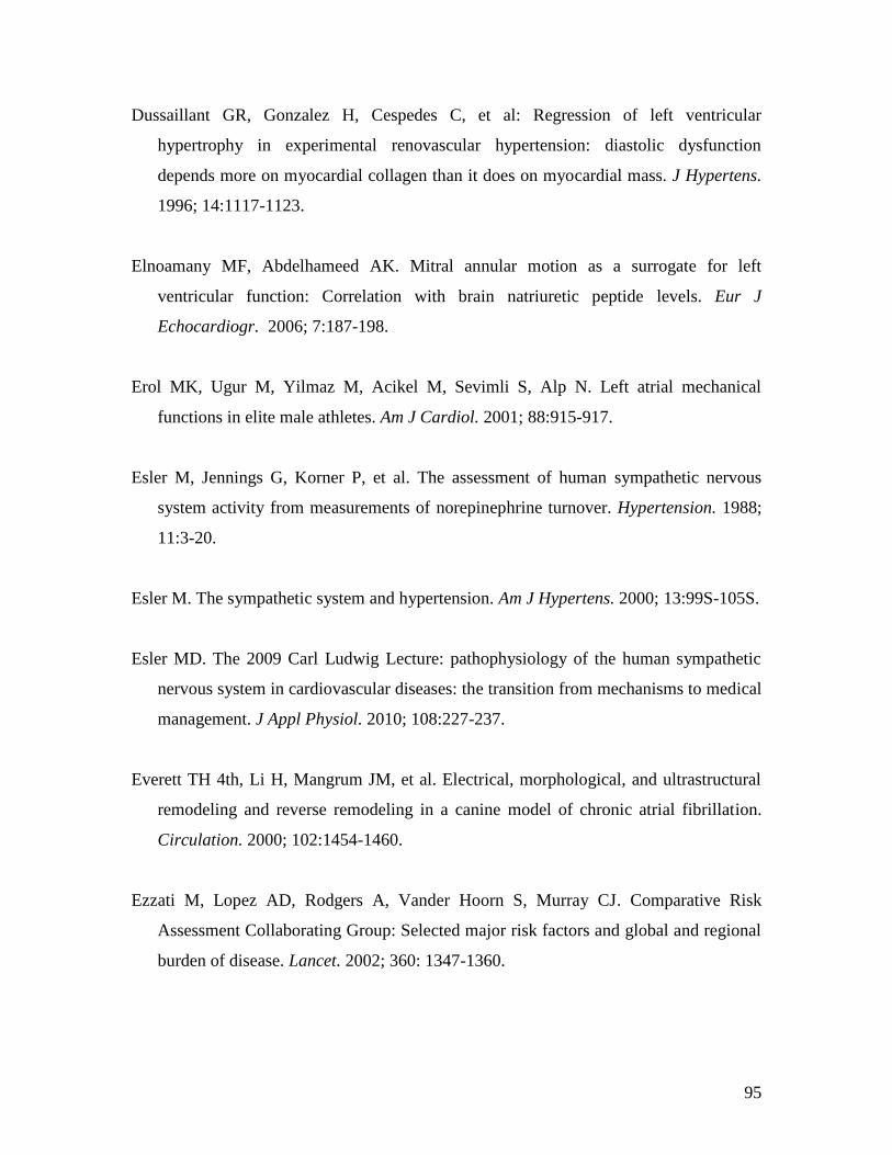
95
Dussaillant GR, Gonzalez H, Cespedes C, et al: Regression of left ventricular
hypertrophy in experimental renovascular hypertension: diastolic dysfunction
depends more on myocardial collagen than it does on myocardial mass. J Hypertens.
1996; 14:1117-1123.
Elnoamany MF, Abdelhameed AK. Mitral annular motion as a surrogate for left
ventricular function: Correlation with brain natriuretic peptide levels. Eur J
Echocardiogr. 2006; 7:187-198.
Erol MK, Ugur M, Yilmaz M, Acikel M, Sevimli S, Alp N. Left atrial mechanical
functions in elite male athletes. Am J Cardiol. 2001; 88:915-917.
Esler M, Jennings G, Korner P, et al. The assessment of human sympathetic nervous
system activity from measurements of norepinephrine turnover. Hypertension. 1988;
11:3-20.
Esler M. The sympathetic system and hypertension. Am J Hypertens. 2000; 13:99S-105S.
Esler MD. The 2009 Carl Ludwig Lecture: pathophysiology of the human sympathetic
nervous system in cardiovascular diseases: the transition from mechanisms to medical
management. J Appl Physiol. 2010; 108:227-237.
Everett TH 4th, Li H, Mangrum JM, et al. Electrical, morphological, and ultrastructural
remodeling and reverse remodeling in a canine model of chronic atrial fibrillation.
Circulation. 2000; 102:1454-1460.
Ezzati M, Lopez AD, Rodgers A, Vander Hoorn S, Murray CJ. Comparative Risk
Assessment Collaborating Group: Selected major risk factors and global and regional
burden of disease. Lancet. 2002; 360: 1347-1360.
96
Fitzgerald PJ. Is elevated noradrenaline an aetiological factor in a number of diseases?
Auton Autacoid Pharmaco.l 2009; 29:143-156.
Florian Andre, Dirk Lossnitzer, Sebastian Buss, Henning Steen. Reference values of
mitral and tricuspid annular plane systolic excursion for the evaluation of left and
right ventricular performance. J Cardiovasc Magn Reson. 2012, 14(Suppl 1):M3.
(Abstract).
Franco V, Chen YF, Oparil S, et al. Atrial natriuretic peptide dose-dependently inhibits
pressure overload-induced cardiac remodeling. Hypertension. 2004; 44:746 -750.
Garcia-Fernandez MA, Azevedo J, Moreno M, Bermejo J and Moreno R. Regional left
ventricular diastolic dysfunction evaluated by pulsed-tissue Doppler
echocardiography. Echocardiography. 1999; 16:491-500.
Gerdts E, Wachtell K, Omvik P, Otterstad JE, Oikarinen L, Boman K, et al. Left atrial
size and risk of major cardiovascular events during antihypertensive treatment.
Losartan Intervention for Endpoint in Hypertension Trial. Hypertension. 2007;
49:311-316
Goto K, Mikami T, Onozuka H, Kaga S, Inoue M, Komatsu H et al. Role of left
ventricular regional diastolic abnormalities for global diastolic dysfunction in patients
with hypertrophic cardiomyopathy. J Am Soc Echocardiogr. 2006; 19: 857-864.
Gottdiener JS: Measuring diastolic function. J Am Coll Cardiol. 1991; 18:83-84.
Gottdiener JS: Left ventricular mass, diastolic dysfunction, and hypertension. Adv Intern
Med. 1993; 38:31- 56.

97
Greenberg NL, Firstenberg MS, Castro PL, Main ML, Travaglini A, Odabashian JA et al.
Doppler-derived myocardial systolic strain rate is a strong index of left ventricular
contractility. Circulation. 2002; 105: 99-105.
Hanna N, Cardin S, Leung TK, Nattel S. Differences in atrial versus ventricular
remodeling in dogs with ventricular tachypacing-induced congestive heart failure.
Cardiovasc Res. 2004; 63:236-244.
Hausberg M, Kosch M, Harmelink P, et al. Sympathetic nerve activity in end-stage renal
disease. Circulation. 2002; 106: 1974–1979.
Heagerty AM. Effect of AT1-receptor blockade on cardiovascular structure and function.
Eur Heart J. Supplements 2004 ;6:H17–H21.
Heidenreich PA, Trogdon JG, Khavjou OA, Butler J, Dracup K, Ezekowitz MD,
Finkelstein EA, Hong Y, Johnston SC, Khera A, Lloyd-Jones DM, Nelson SA,
Nichol G, Orenstein D, Wilson PW, Woo YJ. Forecasting the future of cardiovascular
disease in the United States: a policy statement from the American Heart Association.
Circulation. 2011; 123: 933-944.
Hellenic J. The Constellation of Hypertensive Heart Disease. Cardiol. 2008; 49: 92-99.
Hill JA, Olson EN. Cardiac plasticity. N Engl J Med. 2008; 358: 1370-1380.
Hillis GS, Moller JE, Pellikka PA, Gersh BJ, Wright RS, Ommen SR, Reeder GS, Oh JK.
Noninvasive estimation of left ventricular filling pressure by E/e´ is a powerful
predictor of survival after acute myocardial infarction. J Am Coll Cardiol. 2004;
43:360-367.
Höglund C, Alam M, Thorstrand C. Atrioventricular valve plane displacement in healthy
persons. An echocardiographic study. Acta Med Scand. 1988; 224:557-562.
98
Höglund C, Alam M, Thorstrand C. Effects of acute myocardial infarction on the
displacement of the atrioventricular plane: an echocardiographic study. J Int Med.
1989; 226:251-256.
Horton KD, Meece RW, Hill JC. Assessment of the Right Ventricle by
Echocardiography: A Primer for Cardiac Sonographers. J Am Soc Echocardiogr.
2009; 22:776-792.
Imbalzano E, Zito C, Carerj S, Oreto G, Mandraffino G, Cusm`a-Piccione M, Bella GD,
Saitta C, and Saitta A. Left ventricular function in hypertension: new insight by
speckle tracking echocardiography. Echocardiography. 2011; 28:649-657.
Inoue S, Murakami Y, Sano K, Katoh H, Shimada T. Atrium as a source of brain
natriuretic polypeptide in patients with atrial fibrillation. J Card Fail. 2000; 6:92-96.
Iwahashi N, Nakatani S, Kanzaki H, Hasegawa T, Abe H, Kitakaze M. Acute
improvement in myocardial function assessed by myocardial strain and strain rate
after aortic valve replacement for aortic stenosis. J Am Soc Echocardiogr. 2006; 19:
1238-1244.
Jones CJ, Song GJ, Gibson DG. An echocardiographic assessment of atrial mechanical
behavior. Br Heart J. 1991; 65:31-36.
Kandzari DE, Bhatt DL, Sobotka PA, O’Neill WW, Esler M, Flack JM, Katzen BT, Leon
MB, Massaro JM, Negoita M, Oparil S, Rocha-Singh K, Straley C, Townsend RR,
Bakris G. Catheter-based renal denervation for resistant hypertension: rationale and
design of the SYMPLICITY HTN-3 Trial. Clin Cardiol. 2012; 35, 528-535.

99
Kasner M, Gaub R, Sinning D, Westermann D, Steendijk P, Hoffmann W, Schultheiss
HP, Tscho¨pe1 C. Global strain rate imaging for the estimation of diastolic function
in HFNEF compared with pressure–volume loop analysis. Eur J Echocardiogr. 2010;
11, 743-751.
Kearney PM, Whelton M, Reynolds K, Muntner P, Whelton PK, He J Global burden of
hypertension: analysis of worldwide data. Lancet. 2005; 365, 217-223.
Khan A, Moe GW, Nili N, et al. The cardiac atria are chambers of active remodeling and
dynamic collagen turnover during evolving heart failure. J Am Coll Cardiol. 2004;
43:68-76.
Kim H, Cho HO, Cho YK, Nam CW, Han SW, Hur SH, Kim KS, Kim YM and Kim
KB. Relationship between early diastolic strain rate imaging and left ventricular
geometric patterns in hypertensive patients. Heart Vessels. 2008 23:271-278.
Kim BS, Lee HJ, Kim JH, Jang HS, Bae BS, Kang HJ, et al. Relationship between left
atrial size and stroke in patients with sinus rhythm and preserved systolic function.
Korean J Intern Med. 2009; 24:24-32.
Klingbeil AU, Muller HJ, Delles C, et al: Regression of left ventricular hypertrophy by
AT1 receptor blockade in renal transplant recipients. Am J Hypertens. 2000; 13:1295-
1300.
Klingbeil AU, Schneider M, Martus P, Messerli FH, Schmieder RE. A meta-analysis of
the effects of treatment on left ventricular mass in essential hypertension. Am J Med.
2003; 115:41-46.
Kolloch R, Legler UF, Champion A , et al. Impact of resting heart on outcomes in
hypertensive patients with coronary artery disease: finding from the INternational
VErapamil –SR/trandopril Study (INVEST) . Eur Heart J. 2008; 29:1327-1334.
100
Komuro I, Katoh Y, Kaida T, Shibazaki Y, Kurabayashi M, Takaku F et al. Mechanical
loading stimulates cell hypertrophy and specific gene expression in cultured rat
cardiac myocytes. J Biol Chem. 1991; 266: 1265-1268.
Koyama J, Ray-Sequin P, Falk RH. Longitudinal myocardial function assessed by tissue
velocity, strain and strain rate tissue Doppler echocardiography in patients with AL
(primary) cardiac amyloidosis. Circulation. 2003; 107: 2446-2452.
Krahn AD, Manfreda J, Tate RB, Mathewson FA, Cuddy TE. The natural history of atrial
fibrillation: incidence, risk factors, and prognosis in the Manitoba follow-up study.
Am J Med. 1995; 98:476-484.
Krum H, Schlaich M, Whitbourn R, et al. Catheter-based renal sympathetic denervation
for resistant hypertension: a multicentre safety and proof-of-principle cohort study.
Lancet. 2009; 373:1275-1281.
Krum H, Sobotka P, Mahfoud F, Böhm M, Esler MD, Schlaich M. Device-based
antihypertensive therapy: therapeutic modulation of the autonomic nervous system.
Circulation. 2011; 123: 209-215.
Kumagai K, Nakashima H, Urata H, Gondo N, Arakawa K, Saku K. Effects of
angiotensin II type 1 receptor antagonist on electrical and structural remodeling in
atrial fibrillation. J Am Coll Cardiol. 2003; 41:2197-2204.
Lang RM, Bierig M, Devereux RB, Flachskampf FA, et al. Recommendations for
chamber quantification: a report from the American Society of Echocardiography’s
Guidelines and Standards Committee and the Chamber Quantification Writing Group,
developed in conjunction with the European Association of Echocardiography a
branch of the European Society of Cardiology. J Am Soc Echocardiogr. 2005;
18:1440-1463.
101
Langeland S, D’Hooge J, Claessens T, Claus P, Verdonck P, Suetens P, Sutherland GR,
Bijnens B. RF-based two dimensional cardiac strain estimation: a validation study in
a tissue-mimicking phantom. IEEE Trans Ultrason Ferroelectr Freq Control. 2004;
51:1537-1546.
Lawes CM, Vander Hoorn S, Rodgers A. Global burden of blood-pressure-related
disease, 2001. Lancet. 2008; 371:1513-1518.
Leibowitz D, Planer D, Ben-Ibgi F, Rott D, Weiss AT, Bursztyn M. Measurement of
wall thickness alone does not accurately assess the presence of left ventricular
hypertrophy. Clin Exp Hypertens. 2007; 29:119-125.
Levick SP, Murray DB, Janicki JS, Brower GL. Sympathetic nervous system modulation
of inflammation and remodeling in the hypertensive heart. Hypertension. 2010;
55:270-276.
Li DS, Fareh S, Leung TK, Nattel S. Promotion of atrial fibrillation by heart failure in
dogsatrial remodeling of a different sort. Circulation. 1999; 100:87-95.
Lim TK, Ashrafian H, Dwivedi G, Collinson PO, Senior R. Increased left atrial volume
index is an independent predictor of raised serum natriuretic peptide in patients with
suspected heart failure but normal left ventricular ejection fraction: implication for
diagnosis of diastolic heart failure. Eur J Heart Fail. 2006; 8:38-45.
Lind L, Anderson PE, Andren B, Hanni A, Lithell HO. Left ventricular hypertrophy in
hypertension is associated with insulin-resistance metabolic syndrome. J Hypertens.
1995; 13:433-438.

102
Lindholm LH, Carlberg B, Samuelsson O. Should beta blockers remain first choice in
the treatment of primary hypertension? A meta-analysis. Lancet. 2005; 366: 1545-
1553.
Little WC, Ohno M, Kitzman DW, et al. Determination of left ventricular chamber
stiffness from the time for deceleration of early left ventricular filling. Circulation.
1995; 92:1933-1939.
Lloyd-Jones D et al. Heart disease and stroke statistics–2010 update: A report from the
American Heart Association. Circulation. 2010; 121:e46-e215.
Lohmeier TE. The sympathetic nervous system and long-term blood pressure regulation.
Am J Hypertens. 2001; 14:147S-154S.
Lorell BH, Carabello BA. Left ventricular hypertrophy: pathogenesis, detection, and
prognosis. Circulation. 2000; 102:470-479.
Mahfoud F, Schlaich MP, Kindermann I, Ukena C, Cremers B, Brandt MC, Hoppe UC,
Vonend O, Rump LC, Sobotka PA, Krum H, Esler MD, Böhm M. Circulation. 2011;
123:1940-1946.
Mancia G, Bousquet P, Elghozi JL, et al. The sympathetic nervous system and the
metabolic syndrome. J Hypertens. 2007; 25:909-920.
Mandinov L, Eberli FR, Seiler C, Hess OM: Diastolic heart failure. Cardiovasc Res.
2000; 45: 813-825.
Martinez DA, Guhl DJ, Stanley WC, Vailas AC. Extracellular matrix maturation in the
left ventricle of normal and diabetic swine. Diabetes Res Clin Pract. 2003; 59:1-9.

103
Marwick TH. Measurement of strain and strain rate by echocardiography: ready for
prime time? J Am Coll Cardiol. 2006; 47: 1313-1327.
Mattioli AV, Bonatti S, Monopoli D, Zennaro M, Mattiolli G. Influence of regression of
left ventricular hypertrophy on left atrial size and function in patients with moderate
hypertension. Blood Pressure. 2005; 14: 273-278.
McMurray JJV, Adamopoulos S, Anker SD, Auricchio A, Böhm M, Dickstein K, Falk V,
Filippatos G, Fonseca C, Gomez-Sanchez MA, Jaarsma T, Køber L, Lip GYH,
Maggioni AP, Parkhomenko A, Pieske BM, Popescu BA, Rønnevik PK, Rutten FH,
Schwitter J, Seferovic P, Stepinska J, Trindade PT, Voors AA, Zannad F, Zeiher A.
ESC Guidelines for the diagnosis and treatment of acute and chronic heart failure.
Eur Heart J. 2012; 33: 1787–1847.
Miyauchi Y, Zhou S, Okuyama Y, et al. Altered atrial electrical restitution and
heterogeneous sympathetic hyper innervation in hearts with chronic left ventricular
myocardial infarction: implications for atrial fibrillation. Circulation. 2003; 108:360-
366.
Mizushige K, Yao L, Noma T, et al. Alteration in left ventricular diastolic filling and
accumulation of myocardial collagen at insulin-resistant prediabetic stage of a type II
diabetic rat model. Circulation. 2000; 101: 899-907.
Moen CA, Salminen PR, Grong K, Matre K. Left ventricular strain, rotation, and torsion
as markers of acute myocardial Ischemia. Am J Physiol Heart Circ Physiol. 2011;
300: H2142–H2154.
Muiesan ML, Agabiti-Rosei E, Romanelli G, et al: Improved left ventricular systolic and
diastolic function after regression of cardiac hypertrophy, treatment withdrawal, and
redevelopment of hypertension. J Cardiovasc Pharmacol. 1991; 17:S179- S181.
104
Nagueh SF, Middleton KJ, Kopelen HA, Zoghbi WA, Quinones MA. Doppler tissue
imajng: A noninvasive technique for evaluation of left ventricular relaxation and
estimation of filling pressures. J AM Coll Cardiol. 1997; 30: 1527-1533.
Nagueh SF, Appleton P, Gillebert TC, Marino PN, Oh JK, Smiseth OA, Waggoner AD,
Flachskampf FA, Pellikka PA, Evangelisa A. Recommendations for the Evaluation of
Left Ventricular Diastolic Function by Echocardiography. Eur J Echocardiogr. 2009;
10, 165-193.
Narkiewicz K, Pesek CA, Kato M, et al. Baroreflex control of sympathetic nerve activity
and heart rate in obstructive sleep apnea. Hypertension. 1998; 32:1039-1043.
Ohno M, Cheng CP, Little WC. Mechanism of altered patterns of left ventricular filling
during the development of congestive heart failure. Circulation. 1994; 89: 2241-2250.
Okin PM, Devereux RB, Jern S, et al; LIFE Study Investigators. Regression of
electrocardiographic left ventricular hypertrophy during antihypertensive treatment
and the prediction of major cardiovascular events. JAMA. 2004; 292:2343-2349.
Okin PM, Wachtell K, Devereux RB, et al. Regression of electrocardiographic left
ventricular hypertrophy and decreased incidence of new-onset atrial fibrillation in
patients with hypertension. JAMA. 2006; 296: 1242-1248.
Okin PM, Devereux RB, Harris KE, et al; LIFE Study Investigators. In-treatment
resolution or absence of electrocardiographic left ventricular hypertrophy is
associated with decreased incidence of new-onset diabetes mellitus in hypertensive
patients: the Losartan Intervention for Endpoint Reduction in Hypertension (LIFE)
Study. Hypertension. 2007; 50:984-990.

105
Ommen SR, Nishimura RA, Appleton CP, Miller FA, Oh JK, Redfield MM, et al.
Clinical utility of Doppler echocardiography and tissue Doppler imaging in the
estimation of left ventricular filling pressures. Circulation. 2000; 102:1788-1794.
Omvik P, Gerdts E, Myking OL, et al: Long-term central hemodynamic effects at rest
and during exercise of losartan in essential hypertension. Am Heart J. 2000; 140:624-
630.
Owan TE, Hodge DO, Herges RM, Jacobsen SJ, Roger VL, Redfield MM. Trends in
prevalance and outcome of heart failure with preserved ejection fraction. N Engl J
Med. 2006; 355:251–259.
Oxborough D, Batterham AM, Shave R, Artis N, Birch KM, Whyte G, et al.
Interpretation of two-dimensional and tissue Doppler-derived strain (epsilon) and
strain rate data: is there a need to normalize for individual variability in left
ventricular morphology? Eur J Echocardiogr. 2009; 10:677-682.
Pai RG, Bodenheimer MM, Pai SM, Koss JH, Adamick RD. Usefulness of systolic
excursion of the mitral annulus as an index of left ventricular systolic function. Am J
Cardiol. 1991; 67:222-224.
Park TH, Nagueh SF, Khoury DS, Kopelen HA, Akrivakis S, Nasser K et al. Impact of
myocardial structure and function post infarction on diastolic strain measurements:
implications for assessment of myocardial viability. Am J Physiol Heart Circ Physiol.
2006; 290: H724–H731.
106
Paulus WJ, Tschöpe C, Sanderson JE, Rusconi C, Flachskampf FA, Pademakers FE,
Marino P, Smiseth OA, De Keulenaer G, Leite-Moreira AF, Borbely A, Edes I,
Handoko ML, Heymans S, Pezzali N, Pieske B, Dickstein K, Fraser AG, Brutsaert
DL. How to diagnose diastolic heart failure: a consensus statement on the diagnosis
of heart failure with normal left ventricular ejection fraction by the Heart Failure and
Echocardiography Associations of the European Society of Cardiology. Eur Heart J.
2007; 28:2539-2550.
Pavlopoulos H, Grapsa J, Stefanadi E, Philippou E, Dawson D, Nihoyannopoulos P. Is it
only diastolic dysfunction? Segmental relaxation patterns and longitudinal systolic
deformation in systemic hypertension. Eur J Echocardiogr. 2008; 9:741-747.
Pellikka PA, Ritman EL, Greenleaf JF. Ultrasound strain imaging of altered myocardial
stiffness: stunned versus infarcted reperfused myocardium. Circulation. 2004;
109:2905-2910.
Perk G, Tunick PA, Kronzon I. Non-Doppler two-dimensional strain imaging by
echocardiography – from technical considerations to clinical applications. J Am Soc
Echocardiogr. 2007; 49:1903-1914.
Perkovic V, Huxley R, Wu Y, Prabhakaran D, Macmahon S. The burden of blood
pressure-related disease: a neglected priority for global health. Hypertension. 2007
50(6), 991-997.
Perlini S, Palladini G, Ferrero I, et al. Sympathectomy or doxazosin, but not propranolol,
blunt myocardial interstitial fibrosis in pressure overload hypertrophy. Hypertension.
2005; 46:1213-1218.
Perry R, De Pasquale CG, Chew DP, Joseph MX. Assessment of early diastolic left
ventricular function by two-dimensional echocardiographic speckle tracking. Eur J
Echocard. 2008; 9, 791-795.

107
Pierdomenico SD, Cuccurullo F. Risk reduction after regression of echocardiographic left
ventricular hypertrophy in hypertension: a meta-analysis. Am J Hypertens. 2010;
23:876-881.
Pislaru C, Bruce CJ, Anagnostopoulos PC, Allen JL, Seward JB, Pellikka PA et al.
Ultrasound strain imaging of altered myocardial stiffness: stunned versus infarcted
reperfused myocardium. Circulation. 2004; 109: 2905-2910.
Poulsen SH, Andersen NH, Ivarsen PI, et al: Doppler tissue imaging reveals systolic
dysfunction in patients with hypertension and apparent “isolated” diastolic
dysfunction. J Am Soc Echocardiogr. 2003; 16:724-731.
Pritchett AM, Jacobsen SJ, Mahoney DW, Rodeheffer RJ, Bailey KR and Redfield MM.
Left atrial volume as an index of left atrial size: A population-based study. J Am Coll
Cardiol. 2003; 41:1036-1043.
Qin JX, Shiota T, Tsujino H, et al. Mitral annular motion as a surrogate for left
ventricular ejection fraction: real e time three-dimensional echocardiography and
magnetic resonance imaging studies. Eur J Echocardiogr. 2004; 5:407-415.
Redfield MM, Jacobsen SJ, Burnett JC, Jr, Mahoney DW, Bailey KR and Rodeheffer RJ.
Burden of systolic and diastolic ventricular dysfunction in the community:
Appreciating the Scope of the Heart Failure Epidemic. JAMA. 2003; 289:194-202.
Reil JC, Custodis F, Swedberg K, et al. Heart rate reduction in cardiovascular disease and
therapy. Clin Res Cardiol 2011; 10011-10019.
Reisner SA, Lysyansky P, Agmon Y, Mutlak D, Lessick J, Friedman Z. Global
longitudinal strain: a novel index of left ventricular systolic function. J Am Soc
Echocardiogr. 2004; 17:630-633.

108
Rivas-Gotz C, Khoury DS, Manolios M, Rao L, Kopelen HA, Nagueh SF. Time interval
between onset of mitral inflow and onset of early diastolic velocity by tissue Doppler:
a novel index of left ventricular relaxation: experimental studies and clinical
application. J Am Coll Cardiol. 2003; 42:1463-1470.
Roger VL, Go AS, Lloyd-Jones DM, et al. Heart disease and stroke statistics 2012
update; a report from the American Heart Association. Circulation. 2012; 125:e12-
e230
Ruilope LM, Schmieder RE. Left ventricular hypertrophy and clinical outcomes in
hypertensive patients. Am J Hypertens. 2008; 21:500-508.
Rusconi C, Sabatini T, Faggiano P, Ghizzoni G, Oneglia C, Simoncelli U, Gualeni A,
Sorgato A, Marchetti A. Prevalence of isolated left ventricular diastolic dysfunction
in hypertension as assessed by combined transmitral and pulmonary vein flow
Doppler study. Am J Cardiol. 2001; 87:357-360.
Sagie A, Benjamin EJ, Galderisi M. et al., “Echocardiographic assessment of left
ventricular structure and diastolic filling in elderly subjects with borderline isolated
systolic hypertension (the Framingham Heart Study),” Am J Cardiol. 1993; 72: 662-
665.
Salles GF, Cardoso CR, Fiszman R, Muxfeldt ES. Prognostic impact of baseline and
serial changes in electrocardiographic left ventricular hypertrophy in resistant
hypertension. Am Heart J. 2010; 159:833-840.
Sarafidis PA, Bakris GL. Resistant hypertension: an overview of evaluation and
treatment. J Am Coll Cardiol. 2008; 52:1749-1757.

109
Schlaich MP, Lambert E, Kaye DM, Krozowski Z, Campbell DJ, Lambert G, Hastings J,
Aggarwal A, Esler MD. Sympathetic augmentation in hypertension: role of nerve
firing, norepinephrine reuptake, and angiotensin neuromodulation. Hypertension.
2004; 43:169-175
Schlaich MP, Sobotka PA, Krum H, Lambert E, Esler MD. Renal sympathetic-nerve
ablation for uncontrolled hypertension. N Engl J Med. 2009; 361:932-934..
Schlaich MP, Krum H, Sobotka PA, Esler MD. Renal denervation and hypertension. Am
J Hypertens. 2011; 24, 635-642. (a)
Schlaich MP, Straznicky N, Grima M, et al Renal denervation: a potential new treatment
modality for polycystic ovary syndrome? J Hypertens. 2011; 29:991-996. (b)
Schoonderwoerd BA, Van Gelder IC, Van Veldhuisen DJ, Van den Berg MP, Crijns HJ.
Electrical and structural remodeling: role in the genesis and maintenance of atrial
fibrillation. Prog Cardiovasc Dis. 2005; 48:153-168.
Sen S, Bumpus FM: Collagen synthesis in development and reversal of cardiac
hypertrophy in spontaneously hypertensive rats. Am J Cardiol. 1979; 44:954-958.
Seo JM, Park TH, Lee DY, Cho YR, Baek HK, Park JS, Kim MH, Kim YD, Choi SY,
Lee SM, Hong YS. Subclinical Myocardial Dysfunction in Metabolic Syndrome
Patients without Hypertension. J Cardiovasc Ultrasound. 2011; 19:134-139.
Sheehan FH. Quantitative evaluation of regional left ventricular systolic function. In:
Otto CM, editor. The Practice of Clinical Echocardiography. Philadelphia: WB
Saunders Company; 2002. pp. 65–87.

110
Shi Y, Ducharme A, Li D, Gaspo R, Nattel S, Tardif JC. Remodeling of atrial dimensions
and emptying function in canine models of atrial fibrillation. Cardiovasc Res. 2001;
52:217-225.
Signolet IL, Bousquet PP, Monassier LJ. Improvement of cardiac diastolic function by
long-term centrally mediated sympathetic inhibition in one-kidney, one-clip
hypertensive rabbits. Am J Hypertens. 2008; 21:54-60. (Abstract).
Simonson J, Schiller NB. Descent of the base of the left ventricle: an echocardiographic
index of left ventricular function. J Am Soc Echocardiogr. 1998; 2:25-35.
Smithwick RH, Thompson JE. Splanchnicectomy for essential hypertension. J Am Med
Assoc. 1953; 152:1501-1504.
Sobotka PA, Mahfoud F, Schlaich MP, Hoppe UC, Bohm M and Krum H. Sympatho-
renal axis in chronic disease. Clin Res Cardiol. 2011; 100:1049-1057.
Sohn DW, Chai IH, Lee DJ, Kim HC, Kim HS, Oh BH, Lee MM, Park YB, Choi YS,
Seo JD and Lee YW. Assessment of mitral annulus velocity by Doppler tissue
imaging in the evaluation of left ventricular diastolic function. JACC. 1997; 30:474-
480.
Støylen A, Slørdahl S, Skjelvan GK, Heimdal A and Skjaerpe T. Strain Rate Imaging in
Normal and Reduced Diastolic Function: Comparison with Pulsed Doppler Tissue
Imaging of the Mitral Annulus. J Am Soc Echocardiogr. 2001; 14:264-274.
Symplicity HTN-2 Investigators. Renal sympathetic denervation in patients with
treatment-resistant hypertension (the SymplicityHTN-2 trial): a randomized
controlled trial. Lancet. 2010; 376:1903-1909.
111
Takemoto Y, Pellikka PA, Wang J, Modesto KM, Cauduro S, Belohlavek M, Seward JB,
Thomson HL, Khandheria B, Abraham TP. Analysis of the interaction between
segmental relaxation patterns and global diastolic function by strain
echocardiography. J Am Soc Echocardiogr. 2005; 18:901-906.
Tanaba Y, Kawamura Y, Sakamoto N, Sato N, Kikushi K, Hasebe N. Blood pressure
control and the reduction of left atrial overload is essential for controlling atrial
fibrillation. Int Heart J. 2009; 50:445-456.
Timek TA, Miller DC. Mitral annular ring is an essential, dynamic structure.
Experimental and clinical assessment of mitral annular area and dynamics: what are
we actually measuring? Ann Thorac Surg. 2001; 72:966-974.
Trimarco B, DeLuca N, Rosiello G, et al: Improvement of diastolic function after reversal
of left ventricular hypertrophy induced by long-term antihypertensive treatment with
tertatolol. Am J Cardiol. 1989; 64: 278S-283S.
Triposkiadis F, Karayannis G, Giamouzis G, et al. The sympathetic nervous system in
heart failure physiology, pathophysiology, and clinical implications. J Am Coll
Cardiol. 2009;54:1747-1762.
Tsang TS, Barnes ME, Gersh BJ, Bailey KR, Seward JB. Left atrial volume as a
morphophysiologic expression of left ventricular diastolic dysfunction and relation to
cardiovascular risk burden. Am J Cardiol. 2002; 90:1284-1289.
Tsang TS, Barnes ME, Abhayaratna WP, et al. Effects of quinapril on left atrial structural
remodeling and arterial stiffness. Am J Cardiol. 2006; 97:916 -920.
Tsioufis C, Stougiannos P, Taxiarchou E, et al. The interplay between haemodynamic
load, brain natriuretic peptide and left atrial size in the early stages of essential
hypertension. J Hypertens. 2006; 24:965-972.

112
Ukena C, Mahfoud F, Kindermann I, Barth C, Lenski M, Michael Kindermann, ,
Hoppe UC, Krum H, Esler MD, Sobotka PA, Böhm M. Cardiorespiratory Response
to Exercise After Renal Sympathetic Denervation in Patients With Resistant
Hypertension. J Am Coll Cardiol. 2011; 58:1176-1182.
Vasan RS, Daniel Levy. Defining Diastolic Heart Failure: A Call for Standardized
Diagnostic Criteria. Circulation. 2000; 101:2118-2121.
Verdecchia P, Schillaci G, Reboldi GP, et al: Long term effects of losartan and enalapril,
alone or with a diuretic, on ambulatory blood pressure and cardiac performance in
hypertension: A case-control study. Blood Press Monit. 2000; 5:187-193.
Verdecchia P, Reboldi GP, Gattobigio R, Bentivoglio M, Borgioni C, Angeli F, Carluccio
E, Sardone MG, Porcellati C. Atrial fibrillation in hypertension : Predictors and
outcome. Hypertension. 2003; 41:218-223.
Villari B, Vassalli G, Monrad ES, et al. Normalization of diastolic dysfunction in aortic
stenosis late after valve replacement. Circulation. 1995; 91:2353-2358.
Vinereanu D, Lim PO, Frenneaux MP, Fraser AG. Reduced myocardial velocities of left
ventricular long-axis contraction identify both systolic and diastolic heart failure - a
comparison with brain natriuretic peptide. Eur J Heart Fail. 2005; 7: 512-519. (a)
Vinereanu D, Nicolaides E, Tweddel AC, Fraser AG. ‘‘Pure’’ diastolic dysfunction is
associated with long-axis systolic dysfunction. Implications for the diagnosis and
classification of heart failure. Eur Heart Fail. 2005; 7: 820 -828. (b)
Vonend O, Marsalek P, Russ H, Wulkow R, Oberhauser V, Rump LC. Moxonidine
treatment of hypertensive patients with advanced renal failure. J Hypertension. 2003;
21:1709-1717.

113
Wachtell K, Hornestam B, Lehto M, Slotwiner DJ, Gerdts E, Olsen MH, et al.
Cardiovascular morbidity and mortality in hypertensive patients with a history of
atrial fibrillation: the losartan intervention for end point reduction in hypertension
(LIFE) study. J Am Coll Cardiol. 2005; 45:705-711. (a)
Wachtell K, Lehto M, Gerdts E, et al. Angiotensin II receptor blockade reduces new-
onset atrial fibrillation and subsequent stroke compared to atenolol: the Losartan
Intervention For End Point Reduction in Hypertension (LIFE) study. J Am Coll
Cardiol. 2005; 45:712-719. (b)
Wang J, Khoury DS, Thohan V, Torre-Amione G, Nagueh SF. Global diastolic strain rate
for the assessment of left ventricular relaxation and filling pressures. Circulation.
2007; 115:1376-1383.
Wang J, Khoury DS, Yue Y, Torre-Amione G, Nagueh SF. Preserved left ventricular
twist and circumferential deformation, but depressed longitudinal and radial
deformation in patients with diastolic heart failure. Euro Heart J. 2008; 29:1283-
1289. (a)
Wang J, Nagueh SF. Echocardiographic assessment of left ventricular filling pressures.
Heart Fail Clin. 2008; 4:57-70. (b)
Weidemann F, Jamal F, Kowalski M, Kukulski T, D’Hooge J, Bijnens B, et al. Can strain
rate and strain quantify changes in regional systolic function during dobutamine
infusion, b-blockade, and atrial pacing? Implications for quantitative stress
echocardiography. J Am Soc Echocardiogr. 2002; 15:416-424.
Whelton PK et al. Primary prevention of hypertension: Clinical and public health
advisory from the National High Blood Pressure Education Program. JAMA. 2002;
288:1882-1888.
114
White WB, Schulman P, Karimeddini MK, et al: Regression of left ventricular mass is
accompanied by improvement in rapid left ventricular filling following
antihypertensive therapy with metoprolol. Am Heart J. 1989; 117:145-150.
Williams B, Lacy PS, Thom SM, et al. Differential impact of blood pressure-lowering
drugs on central aortic pressure and clinical outcomes: principal results of the Conduit
Artery Function Evaluation (CAFE) study. Circulation. 2006; 113:1213-1225.
Willenheimer R, Israelsson B, Cline C, Rydberg E, Broms K, Erhardt L. Left
atrioventricular plane displacement is related to both systolic and diastolic left
ventricular performance in patients with chronic heart failure. Euro Heart J. 1999; 20,
612-618
World Health Organization. World Health Report: Reducing Risks, Promoting Healthy
Life. 2002. Geneva, Switzerland.
World Health Organization. Global Health Risks: Mortality and Burden of Disease
Attributable to Selected Major Risks. 2009. WHO Press, Geneva, Switzerland.
Yamamoto K, Masuyama T, Sakata Y, et al: Myocardial stiffness is determined by
ventricular fibrosis, but not by compensatory or excessive hypertrophy in
hypertensive heart. Cardiovasc Res. 2002; 55: 76-82.
Yoshida C, Nakao S, Goda A, et al. Value of assessment of left atrial volume and
diameter in patients with heart failure but with normal left ventricular ejection
fraction and mitral flow velocity pattern. Eur J Echocardiogr. 2009; 10:278-281.
Yu CM, Lin H, Yang H, Kong SL, Zhang Q, Lee SW. Progression of systolic
abnormalities in patients with ‘‘isolated’’ diastolic heart failure and diastolic
dysfunction. Circulation. 2002; 105:1195-1201.

115
Yu CM, Sanderson JE, Marwick TH, Oh JK. Tissue Doppler imaging a new
prognosticator for cardiovascular diseases. J Am Coll Cardiol. 2007; 20:234-243.
Yusuf S, Pfeffer MA, Swedberg K, Granger CB, Held P, McMurray JJ, Michelson EL,
Olofsson B, Ostergren J, CHARM Investigators and Committees. Effects of
candesartan in patients with chronic heart failure and preserved left-ventricular
ejection fraction: the CHARM-Preserved Trial. Lancet. 2003; 362: 777-781.
Zaky A, Grabhorn L, Feigenbaum H. Movement of the mitral ring: a study in ultrasound
cardiography. Cardiovasc Res. 1967; 1:121-131.
Zakynthinos E, Pierutsakos CH, Konstantinidis K, Zakynthinos S, Papadogiannis D.
Losartan reduces left ventricular hypertrophy proportionally to blood pressure
reduction in hypertensives, but does not affect diastolic cardiac function. Angiology.
2004; 55: 669-678.

116
11. PUPLICATIONS
- Thesis for partial fulfilment for the Master degree in Cardiology from Assiut University
under the title of “Links between Cardiovascular Risks and Hormonal Contraception
in Women”. In 2007.
- Marwa A. Sayed, SH Schirmer, F. Mahfoud, M. Böhm. Diastolic strain parameters
improve in resistant hypertensive patients treated with renal sympathetic denervation.
(Abstract-oral presentation). ESC Congress, Munich, August 2012.

117
Acknowledgments
I am greatly indebted to the staff of the cardiology department at Uniklinikum- Saarland
University who helped me a lot during my stay as a cardiology fellow in
echocardiography lab. I would like to thank them all very much for their support that
finally led to this thesis. It is a very important step in pursuing my career and scientific
achievements.
First and foremost, I would like to express my sincere gratitude and appreciation to my
supervisor (Doktorvater) Prof. Dr. M. Böhm, the head of Innere Medizin III department
(Kardiologie, Angiologie und internistische Intensivmedizin)–UKS, for offering me the
opportunity to join his working group, for his support and encouragement, for his fruitful
discussions especially in the time of new ideas and difficulties and for being always
willing to help. Want to thank him for his friendly manner and for being full
understanding, which create a calm atmosphere with absolutely no pressure on his
students. It’s a pleasure and owner to me to work under his supervision.
I really also appreciate Dr. Stephan Schirmer for giving effort and time to guide and
support me during my study in Germany. Thank you Stephan!
I can not sufficiently express my gratitude and appreciation to my colleagues Dr.
Christian Werner and Dr. Andreas Zimmermann for their kind help and for sharing with
me their clinical experience during my work in the echo lab. I would like to express my
sincere thanks also to Otto, (Krankenpfleger – male nurse) in the Innere Medizin III for
his kind help.
Thanks are extended to Prof. Dr. med. S. Gräber and Dr. M.F. Ong from medical
biomathematics, epidemiology and medical statistics, University of Saarland for review
and supervision of statistical analysis.

118
I would also like to express my appreciation to the secretary staff at the cardiology
department - UKS and at renal denervation project, especially Mrs. B. Kratz, Mrs. D.
Allar and Mrs. R. Kraus.
Coming back to Egypt, I am particular indebted to Prof. Salwa R. Dimetry, the main
supervisor of my egyptian research project, Prof. Hassan k. Nagy (Cairo university) and
Prof. Doaa F. Abdel Gawad, the co-supervisors of my egyptian research project and all
staff members of the cardiology department at Assiut University, Assiut-Egypt for their
support and encouragement during my fellowship period at Saarland university. I would
like to acknowledge the Egyptian Ministry of Higher Education and Scientific Research
as well as the DAAD “Deutscher Akademischer Austauch Dienst” for their financial
support during my stay in Germany.
Words are not there to express my gratitude to the people who shaped my life, my family.
The word “Thanks” is simply meaningless, but sorry I have to say “thanks” to you my
parents to whom I owe everything. I would like to thank, deeply from my heart my
husband Khaled for his unconditional love, support and help, for his infinite patience, for
his trust and faith in me. Last but not least I would hearty thanks my kids for their
patience and encouragement during the work. Aya and Ahmed you are bringing the sense
of balance and happiness to my life, you are the best thing that ever happened to me.
It is impossible to end the acknowledgments…. I finally want to thank all the patients
enrolled in this study and also the people that I could not mention them here. Please
accept my respect and gratitude!
Thanks!
Marwa Mohamme Yehia Abdelmohsen Sayed
2012

119
13. Curriculum Vitae
NAME: Marwa Mohammed Yehia Abdelmohsen Sayed.
BIRTH DATE AND PLACE: 8 March 1979- Abu-Dhabi, UAE.
GENDER: Female.
NATIONALITY: Egyptian.
MARITAL STATUS: Married.
E-MAIL: [email protected].
CONTACT ADDRESS:
Germany:
Klinik für Innere Medizin III
Gebäude 40, Gebäude 24
Universitätsklinikum des Saarlandes
Kirrberger Straße
D-66421 Homburg/Saar.
Phone: 06841/16-23372 - 06841/16-23269
Egypt
Cardiovascular Medicine (Cardiology) departement, Assiut University hospital, Assiut
University,
Assiut 71515, Egypt.
Phone: 002-0882335659. Fax: 002-0882333327.
CURRENT OCCUPATION:
–Cardiology Fellow at UKS, Homburg-Saar, Germany.
– Associate Lecturer of Cardiology, Assiut University, Assiut, Egypt.
LANGUAGES:
– Arabic (mother language)
– English (fluent written and spoken)
– German (as a second foreign language, B2 Zertifikat- Goette Insitute).

120
EDUCATION
– M.B.B.Ch in medicine and surgery in 2002, Faculty of Medicine, Assiut University,
Assiut, Egypt.
– Master Degree (M.Sc) in Cardiology on 01/07/2007 from Assiut University, Assiut,
Egypt.
– The thesis was entitled: "Links between Cardiovascular Risks and Hormonal
Contraception in Women ".
PROFESSIONAL RECORD
– House officer in Assiut University Hospital from 1/03/2002 to 28/02/2003.
– Resident (Registrar) in Assiut University hospital from 01/03/2003 to 29/02/2007.
– Demonstrator from 01/03/2007 to 01/07/2007 in Assiut University Hospital.
– Associate lecturer from 12/2007 till the present time.
– Cardiology clinical fellow at Uniklinikum des Saarlandes from 01/10/2010 –
30/09/212.


![Publikationen der UdS: Zur Startseite · 2019. 1. 10. · ment (refractive index gradients) is decisive [7— 10]. To take into account these factors in the development of workable](https://static.fdocuments.in/doc/165x107/60acb21fb8c1263a460559f3/publikationen-der-uds-zur-startseite-2019-1-10-ment-refractive-index-gradients.jpg)