17228849-Case-Report-VKH-Iqbal-Edited-080709.doc
25
Case report Colchicine treatment for steroid-resistant VKH disease The first and successful case reported in Cipto Mangunkusumo Hospital: shall we follow this? Muhammad Iqbal Sofyan, Lukman Edwar, Made Susiyanti, Soedarman Sjamsoe Department of Ophthalmology, Faculty of Medicine University of Indonesia, Cipto Mangunkusumo Hospital, Jakarta, Indonesia Abstract Introduction: Steroid-resistant Vogt-Koyanagi-Harada (VKH) disease is VKH disease which does not respond adequately with high dose steroid in certain time in its clinical course. Second and, if necessary, third line immunosuppressive agents are mandatory. Colchicine as immunosuppressive agent can be used for ocular inflammation resistant to steroid. This case report is aimed to show the role of colchicine as additional immunosuppressive agent in resistant VKH disease. Case: Steroid-resistant VKH disease with bilateral exudative macular detachment was treated by colchicine. Obsevations: Male, 35 years old, with resistant VKH disease and bilateral exudative macular detachment had received high dose steroid and second line immunosuppressive agent (Azathioprine) did not show any improvement. Visual acuity was OD: 1/300 and OS: 3/60. Colchicine as third line agent then was given. Colchicine can improve the left eye to 6/6, but the right eye cannot be helped as ischemia of posterior segment had already occurred. Conclusion: Colchicine as immunosuppressive agent has potential role for VKH disease. 1
-
Upload
licservernoida -
Category
Documents
-
view
214 -
download
2
Transcript of 17228849-Case-Report-VKH-Iqbal-Edited-080709.doc

Case report
Colchicine treatment for steroid-resistant VKH diseaseThe first and successful case reported in Cipto Mangunkusumo Hospital: shall we follow this?
Muhammad Iqbal Sofyan, Lukman Edwar, Made Susiyanti, Soedarman Sjamsoe
Department of Ophthalmology, Faculty of Medicine University of Indonesia, Cipto Mangunkusumo
Hospital, Jakarta, Indonesia
Abstract
Introduction: Steroid-resistant Vogt-Koyanagi-Harada (VKH) disease is VKH disease which does not
respond adequately with high dose steroid in certain time in its clinical course. Second and, if necessary,
third line immunosuppressive agents are mandatory. Colchicine as immunosuppressive agent can be used
for ocular inflammation resistant to steroid. This case report is aimed to show the role of colchicine as
additional immunosuppressive agent in resistant VKH disease.
Case: Steroid-resistant VKH disease with bilateral exudative macular detachment was treated by
colchicine.
Obsevations: Male, 35 years old, with resistant VKH disease and bilateral exudative macular detachment
had received high dose steroid and second line immunosuppressive agent (Azathioprine) did not show any
improvement. Visual acuity was OD: 1/300 and OS: 3/60. Colchicine as third line agent then was given.
Colchicine can improve the left eye to 6/6, but the right eye cannot be helped as ischemia of posterior
segment had already occurred.
Conclusion: Colchicine as immunosuppressive agent has potential role for VKH disease.
Keywords: colchicine, Vogt-Koyanagi-Harada disease, steroid-resistant
1

Introduction
Vogt-Koyanagi-Harada (VKH) disease, formerly known as uveomeningitic syndrome, is a systemic
disorder involving multiple organ systems, including ocular, auditory, nervous, and integumentary
systems.1 Severe bilateral panuveitis associated with exudative retinal detachment is the hallmark of this
ocular disease. It was first described by a Persian physician, Ali-ibn-Isa (940 to 1010 AD), who report
whitening of of the eyelashes, eyebrows and hair (poliosis) associated with inflammation of the eyes. The
current tripartite eponym stems from descriptions of the chronic disease phase by Vogt in 1906 and
Koyanagi in 1929, and of the acute disease phase by Harada in 1926. Babel in 1932 and Bruno and
McPherson in 1949 realized that these descriptions were points on a disease spectrum and grouped these
entities under the rubric of VKH disease. Although the acute presentation is still occasionally referred to as
Harada’s disease, recently published revised diagnostic criteria favor always using the full name, given the
knowledge that this is one disease process.1,2
The disease has a worldwide distribution but has predilection for darkly pigmented races such as Asians,
Hispanics, and Native Americans. The incidence of VKH disease is variable worldwide. It is more common
in Japan, where it accounts for 6.8-9.2% of all uveitis referrals, than in the United States, where it accounts
for only 1-4% of all uveitis clinic referrals.3 Even in the United States there is variability in the racial
distributions of patients with VKH disease. Ohno and colleagues reported that among patients in northern
California with VKH disease, 71% were dark pigmented races (Asian, Hispanic, Black) and 29% white.3 In
our hospital, Wulandari (2008) reported that there were 181 uveitic patients in a year, and VKH disease
accounts for 4.5% (8 patients).4
The exact cause of VKH disease is unknown. Ophthalmic histopathology has shown VKH disease to be a
granulomatous panuveitis that has many similarities with sympathetic ophthalmia. Immunohistochemical
investigations have shown the predominant infiltrating cell in the choroid is the T lymphocyte, with a larger
proportion of helper (CD4+) cells than cytotoxic (CD8+) cells. Existing evidence points to a T-
lymphocyte–mediated autoimmune process directed against a melanocyte-associated antigen or antigens,
occurring in a permissive immune environment, more commonly found in certain groups.2
Vogt-Koyanagi-Harada disease typically consists of four phases: (1) prodromal, characterized by
neurologic and auditory manifestations; (2) acute uveitic, characterized by a diffuse choroiditis, which may
result in exudative retinal detachments and papillitis, and possibly an intraocular cellular reaction; (3)
chronic, characterized by depigmentation of various structures, including ocular (fundus and limbus) and
integumentary (poliosis and vitiligo, also possibly with alopecia); and (4) chronic-recurrent, with an
iridocyclitis that may be recurrent, chronic, or both.1,2 Based on these features and their distinctive timing
within the overall disease course, diagnostic criteria were recently revised and published divided VKH
disease into three types: complete, incomplete, and probable.5
2

Typically, VKH disease shows good response with high dose steroid. However, it could be resistant to
steroid in certain time in its clinical course, especially if the initial steroid treatment is tapered too rapid, or
the inflammation relapsed and it manifests as VKH disease at chronic recurrent phase. 1-3, 6 In this situation
second line, and if necessary, third line immunosuppressive agent are needed, and might have good respond
to reduce inflammation. Multiple therapeutic classes and agents have been used and recommended,
including the antimetabolite azathioprine, and the alkylating agents cyclophosphamide and chlorambucil.
Other agents such as cyclosporine and tacrolimus, which target T-lymphocyte function, also have been
proved to be effective.1-3, 6
Colchicine is immunosuppressive and anti-inflammatory agent best known for its use in the treatment of
gout. Several studies have reported its clinical efficacy for ocular inflammation associated with systemic
disease such as Behcet syndrome and scleritis.7 Clinical trial or case reports of its use for VKH disease are
rare. In our hospital, colchicine as immunosuppressive agent or steroid-sparing agent is seldom (or perhaps
never) been used in chronic uveitis cases because of several reasons. Colchicine is considered an ‘obsolete’
drug and limited studies have been published to support its efficacy. If there is any randomized clinical trial
which support its efficacy, it was enrolled more than 15 years ago.8 On the other hand, several
immunosuppressive agents which considered ‘newer’ drugs have already available in our hospital, like
azathioprine, and cyclosporine are tend to be the drug of choice.
Colchicine acts by binding to microtubular proteins. It interferes with the function of the mitotic spindle
and causes depolymerization and disappearance of fibrillar microtubules in granulocytes and other motile
cells. Thus, it inhibits migration of granulocytes, which release lactic acid and inflammatory enzymes, to
sites of inflammation. A role for colchicine as an antimitotic agent has been proposed since its prevention
spindle formation can arrest cell division in metaphase. Cells with the highest rate of division are affected
earliest.7
Masuda (1989) reported in his double-masked randomized clinical trial comparing colchicine and
cyclosporine for treating Behcet disease that colchicine is less effective than cyclosporine to control ocular
inflammation in long term fashion. However, in this study also reported that colchicine had significantly
minimal side effect than cyclosporine, and the research group recommended colchicine as first line agent in
treating Behcet disease.8 Since the mechanism of action of colchicine and cyclosporine has some
similarities in controlling migration and replication of immunocompetent lymphocytes responsible for
inflammation, we can consider that colchicine use in VKH disease is rational.
Case report
Male, 32 years old, married, Javanese, Indonesian, came to policlinic on April 7 th, 2009 with chief
complaint blurred vision on right eye since 2 weeks. Blurred vision was insidious in onset and especially
3

involving partial of nasal side of his visual field. There was no redness or pain on both eyes. There was no
history of promiscuity, no intravenous drug use, and no trauma.
On general examination, the patient looked healthy, with normal posture, and body weight 60 kg. On
ophthalmologic examination, the initial visual acuity was OD:6/20 and OS: 6/5, and anterior segment was
quiet. Fundus examination revealed focal depigmented area with subtle subretinal fluid (SRF) at
perimacula on the right eye and the optic nerve head showing slightly hyperemic. The fundus of left eye
was good. Those features were mistakenly recognized as retinal exudates and edema of optic nerve head
which might be found in retinochoroiditis toxoplasmosis (figure 1). Laboratory examination resulted
toxoplasma IgG (+) IgM (-). The patient then was diagnosed having neuroretinitis toxoplasmosis of the
right eye.
Figure 1. Initial fundus appearance of the right eye showed focal depigmented area with subtle subretinal fluid (SRF) at perimacula and the optic nerve head slightly hyperemic.
After one week received toxoplasmosis medication (trimethoprim/sulfamethoxazole 960 mg bid,
methylprednisolone 48 mg qd starting at day 3, and ranitidine 150 mg bid) patient’s blurred vision slightly
improved (OD became 6/12), but newly exudative retinal detachment at inferior region appeared on the
right eye. Fundus examination of the left eye was still normal. The diagnosis was then added by unknown
origin of exudative retinal detachment of the right eye. The patient was suggested to sleep with semifowler
position, the therapy was continued, and the patient was asked to control every week.
After four weeks of treatment (May 5th, 2009), the retinal detachment of the right eye was worsening, and
newly multifocal subretinal fluid (not involving macula) appeared on the left eye (figure 2). Visual acuity at
that time OD: 1/300 and OS: 6/5. The second laboratory examination showed normal complete blood
count, normal blood glucose (89 mg/dL), normal liver function test (ALT: 39 u/L; AST: 21 u/L), normal
4

renal function test (Ur: 28 mg/dL; Cr: 0.8 mg/dL), VDRL (-) and TPHA (-). Chest X-ray also showed
normal condition (no sign of tuberculosis and sarcoidosis). Due to no sign of infection, bilateral
abnormality did found on fundus examination, and the features were very pathognomonic, the patient then
finally diagnosed with VKH disease. However, re-anamnesis did not show any symptoms/history of
symptoms of menigismus (malaise, fever, headache, abdominal pain, stiffness on neck and back) or
tinnitus. However, he complained of temporary hearing loss before manifestation of his blurred vision.
Physical examination did not show any vitiligo, poliosis or alopecia.
The patient then was hospitalized and received corticosteroid pulse treatment with intravenous
methylprednisolone one gram per day (250 mg four times daily) for three consecutive days. After
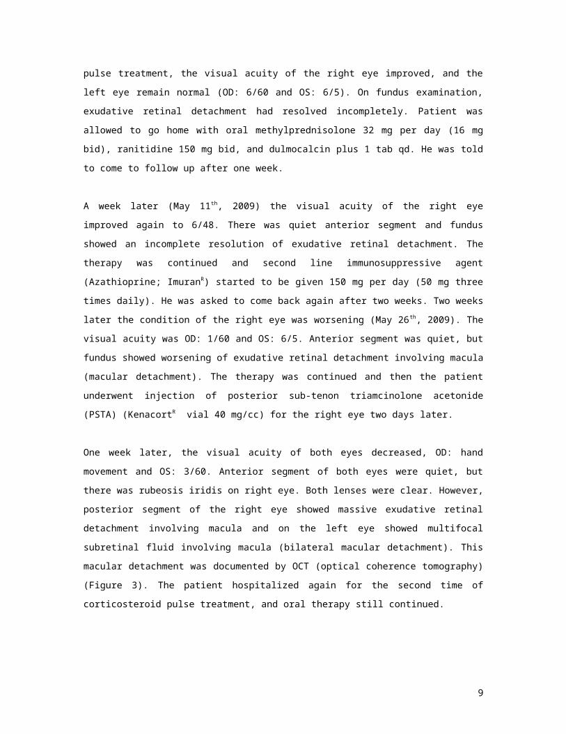
completing 3 days of pulse treatment, the visual acuity of the right eye improved, and the left eye remain
normal (OD: 6/60 and OS: 6/5). On fundus examination, exudative retinal detachment had resolved
incompletely. Patient was allowed to go home with oral methylprednisolone 32 mg per day (16 mg bid),
ranitidine 150 mg bid, and dulmocalcin plus 1 tab qd. He was told to come to follow up after one week.
A week later (May 11th, 2009) the visual acuity of the right eye improved again to 6/48. There was quiet
anterior segment and fundus showed an incomplete resolution of exudative retinal detachment. The therapy
was continued and second line immunosuppressive agent (Azathioprine; ImuranR) started to be given 150
5
Figure 2. Right eye showed enlarging of focal SRF now involving macula (A), and exudative retinal detachment on inferior extending to posterior pole (B). Left eye showed multifocal serous retinal detachment perimacula (C).
A C
B

mg per day (50 mg three times daily). He was asked to come back again after two weeks. Two weeks later
the condition of the right eye was worsening (May 26 th, 2009). The visual acuity was OD: 1/60 and OS:
6/5. Anterior segment was quiet, but fundus showed worsening of exudative retinal detachment involving
macula (macular detachment). The therapy was continued and then the patient underwent injection of
posterior sub-tenon triamcinolone acetonide (PSTA) (KenacortR vial 40 mg/cc) for the right eye two days
later.
One week later, the visual acuity of both eyes decreased, OD: hand movement and OS: 3/60. Anterior
segment of both eyes were quiet, but there was rubeosis iridis on right eye. Both lenses were clear.
However, posterior segment of the right eye showed massive exudative retinal detachment involving
macula and on the left eye showed multifocal subretinal fluid involving macula (bilateral macular
detachment). This macular detachment was documented by OCT (optical coherence tomography) (Figure
3). The patient hospitalized again for the second time of corticosteroid pulse treatment, and oral therapy
still continued.
5Figure 3. OCT image of macula of the left eye showed serous retinal detachment.
Macular thickness measured was around 1000 um.
During hospitalization we perform magnetic resonance imaging (MRI) on the patient’s head for discarding
possibility of masquerade syndrome. However, the MRI did not reveal any tumor or abnormality in the
brain, but just showed posterior eye wall thickening which corresponds to posterior uveitis. After three days
of pulse treatment the visual acuity of both eyes did not improved significantly, OD: still hand movement
and OS: 4/60. The patient was allowed to go home with oral therapy: Methylprednisolone 64 mg per day
6

(32 mg bid), Azathioprine 150 mg per day (50 mg tid), Colchicine 1 mg/day (0.5 mg bid), Ranitidine 150
mg bid, and Dulmocalcin plus 1x1 tab. He was asked to control in two days.
Two days later the visual acuity of left eye return to 6/6. However, the visual acuity of the right eye still
hand movement due to ischemia of posterior segment. On fundus and OCT examination, subretinal fluid at
macula of the left eye had resolved (figure 4). The visual acuity OS: 6/6 is maintained until the last visit of
the patient (it has been three weeks he had colchicine). He is now still on medication.
7
Figure 4. On fundus examination, right eye still shows non-resolved exudative retinal detachment (A); however left eye shows resolved retinal detachment with minimal multifocal serous retinal detachment adjacent to macula (B). OCT image of the keft eye shows macular detachment already resolved and macular thickness return to nearly normal value. We can also notice that some part of RPE layer is damaged (C).
A B
C

Discussion
Diagnosis in this patient is based on revised diagnostic criteria for VKH disease by International
Committee on Nomenclature.5 Clinical findings in this patient correspond to ‘Incomplete VKH’ since we
can found these following conditions: 1) bilateral serous retinal detachment with multifocal pattern; 2) no
history of trauma; 3) no evidence of infectious origin; 4) sign of neurologic symptom that is hearing loss,
but no yet sign of integumentary system.
Extraocular manifestations of VKH disease exclusively occur in prodromal phase and last for few days and
followed by acute uveitic phase.1,2 If the patient ignored those symptoms, he/she will forget to report it to
the doctor at the time of examination. Beniz reported that headache is the most common neurologic
symptoms noticed by patients.1 If we want to reassure the neurologic involvement in patient who did not
noticed neurologic symptoms, we can expect to have positive result from cerebrospinal fluid (CSF)
examination. CSF pleocytosis with predominance of lymphocytes and monocytes and normal glucose have
been found in more than 80% patient with VKH and may persist up to 8 weeks. 1 However, in management
of this patient we didn’t perform CSF examination (lumbar puncture) due to its invasive procedure and its
presence does not give additional diagnostic value because complaint of hearing loss already exists.
We examine this patient in his acute uveitic phase. Usually patient will come to ophthalmologist with
bilateral complaint. However, in this patient we only saw unilateral abnormality at the first visit. In 30% of
cases, there may be delay of 1 to 3 days before the second eye becomes involved.1 Since the patient had
already complaint of his blurred vision for 2 weeks, we consider that the fellow eye might have been
showing subtle abnormalities at his first visit which can only be visualized by fundus fluorescein
angiography. Initially he was diagnosed for having neuroretinitis toxoplasmosis of the right eye. After one
week of treatment, exudative retinal detachment occurred on inferior part, not yet involving macula, while
the left eye showed normal fundus. We can expect that ocular toxoplasmosis will never give sign of
exudative retinal detachment.1,6 Thus, it is a must if we found unilateral exudative retinal detachment; we
then perform fundus fluorescein angiography to investigate whether the fellow eye had subtle retinal or
vascular abnormalities. In early manifestation of VKH disease, a few multiple punctates (pinpoint)
hyperfluorescent dots (leakage) at the level of RPE perhaps will be hard to be evaluated by indirect
ophthalmoscope. In acute uveitic phase, optic disc hyperemia is also early findings. As the disorder
evolves, optic nerve head swelling manifested, and it can be found in 87% cases. On fluorescein
angiography optic nerve head swelling will give disc leakage appearance.1
The classic definition of VKH disease mentioned about bilateral granulomatous inflammation of the eye
which originates from posterior segment and extends to anterior segment.1-3 However, in the acute uveitic
phase of this patient didn’t show any granulomatous inflammation signs on anterior segment. There were
8
no cells and flare on anterior chamber, no mutton fat keratic precipitates and no iris nodule. It seems the
inflammation rather isolated on posterior segment in this patient.
Differential diagnosis of VKH disease in this patient includes sympathetic ophthalmia, primary intraocular
lymphoma, sarcoidosis, acute posterior multifocal placoid pigment epitheliopathy/APMPPE (one of white
dots syndrome), uveal effusion syndrome, posterior scleritis, and other systemic disorder causing exudative
retinal detachment such as renal disease.1 Sympathetic ophthalmia can be discarded because of no history
of trauma. Primary intraocular lymphoma usually manifested as unilateral abnormalities with associated
neurologic and/or systemic deficit (also called intraocular-central nervous system lymphoma), and MRI
will show mass with characteristic pattern of supratentorial and multicentric, oftenly in frontal lobe.
Possibility of sarcoidosis can be discarded since no lung parenchym or lymph node abnormalities in chest
x-ray, no sarcoid nodule on anterior segment, and no vitritis on posterior segment. APMPPE and VKH
disease share many similarities, but visual loss in APMPPE do not preceded by neurologic symptoms
(meningismus, tinnitus, hearing loss). Fluorescein angiography in APMPPE and VKH disease very similar
except in VKH disease there will be accumulation of dye in subretinal space on late phase. APMPPE is a
self-limited disease with no recurrences, and it needs no treatment. Uveal effusion syndrome is a unilateral
disease with no prodromal symptoms. Posterior scleritis usually unilateral and accompany with moderate to
severe eye ball pain. Scleral thickening can also be visualized in MRI, which is do not exist in this patient.
Renal disease in this patient is excluded since the renal function test is normal.1
Treatment of VKH disease in this patient began with high dose steroid administered by ‘pulse treatment’
method. The patient hospitalized and received 1 g/day of intravenous methylprednisolone for three
consecutive days. Usually, initial high dose oral steroid (1-2 mg/kg/day) is sufficient to control
inflammation and followed by slow tapering over 3-6 months. If retinal detachments fail to resolve, then
intravenous therapy is decided.1,2 However, this patient showed clinical course of non-resolved retinal
detachments, and we predicted that the visual prognosis will be poor if it was not treated very aggressively.
So pulse treatment is decided as the initial therapy at the time diagnosis VKH disease had been established.
Fortunately, pulse treatment was effective in resolving the retinal detachment partially and the patient
experienced some visual gain.
Steroid-resistant in this patient began approximately three weeks after the patient had been given pulse dose
steroid therapy. The cause of steroid-resistant is probably because of rapid tapering of steroid given. After
pulse dose steroid (1 g/day intravenous methylprednisolone), the patient was allowed to go home with
relatively low dose steroid, that is 32 mg oral methylprednisolone (16 mg bid). After three weeks, the
visual acuity became worst again, and the retinal detachments which had been partially resolved, became
extensive again. Periocular injection of triamcinolone acetonide (40 mg, posterior sub-tenon) and the
second pulse dose steroid which have been given next, did not make any improvement.
9

Slow tapering over three months is recommended, as Rubsamen and Gass found that recurrences in 43%
and 52% of their patients occurred in the first three and six months of the disease, respectively, and these
were associated in most cases with too rapid tapering of steroids.3 Fujioka et al noted that if patients were
treated within two weeks of onset of the disease, the average duration of uveitis was three to eight months;
if treatment were delayed, however, steroid therapy was required up to 45 months. 3 Other causes that make
VKH disease resistant to steroid therapy are complete VKH disease with severe anterior segment
inflammation and VKH disease in chronic recurrent phase which typically manifests with severe anterior
uveitis.1-3 The VKH with severe anterior uveitis and exudative retinal detachments was more resistant to
treatment, requiring more than 200 mg of intravenous prednisone therapy daily. Conversely, Hayasaka et al
found that most patients required less than 100 mg of prednisone daily for adequate suppression of the
inflammation in the Harada's form (exudative retinal detachments and mild anterior uveitis) of the VKH
disease.3
Immunosuppressive agents in VKH disease are reserved for patient who are resistant to steroid therapy or
those who have developed unacceptable side effects thereof. Their use may also provide steroid-sparing
effect, whereby the dose of steroid necessary to achieve quiescence is dramatically reduced or discontinued
altogether. Immunosuppressive drugs used in the management of VKH disease have been recommended
are cyclophosphamide, chlorambucil, azathioprine, cyclosporine and tacrolimus.1-3 In this patient, we give
azathioprine 150 mg/day (50 mg tid) as second line immunosuppressive agent starting at the second week
of VKH treatment. Oral dose of azathioprine is 1-3 mg/kg/day divided in two or three doses. We give
azathioprine because of its efficacy in choric uveitis especially with combination with steroid. Although
never been used in controlled clinical trial for VKH disease, azathioprine was effective in decreasing the
occurrence in those without ocular involvement and decreasing the occurrence of second eye disease in
those with unilateral disease in Behcet syndrome. Due to long onset of action (1-3 months), azathioprine
must be given very early in order to achieve therapeutic purpose to control inflammation and/or as steroid-
sparing agent. Azathioprine has relatively low cost compare to cyclosporine, so it is affordable for Cipto
Mangunkusumo’s patients who most on middle-low economy status.
This patient’s clinical course showed very dramatic decrease of vision within two weeks after pulse
treatment of steroid and did not respond with periocular steroid injection and also the second pulse dose
steroid. Even though azathioprine had been given for two weeks, we cannot expect the immunosuppressive
effect of this drug because its onset of action takes time 1-3 months. Then colchicine as third line
immunosuppressive agent was given at dose 1 mg/day (0.5 mg bid). Fortunately the visual acuity of left eye
return to normal, but visual acuity of right eye cannot be helped since ischemia of posterior segment had
already occurred. Onset of action colchicine as immunosuppressive agent has not been determined, but its
onset of action for relieving acute Gout arthritis is 12 hours. From this information we can understand that
the visual acuity of left eye dramatically improved from 4/60 to 6/6 by only 2 days.
10

Colchicine is an immunosuppressive and anti-inflammatory drug best known for arthritis gout treatment
and other rheumatic disease. Its use in ocular disease mainly related to systemic disease which also
affecting ocular tissue.7 There were several studies reported using colchicine treatment related to ocular
disease, including Behcet disease,7 postoperative anti-fibrotic treatment for trabeculectomy,9 nodular
scleritis,10 inflammatory Grave’s ophthalmopathy,11 proliferative vitreoretinopathy,12 and relapsing
polychondritis with ocular manifestations.13 Interestingly, we cannot easily find studies reporting colchicine
usage for VKH disease.
The most qualified ophthalmology study with level I (one) evidence (properly conduct randomized
controlled trial) comparing colchicine to other drug (cyclosporine) has been done at 1980’s for Behcet
disease in Japan.8 Before cyclosporine had been produced (1970’s), colchicine was the first line
immunosuppressive agent along with steroid for treating Behcet disease. In 1980’s colchicine still act as
first line agent, but cyclosporine was being introduced for Behcet disease.14 A relatively large randomized
clinical trial then was enrolled comparing colchicine and cyclosporine, and long term follow up was
performed. The study result showed that cyclosporine is effective for treating all manifestations of Behcet
disease, superior to colchicine. But the study noticed that the frequency of side-effects in cyclosporine
group was significantly higher than colchicine group. The side-effect includes (from the more frequent)
renal dysfunction, hirsutism, hepatic dysfunction, fever, gastrointestinal problems, and fatigue.
Interestingly, after this study the research group recommended that cyclosporine should be used in Behcet
disease resistant to other agents, and colchicine still the first line immunosuppressive agent.8 In 1990’s
when data of Behcet disease accumulates, greater proportion of clinician not longer use cyclosporine due to
its adverse reactions (and perhaps its cost). Conversely, colchicine still mainstay as drug of choice in
1990’s in Japan.14
Recommended immunosuppressive agents for VKH disease as mentioned above are azathioprine,
cyclophosphamide, chlorambucil, tacrolimus and cyclosporine. However, we do consider colchicine should
be approved for one of the options. Steroid-resistant VKH should be managed by immunosuppressive
agent. We may use one, two, and if necessary, three immunosuppressive drugs. The therapeutic
strategy to combine medications is employed frequently in the treatment of cancer.15
The immune system also lends itself to a multipronged attack, because a medication
that preferentially affects one arm of the immune system (for example, antigen
presenting cells, T lymphocytes, B lymphocytes, specific cytokines, or cell adhesion
molecules on endothelial cells) could be effectively combined with a medication that
targets a different arm. The hope is that such a combination would lead to enhanced
immunosuppression without encountering dose-limiting toxicity.15
To be rational in combination therapy of immunosuppressive agent we must understand the mechanism of
action of those drugs. Azathioprine is an antimetabolite agent. Cyclophosphamide and chlorambucil are
11

alkylating agents. Cyclosporine and tacrolimus are T-cell inhibitors. Colchicine is an antimitotic agent.
Mechanism of action of these drugs will be explained briefly below along wit its scheme (figure 5.).
1) Azathioprine is a purine nucleoside analog. It interferes with adenine and guanine
ribonucleotides by suppression of inosinic acid synthesis, which in turn
interferes with DNA replication and RNA transcription. Immunologically,
azathioprine decreases the numbers of peripheral T and B lymphocytes, and
reduces mixed lymphocyte reactivity, interleukin-2 synthesis and IgM
production.15
Figure 5. Scheme of mechanism of action several immunosuppressive drugs
which can be given for steroid-resistant VKH disease
2) Cyclophosphamide is a nitrogen mustard-alkylating agent the active
metabolites of which alkylate purines in DNA and RNA, resulting in cross-
linking, aberrant base pairing, ring cleavage and depurination. This process
results in cell death, because the cells are unable to replicate.
Cyclophosphamide is cytotoxic to both resting and dividing lymphocytes. In
patients, it decreases the number of activated T lymphocytes, suppresses
helper T lymphocyte functions, and decreases B lymphocytes for months.15
3) Chlorambucil has same mechanism that is alkylating molecules to make
cross-linking DNA to DNA or DNA to protein, but chlorambucil acts by
12
Purine synthesis
Ribonucleotides
Deoxyribonucleotides
DNA
Proteins
RNA
Azathioprine
Cyclosporine, Tacrolimus
Colchicine
Cyclophosphamide, Chlorambucil
Pyrimidine synthesis

substituting hydrogen ions in organic compound so that alkylating process
then be occurred.15
4) Cyclosporine is a natural product of fungi, named Beauveria nivea.
Cyclosporine (cyclosporine A) is an 11-amino acid cyclic peptide. Cyclosporine
appears to affect preferentially immunocompetent T lymphocytes that are in
the G0 and G1 phase of their cell cycle, and its effect appears to be a specific
transcriptional inhibition in these cells, blocking replication, as well as their
ability to produce lymphokines, such as interleukin-2.15
5) Tacrolimus is a macrolide antibiotic produced by Streptomyces tsukubaensis.
Tacrolimus inhibits the activation of T lymphocytes by a mechanism similar to
that of cyclosporine.15
6) Colchicine acts by binding to microtubular proteins; it interferes with the function of the
mitotic spindle and causes depolymerization and disappearance of fibrillar microtubules in
granulocytes and other motile cells. Thus, it inhibits migration of granulocytes, which release
lactic acid and inflammatory enzymes, to sites of inflammation. A role for colchicine as an
antimitotic agent has been proposed since its prevention of spindle formation can arrest cell
division in metaphase. Cells with the highest rate of division are affected earliest.7
To choice immunosuppressive agent we must consider the side-effects of each drug. Side-effects of
colchicine are relatively minimal and tolerable at its moderate dose (0.5 – 0.6 mg, 2-3 times daily).7
Common side-effects is gastrointestinal upset. In large dose, saute toxicity manifests as hemorrhagic
gastroenteritis, nephrotoxicity, vascular damage, muscular depression, and ascending paralysis of the
central nervous system. Chronic administration can lead to agranulocytosis, aplastic anemia, alopecia,
myopathy, and azospermia.7 Toxicity may appear if the dose exceeding 6 mg/day, or 3 mg/day in risky
patient (elderly, weight less than 50 mg, hepatic dysfunction, and renal dysfunction).16 Side-effects of other
agents are summarized at table below (table 1).
Table 1. Summary of common side-effects of immunosuppressive agents15
Drug Side-effectsAzathioprine Bone marrow suppression (leucopenia thrombocytopenia), GI upset,
hepatitisCyclophosphamide Bone marrow suppression, infection, hematuria and hemmorrhagic
cystitis, increased risk malignancy, sterility, alopeciaChlorambucil Bone marrow suppresson, infection, increased risk malignancy,
sterilityCyclosporine Renal dysfunction, tremor, hirsutism, hypertension, gum hyperplasiaTacrolimus Nephrotoxicity, high blood pressure, neurotoxicity, hyperkalemia,
hypomagnesemia, hepatitis, diabetesColchicine Nausea, vomiting, bone marrow suppression
13

Colchicine treatment offers relatively low cost compare to other treatment. Since the characteristic of
patients in Cipto Mangunkusumo Hospital are in middle-low economy status, we need to consider the cost
aspect of medication. The table below summarizes the cost of immunosuppressive agent in Cipto
Mangunkusumo Hospital in July 2009 (table 2).
14

Table 2. Summary of the cost of immunosuppressive agent for VKH disease patient with weight 60 kgDrug Price per tablet Daily dose Price per month
(30 days)Azathioprine (ImuranR) Rp. 9.362,-; tablet 50 mg 3 x 50 mg (1-3 mg/kg/day) Rp. 842.580,-Cyclophosphamide (EndoxanR) Rp. 4.829,-, tablet 50 mg 3 x 50 mg (1-3 mg/kg/day) Rp. 434.610,-Chlorambucil (LeukeranR) Rp. 3.094,-; tablet 5 mg 1 x 10 mg (0.1-0.2 mg/kg/day) Rp. 185.640,-Cyclosporine (SandimmuneR) Rp. 55.250,-; tablet 50 mg 2 x 150 mg (5 mg/kg/day) Rp. 9.945.000,-Tacrolimus (not available) - - -Colchicine (RecolfarR) Rp. 4538,-; tablet 0.5 mg 2 x 0.5 mg (1 mg/day) Rp. 272.280,-Source: Pharmacy of Cipto Mangunkusumo Hospital, July 7th, 2009
Up until now the patient is still on medication for controlling his ocular inflammation. He received oral
methylprednisolone on slow tapering, azathioprine, colchicine, ranitidine, calcium and vitamin D
supplement. His right eye (OD: hand movement) has poor prognosis since ischemia of posterior segment
has already occurred. We consider giving bevacizumab intravitreal injection to the right eye only if
neovascular glaucoma develops. Afterward, for his left eye, if the inflammation relapse at certain times in
its clinical course (we hope it will not), we consider to give additional immunosuppressive agent which is
cyclosporine.
Conclusions
Steroid-resistant VKH disease should be managed with immunosuppressive agents. Colchicine as
immunosuppressive agent has potential role for steroid-resistant VKH disease. It has minimal and tolerable
side-effect and also has relatively low cost. We think the colchicine treatment cannot yet necessary for
being approved in standard operational procedure in treating VKH disease since the data to support it is still
minimal. However, we consider supporting it usage for VKH disease more frequently in Cipto
Mangunkusumo Hospital so that the data will accumulate and we may analyzed and draw more
comprehensive conclusions from it.
15

References
1. Foster CS, Vitale AT. Diagnosis and treatment of uveitis. WB Saunders Company, USA. 2002.
2. Read, RW. Vogt-Koyanagi-Harada disease. Ophthalmol Clin N Am 15 (2002) 333– 341.
3. Moorthy RS, Inomata H, Rao NA. Vogt-Koyanagi-Harada syndrome. Surv Ophthalmol
1995;39:265-292.
4. Wulandari RS. Karakteristik pasien uveitis posterior dan panuveitis tahun 2008 di RS Cipto
Mangunkusumo. Hasil penelitian dekriptif, belum dipublikasi. 2009.
5. Read RW, Holland GN, Rao NA, et al. Revised diagnostic criteria for Vogt-Koyanagi-Harada
disease: report from international committee on nomenclature. Am J Ophthalmol 2001;131:647-652)
6. Nussenblatt RB, Whitcup SM. Uveitis: fundamentals and clinical practice. Mosby, USA. 2004.
7. Hemady R, Tauber J, Foster CS. Immunosuppressive drugs in immune and inflammatory ocular
disease. Surv Ophthalmol 1991;35:369-385.
8. Masuda K, Nakajima A, Urayama A, Nakae K, Kogure M, Inaba G. Double-masked trial of
cyclosporin versus colchicine and long-term open study of cyclosporin in Behçet's disease. Lancet
1989 May 20;1(8647):1093-6.
9. Ah-Chan JJ, Molteno AC, Bevin TH. Anti-inflammatory fibrosis suppression in threatened
trabeculectomy bleb failure. Arch Ophthalmol 2006 Apr;124(4): 603
10. Wong MH, Su DH, Loh RS. Nodular scleritis and Sweet's syndrome. Clin Experiment Ophthalmol
2007 Dec;35(9):858-60.
11. Stamato FJ, Maciel RM, Manso PG, Wolosker AM, Paiva ER, Lopes AC, Furlanetto RP. Colchicine
in the treatment of the inflammatory phase of Graves' ophthalmopathy: a prospective and
randomized trial with prednisone. Arq Bras Oftalmol 2006 Nov-Dec;69(6):811-6.
12. Berman DH, Gombos GM. Proliferative vitreoretinopathy: does oral low-dose colchicine have an
inhibitory effect? A controlled study in humans. Ophthalmic Surg 1989 Apr;20(4):268-72. Erratum
in: Ophthalmic Surg 1989 Jul;20(7):522.
13. Letko E, Zafirakis P, Baltatzis S, Voudouri A, Livir-Rallatos C, Foster CS. Relapsing
polychondritis: a clinical review. Semin Arthritis Rheum 2002 Jun;31(6):384-95.
14. Yoshida A, Kawashima H, Motoyama Y, et al. Comparison of Patients with Behcet’s Disease in the
1980s and 1990s. Ophthalmology 2004;111:810–815
15. Jabs DA, Rosenbaum JT, Foster CS. Guidelines for the Use of Immunosuppressive Drugs in Patients
With Ocular Inflammatory Disorders: Recommendations of an Expert Panel. Am J Ophthalmology
2000;130:492–513.
16. Medsafe Editorial Team. Colchicine Toxicity Prompts Dosage Change. Downloaded from:
http://www.medsafe.govt.nz/Profs/PUArticles/colchicinetoxicity.htm, July 8th, 2009.
17. Research Paper help
16