12.2 DNA structure - etsu.edu · Franklin’s X-Rays. In the 1950s, British scientist Rosalind...
17
12.2 DNA structure
Transcript of 12.2 DNA structure - etsu.edu · Franklin’s X-Rays. In the 1950s, British scientist Rosalind...

12.2 DNA structure
Lesson Overview The Structure of DNA
Nucleic Acids and NucleotidesNucleic acids are long, slightly acidic molecules originally identified in cell nuclei.
Nucleic acids are made up of nucleotides, linked together to form long chains.
The nucleotides that make up DNA are shown.
Lesson Overview The Structure of DNA
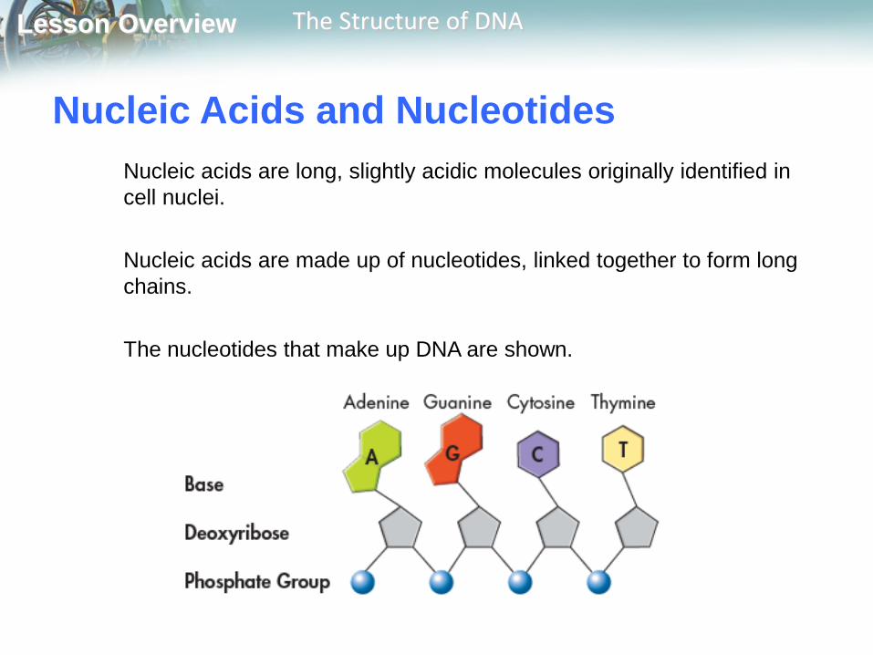
Nucleic Acids and NucleotidesDNA’s nucleotides are made up of three basic components: a 5-carbon sugar called deoxyribose, a phosphate group, and a nitrogenous base.
Lesson Overview The Structure of DNA
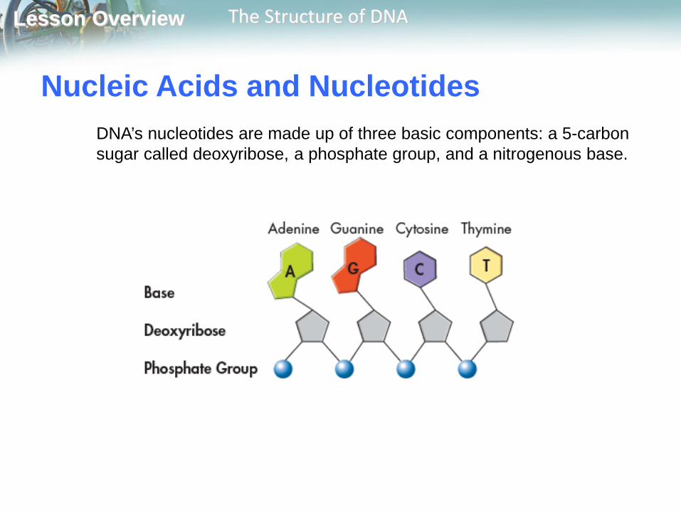
Nitrogenous Bases and Covalent BondsThe nucleotides in a strand of DNA are joined by covalent bonds formed between their sugar and phosphate groups.
Lesson Overview The Structure of DNA
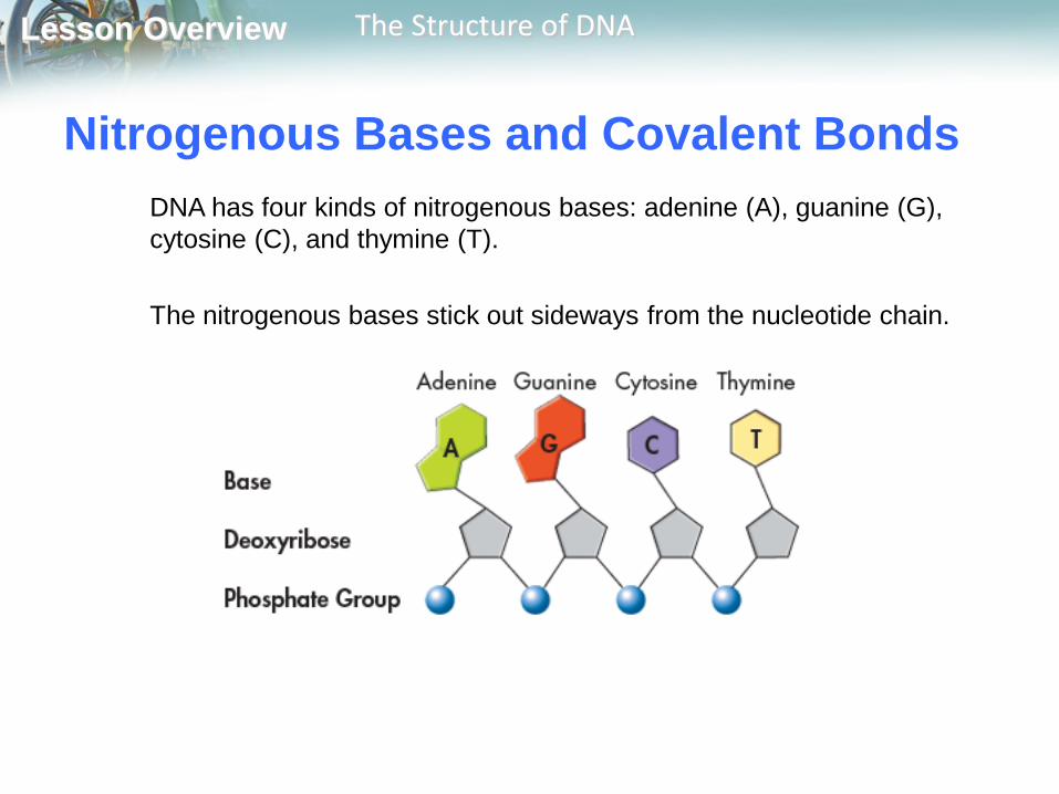
Nitrogenous Bases and Covalent BondsDNA has four kinds of nitrogenous bases: adenine (A), guanine (G), cytosine (C), and thymine (T).
The nitrogenous bases stick out sideways from the nucleotide chain.

Lesson Overview The Structure of DNA
Nitrogenous Bases and Covalent BondsThe nucleotides can be joined together in any order, meaning that any sequence of bases is possible.

Lesson Overview The Structure of DNA
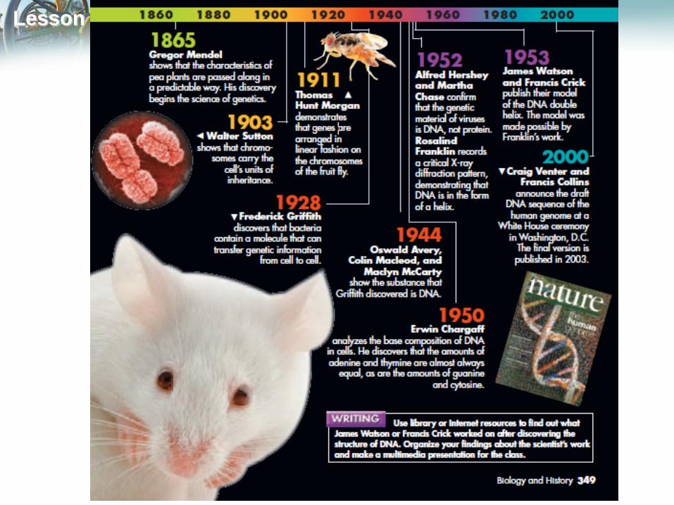
Chargaff’s RulesErwin Chargaff discovered that the percentages of adenine [A] and thymine [T] bases are almost equal in any sample of DNA.
The same thing is true for the other two nucleotides, guanine [G] and cytosine [C].
The observation that [A] = [T] and [G] = [C] became known as one of “Chargaff’s rules.”

Lesson Overview The Structure of DNA
Franklin’s X-RaysIn the 1950s, British scientist Rosalind Franklin used a technique called X-ray diffraction to get information about the structure of the DNA molecule.
Lesson Overview The Structure of DNA
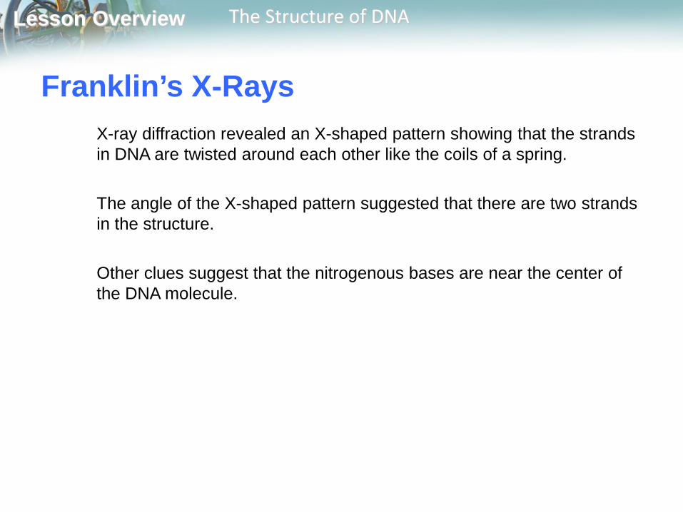
Franklin’s X-RaysX-ray diffraction revealed an X-shaped pattern showing that the strands in DNA are twisted around each other like the coils of a spring.
The angle of the X-shaped pattern suggested that there are two strands in the structure.
Other clues suggest that the nitrogenous bases are near the center of the DNA molecule.
Lesson Overview The Structure of DNA
The Work of Watson and CrickAt the same time, James Watson, an American biologist, and Francis Crick, a British physicist, were also trying to understand the structure of DNA.
They built three-dimensional models of the molecule.
Lesson Overview The Structure of DNA
The Work of Watson and CrickEarly in 1953, Watson was shown a copy of Franklin’s X-ray pattern.
The clues in Franklin’s X-ray pattern enabled Watson and Crick to build a model that explained the specific structure and properties of DNA.
Watson and Crick’s breakthrough model of DNA was a double helix, in which two strands were wound around each other.

Lesson Overview The Structure of DNA
The Double-Helix ModelA double helix looks like a twisted ladder.
In the double-helix model of DNA, the two strands twist around each other like spiral staircases.
The double helix accounted for Franklin’s X-ray pattern and explains Chargaff’s rule of base pairing and how the two strands of DNA are held together.

Lesson Overview The Structure of DNA
Antiparallel StrandsIn the double-helix model, the two strands of DNA are “antiparallel”—they run in opposite directions.
This arrangement enables the nitrogenous bases on both strands to come into contact at the center of the molecule.
It also allows each strand of the double helix to carry a sequence of nucleotides, arranged almost like letters in a four-letter alphabet.
51
end 31
end
31
end51
end

Lesson Overview The Structure of DNA
Hydrogen BondingWatson and Crick discovered that hydrogen bonds could form between certain nitrogenous bases, providing just enough force to hold the two DNA strands together.
Hydrogen bonds are relatively weak chemical forces that allow the two strands of the helix to separate.
The ability of the two strands to separate is critical to DNA’s functions.
Lesson Overview The Structure of DNA
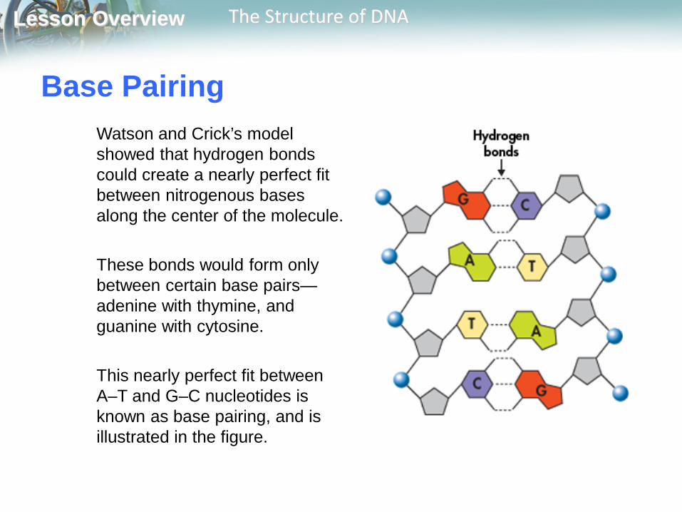
Base PairingWatson and Crick’s model showed that hydrogen bonds could create a nearly perfect fit between nitrogenous bases along the center of the molecule.
These bonds would form only between certain base pairs—adenine with thymine, and guanine with cytosine.
This nearly perfect fit between A–T and G–C nucleotides is known as base pairing, and is illustrated in the figure.
Lesson Overview The Structure of DNA
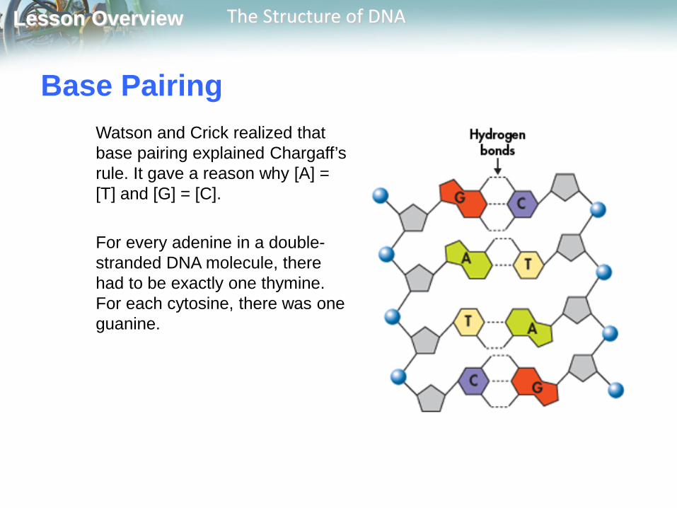
Base PairingWatson and Crick realized that base pairing explained Chargaff’s rule. It gave a reason why [A] = [T] and [G] = [C].
For every adenine in a double-stranded DNA molecule, there had to be exactly one thymine. For each cytosine, there was one guanine.
Lesson Overview The Structure of DNA