1-s2.0-S0006291X120233k40-main
7
c-Myc enhances colon cancer cell-mediated angiogenesis through the regulation of HIF-1a Cheng Chen, Shaoxin Cai, Guihua Wang, Xiaonian Cao, Xi Yang, Xuelai Luo, Yongdong Feng, Junbo Hu ⇑ Cancer Research Institute, Tongji Hospital, Huazhong University of Science and Technology, Wuhan 430030, China article info Article history: Received 19 November 2012 Available online 10 December 2012 Keywords: Angiogenesis Colon cancer c-Myc HIF-1a VEGF abstract Angiogenesis plays a pivotal role in tumor growth. The hypoxia-inducible factor 1, a subunit (HIF-1a)/ vascular endothelial growth factor pathway is the most important pathway for regulating angiogenesis in the tumor microenvironment. c-Myc is an important oncogene that has many biological functions. In this study, we investigated the role of c-Myc in tumor angiogenesis. We found that the overexpression of c-Myc in colon cancer cells could promote the expression of HIF-1a and that of vascular endothelial growth factor. Moreover, we found that c-Myc regulated HIF-1a at the post-transcriptional level. The results revealed c-Myc-dependent regulation of HIF-1a instead of HIF-1a-dependent c-Myc regulation for the first time. They also showed that c-Myc was essential to regulate colon cancer cell-mediated angi- ogenesis and contributed to tumor growth. This research provides the theoretical basis for clinical trials of new therapeutic targets of c-Myc and HIF-1a in colon cancer cells. Ó 2012 Elsevier Inc. All rights reserved. 1. Introduction Angiogenesis is essential to tumor progression: It provides the tumor with oxygen and nutrients by secreting vascular endothelial growth factor (VEGF) [1]. The microenvironment of solid tumors is often exposed to low oxygen tension as a result of an inadequate and poor blood supply [2]. Hypoxia can activate the expression of numerous angiogenic factors such as VEGF by the induction of hypoxia-inducible factor-1 (HIF-1). HIF-1 consists of two subunits, namely, the a subunit (HIF-1a) and the b subunit (HIF-1b). HIF-1b is constitutively expressed in cells in normoxia and hypoxia. HIF-1a is one of the most important regulators of oxygen homeostasis [1]. Its expression is related to the cellular O 2 concentration. The stability and activity of HIF-1a are regulated by post-translational modifications, such as hydrox- ylation, ubiquitination, acetylation, and phosphorylation [3]. In hy- poxia, HIF-1a is stable and translocates from the cytoplasm to the nucleus, where it dimerizes with HIF-1b and becomes transcrip- tionally active [4,5]. However, in normoxia, the von Hippel–Lindau protein identifies the hydroxylation of HIF-1a by prolyl hydroxy- lase enzymes and then leads to rapid proteasomal degradation of HIF-1a through ubiquitination [6]. c-Myc is a major human oncogene that is frequently altered in many forms of cancer [7–10]. It modulates the cell cycle and cell proliferation, increases cell metabolism, and stimulates differenti- ation, among its many other biological functions [11–14]. How- ever, data about c-Myc and tumor angiogenesis are limited. Many studies have reported that HIF-1a inhibits c-Myc activity via direct interactions under physiologic conditions [15–17]. The present study showed that c-Myc inhibited the degradation of HIF-1a in both normoxia and hypoxia. The results demonstrated, for the first time, that c-Myc enhanced tumor angiogenesis by pro- moting the expression of HIF-1a in the LoVo cell line. 2. Materials and methods 2.1. Reagents and antibodies Antibodies for c-Myc, HIF-1a, glyceraldehydes 3-phosphate dehydrogenase (GAPDH), CD31, and VEGF were purchased from Santa Cruz Biotechnology (Santa Cruz, CA). The antibody for HIF- 1b was purchased from Cell Signaling Technology (Beverly, MA). Cobalt chloride (CoCl 2 ), cycloheximide (Chx), MG132, and DMSO were purchased from Sigma (St. Louis, MO). The enzyme-linked immunosorbent assay (ELISA) kit for VEGF was purchased from R&D Systems (Minneapolis, MN). VEGF receptor (VEGFR) tyrosine kinase inhibitor IV (VEGFRi) was purchased from Santa Cruz Bio- technology (sc-356189, Santa Cruz, CA). Growth factor-reduced Matrigel was purchased from BD Bioscience (San Diego, CA). 2.2. Cell culture and treatment LoVo cells and human umbilical vein endothelial cells (HUVECs) were obtained from American Type Culture Collection. LoVo cells were cultured at 37 °C in DMEM supplemented with 10% fetal 0006-291X/$ - see front matter Ó 2012 Elsevier Inc. All rights reserved. http://dx.doi.org/10.1016/j.bbrc.2012.12.006 ⇑ Corresponding author. E-mail address: [email protected] (J. Hu). Biochemical and Biophysical Research Communications 430 (2013) 505–511 Contents lists available at SciVerse ScienceDirect Biochemical and Biophysical Research Communications journal homepage: www.elsevier.com/locate/ybbrc
-
Upload
ottermanerttwb -
Category
Documents
-
view
217 -
download
2
description
goo
Transcript of 1-s2.0-S0006291X120233k40-main

Biochemical and Biophysical Research Communications 430 (2013) 505–511
Contents lists available at SciVerse ScienceDirect
Biochemical and Biophysical Research Communications
journal homepage: www.elsevier .com/locate /ybbrc
c-Myc enhances colon cancer cell-mediated angiogenesis through the regulation ofHIF-1a
Cheng Chen, Shaoxin Cai, Guihua Wang, Xiaonian Cao, Xi Yang, Xuelai Luo, Yongdong Feng, Junbo Hu ⇑Cancer Research Institute, Tongji Hospital, Huazhong University of Science and Technology, Wuhan 430030, China
a r t i c l e i n f o
Article history:Received 19 November 2012Available online 10 December 2012
Keywords:AngiogenesisColon cancerc-MycHIF-1aVEGF
0006-291X/$ - see front matter � 2012 Elsevier Inc. Ahttp://dx.doi.org/10.1016/j.bbrc.2012.12.006
⇑ Corresponding author.E-mail address: [email protected] (J. Hu).
a b s t r a c t
Angiogenesis plays a pivotal role in tumor growth. The hypoxia-inducible factor 1, a subunit (HIF-1a)/vascular endothelial growth factor pathway is the most important pathway for regulating angiogenesisin the tumor microenvironment. c-Myc is an important oncogene that has many biological functions.In this study, we investigated the role of c-Myc in tumor angiogenesis. We found that the overexpressionof c-Myc in colon cancer cells could promote the expression of HIF-1a and that of vascular endothelialgrowth factor. Moreover, we found that c-Myc regulated HIF-1a at the post-transcriptional level. Theresults revealed c-Myc-dependent regulation of HIF-1a instead of HIF-1a-dependent c-Myc regulationfor the first time. They also showed that c-Myc was essential to regulate colon cancer cell-mediated angi-ogenesis and contributed to tumor growth. This research provides the theoretical basis for clinical trialsof new therapeutic targets of c-Myc and HIF-1a in colon cancer cells.
� 2012 Elsevier Inc. All rights reserved.
1. Introduction
Angiogenesis is essential to tumor progression: It provides thetumor with oxygen and nutrients by secreting vascular endothelialgrowth factor (VEGF) [1]. The microenvironment of solid tumors isoften exposed to low oxygen tension as a result of an inadequateand poor blood supply [2]. Hypoxia can activate the expressionof numerous angiogenic factors such as VEGF by the induction ofhypoxia-inducible factor-1 (HIF-1).
HIF-1 consists of two subunits, namely, the a subunit (HIF-1a)and the b subunit (HIF-1b). HIF-1b is constitutively expressed incells in normoxia and hypoxia. HIF-1a is one of the most importantregulators of oxygen homeostasis [1]. Its expression is related tothe cellular O2 concentration. The stability and activity of HIF-1aare regulated by post-translational modifications, such as hydrox-ylation, ubiquitination, acetylation, and phosphorylation [3]. In hy-poxia, HIF-1a is stable and translocates from the cytoplasm to thenucleus, where it dimerizes with HIF-1b and becomes transcrip-tionally active [4,5]. However, in normoxia, the von Hippel–Lindauprotein identifies the hydroxylation of HIF-1a by prolyl hydroxy-lase enzymes and then leads to rapid proteasomal degradation ofHIF-1a through ubiquitination [6].
c-Myc is a major human oncogene that is frequently altered inmany forms of cancer [7–10]. It modulates the cell cycle and cellproliferation, increases cell metabolism, and stimulates differenti-ation, among its many other biological functions [11–14]. How-
ll rights reserved.
ever, data about c-Myc and tumor angiogenesis are limited.Many studies have reported that HIF-1a inhibits c-Myc activityvia direct interactions under physiologic conditions [15–17]. Thepresent study showed that c-Myc inhibited the degradation ofHIF-1a in both normoxia and hypoxia. The results demonstrated,for the first time, that c-Myc enhanced tumor angiogenesis by pro-moting the expression of HIF-1a in the LoVo cell line.
2. Materials and methods
2.1. Reagents and antibodies
Antibodies for c-Myc, HIF-1a, glyceraldehydes 3-phosphatedehydrogenase (GAPDH), CD31, and VEGF were purchased fromSanta Cruz Biotechnology (Santa Cruz, CA). The antibody for HIF-1b was purchased from Cell Signaling Technology (Beverly, MA).Cobalt chloride (CoCl2), cycloheximide (Chx), MG132, and DMSOwere purchased from Sigma (St. Louis, MO). The enzyme-linkedimmunosorbent assay (ELISA) kit for VEGF was purchased fromR&D Systems (Minneapolis, MN). VEGF receptor (VEGFR) tyrosinekinase inhibitor IV (VEGFRi) was purchased from Santa Cruz Bio-technology (sc-356189, Santa Cruz, CA). Growth factor-reducedMatrigel was purchased from BD Bioscience (San Diego, CA).
2.2. Cell culture and treatment
LoVo cells and human umbilical vein endothelial cells (HUVECs)were obtained from American Type Culture Collection. LoVo cellswere cultured at 37 �C in DMEM supplemented with 10% fetal

506 C. Chen et al. / Biochemical and Biophysical Research Communications 430 (2013) 505–511
bovine serum (Hyclone). HUVECs were cultured at 37 �C in EBM-2endothelial cell basal medium with SingleQuot Kit (Lonza/Camb-rex, Walkersville, MD) as per the manufacturer’s instructions. Hyp-oxic conditions were induced by culturing cells for 2 h in a sealedhypoxia chamber (Billups Rothenberg) after flushing with a mix-ture of 1% O2, 94% N2, and 5% CO2, as well as by the addition ofCoCl2 at the concentration of 200 lM for 4 h.
2.3. Transfection
The c-Myc-overexpressing plasmid (pcDNA3.1-c-Myc plasmid)was constructed in our laboratory. Full-length c-Myc was isolatedfrom a human fetal liver cDNA library and cDNA was subclonedinto pCDNA3.1 plasmid. Sequence verified constructs were usedin this experiment. c-Myc siRNA (50-CAGAAATGTCCTGAGCAAT-30)and non-targeting scrambled siRNA (NS) were purchased fromRibobio (Guangzhou, China). LoVo cells were transfected with thesiRNA duplexes and plasmids using Lipofectamine 2000 (Invitro-gen, San Diego, CA) according to the manufacturer’s instructions.
Stable transfections of pCDNA3.1-c-Myc plasmid and pcDNA3.1empty vector were performed with Lipofectamine 2000. Aftertransfection for 24 h, LoVo cells were selected with G418(400 lg/mL).
2.4. Preparation of conditioned medium (CM)
CM was prepared as described previously but with some mod-ifications [18]. Briefly, cells treated differently were seeded at thedensity of 1 � 105 cells/mL in six-well plates. One day later, thecells were washed three times in phosphate-buffered saline andswitched to 2 mL of DMEM without fetal bovine serum. After24 h of incubation, CM was collected from the six-well plates witha cell density of approximately 90%.
2.5. Real-time PCR assay for c-Myc, HIF-1a, and VEGF
Real-time PCR was used to detect the messenger RNA (mRNA)levels of c-Myc, HIF-1a, and VEGF. Total mRNA was extracted usingTrizol (Roche Bioscience, Germany), and reverse transcription wasperformed using an RT-PCR kit (Transgen, Beijing, China). Real-time experiments were conducted on an ABI-7300 Real-time PCRDetection System using SYBR Green Real-time PCR Master Mix(Toyobo, Shanghai, China). The following PCR conditions wereused: 5 min at 95 �C followed by 40 cycles of denaturation for15 s at 95 �C, annealing for 30 s at 60 �C and primer extension for40 s at 72 �C. Primers used were: c-Myc CGAGGAGGAGAACTTC-TACCAGC and CGAGAAGCCGCTCCACATACAGTCC; HIF-1aGGCGCGAACGACAAGAAAAAG and CCTTATCAAGATGCGAACTC-ACA; VEGF CGCAGCTACTGCCATCCAAT and GTGAGGTTTGATCCG-CATAATCT; GAPDH GGTGTGAACCATGAGAAGTATGACAAC andCCAGTAGAGGCAGGGATGATGTTC. The comparative CT methodwas used to quantitate the expression of c-Myc, HIF-1a, and VEGFusing GAPDH as control [19].
2.6. Measurement of VEGF protein level in CM
The protein level of VEGF in every CM was measured using asandwich ELISA kit according to the manufacturer’s protocol.
2.7. MTT assay
Cell proliferation was measured by MTT assay. Briefly, approxi-mately 103 HUVECs were cultured in 96-well plates and the mediawere replaced 24 h later by various CM. After 48 h of incubation,relative cell numbers were quantified via MTT assay. The mediumwas replaced with 10 lL of 5 mg/mL MTT in each well. After 4 h of
incubation at 37 �C, 100 lL of DMSO was added to each well andthe absorbance was measured at 492 nm on a multifunction micro-plate reader (POLARstar OPTIMA; BMG, Offenburg, Germany).
2.8. Scratch-wound assay
HUVECs were cultured in 24-well plates. A scratch-wound wasmade using a 200-lL pipette tip when the cell density was approx-imately 100%. The medium was replaced by CM and 1 lM 5-fluo-uracil (Sigma) was added after scratch wounding to block cellproliferation. The distance of each scratch closure was obtainedby comparing the images from time 0 to the last time point(24 h) and based on the distances measured by software.
2.9. In vitro angiogenesis assay
In vitro angiogenesis was detected by tube formation. Growthfactor-reduced Matrigel was placed in 96-well tissue culture plates(100 lL/well) and allowed to form a gel at 37 �C for 30 min. HU-VECs (2 � 104 cells) were added into each well and incubated inCM for 24 h. Endothelial tubes were examined under a light micro-scope every 4 h by inspecting the overall branch points.
2.10. Western blot analysis
Cells were lysed in NP40 with PMSF, and the protein concentra-tion was determined. Proteins were separated on 10% or 12% pre-made Tris–HCl SDS–PAGE and transferred to a polyvinylidenedifluoride membrane (Bio-Rad). Proteins of interest were detectedby immunoblotting using specific antibodies.
2.11. Animal studies
Male BALB/c nude mice (4 weeks old) were purchased from theWuhan Laboratory Animal Center and maintained in the Labora-tory Animal Center of Huazhong University of Science and Technol-ogy, China. All animal experiments were done in accordance withinstitutional animal research guidelines approved by the local eth-ics committee.
Cells were cultured in fresh medium for 24 h and harvested. Atotal of 5 � 106 cells in 50 lL DMEM were mixed with 50 ll ice-cold Matrigel. Then the mixture was subcutaneously injected intothe nude mice. Tumor dimensions and volumes were determinedevery 7 days. Animals were killed 35 days after injection. The tu-mor xenografts were then removed and immediately weighed.Next, the tumor xenografts were bisected. Half of each tumorwas fixed in 4% paraformaldehyde overnight and analyzed byimmunohistochemistry, and the other half was homogenized togenerate lysates for Western blot analysis.
Microvascular density (MVD) was determined as described byWeidner et al. [20]. First, the stained sections were screened at40� magnification under a light microscope to identify the areasof highest CD31-positive vessel density. These areas were thencounted at 200�magnification in 10 random fields. Data were col-lected by two independent observers unaware of the test. Thenumber of microvessels in each field was determined as the MVD.
2.12. Clinical samples
All colon tumors were collected with informed consent of thepatients under an institutionally approved protocol at the TongjiHospital, Tongji Medical College, Huazhong University of Scienceand Technology. Every tumor was the same cell type and con-firmed by a pathologist. Fresh tumor tissues were homogenizedto generate lysates for Western blot analysis.

C. Chen et al. / Biochemical and Biophysical Research Communications 430 (2013) 505–511 507
2.13. Statistical analysis
Student’s t test was used for pairwise comparisons. Compari-sons between multiple experimental groups were conducted usingthe Bonferroni test with SPSS 13.0 for Windows. In all cases,P < 0.05 was considered statistically significant.
3. Results
3.1. c-Myc suppressed the degradation of HIF-1a
We first detected the mRNA and protein levels of HIF-1a inLoVo cells. Regardless of whether normoxic or hypoxic conditionswere used, the mRNA level of HIF-1a did not exhibit variationswhen c-Myc was overexpressed or knocked down. However, theprotein levels of c-Myc and HIF-1a were positively correlated un-der both normoxia and hypoxia (Fig. 1A–C). As c-Myc could affectthe protein level but had no influence on the mRNA level of HIF-1a,we hypothesized that the increase in protein expression is partlydue to the decrease in the degradation of HIF-1a. To confirm this,we used Chx to inhibit protein translation. We detected HIF-1afold induction by comparing the protein levels at 0, 1, 2, and 3 h.Interestingly, we found that overexpression of c-Myc under hypox-ia significantly stabilized the HIF1a protein (Fig. 1D), suggestingthat c-Myc may affect the degradation of HIF-1a. To further con-firm this result, we used the proteasome inhibitor MG132 to inhi-bit the degradation of HIF-1a. We found that overexpression of
Fig. 1. c-Myc enhances the accumulation of HIF-1a at the post-transcriptional level in boMyc protein level, HIF-1a protein level, and HIF-1b protein level of LoVo cells after they wand cells transfected with pCDNA3.1 plasmid (vector) were determined as control. (B)scramble (NS). Wild-type cells were used as control. The protein levels of HIF-1a and HIFin panels (A) and (B). Real-time PCR was performed using RNA harvested from LoVo cellsHIF-1a RNA levels were normalized to GAPDH. (D) LoVo cells (vector or c-Myc-overexHypoxia was established by treating cells with CoCl2 for 3 h prior to Chx treatment. Theprotein were determined by measuring the density of the HIF-1a protein band and norma1.0. (E) LoVo cells (wild type, vector, and c-Myc-overexpressing) were treated with proteWestern blot analysis. All results were obtained from three independent experiments. ⁄
c-Myc had no impact on the protein level of HIF-1a (Fig. 1E). Takentogether, these results showed that c-Myc was involved in HIF-1astabilization but not transcription.
We also detected the protein level of HIF-1b and found that c-Myc had no impact on its expression.
3.2. c-Myc regulated the expression of VEGF
As VEGF is one of the most important target genes of HIF-1a[21], we detected the effects of c-Myc on VEGF in the LoVo cell line.We found that overexpression of c-Myc promoted the expressionof VEGF at the mRNA level in both normoxia and hypoxia. On thecontrary, the mRNA levels of VEGF in normoxia and hypoxia de-creased when c-Myc was knocked down (Fig. 2A). Next, we de-tected the VEGF protein level by Western blot analysis and ELISA.We found that the impact of c-Myc on the VEGF protein levelwas the same as that on its mRNA level (Fig. 2B and C). Taken to-gether, these results indicated that c-Myc could positively regulatethe expression of VEGF at the mRNA and protein levels.
3.3. c-Myc promoted cell proliferation, migration and tube formationby HUVECs induced by CM from LoVo cells
Research has shown that VEGF could promote cell proliferation,migration, and angiogenesis in endothelial cells [22]. We detectedthe impact of various CM on cell growth, migration, and tube for-mation in HUVECs. To eliminate the possibility that other factor(s)
th normoxia and hypoxia. (A) Western blot analysis was performed to detect the c-ere transfected with pCDNA3.1-c-Myc plasmid (c-Myc). Wide-type (WT) LoVo cellsLoVo cells were transfected with siRNA against c-Myc (sic-Myc) or non-targeting
-1b were detected as described in panel (A). (C) LoVo cells were treated as describedand primers specific to c-Myc, HIF-1a and GAPDH in triplicate. Average c-Myc and
pressing) were treated with the protein translation inhibitor Chx for 1, 2, and 3 h.expression level of HIF-1a was detected by Western blot analysis. Levels of HIF-1alized to that of GAPDH. The relative HIF-1a protein level at time zero was defined asasome inhibitor MG132 under hypoxia. The protein level of HIF-1a was detected byp < 0.05.
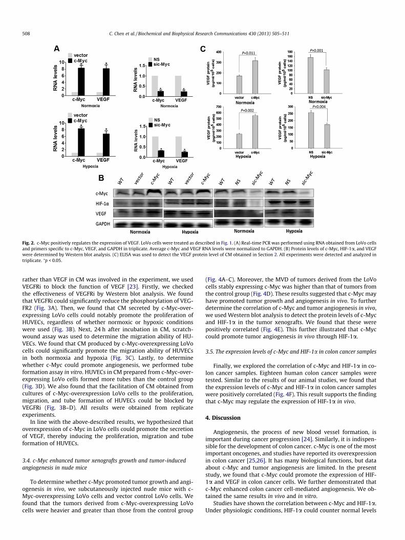
Fig. 2. c-Myc positively regulates the expression of VEGF. LoVo cells were treated as described in Fig. 1. (A) Real-time PCR was performed using RNA obtained from LoVo cellsand primers specific to c-Myc, VEGF, and GAPDH in triplicate. Average c-Myc and VEGF RNA levels were normalized to GAPDH. (B) Protein levels of c-Myc, HIF-1a, and VEGFwere determined by Western blot analysis. (C) ELISA was used to detect the VEGF protein level of CM obtained in Section 2. All experiments were detected and analyzed intriplicate. ⁄p < 0.05.
508 C. Chen et al. / Biochemical and Biophysical Research Communications 430 (2013) 505–511
rather than VEGF in CM was involved in the experiment, we usedVEGFRi to block the function of VEGF [23]. Firstly, we checkedthe effectiveness of VEGFRi by Western blot analysis. We foundthat VEGFRi could significantly reduce the phosphorylation of VEG-FR2 (Fig. 3A). Then, we found that CM secreted by c-Myc-over-expressing LoVo cells could notably promote the proliferation ofHUVECs, regardless of whether normoxic or hypoxic conditionswere used (Fig. 3B). Next, 24 h after incubation in CM, scratch-wound assay was used to determine the migration ability of HU-VECs. We found that CM produced by c-Myc-overexpressing LoVocells could significantly promote the migration ability of HUVECsin both normoxia and hypoxia (Fig. 3C). Lastly, to determinewhether c-Myc could promote angiogenesis, we performed tubeformation assay in vitro. HUVECs in CM prepared from c-Myc-over-expressing LoVo cells formed more tubes than the control group(Fig. 3D). We also found that the facilitation of CM obtained fromcultures of c-Myc-overexpression LoVo cells to the proliferation,migration, and tube formation of HUVECs could be blocked byVEGFRi (Fig. 3B–D). All results were obtained from replicateexperiments.
In line with the above-described results, we hypothesized thatoverexpression of c-Myc in LoVo cells could promote the secretionof VEGF, thereby inducing the proliferation, migration and tubeformation of HUVECs.
3.4. c-Myc enhanced tumor xenografts growth and tumor-inducedangiogenesis in nude mice
To determine whether c-Myc promoted tumor growth and angi-ogenesis in vivo, we subcutaneously injected nude mice with c-Myc-overexpressing LoVo cells and vector control LoVo cells. Wefound that the tumors derived from c-Myc-overexpressing LoVocells were heavier and greater than those from the control group
(Fig. 4A–C). Moreover, the MVD of tumors derived from the LoVocells stably expressing c-Myc was higher than that of tumors fromthe control group (Fig. 4D). These results suggested that c-Myc mayhave promoted tumor growth and angiogenesis in vivo. To furtherdetermine the correlation of c-Myc and tumor angiogenesis in vivo,we used Western blot analysis to detect the protein levels of c-Mycand HIF-1a in the tumor xenografts. We found that these werepositively correlated (Fig. 4E). This further illustrated that c-Myccould promote tumor angiogenesis in vivo through HIF-1a.
3.5. The expression levels of c-Myc and HIF-1a in colon cancer samples
Finally, we explored the correlation of c-Myc and HIF-1a in co-lon cancer samples. Eighteen human colon cancer samples weretested. Similar to the results of our animal studies, we found thatthe expression levels of c-Myc and HIF-1a in colon cancer sampleswere positively correlated (Fig. 4F). This result supports the findingthat c-Myc may regulate the expression of HIF-1a in vivo.
4. Discussion
Angiogenesis, the process of new blood vessel formation, isimportant during cancer progression [24]. Similarly, it is indispen-sible for the development of colon cancer. c-Myc is one of the mostimportant oncogenes, and studies have reported its overexpressionin colon cancer [25,26]. It has many biological functions, but dataabout c-Myc and tumor angiogenesis are limited. In the presentstudy, we found that c-Myc could promote the expression of HIF-1a and VEGF in colon cancer cells. We further demonstrated thatc-Myc enhanced colon cancer cell-mediated angiogenesis. We ob-tained the same results in vivo and in vitro.
Studies have shown the correlation between c-Myc and HIF-1a.Under physiologic conditions, HIF-1a could counter normal levels

Fig. 3. c-Myc promotes the ability of HUVECs to proliferate, migrate, and form tubes as induced by CM from LoVo cells through VEGF. (A) HUVECs were incubated in CM orCM with VEGFRi, the phosphorylation of VEGFR2 and total VEGFR2 were detected by Western blot analysis. HUVECs were incubated in CM from control LoVo cells, CM fromc-Myc-overexpressing LoVo cells or CM from c-Myc-overexpressing LoVo cells with VEGFRi. The abilities of proliferation (B) migration (C) and tube formation (D) weredetected. The branching points per field were counted to quantify the cells’ tube formation ability. ⁄p < 0.05.
C. Chen et al. / Biochemical and Biophysical Research Communications 430 (2013) 505–511 509
of c-Myc by inhibiting its function; however, deregulated onco-genic c-Myc is able to collaborate with HIF-1a [27–29]. The rela-tionship between c-Myc and HIF-1a in colon cancer cells is notclear. In contrast to previous data, the present study found, for
the first time, c-Myc-dependent regulation of HIF-1a, instead ofHIF-1a-dependent c-Myc regulation, in colon cancer cells regard-less of normoxia and hypoxia. We also found that c-Myc andHIF-1a were positively correlated in clinic colon cancer samples.

Fig. 4. c-Myc promotes tumor xenograft growth and tumor-induced angiogenesis in vivo, and the expression levels of c-Myc and HIF-1a in colon cancer samples arepositively correlated. (A–C) Xenograft growth (A) of c-Myc-overexpressing LoVo cells and control LoVo cells (vector) was determined by measuring their tumor weight (B)and tumor volume (C). (D) The number of vessels was determined by MVD as described in Section 2. (E) Correlation of c-Myc and HIF-1a expression in xenograft tumors. Halfof each tumor was homogenized to generate lysates for Western blot analysis. (F) The expression levels of c-Myc and HIF-1a in colon cancer samples were detected byWestern blot analysis.
510 C. Chen et al. / Biochemical and Biophysical Research Communications 430 (2013) 505–511
Furthermore, we found that c-Myc did not regulate HIF-1a at thetranscriptional level. The data demonstrated that c-Myc-inducedHIF-1a accumulation was post-transcriptional. Recent study re-vealed that Myc-dependent stabilization of HIF-1a involved eitherdisruption of binding to the von Hippel–Lindau complex or post-translational protein modifications in breast cancer cells [30].However the concrete mechanism of c-Myc suppressing HIF-1adegradation remains unresolved, warranting further research.
In the process of tumor growth, because of genetic alterations ofoncogenes and tumor suppressor, or in response to the reducedavailability of oxygen, tumor cells expression of many of the pro-angiogenic growth factors, which stimulated endothelial cells(ECs) migration and proliferation to sprout and form new vascula-ture [31,32]. VEGF plays a key role in this manner, which is se-creted by tumor cells [33]. VEGF is one of the most importanttarget genes of HIF-1a [21], and we thus detected the impact ofc-Myc on VEGF in colon cancer cells. As we speculated, c-Myccould promote the mRNA and protein levels in LoVo cells. As VEGFis a kind of secretory cytokines, we used ELISA to detect the secre-tion level of VEGF protein in LoVo cells. We found that c-Myc couldpromote the secretion of VEGF in LoVo cells. We obtained the sameresults in both normoxia and hypoxia. We then found that CM fromc-Myc-overexpressing LoVo cells could facilitate HUVECs prolifera-tion, migration and tube formation. We also found that the inhib-itor of VEGFR could block these functions, which further indicatedthat VEGF was the most important factor involved in the altera-tions. Therefore, we concluded that c-Myc enhances angiogenesisthrough the HIF-1a/VEGF pathway.
In summary, this study showed a new c-Myc/HIF-1a-dependentpathway in colon cancer cells that enhances the secretion of VEGFand induction of colon cancer cell-mediated angiogenesis. In thiscourse, c-Myc induced the accumulation of HIF-1a at the post-transcriptional level. The results indicated that c-Myc promotedtumor growth not only through cell proliferation but also partly
through the modulation of tumor angiogenesis. We thus identifiedimportant roles for c-Myc and HIF-1a in the pathogenesis of coloncancer. This research provides the theoretical basis for clinical tri-als of new therapeutic targets of c-Myc and HIF-1a in colon cancercells.
Acknowledgment
The study was supported by grants of foundation of ‘‘973’’ Pro-gram (No. 2009CB521802), National Natural Science foundation(Nos. 30872472, 30973496, 81172512). The funders had no rolein study design, data collection and analysis, decision to publish,or preparation of the manuscript.
Contribution: C.C., S.C., G.W., X.C., X.Y., X.L., and Y.F. performedexperiments; C.C. analyzed results and made the figures; C.C. andJ.H. wrote the paper; J.H. designed the research.
References
[1] M.C. Brahimi-Horn, J. Pouyssegur, HIF at a glance, J. Cell. Sci. 122 (2009) 1055–1057.
[2] F. Pez, F. Dayan, J. Durivault, B. Kaniewski, G. Aimond, G.S. Le Provost, B. Deux,P. Clezardin, P. Sommer, J. Pouyssequr, C. Reynaud, The HIF-1-inducible lysyloxidase activates HIF-1via the Akt pathway in a positive regulation loop andsynergizes with HIF-1 in promoting tumor cell growth, Cancer Res. 71 (2011)1647–1657.
[3] C. Brahimi-Horn, N. Mazure, J. Pouyssegur, Signaling via the hypoxia-induciblefactor-1 alpha requires multiple posttranslational modifications, Cell.Signalling 17 (2005) 1–9.
[4] L.E. Huang, Z. Arany, D.M. Livingston, H.F. Bunn, Activation of hypoxia-inducible transcription factor depends primarily upon redox-sensitivestabilization of its alpha subunit, J. Biol. Chem. 271 (1996) 32253–32259.
[5] P.J. Kallio, I. Pongratz, K. Gradin, J. McGuire, L. Poellinger, Activation ofhypoxia-inducible factor 1alpha: posttranscriptional regulation andconformational change by recruitment of the Arnt transcription factor, Proc.Nat. Acad. Sci. USA 94 (1997) 5667–5672.

C. Chen et al. / Biochemical and Biophysical Research Communications 430 (2013) 505–511 511
[6] C.W. Pugh, P.J. Ratcliffe, The von Hippel–Lindau tumor suppressor, hypoxia-inducible factor-1 (HIF-1) degradation, and cancer pathogenesis, Semin.Cancer Biol. 13 (2003) 83–89.
[7] D.J. Liao, R.B. Dickson, C-Myc in breast cancer, Endocr. Relat. Cancer 7 (2000)143–164.
[8] W. Weng, Q. Yang, M. Huang, Y. Qiao, Y. Xie, Y. Yu, A. Jing, Z. Li, C-Myc inhibitsTP53INP1 expression via promoter methylation in esophageal carcinoma,Biochem. Biophys. Res. Commun. 405 (2011) 278–284.
[9] C.V. Dang, K.A. O’Donnell, K.I. Zeller, T. Nguyen, R.C. Osthus, F. Li, The c-Myctarget gene network, Semin. Cancer Biol. 16 (2006) 253–264.
[10] S. Adhikary, M. Eilers, Transcriptional regulation and transformation by Mycproteins, Nat. Rev. Mol. Cell Biol. 6 (2005) 635–645.
[11] C.V. Dang, Myc on the path to cancer, Cell 149 (2012) 22–35.[12] K. Takahashi, S. Yamanaka, Induction of pluripotent stem cells from mouse
embryonic and adult fibroblast cultures by defined factors, Cell. 126 (2006)663–676.
[13] R.C. Osthus, H. Shim, S. Kim, Q. Li, R. Reddy, M. Mukherjee, D. Wonsey, L.A. Lee,C.V. Dang, Deregulation of glucose transporter 1 and glycolytic geneexpression by c-Myc, J. Biol. Chem. 275 (2000) 21797–21800.
[14] M. Yuneva, N. Zamboni, P. Oefner, R. Sachidanandam, Y. Lazebnik, Deficiencyin glutamine but not glucose induces Myc-dependent apoptosis in humancells, J. Cell. Biol. 178 (2007) 93–105.
[15] H. Zhang, P. Gao, R. Fukuda, G. Kumar, B. Krishnamachary, K.I. Zeller, C.V. Dang,G.L. Semenza, HIF-1 inhibits mitochondrial biogenesis and cellular respirationin VHL-deficient renal cell carcinoma by repression of C-MYC activity, CancerCell 11 (2007) 407–420.
[16] P.G. Corn, M.S. Ricci, K.A. Scata, A.M. Arsham, M.C. Simon, D.T. Dicker, W.S. EI-Deiry, Mxi1 is induced by hypoxia in a HIF-1-dependent manner and protectscells from c-Myc-induced apoptosis, Cancer Biol. Ther. 4 (2005) 1285–1294.
[17] J.D. Gordan, C.B. Thompson, M.C. Simon, HIF and c-Myc: sibling rivals forcontrol of cancer cell metabolism and proliferation, Cancer Cell 12 (2007) 108–113.
[18] T. Tian, K.J. Nan, S.H. Wang, X. Liang, C.X. Lu, H. Guo, W.J. Wang, Z.P. Ruan,PTEN regulates angiogenesis and VEGF espression through phosphatase-depedent and –independent mechanisms in HepG2 cells, Carcinogenesis 31(2010) 1211–1219.
[19] T.D. Schmittgen, K.J. Livak, Analyzing Real-time PCR data by the comparative C(T) method, Nat. Protoc. 3 (2008) 1101–1108.
[20] N. Weidner, J.P. Semple, W.R. Welch, J. Folkman, Tumor angiogenesis andmetastasis–correlation in invasive breast carcinoma, N. Engl. J. Med. 324(1991) 1–8.
[21] A. Weidemann, R.S. Johnson, Biology of HIF-1a, Cell. Death Differ. 15 (2008)621–627.
[22] D.J. Hicklin, L.M. Ellis, Role of the vascular endothelial growth factor pathwayin tumor growth and angiogenesis, J. Clin. Oncol. 23 (2005) 1011–1027.
[23] K. Nakamura, E. Taquchi, T. Miura, A. Yamamoto, K. Takahashi, F. Bichat, N.Guilbaud, K. Haseqawa, K. Kubo, Y. Fujiwara, R. Suzuki, K. Kubo, M. Shibuya, T.Isae, KRN951, a highly potent inhibitor of vascular endothelial growth factorreceptor tyrosine kinases, has antitumor activities and affects functionalvascular properties, Cancer Res. 66 (2006) 9134–9142.
[24] C. Hwang, E.I. Heath, Angiogenesis inhibitors in the treatment of prostatecancer, J. Hematol. Oncol. 3 (2010) 26–38.
[25] K. Myant, Q.J. Sansom, Wnt/Myc interactions in the intestinal cancer: partnersin crime, Exp. Cell. Res. 317 (2011) 2725–2731.
[26] J.B. Wright, S.J. Brown, M.D. Cole, Upregulation of c-Myc in cis through a largechromatin loop linked to a cancer risk-associated single-nucleotidepolymorphism in colorectal cancerr cells, Mol. Cell. Biol. 30 (2010) 1411–1420.
[27] J.W. Kim, P. Gao, Y.C. Liu, G.L. Semenza, C.V. Dang, HIF-1 and dysregulated c-Myc cooperatively induces VEGF and metabolic switches, HK2 and PDK1, Mol.Cell. Biol. 27 (2007) 7381–7393.
[28] C.V. Dang, J.W. Kim, P. Gao, J. Yustein, The interplay between MYC and HIF incancer, Nat. Rev. Cancer 8 (2008) 51–56.
[29] S.J. Yeung, J. Pan, M.H. Lee, Roles of p53, Myc and HIF-1 in regulatingglycolysis-the seventh hallmark of cancer, Cell. Mol. Life Sci. 65 (2008) 3981–3999.
[30] M.R. Doe, J.M. Ascano, M. Kaur, M.D. Cole, Myc posttranscriptionally inducesHIF1 protein and target gene expression in normal and cancer cells, CancerRes. 72 (2012) 949–957.
[31] G. Bergers, L.E. Benjiamin, Tumorigenesis and the angiogenic switch, Nat. Rev.Cancer 3 (2003) 401–410.
[32] P. Carmeliet, Angiogenesis in health and disease, Nat. Med. 9 (2003) 653–660.[33] L.M. Ellis, D.J. Hichlin, VEGF-targeted therapy: mechanisms of anti-tumor
activity, Nat. Rev. Cancer 8 (2008) 579–591.