

1 Review of Skeletal System. 2 Skeletal System Function: –Protection –Hematopoiesis –Mineral...
81
1 Review of Skeletal System
-
date post
18-Dec-2015 -
Category
Documents
-
view
221 -
download
2
Transcript of 1 Review of Skeletal System. 2 Skeletal System Function: –Protection –Hematopoiesis –Mineral...

1
Review of Skeletal System
2
Skeletal System• Function:
– Protection– Hematopoiesis– Mineral homeostasis
• Calcium• Phosphorus• Carbonate• Magnesium

3
Structure
• Bone is a connective tissue:– Matrix
• Collagen fibers for flexibility and tensile strength
• Calcium for rigidity
• Hydroxyapatite Ca5(PO4)3OH

4
• Cells:– Osteoblast
• Form organic components of matrix
– Osteocyte– Osteoblasts
• From monocytes• Secrete citric and lactic acids• Collagenases and other enzymes• Stimulated by PTH• Inhibited by Calcitonin

5

6

7
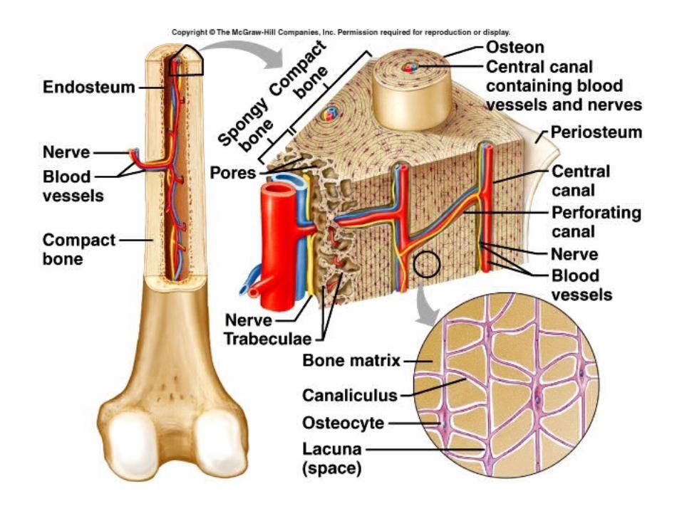
Types of Bone
• Dense or Compact (85%) – Osteon (Haversian System)– Central (Haversian) canal– Lamellae– Lacunae with osteocytes– Canaliculi
• Spongy (cancellous) bone (15%)– trabeculae
8

9

10

11
Periosteum
• Outer layer is dense, irregular CT with nerves and blood vessels
• Inner layer– Osteoblasts– Anchored to bone by collagen fibers that
penetrate into bone
12
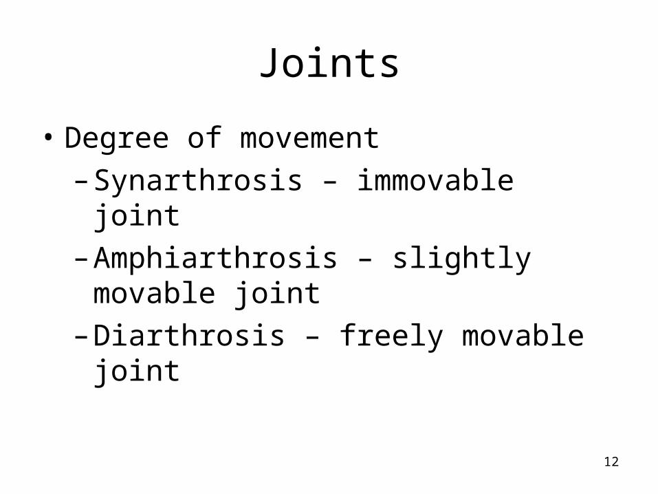
Joints
• Degree of movement
– Synarthrosis – immovable joint
– Amphiarthrosis – slightly movable joint
– Diarthrosis – freely movable joint

13
• Synovial joints
– Joint capsule
• Fibrous CT
• Tendons and ligaments
• Nerves, blood and lymph vessels
– Synovial membrane
• Loose fibrous CT
• Many blood vessels – good repair
– Joint (synovial) Cavity

14
15
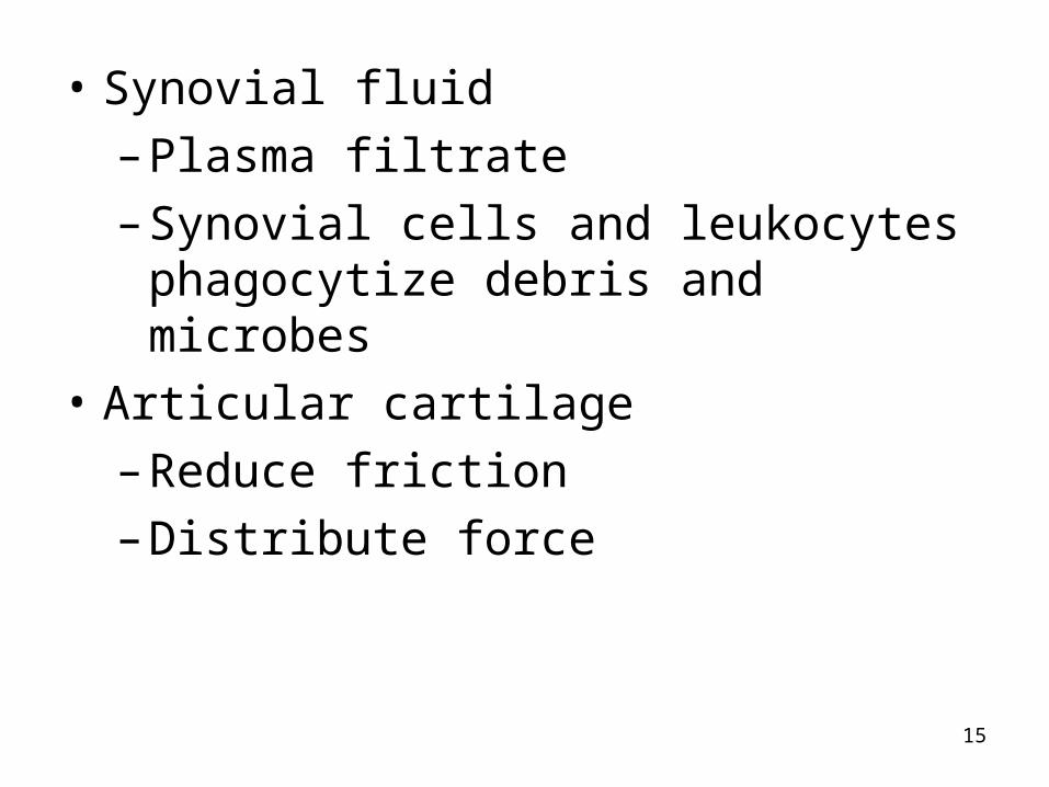
• Synovial fluid
– Plasma filtrate
– Synovial cells and leukocytes phagocytize debris and microbes
• Articular cartilage
– Reduce friction
– Distribute force
16
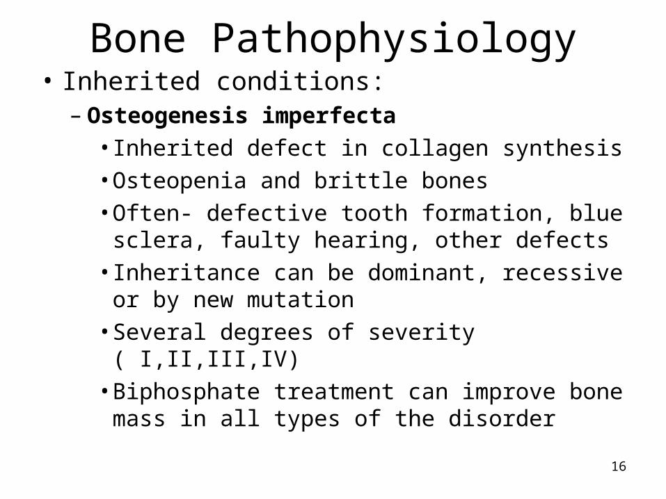
Bone Pathophysiology• Inherited conditions:
– Osteogenesis imperfecta• Inherited defect in collagen synthesis• Osteopenia and brittle bones• Often- defective tooth formation, blue
sclera, faulty hearing, other defects• Inheritance can be dominant, recessive or
by new mutation• Several degrees of severity ( I,II,III,IV)• Biphosphate treatment can improve bone
mass in all types of the disorder
17
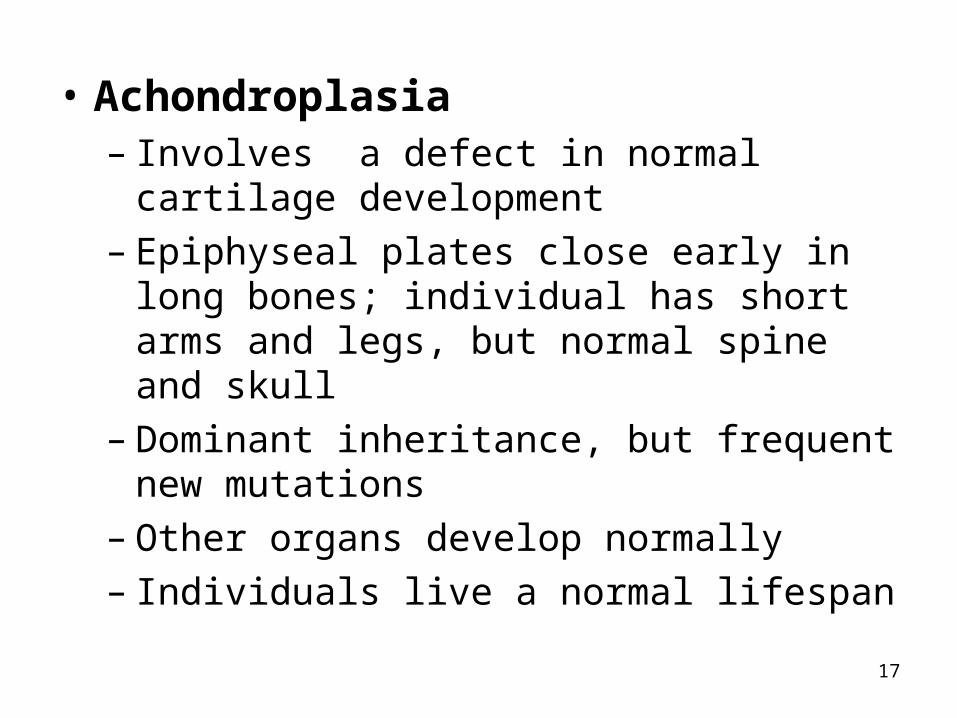
• Achondroplasia– Involves a defect in normal cartilage
development– Epiphyseal plates close early in long bones;
individual has short arms and legs, but normal spine and skull
– Dominant inheritance, but frequent new mutations
– Other organs develop normally– Individuals live a normal lifespan
18
Acquired disorders• Osteoporosis – “porous bone”
– Most common metabolic bone disease in North America
– Can be attributed to genetics, diet or hormones– Most osteoporosis is idiopathic osteoporosis– Bone loss due to an identifiable cause is
secondary osteoporosis– Bone tissue is mineralized normally, but over
time the structural integrity of bone is lost and it becomes thinner and weaker, and more prone to fractures.

19
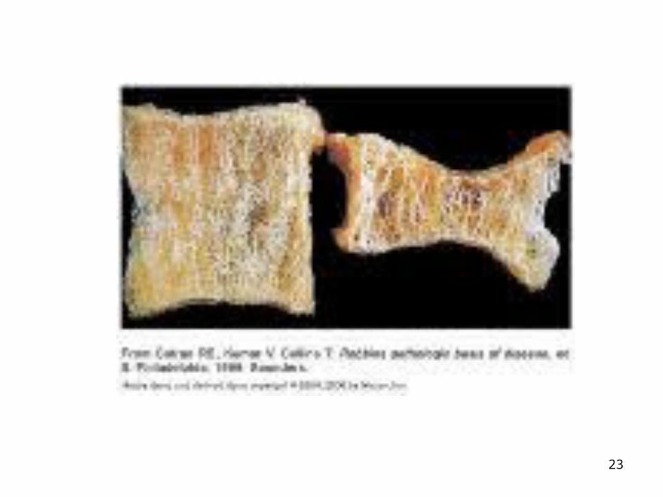
• Key features: bone fracture and the associated pain.
• WHO defines osteoporosis by bone density:– Normal bone > 833 mg/cm2
– Osteopenia 833 to 648 mg/cm2
– Osteoporosis < 648 mg/cm2
• Can be generalized, involving major portions of the axial skeleton
• Can be regional, involving one segment of the appendicular skeleton

20

21
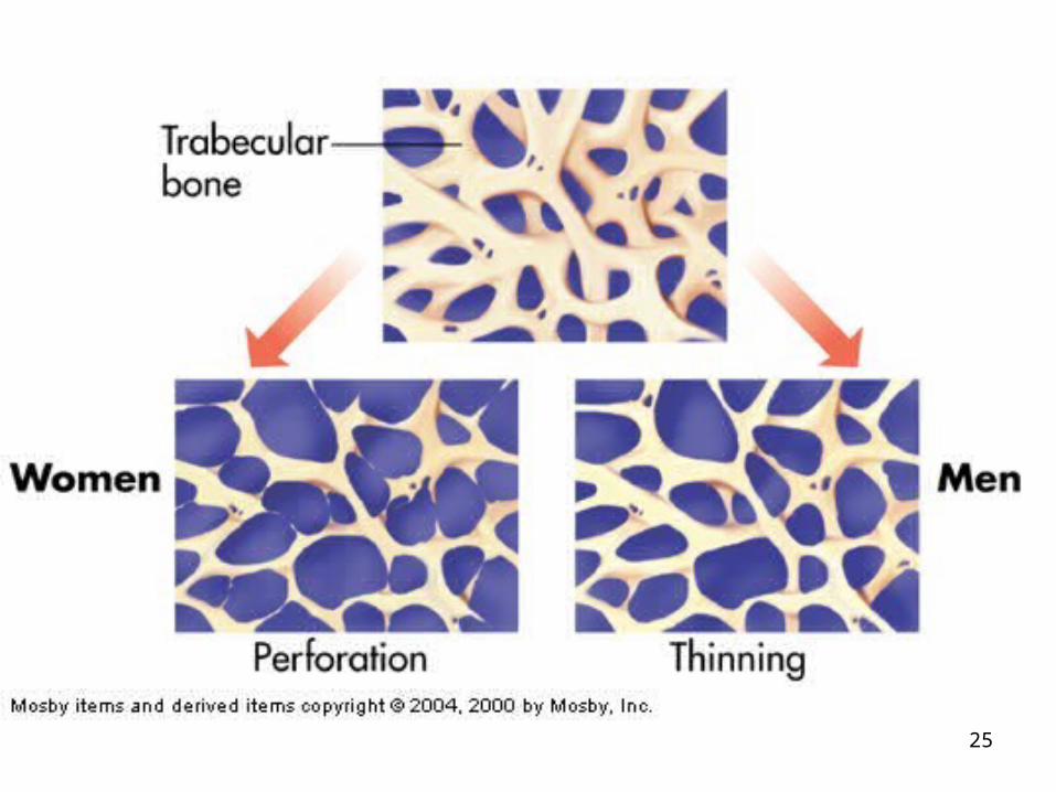
• Remodeling is constant– Teen years more bone is laid down than
reabsorbed– Peak bone mass or maximum density
reached at around 30 years of age– After age 30, bone is reabsorbed faster than it
is laid down (loss of about 0.7% /year)– In women, bone loss is most rapid in the first
years after menopause, but continues throughout postmenopausal years
– Est. 55% of people over 50 have osteoporosis or low bone mass.

22
• Men also lose bone density, but start out with more bone mass so takes longer.
• By age 90 about 17% of males have had a hip fracture, vs. 32 % of females
• Vertebral fractures also occur → kyphosis
• Most common in whites, but affects all races.
• African Americans have about half the fracture rates of whites (higher peak bone mass)
23

24
25
26
Risk factors• Family history
• White race
• Increased age
• Female sex
• Small stature
• Fair or pale skin
• Thin build
• Early menopause (natural or surgical)
• Late menarche

27
Risk factors cont.• Nulliparity• Obesity• Weight below a healthy range• Acidosis • Low dietary calcium and vitamin D• High caffeine intake• Sedentary life style• Smoker• Excessive alcohol consumption• Liver, kidney disease, rheumatoid arthritis, etc.

28
• Often progresses silently for decades until fracture occurs
• Bones can fracture spontaneously
• Most severe in spine, wrist and hips
• Estrogens and androgens may be factors in both sexes– Testosterone is converted into estrogen in
peripheral tissues and decreases bone loss
• Rapid bone loss is osteoclast mediated
• Slow bone loss is osteoblast mediated

29
Clinical manifestations• Pain and bone deformity
• Kyphosis caused by vertebral collapse
• Fractures of long bones
• Fatal complications include fat or pulmonary embolism, pneumonia, hemorrhage and shock
• 20 % die as a result of surgical complications
30
Treatment• No known cure
• Slow bone loss and promote bone deposition
• Calcium and vitamin D supplements
• Nasal or subcutaneous calcitonin
• Hormone replacement therapy
• Biophosphates – inhibit osteoclasts
• Dual x-ray absorptiometry for diagnosis
• PREVENTION

31
Prevention• Intake of calcium, vitamin D, magnesium
and possibly boron
• Regular, weight-bearing exercise
• Avoid tobacco and glucocorticoids
• No alcoholism
• Hormone replacement?
• Parathyroid hormone?
• Testosterone for men and possibly women

32
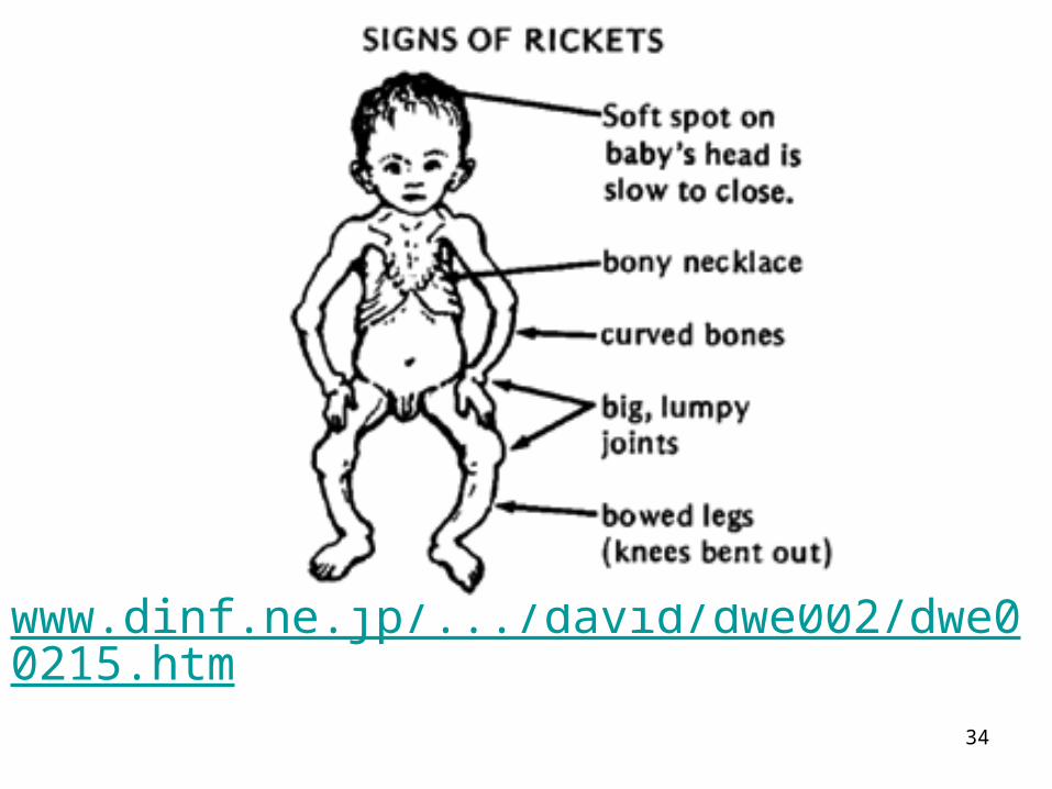
Rickets and Osteomalacia• Inadequate mineral deposition in
essentially normal organic matrix
• Softened bone:– Subject to malformation and distortion –pain

33
Rickets • Dietary vitamin D deficiency causes
inadequate mineralization of the developing skeleton in infants and children
• Rarely seen in Western nations– Poverty– Ignorance
• Bones are soft and easily deformed
• Tendency to fractures
• Therapy: supply vitamin D and calcium

36
Osteomalacia• Rarely due to vitamin D deficiency
• Usually GI malabsorption, renal defect or chronic kidney or liver diseases.
• Elderly often affected due to inadequate diet or lack of outdoor activity
• May accompany and complicate osteoporosis.

37
Joint Disorders• Osteoarthritis
– Most common joint disease in North America– Minimal inflammatory component– Differentiated from inflammatory disease by:
• Absence of synovial membrane inflammation• Lack of systemic signs and symptoms• Normal synovial fluid
– Much of the pain and loss of mobility associated with aging.
38
Osteoarthritis• Incidence increases with age: 85% of people
age 65 have some joint degeneration
• Incidence similar, but women more severely affected
• Exceptional stress on joints: gymnasts, etc.
• Biochemical defect in cartilage
• Malformed joint, obesity and postural defects
• Genetic component
• Torn ACL or meniscectomy
39
Osteoarthritis• When associated with known risk factors it
is secondary OA
• No risk factors – idiopathic OA
• Pathological characteristics:– Erosion of the articular cartilage– Sclerosis of subchondral bone– Formation of bone spurs or osteophytes

40
Osteoarthritis• Begins in articular cartilage
– Yellow-grey or brownish gray– Thin, irregular, frayed– Cracks or fissures develop (fibrillation)– Fluid filled cysts may form– Microfractures of subchondral bone– Formation of fibrocartilage repair plugs– Bone surface exposed– Bone responds by becoming dense and hard

41
Osteoarthritis• Synovial membrane is indirectly affected
– Fragments of fibrocartilage cause inflammation –pain
– Fibrous repair of joint capsule restricts motion– Osteophytes form – pain and loss of motion
• Joint mice

42
Osteoarthritis• Affects one or more weight-bearing joints
– Hand, wrist, lower cervical spine, lumbar spine and sacroiliac, hip, knees, ankles, feet
• Aches and stiffness– Symptoms increase with activity; diminish with
rest
• Usually no swelling or redness of adjacent tissues
• Sometimes nocturnal pain – may be referred

43
OsteoarthritisPrimary signs and symptoms of joint disease
are:
pain, stiffness, enlargement or swelling, tenderness, limited range of motion, muscle wasting, partial dislocation, and deformity, crepitus

44
Osteoarthritis• Evaluation made through clinical
assessment and radiologic studies, CT scan, arthroscopy and MRI
• Treatment:
• Glucosamine may decrease pain and slow or stop progression – 1500 mg/day
• Chondroitin sulfate – questionable absorption

45
Osteoarthritis• Analgesics and antiinflammatory drugs
(NSAIDs)
• Injections of corticosteroids or sodium hyaluronate (to improve lubrication)
• Range of motion exercises
• Reduce aggravating factors– Weight loss– Use of cane, crutches or walker
• Surgical removal of bone spurs, and other
• Replacement of joint

46
Rheumatoid Arthritis• Systemic disease with prominent
involvement of the joints
• Inflammatory joint disease characterized by:– Inflammatory damage in the synovial
membrane or articular cartilage– Systemic signs of inflammation: fever,
leukocytosis, malaise, anorexia, hyperfibrinogenemia)

47
Rheumatoid Arthritis• Systemic autoimmune disease that causes
chronic inflammation of connective tissue• Initially affects synovial membrane• Later articular cartilage, joint capsule,
ligaments and tendons, and bone• Affects joints of hands, wrists, ankles, and
feet, but shoulders, hips and cervical spine may also be involved
• Systemic effects on heart, kidney, lungs, skin and other organs

48
Rheumatoid Arthritis• Mild to severe
• Destroys and distorts joints
• Reduces life expectancy
• Remission and exacerbation
• 1 – 2% of adult population
• Women : men = 3:1
• Onset usually in 20’s or 30’s
• Symptoms lessen during pregnancy
• Seasonal variation

49
Rheumatoid Arthritis
• Idiopathic disease
• Immune-mediated destruction of joints
• Rheumatoid factors (IgM and IgG) target blood cells and synovial membranes forming antigen-antibody complexes
• Genetic predisposition
• Possibly bacterial or viral infection (Epstein-Barr)

50
Rheumatoid Arthritis
• Chronic inflammation of synovial membrane
• Cellular proliferation and damage to the microcirculation
• Synovial membrane becomes irregular
• Swelling, stiffness and pain
• Cartilage and bone destruction
• Ankylosis or fusing of joint
• Ligaments and tendons also affected

51
Rheumatoid Arthritis• Systemic effects:
– Generalized weakness and malaise– Up to 35% develop granulomas called
rheumatoid nodules– Systemic inflammation of blood vessels –
rheumatoid vasculitis– Serous membranes may be affected

52
Rheumatoid Arthritis• Evaluation :
– history– Physical examination– X-ray– Serologic tests for rheumatoid factor and
circulating antigen-antibody complexes, esp. antibodies against cyclic citrullinated peptide (CCP)
• No cure

53
Rheumatoid Arthritis• Therapy:
• Physical and emotional rest
• Relieve pain and swelling and retain as much joint function as possible
• Resting the joint, or binding or splinting
• Use of hot and cold packs
• Diet high in calories and vitamins
• Strengthening of associated muscles

54
Rheumatoid Arthritis• Drug therapy:
– NSAIDS– Methotrexate– Antimalarial drugs and immunosuppression
• Surgical– Synovectomy– Correction of deformities– Joint replacement– Joint fusion

55
Review of Muscular System

56
Muscle
• Skeletal muscle – > 600 muscles in body
• Cardiac muscle
• Smooth muscle
57
Muscle cell structure
• Sarcolemmamotor end platetransverse ( t- ) tubules
• Sarcoplasm
• Sarcoplasmic Reticulum – Stores Ca++

58

59
60
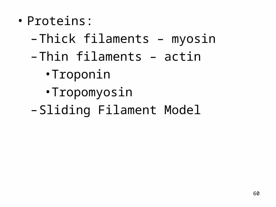
• Proteins:
– Thick filaments – myosin
– Thin filaments – actin
• Troponin
• Tropomyosin
– Sliding Filament Model

61

62

63

64

65
Muscular Dystrophy
• Group of rare diseases characterized by a genetic etiology and progressive degeneration of skeletal muscle.
• X-linked recessive defect
• Most common of the muscular dystrophies
• 1 in 3,500 live male births
• Affects males
• Gene located on the short arm of the X chromosome.

66
• 30% of cases arise as a new mutation
• Can be diagnosed immediately after birth by high serum creatine kinase
• Muscle weakness and delayed motor skills can be detected early – obvious by age 5
• Age 10 – require leg bracing
• Age 12 – wheelchair
• Age 15 completely bedridden
• Death by 20 – 30 of cardiac arrest or respiratory failure.

67
• Fibrosis → contracture distorts skeletal development– Lordosis– Scoliosis– Compromised respiration
• Respiratory insufficiency– Respiratory infection
• Cardiac muscle– Dysrythmias– Congestive heart failure
• Mental sluggishness

68
• Dystrophin is lacking– Membrane damage– Replaced by fibrous connective tissue and
fatty deposits
• Therapy– Passive stretching, splints to prevent
deformities– Sustain mobility– Sustain respiratory function– Possibly gene therapy

69
Myesthenia gravis• Autoimmune disease in which antibodies
(IgG) bind with acetylcholine receptors on muscle cells. (T-lyphmocyte abnormalities)
• Reduces the number of acetylcholine receptors at the neuromuscular junction
• Characterized by muscle weakness and fatigability
• Also associated with other autoimmune disorders, such as SLE, rheumatoid arthritis, and thyrotoxicosis

70
• In 10-25% of people with MG thymic tumors are found
– More common in males than females
• 70 – 80 % have pathologic changes in the thymus

71
Classification of myasthenia• Neonatal myasthenia
– Transitory condition in which 10-15 % of infants born to mothers with MG show symptoms of the disease
• Congenital myasthenia• Juvenile myasthenia – onset us.about 10
years• Ocular myasthenia
– More common in males– Weakness of eye muscles and eyelids, may
also include swallowing difficulties and slurred speech

72
• Generalized autoimmune myasthenia– Involves proximal musculature throughout the
body, and has several courses:• A course with periodic remissions• Slowly progressive course• Rapidly progressive course• Fulminating course

73
Pathophysiology• Defect in the nerve impulse transmission at
the NMJ
• Postsynaptic acetylcholine receptors are no longer recognized as “self” and antibodies are produced against them.
• IgG blocks the binding of ACh
• Eventually destroys the receptor
• Causes diminished transmission of nerve impulse across the NMJ and lack of muscle depolarization
• Cause is unknown.
74
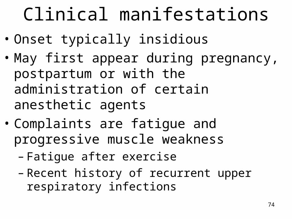
Clinical manifestations• Onset typically insidious
• May first appear during pregnancy, postpartum or with the administration of certain anesthetic agents
• Complaints are fatigue and progressive muscle weakness– Fatigue after exercise– Recent history of recurrent upper respiratory
infections

75
Clinical manifestations• Muscles of the eyes, face, mouth, throat and
neck are usually affected first– Levator and extraocular muscles affected most -
Diplopia, ptosis, and ocular palsies– Muscles of facial expression, mastication,
swallowing and speech are the next most involved
• Facial droop, expressionless face; difficulties in chewing and swallowing, drooling, episodes of choking and aspiration
• Nasal, low volume, high-pitched monotonous speech pattern

76
• Less frequently involved are the muscles of the neck, shoulder girdle and hip flexors– Fatigue requires periods of rest– Weakness of arms and legs– Difficulty maintaining head position– Respiratory muscles of chest wall and
diaphragm become weak
• In advanced stage all muscles are weak

77
Myasthenic crisis• Severe weakness causes quadriparesis or
quadriplegia, respiratory insufficiency and extreme difficulty in swallowing

78
Cholinergic crisis• Anticholinesterase drug toxicity
• Intestinal motility increases
• Fasciculation
• Bradycardia
• Pupillary constriction
• Increased salivation
• Increased sweating

79
Evaluation• Improvement with edrophonium chloride
(Telison) for several minutes
• EMG – amplitude of action potentials declines
• Antiacetylcholine receptor antibody titers
• Antistriated muscle antibody titers
• MRI to rule out thymoma
80
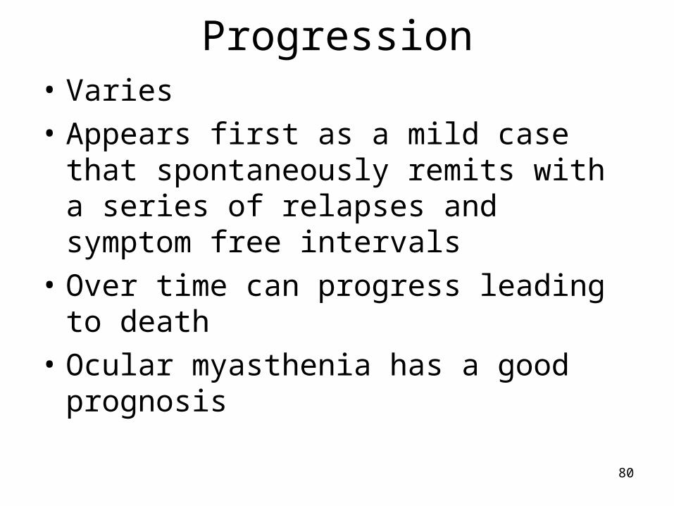
Progression• Varies
• Appears first as a mild case that spontaneously remits with a series of relapses and symptom free intervals
• Over time can progress leading to death
• Ocular myasthenia has a good prognosis

81
Treatment• Anticholinesterase drugs
• Steroids
• Immunosuppressant drugs
• Cyclophosphamide
• Plasmapheresis during myasthenic crisis
• Thymectomy is treatment of choice for individuals with thymoma