Growth & Development of Skeletal Muscle. Skeletal, Striated, Voluntary Muscle.

A MED13-dependent skeletal muscle geneprogram controls systemic glucosehomeostasis and hepatic metabolismLeonela Amoasii,1,2,3 William Holland,4 Efrain Sanchez-Ortiz,1,2,3 Kedryn K. Baskin,1,2,3
Mackenzie Pearson,4 Shawn C. Burgess,5,6 Benjamin R. Nelson,1,2,3 Rhonda Bassel-Duby,1,2,3
and Eric N. Olson1,2,3
1Department of Molecular Biology, University of Texas SouthwesternMedical Center, Dallas, Texas 75390, USA; 2Hamon Centerfor Regenerative Science and Medicine, University of Texas Southwestern Medical Center, Dallas, Texas 75390, USA; 3SenatorPaul D. Wellstone Muscular Dystrophy Cooperative Research Center, University of Texas Southwestern Medical Center, Dallas,Texas 75390, USA; 4Department of Internal Medicine, University of Texas Southwestern Medical Center, Dallas, Texas 75390,USA; 5Advanced Imaging Research Center, University of Texas Southwestern Medical Center, Dallas, Texas 75390, USA;6Department of Pharmacology, University of Texas Southwestern Medical Center, Dallas, Texas 75390, USA
The Mediator complex governs gene expression by linking upstream signaling pathways with the basal transcrip-tional machinery. However, how individual Mediator subunits may function in different tissues remains to be in-vestigated. Through skeletal muscle-specific deletion of theMediator subunitMED13 inmice, we discovered a generegulatory mechanism by which skeletal muscle modulates the response of the liver to a high-fat diet. Skeletalmuscle-specific deletion of MED13 in mice conferred resistance to hepatic steatosis by activating a metabolic geneprogram that enhances muscle glucose uptake and storage as glycogen. The consequent insulin-sensitizing effectwithin skeletal muscle lowered systemic glucose and insulin levels independently of weight gain and adiposity andprevented hepatic lipid accumulation. MED13 suppressed the expression of genes involved in glucose uptake andmetabolism in skeletal muscle by inhibiting the nuclear receptor NURR1 and theMEF2 transcription factor. Thesefindings reveal a fundamental molecular mechanism for the governance of glucose metabolism and the control ofhepatic lipid accumulation by skeletal muscle. Intriguingly, MED13 exerts opposing metabolic actions in skeletalmuscle and the heart, highlighting the customized, tissue-specific functions of the Mediator complex.
[Keywords: glucose homeostasis; mediator complex; skeletal muscle; NURR1/NR4A2]
Supplemental material is available for this article.
Received October 7, 2015; revised version accepted December 29, 2015.
Mediator is a multiprotein complex that links signal-dependent transcription factors and other transcriptionalregulators with the basal transcriptional machinery(Malik and Roeder 2010; Taatjes 2010; Allen and Taatjes2015). There are 26 subunits within the core of the Medi-ator complex. In addition, the kinase submodule, consist-ing of MED12, MED13, CDK8 and Cyclin C, serves as amodulator of the activity of the core Mediator complex.The kinase submodule can repress theMediator by block-ing its association with RNA polymerase II (Pol II) (Tsaiet al. 2013) and can also enhance transcription by the thy-roid hormone receptor (TR) by promoting Pol II recruit-ment to TR-dependent genes in a context-dependentmanner (Kornberg 2005; Furumoto et al. 2007; Belakavadiand Fondell 2010).
Through physical interactions between various tran-scription factors, nuclear receptors, and specific Mediatorsubunits, the Mediator complex controls different signal-ing pathways and processes. Recent studies have begun toreveal unexpected specificity with which various Media-tor subunits influence metabolism. For example, MED1,a core Mediator subunit, binds to nuclear receptors, andits deletion in skeletal muscle enhances metabolism(Chen et al. 2010; Jia et al. 2014). Additional componentsof theMediator complex are involved in fatty acid, choles-terol, and lipid homeostasis (Yang et al. 2006; Zhao et al.2012; Tsai et al. 2013; Chu et al. 2014). We reported thatcardiac overexpression of MED13 increases energy con-sumption and confers a lean phenotype in mice (Grueter
Corresponding author: [email protected] is online at http://www.genesdev.org/cgi/doi/10.1101/gad.273128.115.
© 2016 Amoasii et al. This article is distributed exclusively by ColdSpring Harbor Laboratory Press for the first six months after the full-issuepublication date (see http://genesdev.cshlp.org/site/misc/terms.xhtml).After six months, it is available under a Creative Commons License (At-tribution-NonCommercial 4.0 International), as described at http://creativecommons.org/licenses/by-nc/4.0/.
434 GENES & DEVELOPMENT 30:434–446 Published by Cold Spring Harbor Laboratory Press; ISSN 0890-9369/16; www.genesdev.org
Cold Spring Harbor Laboratory Press on February 24, 2020 - Published by genesdev.cshlp.orgDownloaded from
et al. 2012; Baskin et al. 2014). However, the functions ofMED13 in other tissues have not been explored.As themajor tissue in the body and a site for glucose up-
take and glycogen storage, skeletal muscle plays a funda-mental role in the control of systemic energy metabolism(Mootha et al. 2003; Izumiya et al. 2008;Meng et al. 2013).Sensitization of skeletal muscle to insulin signaling repre-sents a potential means of normalizing glucose homeosta-sis andmetabolism under conditions of obesity and type 2diabetes (T2D). The liver also modulates glucose homeo-stasis by producing glucose during periods of fasting,thereby preventing hypoglycemia and maintaining brainfunction and survival (Oosterveer and Schoonjans 2013).Impaired glucose uptake in skeletalmuscle during obesityand T2D results in hepatic steatosis, a rising health con-cern (Ranalletta et al. 2007; Turner et al. 2013). Enhancedskeletal muscle metabolism and exercise reduce the inci-dence of metabolic syndrome, T2D, and hepatic steatosis(Izumiya et al. 2008; Egan and Zierath 2013; Meng et al.2013). Therefore, a better understanding of the molecularmechanisms that regulate muscle energy homeostasismay reveal new strategies for metabolic control.Here, we show that MED13 governs insulin sensitivity,
glucose uptake, and glycogen storage in skeletal muscleby repressing the activity of the nuclear receptorNURR1 and theMEF2 transcription factor, thereby damp-ening the expression of numerous metabolic genes. Theabsence of MED13 in skeletal muscle promotes glucoseuptake and storage, sensitizing muscle to insulin actionand preventing hyperinsulinemia and hepatic steatosis.These findings expose a transcriptional regulatorymecha-nism for the control of skeletal muscle glucose homeosta-sis controlled by MED13 and its repressive effect onNURR1 and MEF2. Genetic manipulation of this tran-scriptional circuit in skeletal muscle evokes long-rangeeffects on systemic energy homeostasis, therebymodulat-ing the response of the liver to a high-fat diet (HFD).
Results
Muscle-specific deletion of MED13 improves glucosetolerance on a HFD
To explore the functions ofMED13 in skeletal muscle, wedeleted a conditional “floxed”Med13 allele (Grueter et al.2012) in mice by breeding with the Myo-Cre mouse line(Li et al. 2005), which expresses Cre recombinase specifi-cally in skeletal muscle (referred to as MED13-mKOmice). Muscle-specific deletion of Med13 exons 7 and 8(Supplemental Fig. S1A) results in a frameshift and lossof a majority of the MED13-coding sequence, includingthe nuclear localization sequence, nuclear receptor-binding motifs, leucine zipper, and FoxO-like domain.Med13 depletion did not affect the expression level of oth-er Mediator subunits (Supplemental Fig. S1B).On a normal chow (NC) diet, MED13-mKO and control
(CTL) mice displayed similar body weight, body composi-tion, muscle fiber type composition, and glucose toler-ance (Supplemental Fig. S2A–F,K). MED13-mKO andCTL mice also had similar exercise performance and lac-
tate release when subjected to voluntary wheel runningand treadmill experiments (Supplemental Fig. S2G–J).After 12 wk of a HFD, MED13-mKO and CTL mice
showed similar weight gains and body composition (Fig.1A,B). However, MED13-mKO mice showed enhancedglucose tolerance relative to CTL littermates (Fig. 1C).In insulin tolerance tests, the MED13-mKO mice alsoshowed improved glucose clearance (Fig. 1D). Serum-freefatty acid analyses were similar between MED13-mKOand CTL mice (Fig. 1E). Serum glucose analyses in thefasted state were also similar between MED13-mKO andCTL mice on a NC diet; however, serum glucose levelswere significantly lower in MED13-mKO mice than inCTL mice on a HFD (Fig. 1F). As expected, obesity in re-sponse to a HFD led to hyperinsulinemia in CTL mice(Fig. 1G). However, serum insulin levels of MED13-mKO mice on a HFD were significantly lower than inCTL mice (Fig. 1G). Thus, although MED13-mKO micegained weight similar to CTL mice on a HFD, they dis-played improved glucose tolerance and protection fromhyperinsulinemia.
Skeletal muscle deletion of MED13 preventshepatic steatosis
HFD-induced obesity leads to triglyceride (TG) accumula-tion in the liver. To determine whether improved glucosetolerance and reduced insulin levels affected the responseof the liver to a HFD, we analyzed liver TG accumulationbyOil RedO staining.MED13-mKOmice displayed a dra-matic reduction in hepatocyte TG accumulation com-pared with CTL mice on a HFD (Fig. 2A). Biochemicalmeasurements confirmed a pronounced 70% decrease inTG levels in livers from MED13-mKO mice comparedwith CTL mice on a HFD (Fig. 2B). To assess whetherthe decrease in TG accumulation influenced the hepaticglycogen storage response, we measured liver glycogencontent. Hepatic glycogen content was similar betweenMED13-mKO and CTL mice (Supplemental Fig. S3). Thereduced liver TG accumulation in MED13-mKO miceon aHFD suggested thatMED13 actswithin skeletalmus-cle to modulate hepatic lipid handling.To determine whether changes in activity, food intake,
or body temperature might account for the resistance ofMED13-mKO mice to hepatic TG accumulation, weused metabolic cages to monitor the mice on a HFD.MED13-mKO mice and CTL mice showed similar activi-ty, food intake, heat production, O2 consumption, andCO2 production (Supplemental Fig. S4). Additionally,MED13-mKO mice showed no difference in adipocytesize, fat deposition, or the expression of markers of brow-ning, thermogenesis, or adipogenesis in white adipose tis-sue (WAT) or brown adipose tissue (BAT) (SupplementalFig. S5). These results suggest that MED13 deletion inskeletal muscle decreases free fatty acid uptake and denovo lipogenesis in the liver, leading to protection fromhepatic steatosis independent of adipose tissues.Reduced hepatic TG accumulation could result from
decreased fatty acid import, diminished de novo fattyacid lipogenesis, increased fatty acid oxidation, or
Control of systemic glucose homeostasis by muscle
GENES & DEVELOPMENT 435
Cold Spring Harbor Laboratory Press on February 24, 2020 - Published by genesdev.cshlp.orgDownloaded from

enhanced very-low-density lipoprotein-mediated TGexport (Brown and Goldstein 2008; Denechaud et al.2008; Choi and Ginsberg 2011; Moon et al. 2012). Todetermine which of these pathways might be altered inMED13-mKO mice, we analyzed the expression profileof selected metabolic genes in the liver. The free fattyacid transporter CD36/FAT and fat-specific protein 27(Fsp27), a lipid droplet-associated protein, were up-regu-lated in CTLmice under HFD conditions, as reported pre-viously (Fig. 2C; Inoue et al. 2005; Buque et al. 2010). Incontrast, both genes were down-regulated in livers fromMED13-mKOmice on aHFD (Fig. 2C). As expected, genesinvolved in fatty acid biosynthesis, cholesterol synthesis,and glycolysis were up-regulated in the livers of CTLmiceon a HFD compared with mice on a NC diet (Fig. 2D). Incontrast, expression of these genes was significantly de-creased in the livers of MED13-mKO mice (Fig. 2D). Amodest decrease in the expression of genes involved inβ-oxidation of fatty acids was also seen in MED13-mKOmice on a HFD compared with CTL mice on a HFD (Sup-plemental Fig. S6A).
In order to functionally assess changes in liver metabo-lism,we analyzedmitochondrial energetics in isolated he-patic mitochondria using the Seahorse extracellular fluxanalyzer. Under basal conditions, in the presence of sub-strates pyruvate and malate, hepatic mitochondria fromMED13-mKOmice displayed amodest increase in oxygenconsumption rates (OCRs) and in the response to rote-none, succinate, antimycin A, ascorbate, and tetrapheny-
lenediamine (TMPD) treatments (Supplemental Fig. S6B).These experiments demonstrate that hepatic mitochon-dria from MED13-mKO mice contain fully intact andfunctional electron transport chains and exhibit a moder-ate increase in mitochondrial respiratory capacity in re-sponse to a variety of substrates.
Taken together, our results suggest thatMed13 deletionin skeletal muscle decreases free fatty acid uptake and denovo lipogenesis in the liver, leading to protection fromhepatic steatosis independent of adipose tissues. Theseobservations also point toward a MED13-dependent sig-naling pathway that mediates metabolic communicationbetween skeletal muscle and the liver.
Enhanced glucose metabolism in MED13-mKO mice
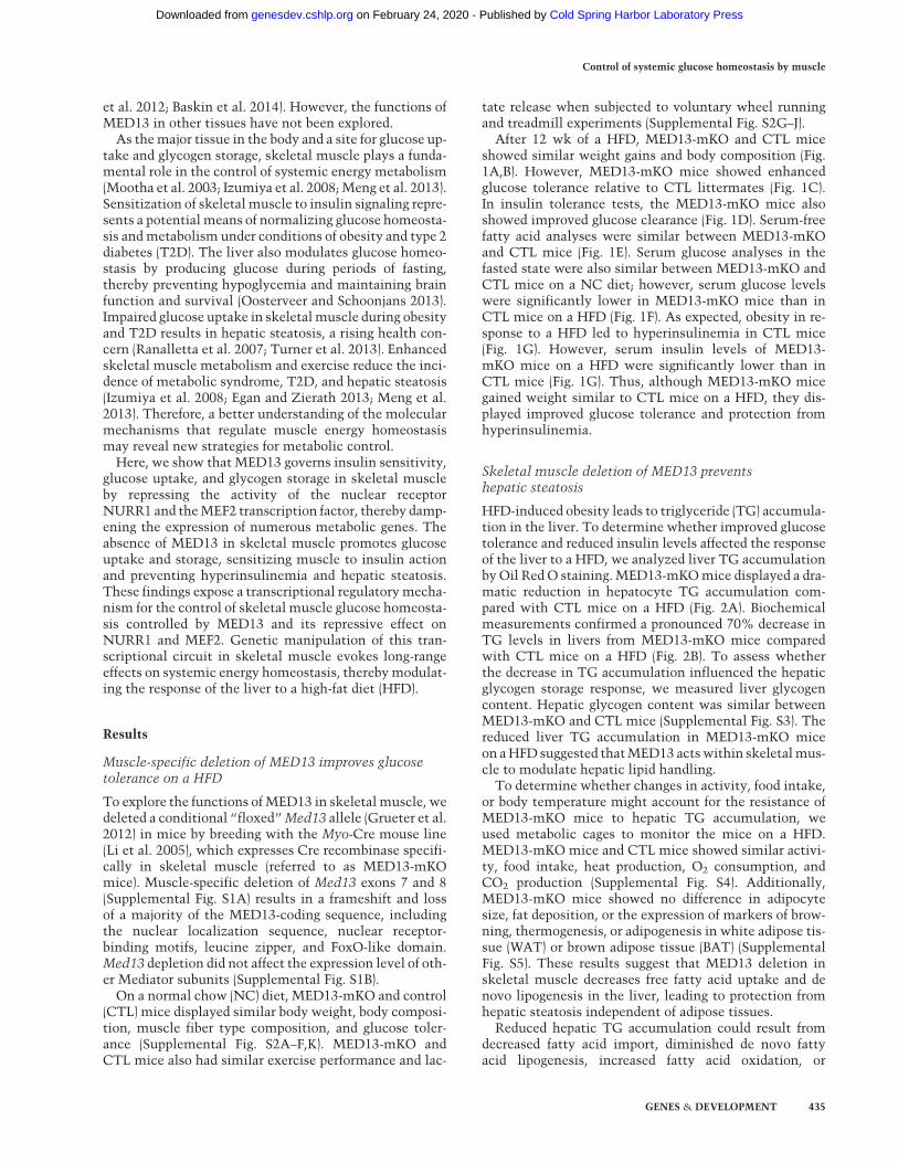
In spite of their similar body weights and adiposity, theMED13-mKOmice displayed improved glucose toleranceand insulin responsiveness compared with CTL mice. Toquantify peripheral insulin sensitivity and evaluate the ef-fect of insulin action on muscle, adipose tissue, and theliver under matched conditions, we performed hyperinsu-linemic–euglycemic clamp studies on MED13-mKO andCTL mice on a HFD diet (Ayala et al. 2006; Kim 2009).The glucose infusion rate required for maintaining eugly-cemia, a measure of whole-body insulin sensitivity, wassubstantially higher in MED13-mKO compared withCTL mice (Fig. 3A). In addition, the insulin-stimulatedglucose disposal rate was higher in MED13-mKO mice,
Figure 1. Glucose tolerance and insulinsensitivity of MED13-mKO and CTLmice. (A) Body weights of mice on a HFDfor 12 wk. (B) Body composition of micemeasured by nuclear magnetic resonance(NMR) after 12 wk on a HFD. (C ) Glucosetolerance test measured after 8 wk on aHFD. n = 10. (D) Insulin tolerance test mea-sured after 10 wk on a HFD. n = 10. (E) Se-rum nonessential fatty acid levels (NEFA)after 12 wk on a HFD in a post-pandrialstate. (F ) Serum glucose levels after 12 wkon a HFD in a fasted state. (G) Serum insu-lin levels after 12 wk on a HFD in a post-pandrial state. Data are represented asmean ± SEM. n = 16 for a HFD for all exper-iments unless otherwise stated. (∗) P < 0.05;(∗∗) P < 0.005.
Amoasii et al.
436 GENES & DEVELOPMENT
Cold Spring Harbor Laboratory Press on February 24, 2020 - Published by genesdev.cshlp.orgDownloaded from

suggesting markedly improved muscle insulin sensitivity(Fig. 3B). Tissue-specific glucose uptake into skeletalmus-cle was also dramatically higher in MED13-mKO micethan in CTL mice (Fig. 3E), whereas glucose uptake intoWAT (Fig. 3E) and hepatic glucose production (Fig. 3D)were unaffected byMED13 loss inmuscle. Such differenc-es could not be explained by differences in body weight,circulating insulin, or blood glucose during the clampedstate.Tomeasure glycogen synthesis inmuscle, wemeasured
glycogen content in muscle biopsies from the hyperinsu-lemic–euglycemic clamp study. Remarkably, our resultsshow a twofold increase in newly synthesized glycogenin muscle of MED13-mKO mice compared with CTLmice on aHFD (Fig. 3C). Similarly, in the fed state,musclefrom MED13-mKO mice displayed an increase in glyco-gen content compared with CTL mice on a HFD (Fig.3F). Histological (Supplemental Fig. S7A) and biochemical(Supplemental Fig. S7B) analyses showed no difference inTG content between MED13-mKO and CTL muscle.There were also no significant differences detected inthe lipid content of MED13-mKO and CTL muscle on aHFD or NC diet, as measured by untargeted lipidomics(Supplemental Fig. S8).
To gain further insight into mitochondrial substrateutilization in skeletal muscle, we used [U-13C6]-glucose-based metabolic flux analysis in vivo. Analysis of quadri-ceps, gastrocnemius plantaris muscle, and the livershowed no difference in the accumulation of 13C-enrichedTCA cycle intermediates betweenCTL andMED13-mKOmice on either a NC diet or HFD (Supplemental Fig. S9).These findings were in agreement with measurementsof O2 consumption and CO2 production during metaboliccage experiments (Supplemental Fig. S4E,F) and suggestedthat glucose oxidation was similar between CTL andMED13-mKO mice in HFD conditions.Overall, these results suggest that Med13 deletion in
skeletal muscle enhances glucose metabolism by increas-ing glucose uptake and disposal and increasing glycogenstorage. This insulin-sensitizing effect improves thewhole-body insulin response and renders the liver protect-ed from hepatic steatosis.
Metabolic pathways modified by MED13 deletionin skeletal muscle
Given the impact of muscle deletion ofMed13 on the liv-er, as indicated by prevention of hepatic steatosis, we
Figure 2. MED13-mKOmice are protected fromHFD-induced hepatic steatosis. (A) H&E and lipid (Oil Red O) stain of liver tissue frommice on a HFD for 12 wk. Bar, 50 µm. (B) Liver TG levels. (C ) Real-time quantitative RT–PCR (qRT–PCR) of genes involved in fatty acidtransport (CD36/FATP) and synthesis (Fsp27) in liver tissue. (D) Real-time qRT–PCRof genes involved in fatty acid biosynthesis (stearoyl-CoA destaurase [Scd1], fatty acid synthase [Fas], elongation of long chain fatty acid familymember [Elovl6], and acetyl-CoA carboxylase α[Acaca]), cholesterol synthesis (HMG-CoA-synthase [HCoASynt], HMG-CoA-reductase [HCoARed], and proprotein convertase subtili-sin/kexin type 9 [Pcsk9]), and gluconeogenesis (glucose 6-phosphatase c [G6Pc], glucose 6-phosphatase d [G6Pd], and liver-type pyruvatekinase [Lpk]) and sterol regulatory element-binding transcription factor 1c (SREBP1c) in liver tissue. Data are represented as mean ± SEM.n = 10 for all experiments unless otherwise stated. (∗) P < 0.05; (∗∗) P < 0.005; (∗∗∗) P < 0.0005.
Control of systemic glucose homeostasis by muscle
GENES & DEVELOPMENT 437
Cold Spring Harbor Laboratory Press on February 24, 2020 - Published by genesdev.cshlp.orgDownloaded from
analyzed the expression of secreted myokines, such as iri-sin, interleukin-6, and several members of the FGF family(Supplemental Fig. S10). MED13-mKOmuscle showed nosignificant changes in expression of any of these muscle-derived regulators of metabolism, pointing to a distinctmechanism of systemic metabolic regulation by skeletalmuscle.
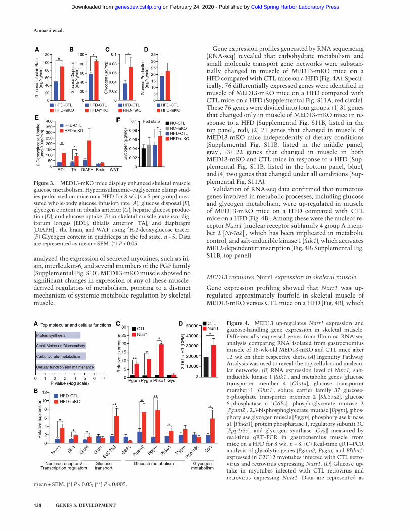
Gene expression profiles generated by RNA sequencing(RNA-seq) revealed that carbohydrate metabolism andsmall molecule transport gene networks were substan-tially changed in muscle of MED13-mKO mice on aHFD compared with CTLmice on a HFD (Fig. 4A). Specif-ically, 76 differentially expressed genes were identified inmuscle of MED13-mKO mice on a HFD compared withCTL mice on a HFD (Supplemental Fig. S11A, red circle).These 76 genes were divided into four groups: (1) 31 genesthat changed only in muscle of MED13-mKO mice in re-sponse to a HFD (Supplemental Fig. S11B, listed in thetop panel, red), (2) 21 genes that changed in muscle ofMED13-mKO mice independently of dietary conditions(Supplemental Fig. S11B, listed in the middle panel,gray), (3) 22 genes that changed in muscle in bothMED13-mKO and CTL mice in response to a HFD (Sup-plemental Fig. S11B, listed in the bottom panel, blue),and (4) two genes that changed under all conditions (Sup-plemental Fig. S11A).
Validation of RNA-seq data confirmed that numerousgenes involved in metabolic processes, including glucoseand glycogen metabolism, were up-regulated in muscleof MED13-mKO mice on a HFD compared with CTLmice on aHFD (Fig. 4B). Among thesewere the nuclear re-ceptorNurr1 (nuclear receptor subfamily 4 group Amem-ber 2 [Nr4a2]), which has been implicated in metaboliccontrol, and salt-inducible kinase 1 (Sik1), which activatesMEF2-dependent transcription (Fig. 4B; Supplemental Fig.S11B, top panel).
MED13 regulates Nurr1 expression in skeletal muscle
Gene expression profiling showed that Nurr1 was up-regulated approximately fourfold in skeletal muscle ofMED13-mKO versus CTL mice on a HFD (Fig. 4B), which
Figure 4. MED13 up-regulates Nurr1 expression andglucose-handling gene expression in skeletal muscle.Differentially expressed genes from Illumina RNA-seqanalysis comparing RNA isolated from gastrocnemiusmuscle of 18-wk-old MED13-mKO and CTL mice after12 wk on their respective diets. (A) Ingenuity PathwayAnalysis was used to reveal the top cellular and molecu-lar networks. (B) RNA expression level of Nurr1, salt-inducible kinase 1 (Sik1), and metabolic genes (glucosetransporter member 4 [Glut4], glucose transportermember 1 [Glut1], solute carrier family 37 glucose-6-phosphate transporter member 2 [Slc37a2], glucose6-phosphatase c [G6Pc], phosphoglycerate mutase 2[Pgam2], 2,3-bisphosphoglycerate mutase [Bpgm], phos-phorylase glycogenmuscle [Pygm], phosphorylase kinaseα1 [Phka1], protein phosphatase 1, regulatory subunit 3C[Ppp1r3c], and glycogen synthase [Gys]) measured byreal-time qRT–PCR in gastrocnemius muscle frommice on a HFD for 8 wk. n = 8. (C ) Real-time qRT–PCRanalysis of glycolytic genes (Pgam2, Pygm, and Phka1)expressed in C2C12 myotubes infected with CTL retro-virus and retrovirus expressing Nurr1. (D) Glucose up-take in myotubes infected with CTL retrovirus andretrovirus expressing Nurr1. Data are represented as
mean ± SEM. (∗) P < 0.05; (∗∗) P < 0.005.
Figure 3. MED13-mKO mice display enhanced skeletal muscleglucose metabolism. Hyperinsulinemic–euglycemic clamp stud-ies performed on mice on a HFD for 8 wk (n = 5 per group) mea-sured whole-body glucose infusion rate (A), glucose disposal (B),glycogen content in tibialis anterior (C ), hepatic glucose produc-tion (D), and glucose uptake (E) in skeletal muscle (extensor dig-itorum longus [EDL], tibialis anterior [TA], and diaphragm[DIAPH]), the brain, and WAT using 3H-2-deoxyglucose tracer.(F ) Glycogen content in quadriceps in the fed state. n = 5. Dataare represented as mean ± SEM. (∗) P < 0.05.
Amoasii et al.
438 GENES & DEVELOPMENT
Cold Spring Harbor Laboratory Press on February 24, 2020 - Published by genesdev.cshlp.orgDownloaded from

was also confirmed by Western blot analysis (Supplemen-tal Fig. S13). Nurr1 belongs to a nuclear receptor familythat contains three members (NR4A1/Nur77, NR4A2/NURR1, and NR4A3/NOR-1). Nur77 and NOR-1 havebeen reported to be involved in muscle metabolism, butthe function of NURR1 in skeletal muscle remains un-known (Chao et al. 2007, 2012; Pearen and Muscat2012). To explore the potential effect on muscle metabo-lism, NURR1 was overexpressed in C2C12 muscle cellsusing retroviral transduction. NURR1 overexpressionstrongly induced genes involved in glucose and glycogenmetabolism, such as Pgam, Pygm, and Phka1 (Fig. 4C),and increased glucose uptake into C2C12 myotubes asmeasured by 3H-2-deoxyglucose uptake (Fig. 4D). Theseresults indicate that genetic deletion of Med13 in muscleincreases expression of NURR1, which in turn acti-vates the expression of key genes involved in glucosemetabolism.
Regulation of Glut4 transcription by NURR1,MEF2, and MED13
GLUT4, the main effector of insulin-stimulated glucosetransport in skeletal muscle, was up-regulated in musclefrom MED13-mKO mice on a HFD compared withCTL mice (Fig. 4B). To understand how MED13 regu-lates Glut4 expression, we analyzed the Glut4 promoterfor functional regulatory elements and identified a con-sensus sequence for binding of NURR1 (referred toas a NURR1 response element [NRE]) adjacent to a bind-ing site for MEF2 (MEF2 regulatory element [MRE]),shown previously to regulate the Glut4 promoter (Fig.5A; Santalucía et al. 2001; Knight et al. 2003; Pei et al.2006). Chromatin immunoprecipitation (ChIP) assaysconfirmed the occupancy of NRE and MRE sites byNURR1 andMEF2 proteins, respectively, in skeletal mus-cle (Fig. 5B).NURR1 andMEF2 individually activated theGlut4 pro-
moter linked to a luciferase reporter, and the combinationof NURR1 and MEF2 resulted in synergistic activation ofthe Glut4 promoter (Fig. 5C). This synergistic effect waslost upon mutation of the NRE or MRE sites (Fig. 5C).MED13 expression significantly repressed the activationof the Glut4 promoter by MEF2 and NURR1 (Fig. 5C).Interestingly, mutation of the MRE in the Glut4 promo-ter strongly diminished activation by both MEF2 andNURR1. Similarlymutation of theNRE prevented activa-tion by NURR1 and also affected MEF2 activation, sug-gesting cooperative transcriptional activation by the twofactors. In light of their synergistic activation of theGlut4 promoter, we tested whetherMEF2 andNURR1 in-teracted directly. Indeed, in coimmunoprecipitation (co-IP) assays, the anti-Myc antibody specifically immunopre-cipitated MEF2-Myc with Flag-tagged NURR1 (Fig. 5D).We also identified a highly conserved MEF2 site in the
Nurr1 cis-proximal enhancer region (Fig. 5E; Castilloet al. 1997; Saucedo-Cardenas et al. 1997). This site boundMEF2 in muscle cells, as shown by ChIP assay (Fig. 5F).The Nurr1 enhancer with the MRE linked to a luciferasereporter was activated by MEF2 (Fig. 5G). MED13 expres-
sion significantly repressed the activation of theNurr1 en-hancer by MEF2 (Fig. 5G).To investigate whether repression of theGlut4 promot-
er by MED13 occurred through a direct interaction withNURR1 or MEF2, we performed co-IP assays using GFP-tagged MED13, Flag-tagged NURR1, and Myc-taggedMEF2. These experiments revealed direct association ofMED13 with NURR1 (Fig. 5H) but not with MEF2 (Sup-plemental Fig. S14).Genetic deletion of Med13 in muscle increases expres-
sion of NURR1 and glucose-handling genes such asGlut4 in response to a HFD. To determine whether mus-cle deletion of Med13 leads to increased occupancy ofMEF2 and NURR1 with target sites associated with theirrespective target genes, we performed ChIP assays in skel-etal muscle of CTL and MED13-mKO mice on a NC dietandHFD. ChIP-qPCR analysis ofMEF2 andNURR1 bind-ing showed similar binding to the Nurr1 and Glut4 genesin skeletal muscle of CTL mice on a NC diet comparedwith a HFD (Fig. 5I,K). Remarkably, ChIP-qPCR analysisof MEF2 and NURR1 binding to their target genes re-vealed increased binding in skeletal muscle of MED13-mKO mice compared with CTL mice on a HFD (Fig. 5J,L). Overall, these results demonstrate that MED13 sup-presses NURR1 expression and prevents activation ofthe Glut4 promoter by NURR1 and MEF2, thereby sup-pressing glucose utilization pathways in skeletal muscle.
Discussion
Our results uncover a new regulatory pathway for meta-bolic control and an unanticipated role of the Mediatorsubunit MED13 in the control of skeletal muscle metabo-lism and systemic energy homeostasis. The absence ofMED13 in skeletal muscle results in enhanced glucoseutilization and storage as glycogen, rendering skeletalmuscle hyperresponsive to insulin under HFD conditionsand thereby preventing fatty liver disease. Our resultsshow that NURR1 acts downstream fromMED13 in skel-etal muscle to regulate expression of genes linked to glu-cose metabolism. The increase in NURR1 expression inMED13-mKO skeletal muscle provides a mechanisticbasis for the enhanced glucose metabolism and the insu-lin sensitizer effect in these mutant mice.
A metabolic gene circuit downstream from MED13
Increased expression ofNurr1, Sik1, and glucose metabol-ic genes in MED13-mKOmice suggests a repressive func-tion of MED13 under HFD-induced insulin-resistantconditions in skeletal muscle. These observations alsoimply that nutritional signals regulate MED13 functionand, consequently, NURR1 and MEF2 activity. These ob-servations are in agreement with a previous study that re-ported a decline inNurr1 expression in skeletal muscle ofmice on aHFD, suggesting that conditions of insulin resis-tance suppress its expression (Fu et al. 2007). Nurr1 hasalso been identified as one of the most up-regulated genesin human muscle biopsies after exercise (Catoire et al.
Control of systemic glucose homeostasis by muscle
GENES & DEVELOPMENT 439
Cold Spring Harbor Laboratory Press on February 24, 2020 - Published by genesdev.cshlp.orgDownloaded from

Figure 5. Regulation of the Glut4 and Nurr1 promoters. (A) Structure of the Glut4 promoter and sequences of NRE and MRE sites. (B)ChIP assays showing binding of NURR1 and MEF2 to the Glut4 promoter in mouse skeletal muscle. Antibodies against NURR1 andMEF2 were used in ChIP assays for the Glut4 promoter. Graphs display mean quantification of ChIP (percentage of input) normalizedto IgG control. n = 3. (C ) Transcriptional activation of the Glut4 promoter linked to the luciferase reporter in COS7 cells by NURR1,MEF2, and MED13. Promoters with mutations in the NRE- and MRE-binding sites were also included as indicated. (D) Coimmunopre-cipitation (co-IP) experiments were performed by cotransfecting Myc-tagged MEF2 and Flag-tagged NURR1 in HEK293 cells. Antibodiesagainst theMyc epitope were used for co-IP. The extracts (input) fromHEK293 cells and the proteins from the immunoprecipitation wereanalyzed by immunoblotting (IB). Representative results for co-IP (repeated three times) are shown. (E) SchematicMRE site in the cis-prox-imal enhancer region of the Nurr1 gene. (F ) ChIP assays showing binding of MEF2 to the Nurr1 promoter in mouse skeletal muscle. An-tibodies against MEF2 or IgG were used in ChIP assays for the Nurr1 promoter. Graphs display the mean quantification of ChIP(percentage of input) normalized to IgG control. n = 3. (G) Transcriptional activation of the Nurr1 cis-proximal enhancer region linkedto the luciferase reporter in COS7 cells by MEF2. A cis-proximal enhancer region with a mutation in the MRE site was also tested. (H)Co-IP experiments were performed by cotransfecting Flag-tagged Nurr1 and GFP-tagged Med13 in HEK293 cells. Antibodies againsttheGFP epitopewere used for co-IP. The extracts (input) fromHEK293 cells and the proteins from the immunoprecipitationwere analyzedby immunoblotting (IB). Representative results for co-IP (repeated three times) are shown. (I–L) ChIP assays showing binding of NURR1and MEF2 to the Glut4 promoter and r18S gene in skeletal muscle of CTL and MED13-mKO mice on a NC diet and HFD, as indicated.Data are represented as mean ± SEM. Significant differences between conditions are indicated by asterisks ([∗] P < 0.05; [∗∗ ]] P < 0.005;[∗∗∗] P < 0.0005), and significant differences in the same condition between wild-type constructs and mutants are indicated by doubleS ([§] P < 0.05; [§§] P < 0.005; [§§§] P < 0.0005).
Amoasii et al.
440 GENES & DEVELOPMENT
Cold Spring Harbor Laboratory Press on February 24, 2020 - Published by genesdev.cshlp.orgDownloaded from

2012). Although changes in expression of Nurr1 and therelated nuclear receptorsNor-1 andNur77 have been asso-ciated with changes in lipid, carbohydrate, and energy ho-meostasis (Chao et al. 2007, 2012; Pearen et al. 2012), thephysiological significance of such changes was not previ-ously established. Our study reveals for the first timethe involvement of Nurr1 in skeletal muscle glucose me-tabolism and reveals the mechanistic basis for metaboliccontrol by this orphan nuclear receptor.Skeletal muscle deletion of MED13 and enhanced mus-
cle glucose disposal affect the liver by preventing thedevelopment of hepatic steatosis. Analysis of several po-tentialmyokines, such as irisin, interleukin-6, and severalmembers of the FGF family, showed no evidence for theirinvolvement in signaling from muscle to the liver in re-sponse to skeletal muscle deletion of Med13. Numerousstudies have uncovered the importance of myokines inthe interorgan metabolic communication in the body;however, our results reveal a distinct mechanism for sys-temic metabolic control and prevention of hepatic steato-sis as a result of an insulin-sensitizing effect generated bythe muscle deletion of Med13.Our results also identify MED13 as a repressor of a
NURR1–MEF2 cooperative pathway that exerts transcrip-tional control of glucose handling in response to nutri-tional signals. MED13, NURR1, and MEF2 form anintegrated feed-forward loop that couples transcriptionaland post-translational controls to amplify the effectsof nutritional stimuli such as a HFD on expression ofGlut4 and other glucose-handling genes.A model to account for our results is shown in Figure
6A. According to thismodel,MED13 represses the expres-sion and activity of NURR1, which acts as an activator ofGlut4 and other genes involved in glucose metabolism inmuscle. The repressive influence of MED13 occursthrough regulation of Nurr1 expression and also througha direct interaction with the NURR1 protein. In theabsence of MED13, MEF2 activates Nurr1 transcrip-tion through an increased binding at the Nurr1 gene.Consequently, increased NURR1 expression activatesGlut4 transcription. Additionally, our results suggestthat MED13 represses expression of SIK1. Since SIK1 isa class II histone deacetylase kinase that enhances MEF2activity, it is a good candidate for indirect regulation ofMEF2 activity byMED13 (Berdeaux et al. 2007; Takemoriet al. 2009). Several metabolic genes up-regulated in mus-cle of MED13-mKO mice on a HFD have been identifiedas direct targets of MEF2, including the genes encodingGLUT4 and members of the NR4A family (Blaeser et al.2000; Youn and Liu 2000; Santalucía et al. 2001; Grayet al. 2002; Potthoff and Olson 2007). Therefore, MEF2represents an important partner for MED13 in the tran-scriptional control of glucose handling in response to nu-tritional signals.
Modulation of hepatic metabolism by skeletalmuscle MED13
Skeletal muscle deletion of Med13 and enhanced muscleglucose disposal affect the liver by preventing the develop-
ment of hepatic steatosis. As to how the absence ofMED13 in muscle evokes such remarkable effects onthe liver, it is important to note that the liver and skeletalmuscle share an intimate relationship with respect to gly-cemic control in the body. The liver is a pivotal site for nu-tritional adaptation, since it can store glucose (viaglycogen synthesis and de novo lipogenesis), release glu-cose (via glycogenolysis and gluconeogenesis), or releaselipid fuels (as lipoproteins) and is one of the first tissuesto respond and adapt to nutritional changes (Ranallettaet al. 2007; Turner et al. 2013).We show that skeletalmus-cle lacking MED13 displays enhanced glucose uptake anddisposal and stores the energy as glycogen. Muscle ofMED13-mKO mice shows an approximately twofold in-crease in glycogen storage compared with CTL mice ona HFD. Skeletal muscle accounts for 40% of total bodymass, which represents 70%–80% of total glycogen inthe body. Therefore, a twofold increase in muscle glyco-gen content has a major impact on whole-body metabo-lism. These observations are in agreement with previousstudies that reported enhanced glucose tolerance and in-sulin sensitivity in mice with a 1.3-fold increase of glyco-gen content in skeletal muscle due to enhanced glycogensynthesis (Patel et al. 2008). Similarly, skeletal musclefrom transgenic mice overexpressing Glut4 shows an in-crease in glycogen content accompanied by enhanced glu-cose tolerance and insulin sensitivity (Liu et al. 1993).Although changes in expression of Glut4 and glycogenstorage have been associated with enhanced glucose toler-ance and insulin sensitivity, our study reveals for the firsttime the involvement of MED13 in skeletal muscle glu-cose metabolism and its impact on hepatic steatosis inthe HFD-induced insulin-resistant state.As depicted in Figure 6B, in the HFD-induced insulin-
resistant state, skeletal muscle loses the ability to removeglucose from the blood, which is therefore diverted to theliver for storage. Additionally, the increased insulin andglucose levels in the HFD-induced insulin-resistant statestimulate hepatic nutrient sensors to drive the expressionof genes involved in lipogenesis. Increased glucose diver-sion from muscle to the liver and hyperinsulinemia pro-voke hepatic steatosis under HFD conditions. Theinsulin-sensitizing effect generated by Med13 deletion inskeletal muscle enhances systemic glucose disposal andstorage and improves hyperinsulinemia, which is sensedprimarily by the liver (Fig. 6C).Med13 deletion in skeletalmuscle influences the liver in several ways. First, sinceskeletal muscle lacking MED13 processes glucose andstores it as glycogen, less glucose is diverted to the liverfor lipid storage. Second, enhanced glucose uptake by skel-etal muscle deficient in MED13 leads to a decrease incompensatory insulin secretion by the pancreas and a re-duction of hyperinsulinemia. Combined, these two out-comes are sensed primarily by the liver nutrient sensors,leading to a decrease in TG accumulation and preventionfrom hepatic steatosis.Despite preventing hepatic steatosis and improving glu-
cose and insulin tolerance, deletion of Med13 in skeletalmuscle does not impact whole-body adiposity. The blockto hepatic steatosis, despite normal adiposity of MED13-
Control of systemic glucose homeostasis by muscle
GENES & DEVELOPMENT 441
Cold Spring Harbor Laboratory Press on February 24, 2020 - Published by genesdev.cshlp.orgDownloaded from

mKO mice, can be explained by the enhanced glucosedisposal and glycogen storage in muscle without the in-creased expression of genes involved in lipid oxidationin muscle, the liver, or adipose tissues.
Opposing metabolic functions of MED13 in skeletalmuscle and the heart
We showed previously that cardiac MED13 overexpres-sion impacts systemic metabolism and enhances lipid ox-idation and mitochondrial activity in WAT, BAT, and theliver (Grueter et al. 2012; Baskin et al. 2014). The findingsof the present study reveal an antithetical function ofMED13 in skeletal muscle, in which it suppresses glucosemetabolism under conditions of HFD-induced insulin re-sistance. These observations suggest opposing metabolicfunctions of MED13 in cardiac and skeletal muscle tis-sues. It is believed that distinct Mediator subunits are re-cruited by different transcription factors, leading to gene-specific physiological effects, including regulation of lipidmetabolism and energy homeostasis. Previous studieshave also suggested dual, opposing functions for anotherMediator complex subunit, MED23, in regulating cyto-skeletal and adipogenic gene programs (Yin et al. 2012).Our results show that the Mediator complex performscustomized, tissue-specific functions inmetabolic regula-tion by cardiac and skeletal muscle.
Skeletal muscle is a more glycolytic tissue than theheart, which is a predominantly oxidative metabolic tis-sue. Skeletal muscle represents ∼40% of body mass andaccounts for 80% of glucose uptake in the body. More-over, skeletal muscle and the liver display an active met-abolic communication that mediates glycemic control inresponse to changing energy demands, which requires ad-ditional nutrition- and signal-sensing pathways that willallow rapid adaptation of skeletal muscle metabolism.MED13 appears to occupy a central role in the governanceof these metabolic functions of skeletal muscle.
Potential therapeutic implications
Hepatic steatosis, resulting frommetabolic syndrome andT2D, is the most common cause of chronic liver disease.The phenotype of mice with skeletal muscle-specificdeletion of MED13 underscores the importance of thisMediator subunit in the control of glucose homeostasis,providing new insight into the molecular basis of the reg-ulation of skeletal muscle glucose metabolism, whichaffects fatty liver disease, insulin resistance, and T2D. Inthis regard, future therapeutic strategies should take intoaccount the possible opposing functions that agonist andantagonist drugs could exert on metabolic coactivatorsin different tissues. Further understanding of the variousinteractions between different activators and Mediator
Figure 6. A model depicting MED13 regulation ofNURR1–MEF2 activity and the influence of skeletalmuscleMED13 on liver steatosis. (A) MED13 repress-es SIK1 expression, which allows activation of MEF2and its target genes, includingNurr1. MED13 repress-es the expression and activity ofNurr1, which acts to-gether with MEF2 as an activator of Glut4 and othergenes involved in glucose metabolism in muscle.In the absence of MED13, MEF2 activates Nurr1transcription. Consequently, increased NURR1 ex-pression, together with MEF2, activates Glut4 tran-scription. (B) In HFD-induced insulin resistance,insulin fails to promote muscle glucose uptake anddisposal, therefore directing glucose to de novo lipo-genesis in the liver. Increased insulin and glucoselevels in the HFD-induced insulin-resistant statestimulate hepatic nutrient sensors to drive the expres-sion of genes involved in de novo lipogenesis. UnderHFD conditions, MED13 exerts a repressive effecton glucose handling genes through the NURR1/MEF2 regulatory pathway. (C ) Med13 deletion inskeletal muscle leads to an increase in the expressionof glucose-handling genes and Nurr1 expression thatgenerates an insulin-sensitizing effect with enhancedskeletal muscle glucose disposal and glycogen stor-age, which improves hyperinsulinemia. This effectis sensed primarily by the liver and influences it intwo ways. First, since MED13-mKO skeletal muscledisposes of and stores glucose as glycogen, less glu-cose is diverted to the liver for lipid storage. Second,it leads to a decrease in compensatory insulin secre-tion by the pancreas followed by a reduction of hyper-
insulinemia that results in decreased hepatic lipogenesis. Red arrows represent the processes occurring in CTL mice in HFD conditions.Green arrows represent the processes occurring in MED13-mKO mice in HFD conditions.
Amoasii et al.
442 GENES & DEVELOPMENT
Cold Spring Harbor Laboratory Press on February 24, 2020 - Published by genesdev.cshlp.orgDownloaded from

subunits in individual metabolic tissues may offer novelstrategies to manipulate these diverse biological func-tions, which may represent a potential therapeutic inroadinto the treatment of obesity and its associated diseases.
Materials and methods
Animals
Animals were housed in a pathogen-free barrier facility witha 12-h light/dark cycle and maintained on standard chow(2916 Teklad Global).Med13flox/flox micewere generated throughhomologous recombination (Grueter et al. 2012). Exons 7 and8 of the Med13 gene were flanked by two loxP sites. TheMed13flox/flox mice were backcrossed with C57/BL6J mice formore than three generations. To inactivate MED13 in skeletalmuscle, we crossed Med13flox/flox mice with Myo-Cre transgenicmice in the C57BL/6J genetic background (Li et al. 2005). The lit-termates were screened by genotyping, and mice with two copiesof loxP sites and Cre recombinase were characterized as MED13-mKO (Med13flox/flox;Myo-Cre). Male mice were used in all exper-iments. For a HFD (60% fat calories; Research Diet, D12492),micewere fed from the age of 5wk to the indicated times. Tissueswere taken in the fed state except when otherwise mentioned.
Study approval
All experimental procedures involving animals in this studywerereviewed and approved by the University of Texas SouthwesternMedical Center’s Institutional Animal Care and Use Committee.
Plasmids
DNA fragments from the promoter region of Glut4 and Nurr1were isolated by PCR using mouse genomic DNA as a templateand cloned into the luciferase reporter pGL3 (Promega).Mutagen-esis ofMEF2 andNRE siteswas performed using theQuikChangeII site-directed mutagenesis kit (Agilent Technologies) accordingto the manufacturer’s instructions. The pcDNA3.1 Myc-basedMEF2 expression vectors were reported previously (McKinseyet al. 2000). Primer sequences and plasmid construct designsare available on request. Plasmids encoding Nurr1 and Nur77were obtained from the Invitrogen library.
Antibodies
Antibodies to MYH1 (1:3000; Noq7 and M8421; Sigma-Aldrich),NURR1 (1:1000; Abcam, ab41917), MEF2 (1:1000; Santa CruzBiotechnology, sc-313), GFP (1:1000; Life Technology, A11122),Flag (1:1000; clone M2; Sigma-Aldrich), MYC (1:3000; clone9E10; Sigma-Aldrich), and GAPDH (1:8000; Millipore, MAB374)and goat anti-mouse and goat-anti rabbitHRP-conjugated second-ary antibodies (1:3000; Bio-Rad) were used for the describedexperiments.
Retrovirus production and C2C12 infection
Retrovirus production and C2C12 myotube infection were per-formed as previously described (Millay et al. 2013). See the Sup-plemental Material for a more detailed protocol.
RNA analysis
RNAwas isolated frommouse tissues using TRIzol reagent (Invi-trogen). RT–PCR was performed to generate cDNA. Primers forribosomal 18S RNA served as internal controls for the quality
of RNA. The sequence of primers is available on request. IlluminaRNA-seq analysis was performed by the University of TexasSouthwestern Microarray Core Facility using RNA extractedfrom tissues of 12-wk-old CTL or MED13-mKO mice on a HFDor NC diet. See the Supplemental Material for a more detailedprotocol.
ChIP assays
ChIP assays were performed as described previously by Tutejaet al. (2008). See the Supplemental Material for a more detailedprotocol.
Luciferase reporter assays
Luciferase assays were performed as described previously(Grueter et al. 2012). See the SupplementalMaterial for amore de-tailed protocol.
Immunoprecipitation
HEK293 cells were grown in DMEM containing 10% FBS. Trans-fections were performed with FuGENE 6 transfection reagent(Promega) according to the manufacturer’s instructions. Twen-ty-four hours after transfection, the cells were lysed in lysis buffer(50 mM Tris at pH 7.5, 150 mM NaCl, 1% TritonX-100, Com-plete protease inhibitor cocktail [Roche], 1 mM PMSF, 10 mMNaF) with a syringe and a Fischer Scientific 550 sonicator. Forthe co-IP analysis of NURR1 and MEF2 (using Flag-taggedNURR1 and Myc-tagged-MEF2), anti-MYC agarose beads(Sigma-Aldrich) were incubated with extract. The immunopre-cipitated proteins were analyzed by immunoblotting. For theco-IP analysis of NURR1 and MED13 (using Flag-taggedNURR1 and GFP-tagged-MED13), 1 μg of anti-GFP antibodywas incubated with extract and Dynabeads protein G-conjugatedmagnetic beads (Life Technologies), and the immunoprecipitatedproteins were analyzed by immunoblotting. All experimentswere repeated at least twice.
Histology
WAT, BAT, and the liver were isolated and fixed in 4% parafor-maldehyde (PFA) and processed for H&E staining. For Oil RedO staining, liver tissues were fixed in 4%PFA overnight and incu-bated in 12% sucrose for 12 h and then in 18% sucrose overnightbefore being cryoembedded and sectioned by the University ofTexas Southwestern Histology Core Facility. For skeletal musclefiber analysis, tissueswere frozen in liquid nitrogen precooled iso-pentane, and 8-µm sections were used for H&E and fiber typestaining.
Metabolic chambers and whole-body composition analysis
Metabolic phenotyping of CTL andMED13-mKOmice on a HFDwas performed using TSE metabolic chamber analysis by theMouseMetabolic PhenotypingCore Facility atUniversity of Tex-as Southwestern Medical Center. Thirteen-week-old mice on aHFDwere placed in TSEmetabolic chambers for an initial 5-d ac-climation period followed by a 4.5-d experimental period withdata collection. Whole-body composition parameters were mea-sured by magnetic resonance imaging (MRI) using a Bruker Min-ispec mq10 system.
Plasma and tissue chemistry
Blood was collected using a 1-mL syringe coated in 0.5 MK2EDTA, and serum was collected by centrifugation at 1000g
Control of systemic glucose homeostasis by muscle
GENES & DEVELOPMENT 443
Cold Spring Harbor Laboratory Press on February 24, 2020 - Published by genesdev.cshlp.orgDownloaded from

for 20 min. Insulin and leptin levels were measured by ELISA.Serum TG levels were measured using the Ortho Vitros 250chemistry system. To measure TG in the liver and skeletal mus-cle, tissue specimens were frozen immediately after isolationand pulverized in liquid nitrogen with a cell crusher. Serumand tissue TG levels were measured by the Mouse MetabolicPhenotyping Core Facility at University of Texas SouthwesternMedical Center.
Muscle lipidomics analysis
Lipids were quantified by shotgun lipidomics using an ABI 5600+
(AB Sciex). See the Supplemental Material for a more detailedprotocol.
13C-glucose isotope metabolic tracing analysis
After a 6-h fast, mice underwent a glucose tolerance test in which10% of [U-13C6]-glucose (99% CLM-1396 D-Glc-U-13C6 fromCambridge Isotope) was administered via intraperitoneal injec-tion. Tissues were collected at 60min following glucose adminis-tration and rapidly frozen in liquid nitrogen. Glucose (Sunny andBequette 2010), organic, and amino acid (Sobolevsky et al. 2003)13C-mass isotopomer enrichments were determined as describedpreviously. M+3 enrichments of tissue lactate and pyruvate andtotal mass enrichment of TCA cycle organic acids were used assemiquantitative indicators of glucose oxidation.
Glucose uptake and insulin tolerance
Glucose tolerance and insulin tolerance tests were performed asdescribed previously (Grueter et al. 2012). See the SupplementalMaterial for a more detailed protocol.
Glycogen measurements
A glycogen colorimetric/fluorometric assay kit (Abcam, 65620)was used per the manufacturer’s protocol to measure the quadri-cep glycogen content in CTL and MED13-mKO mice on a HFDand NC diet.
Hyperinsulemic–euglycemic clamps
Clamps were performed in conscious unrestrained animals asdescribed previously (Ayala et al. 2006; Kim 2009). See the Sup-plemental Material for a more detailed protocol.
Voluntary wheel running
Ten-week-old MED13-mKO mice and corresponding CTL litter-mates were randomly assigned to housing in individual cageswith or without a running wheel for a total of 6 wk. Completedwheel revolutions and time spent running were continuouslymonitored and recorded. Run distances over 24-h periodswere de-termined at the end.
Treadmill exercise
Mice were run on anExer-6 treadmill apparatus (Columbus In-struments) with mild electrical stimulus at 5% inclination.Two days before the experiment, micewere acclimatized to a sin-gle-lane treadmill by performing a 10-m/min run for 30 min. Totest maximal running speed, mice were acclimated for a periodof 30min at 10m/min followed by acceleration for 1m/min untilexhaustion. For endurance tests,micewere acclimated for 60min
at 10 m/min followed by incremental acceleration (1 m/minevery 5 min) to a maximum speed of 20 m/min until exhaustion.Exhaustion was defined by failure to run for >10 sec. Time to ex-haustion was determined.
Immunoblot analysis
Proteins were extracted from skeletal muscle of mice. Muscleswere homogenized in RIPA buffer, 10 mM NaF, 1 mM Na3VO4,1 mM PMSF, and protease inhibitor tablet (Roche Diagnostics).Protein concentration was determined using a BCA protein assaykit (Thermo Scientific), and lysates were analyzed by SDS–poly-acrylamide gel electrophoresis and Western blot analysis on aPVDF membrane.
Statistical analysis
All values are given as mean standard error. Differences betweentwo groups were assessed using unpaired two-tailed Student’st-tests. P < 0.05 was regarded as significant. Statistical analysiswas performed in Excel (Microsoft).
Acknowledgments
We thank John Shelton for help with histology. We thank LillianB. Sutherland, Jose Cabrera, Cheryl Nolen, John McAnally, AlexMireault, Evelyn Tennison, Stephen Johnson, Xiaorong Fu, EricD. Berglund, Guosheng Liang, and Jennifer Brown for their assis-tance. We appreciate the services of the University of TexasSouthwestern Mouse Metabolic Phenotyping Core Facility.This work was supported by grants from the National Institutesof Health (HL-077439, HL-111665, HL093039, DK-099653,DK-094973, and U01-HL-100401), Foundation Leducq Networksof Excellence, Cancer Prevention andResearch Institute of Texas,and the Robert A. Welch Foundation (grant 1-0025 to E.N.O.).
References
Allen BL, Taatjes DJ. 2015. The Mediator complex: a centralintegrator of transcription. Nat Rev Mol Cell Biol 16: 155–166.
Ayala JE, BracyDP,McGuinness OP,WassermanDH. 2006. Con-siderations in the design of hyperinsulinemic-euglycemicclamps in the conscious mouse. Diabetes 55: 390–397.
Baskin KK, Grueter CE, Kusminski CM, Holland WL, BookoutAL, Satapati S, Kong YM, Burgess SC, Malloy CR, SchererPE, et al. 2014. MED13-dependent signaling from the heartconfers leanness by enhancing metabolism in adipose tissueand liver. EMBO Mol Med 6: 1610–1621.
Belakavadi M, Fondell JD. 2010. Cyclin-dependent kinase 8 posi-tively cooperates with mediator to promote thyroid hormonereceptor-dependent transcriptional activation. Mol Cell Biol30: 2437–2448.
Berdeaux R, Goebel N, Banaszynski L, Takemori H, Wandless T,Shelton GD, Montminy M. 2007. SIK1 is a class II HDAC ki-nase that promotes survival of skeletalmyocytes.NatMed 13:597–603.
Blaeser F, Ho N, Prywes R, Chatila TA. 2000. Ca2+-dependentgene expression mediated by MEF2 transcription factors. JBiol Chem 275: 197–209.
Brown MS, Goldstein JL. 2008. Selective versus total insulin re-sistance: a pathogenic paradox. Cell Metab 7: 95–96.
Buque X, Martinez MJ, Cano A, Miquilena-Colina ME, Garcia-Monzon C, Aspichueta P, Ochoa B. 2010. A subset of
Amoasii et al.
444 GENES & DEVELOPMENT
Cold Spring Harbor Laboratory Press on February 24, 2020 - Published by genesdev.cshlp.orgDownloaded from
dysregulated metabolic and survival genes is associated withseverity of hepatic steatosis in obese Zucker rats. J Lipid Res51: 500–513.
Castillo SO, Xiao Q, LyuMS, Kozak CA, NikodemVM. 1997. Or-ganization, sequence, chromosomal localization, and promot-er identification of the mouse orphan nuclear receptor Nurr1gene. Genomics 41: 250–257.
CatoireM,MensinkM, BoekschotenMV,Hangelbroek R,MüllerM, Schrauwen P, Kersten S. 2012. Pronounced effects of acuteendurance exercise on gene expression in resting and exercis-ing human skeletal muscle. PLoS One 7: e51066.
Chao LC, Zhang Z, Pei L, Saito T, Tontonoz P, Pilch PF. 2007.Nur77 coordinately regulates expression of genes linked toglucose metabolism in skeletal muscle. Mol Endocrinol 21:2152–2163.
Chao LC, Wroblewski K, Ilkayeva OR, Stevens RD, Bain J, MeyerGA, Schenk S, Martinez L, Vergnes L, Narkar VA, et al. 2012.Skeletal muscle Nur77 expression enhances oxidative metab-olism and substrate utilization. J Lipid Res 53: 2610–2619.
Chen W, Zhang X, Birsoy K, Roeder RG. 2010. A muscle-specificknockout implicates nuclear receptor coactivator MED1 inthe regulation of glucose and energy metabolism. Proc NatlAcad Sci 107: 10196–10201.
Choi SH, Ginsberg HN. 2011. Increased very low density lipopro-tein (VLDL) secretion, hepatic steatosis, and insulin resis-tance. Trends Endocrinol Metab 22: 353–363.
Chu Y, Rosso LG, Huang P, Wang Z, Xu Y, Yao X, Bao M, Yan J,Song H, Wang G. 2014. Liver Med23 ablation improves glu-cose and lipid metabolism through modulating FOXO1 activ-ity. Cell Res 24: 1250–1265.
Denechaud P-D, Bossard P, Lobaccaro J-MA, Millatt L, Staels B,Girard J, Postic C. 2008. ChREBP, but not LXRs, is requiredfor the induction of glucose-regulated genes in mouse liver. JClin Invest 118: 956–964.
Egan B, Zierath JR. 2013. Exercise metabolism and the molecularregulation of skeletal muscle adaptation. Cell Metab 17:162–184.
Fu Y, Luo L, Luo N, Zhu X, Garvey WT. 2007. NR4A orphan nu-clear receptors modulate insulin action and the glucose trans-port system: potential role in insulin resistance. J Biol Chem282: 31525–31533.
Furumoto T, Tanaka A, Ito M, Malik S, Hirose Y, Hanaoka F,Ohkuma Y. 2007. A kinase subunit of the human mediatorcomplex, CDK8, positively regulates transcriptional activa-tion. Genes Cells 12: 119–132.
Gray S, FeinbergMW,Hull S, KuoCT,WatanabeM, Sen-BanerjeeS, DePina A, Haspel R, JainMK. 2002. The Krüppel-like factorKLF15 regulates the insulin-sensitive glucose transporterGLUT4. J Biol Chem 277: 34322–34328.
Grueter CE, van Rooij E, Johnson BA,DeLeon SM, Sutherland LB,Qi X, Gautron L, Elmquist JK, Bassel-DubyR,Olson EN. 2012.A cardiacmicroRNA governs systemic energy homeostasis byregulation of MED13. Cell 149: 671–683.
Inoue M, Ohtake T, Motomura W, Takahashi N, Hosoki Y,Miyoshi S, Suzuki Y, Saito H, Kohgo Y, Okumura T. 2005.Increased expression of PPARγ in high fat diet-induced liversteatosis in mice. Biochem Biophys Res Commun 336:215–222.
Izumiya Y, Hopkins T, Morris C, Sato K, Zeng L, Viereck J, Ham-ilton JA, Ouchi N, LeBrasseur NK, Walsh K. 2008. Fast/glyco-lytic muscle fiber growth reduces fat mass and improvesmetabolic parameters in obese mice. Cell Metab 7: 159–172.
Jia Y, Viswakarma N, Reddy JK. 2014. Med1 subunit of the medi-ator complex in nuclear receptor-regulated energy metabo-
lism, liver regeneration, and hepatocarcinogenesis. GeneExpr 16: 63–75.
Kim JK. 2009. Hyperinsulinemic–euglycemic clamp to assess in-sulin sensitivity in vivo. Methods Mol Biol 560: 221–238.
Knight JB, Eyster CA, Griesel BA, Olson AL. 2003. Regulation ofthe human GLUT4 gene promoter: interaction between atranscriptional activator and myocyte enhancer factor 2A.Proc Natl Acad Sci 100: 14725–14730.
Kornberg RD. 2005. Mediator and the mechanism of transcrip-tional activation. Trends Biochem Sci 30: 235–239.
Li S, Czubryt MP, McAnally J, Bassel-Duby R, Richardson JA,Wiebel FF, NordheimA, Olson EN. 2005. Requirement for se-rum response factor for skeletal muscle growth and matura-tion revealed by tissue-specific gene deletion in mice. ProcNatl Acad Sci 102: 1082–1087.
Liu ML, Gibbs EM, McCoid SC, Milici AJ, Stukenbrok HA,McPherson RK, Treadway JL, Pessin JE. 1993. Transgenicmice expressing the human GLUT4/muscle-fat facilitativeglucose transporter protein exhibit efficient glycemic control.Proc Natl Acad Sci 90: 11346–11350.
Malik S, Roeder RG. 2010. The metazoan Mediator co-activatorcomplex as an integrative hub for transcriptional regulation.Nat Rev Genet 11: 761–772.
McKinseyTA,ZhangCL,Olson EN. 2000.Activation of themyo-cyte enhancer factor-2 transcription factor by calcium/cal-modulin-dependent protein kinase-stimulated binding of14-3-3 to histone deacetylase 5. Proc Natl Acad Sci 97:14400–14405.
MengZ-X, Li S,Wang L, KoHJ, Lee Y, JungDY,OkutsuM, YanZ,Kim JK, Lin JD. 2013. Baf60c drives glycolytic metabolismin the muscle and improves systemic glucose homeostasisthrough Deptor-mediated Akt activation. Nat Med 19:640–645.
Millay DP, O’Rourke JR, Sutherland LB, Bezprozvannaya S, Shel-ton JM, Bassel-Duby R, Olson EN. 2013.Myomaker is amem-brane activator of myoblast fusion and muscle formation.Nature 499: 301–305.
Moon Y-A, Liang G, Xie X, Frank-Kamenetsky M, Fitzgerald K,Koteliansky V, Brown MS, Goldstein JL, Horton JD. 2012.The Scap/SREBP pathway is essential for developing diabeticfatty liver and carbohydrate-induced hypertriglyceridemia inanimals. Cell Metab 15: 240–246.
MoothaVK, LindgrenCM, ErikssonK-F, SubramanianA, Sihag S,Lehar J, Puigserver P, Carlsson E, Ridderstråle M, Laurila E,et al. 2003. PGC-1α-responsive genes involved in oxidativephosphorylation are coordinately downregulated in human di-abetes. Nat Genet 34: 267–273.
OosterveerMH, SchoonjansK. 2013. Hepatic glucose sensing andintegrative pathways in the liver. Cell Mol Life Sci 71:1453–1467.
Patel S, Doble BW, MacAulay K, Sinclair EM, Drucker DJ, Wood-gett JR. 2008. Tissue-specific role of glycogen synthase kinase3β in glucose homeostasis and insulin action.MolCell Biol 28:6314–6328.
Pearen MA, Muscat GEO. 2012. Orphan nuclear receptors andthe regulation of nutrientmetabolism: understanding obesity.Physiology 27: 156–166.
PearenMA, ErikssonNA, Fitzsimmons RL, Goode JM,Martel N,Andrikopoulos S, Muscat GEO. 2012. The nuclear receptor,Nor-1, markedly increases type II oxidative muscle fibersand resistance to fatigue. Mol Endocrinol 26: 372–384.
Pei L,WakiH, Vaitheesvaran B,WilpitzDC,Kurland IJ, TontonozP. 2006. NR4A orphan nuclear receptors are transcriptionalregulators of hepatic glucose metabolism. Nat Med 12:1048–1055.
Control of systemic glucose homeostasis by muscle
GENES & DEVELOPMENT 445
Cold Spring Harbor Laboratory Press on February 24, 2020 - Published by genesdev.cshlp.orgDownloaded from

Potthoff MJ, Olson EN. 2007.MEF2: a central regulator of diversedevelopmental programs. Development 134: 4131–4140.
RanallettaM, Du XQ, Seki Y, Glenn AS, KruseM, Fiallo A, Estra-da I, Tsao TS, Stenbit AE, Katz EB, et al. 2007. Hepatic re-sponse to restoration of GLUT4 in skeletal muscle ofGLUT4 null mice. Am J Physiol Endocrinol Metab 293:E1178–E1187.
Santalucía T, Moreno H, Palacı´n M, Yacoub MH, Brand NJ, Zor-zano A. 2001. A novel functional co-operation betweenMyoD, MEF2 and TRα1 is sufficient for the induction ofGLUT4 gene transcription. J Mol Biol 314: 195–204.
Saucedo-Cardenas O, Kardon R, Ediger TR, Lydon JP, ConneelyOM. 1997. Cloning and structural organization of the gene en-coding the murine nuclear receptor transcription factor,NURR1. Gene 187: 135–139.
SobolevskyTG, Revelsky AI,Miller B, Oriedo V, Chernetsova ES,Revelsky IA. 2003. Comparison of silylation and esterifica-tion/acylation procedures in GC-MS analysis of amino acids.J Sep Sci 26: 1474–1478.
SunnyNE, Bequette BJ. 2010. Gluconeogenesis differs in develop-ing chick embryos derived from small compared with typicalsize broiler breeder eggs. J Anim Sci 88: 912–921.
Taatjes DJ. 2010. The human Mediator complex: a versatile, ge-nome-wide regulator of transcription. Trends Biochem Sci35: 315–322.
Takemori H, Katoh Hashimoto Y, Nakae J, Olson EN, OkamotoM. 2009. Inactivation of HDAC5 by SIK1 in AICAR-treatedC2C12 myoblasts. Endocr J 56: 121–130.
Tsai K-L, Sato S, Tomomori-Sato C, Conaway RC, Conaway JW,Asturias FJ. 2013. A conserved Mediator–CDK8 kinase mod-ule association regulates Mediator–RNA polymerase II inter-action. Nat Struct Mol Biol 6: 611–619.
Turner N, Kowalski GM, Leslie SJ, Risis S, Yang C, Lee-YoungRS, Babb JR, Meikle PJ, Lancaster GI, Henstridge DC, et al.2013. Distinct patterns of tissue-specific lipid accumulationduring the induction of insulin resistance in mice by high-fat feeding. Diabetologia 56: 1638–1648.
Tuteja G, Jensen ST, White P, Kaestner KH. 2008. Cis-regulatorymodules in the mammalian liver: composition depends onstrength of Foxa2 consensus site. Nucleic Acids Res 36:4149–4157.
Yang F, Vought BW, Satterlee JS, Walker AK, Jim Sun ZY, WattsJL, DeBeaumont R, Mako Saito R, Hyberts SG, Yang S, et al.2006. An ARC/Mediator subunit required for SREBP controlof cholesterol and lipid homeostasis. Nature 442: 700–704.
Yin JW, LiangY, Park JY, ChenD, YaoX, XiaoQ, LiuZ, Jiang B, FuY, BaoM, et al. 2012.MediatorMED23 plays opposing roles indirecting smooth muscle cell and adipocyte differentiation.Genes Dev 26: 2192–2205.
Youn HD, Liu JO. 2000. Cabin1 represses MEF2-dependentNur77 expression and T cell apoptosis by controlling associa-tion of histone deacetylases and acetylases with MEF2. Im-munity 13: 85–94.
Zhao J, Ramos R, Demma M. 2012. CDK8 regulates E2F1 tran-scriptional activity through S375 phosphorylation.Oncogene32: 3520–3530.
Amoasii et al.
446 GENES & DEVELOPMENT
Cold Spring Harbor Laboratory Press on February 24, 2020 - Published by genesdev.cshlp.orgDownloaded from

10.1101/gad.273128.115Access the most recent version at doi: 30:2016, Genes Dev.
Leonela Amoasii, William Holland, Efrain Sanchez-Ortiz, et al. glucose homeostasis and hepatic metabolismA MED13-dependent skeletal muscle gene program controls systemic
Material
Supplemental
http://genesdev.cshlp.org/content/suppl/2016/02/16/30.4.434.DC1
References
http://genesdev.cshlp.org/content/30/4/434.full.html#ref-list-1
This article cites 55 articles, 16 of which can be accessed free at:
License
Commons Creative
.http://creativecommons.org/licenses/by-nc/4.0/at Creative Commons License (Attribution-NonCommercial 4.0 International), as described
). After six months, it is available under ahttp://genesdev.cshlp.org/site/misc/terms.xhtmlsix months after the full-issue publication date (see This article is distributed exclusively by Cold Spring Harbor Laboratory Press for the first
ServiceEmail Alerting
click here.right corner of the article or
Receive free email alerts when new articles cite this article - sign up in the box at the top
© 2016 Amoasii et al.; Published by Cold Spring Harbor Laboratory Press
Cold Spring Harbor Laboratory Press on February 24, 2020 - Published by genesdev.cshlp.orgDownloaded from