First Exam this coming Thursday Covers first nine lectures, discussion
Upload
mary-websterCategory
view
217download
0
1
A&P IFinal Exam Cumulative Review Slides
Spring 2014
Lectures 1-17

2
Body Regions
Figure 1.7 in Textbook

3
Anatomical Position – body standing erect, facing forward, upper limbs at the sides, palms facing forward
Terms of Relative Position• Superior versus Inferior•Anterior versus Posterior•Medial versus Lateral•Ipsilateral versus Contralateral•Proximal versus Distal•Superficial versus Deep
Anatomical Terminology

4
Body’s maintenance of a stable internal environment**Absence of homeostasis = DISEASE
Homeostatic Mechanisms – monitor aspects of the internal environment and corrects any changes
•Receptors - provide information about environment•Control center - tells what a particular value should be•Effectors - causes responses to change internal environment
HomeostasisA CRITICAL (and very testable) concept in physiology
Negative feedback – deviation from set point progressively lessens
Positive feedback – deviation from set point gets progressively greater

5
Homeostasis
• Remember that homeostasis does NOT mean constant!– Continual variations occur in body systems– Gives rise to ‘normal ranges’ (See Appendix B)
• Examples of negative feedback– Temperature regulation, blood pressure, blood
glucose levels
• Examples of positive feedback– Blood clotting, milk production, uterine contraction

6
Chemical Bond Summary
TYPE OF BOND DEFINITION DESCRIPTION EXAMPLE
IONIC when atoms lose or gain electrons becoming ions, and then oppositely charged ions are attracted to one another
bond is broken by water salts, NaCl
COVALENT when 1 or more pair(s) of electrons is/are shared by atoms
(single, double, triple)
strong bond the bonds holding a molecule of H20 together, CO2
HYDROGEN when a (slightly positive) hydrogen atom that is already covalently bonded to a molecule is attracted to a slightly negative atom.
(typically with O, N)
Very weak bond; in molecules whose purpose is to easily break and then come back together
reactions between water molecules (i.e. ice to water to gas);
DNA chains

7
Acids, Bases, and Salts
Electrolytes – soluble inorganic substances that release ions in water (aqueous) and will conduct an electrical current
Acids – substances that release hydrogen ions (protons) in waterHCl H+ + Cl-
Bases – substances that release OH- (or other negative) ions in water that can combine with, and remove, H+ from solution
NaOH Na+ + OH-
Salts – electrolytes formed by the reaction between an acid and a base (anions/cations EXCEPT H+ or OH-)
NaCl Na+ + Cl-
HCl + NaOH H2O + NaCl

8
pH (H+ concentration)
*pH of human blood plasma = 7.35 – 7.45 (AVG = 7.4)
pH scale - indicates the concentration of FREE hydrogen ions insolution (think: “power of Hydrogen”)
*Notice: [H+], pH, [OH-] *Notice: [H+], pH, [OH-]
Acids – substances that release hydrogen ions (protons) in water
Bases – substances that release OH- (or other negative) ions in water that can combine with, and remove, H+ from solution

9
Organic Molecule Carbohy-drates (sugars)
Lipids (Fats) Proteins NucleicAcids
Composed of what atoms?
C, H, O C, H, O C, H, O, N, S C, H, O, N, P
Building Blocks (monomers)
Monosaccharides, e.g. hexoses
Triglycerides: glycerol and 3 fatty acids
Phospholipid: glycerol, 2 FA, phosphate
amino acids nucleotides: pentose sugar, phosphate, nitrogen base
Specific types & functions of monomers
Mono-; glucose, fructose, galactose
Glucose = body’s energy source
TG: energyPhospholipid: cell
membrane component
Steroid: cell membrane component and chemical messenger (i.e. cholesterol)
20 different amino acids; each differs from the others because of its unique R group
N/A
Specific types and functions of polymers
Disaccharides:sucrose, lactose,
maltose; energy_____________PolysaccharidesStarch (plant);Glycogen (animal);
energy storage.
N/A proteins (>100 amino acids);
Many functions:ENZYMES,antibodies, structure,
transport, chemical messengers,
storage
DNA: deoxy-ribonucleic acid; genetic material; RNA:
ribonucleic acid; aids DNA in protein synthesis.
OtherInformation
Saturated (only single bonds between C’s in fa chain) vs. Unsaturated (at least 1 double bond in fa chain)
Amino acids are joined together by peptide bonds
Dipeptide = two aa
Tripeptide = three aa
DNA controls cellular activity by instructing our cells what proteins to make (i.e. Enzymes through protein synthesis).

10
Summary of Transport ProcessesTRANSPORTPROCESS
ISENERGYNEEDED?
CONCEN-TRATIONGRADIENT
GENERALDESCRIPTION
EXAMPLEIN HUMANS
SIGNIFICANCE
SIMPLEDIFFUSION
NO [HIGH]TO[LOW]
spreading out of molecules to equilibrium
O2 into cells; CO2
out of cells.
Cellular Respiration
FACILITATED DIFFUSION
NO [HIGH]TO[LOW]
Using a special cm carrier protein to move something through the cell membrane (cm)
Process by which glucose enters cells
OSMOSIS NO [HIGH]TO[LOW]
water moving through the cm to dilute a solute
maintenance of osmotic pressure of 0.9%.
Same
FILTRATION NO [HIGH]TO[LOW]
using pressure to push something through a cm (sprinkler hose)
manner in which the kidney filters things from blood
removal of metabolic wastes

11
Summary of Transport ProcessesTRANSPORTPROCESS
ISENERGYNEEDED?
CONCEN-TRATIONGRADIENT
GENERALDESCRIPTION
EXAMPLEIN HUMANS
SIGNIFICANCE
ACTIVE TRANSPORT
YES [LOW]TO[HIGH]
opposite of diffusion at the expense of energy
K+-Na+-ATPase pump
maintenance of the resting membrane potential
ENDOCYTOSIS YES [LOW]TO[HIGH]
bringing a substance into the cell that is too large to enter by any of the above ways;
Phagocytosi: cell eating;
Pinocytosis: cell drinking.
Phagocytosed (foreign) particles fuse with lysosomes to be destroyed
help fight infection
EXOCYTOSIS YES [LOW]TO[HIGH]
expelling a substance from the cell into ECF
Exporting proteins; dumping waste
Same

12
Osmolarity and Tonicity
• Osmolarity of a solution is a measure of a solution’s attraction for water and depends on the number of particles ‘trapped’ in that solution– Higher the osmolarity, the higher the solute concentration– Higher the osmolarity, more strongly water is attracted
• Tonicity is a comparison of the osmolarity between two solutions– Equal osmolarity, no net water movement– Unequal osmolarity, water will always move into the
more concentrated solution (from hypotonic to hypertonic)

13
Osmotic Pressure/Tonicity
Osmotic Pressure (Osmolarity) – ability of solute to generate enough pressure to move a volume of water by osmosis
*Osmotic pressure increases as the number of nonpermeable solutes particles increases
• isotonic – same osmotic pressure as a second solution
• hypertonic – higher osmotic pressure
• hypOtonic – lower osmotic pressure
0.9% NaCl5.0% Glucose
Crenation
The O in
hypotonic

14
Passage of Materials through the Cell Membrane
oxygen, carbon dioxide and other lipid-soluble substances diffuse freely through the membrane
Carrier/channel proteins required for all but fat-soluble molecules and small uncharged molecules

15
Cellular Organelles
CELL COMPONENT DESCRIPTION/STRUCTURE
FUNCTION(S)
CELL MEMBRANE Bilayer of phospholipids with proteins dispersed throughout
cell boundary; selectively permeable (i.e. controls what enters and leaves the cell; membrane transport)
CYTOPLASM jelly-like fluid (70% water) suspends organelles in cell
NUCLEUS Central control center of cell; bound by lipid bilayer membrane; contains chromatin (loosely colied DNA and proteins)
controls all cellular activity by directing protein synthesis (i.e. instructing the cell what proteins/enzymes to make.
NUCLEOLUS dense spherical body(ies) within nucleus; RNA & protein
Ribosome synthesis
RIBOSOMES RNA & protein; dispersed throughout cytoplasm or studded on ER
protein synthesis
ROUGH ER Membranous network studded with ribosomes
protein synthesis
SMOOTH ER Membranous network lacking ribosomes
lipid & cholesterol synthesis
GOLGI “Stack of Pancakes”; cisternae modification, transport, and packaging of proteins
Table 1 of 2

16
Cellular Organelles
CELL COMPONENT DESCRIPTION/STRUCTURE
FUNCTION(S)
LYSOSOMES Membranous sac of digestive enzymes destruction of worn cell parts (“autolysis) and foreign particles
PEROXISOMES Membranous sacs filled with oxidase enzymes (catalase)
detoxification of harmful substances (i.e. ethanol, drugs, etc.)
MITOCHONDRIA Kidney shaped organelles whose inner membrane is folded into “cristae”.
Site of Cellular Respiration; “Powerhouse of Cell”
FLAGELLA long, tail-like extension; human sperm locomotion
CILIA short, eyelash extensions;human trachea & fallopian tube
to allow for passage of substances through passageways
MICROVILLI microscopic ruffling of cell membrane increase surface area
CENTRIOLES paired cylinders of microtubules at right angles near nucleus
aid in chromosome movement during mitosis
Table 2 of 2

17
Some Definitions…
Gene – segment of DNA that codes for a protein or RNA- About 30,000 protein-encoding genes in humans- DNA’s instructions are ultimately responsible for the ability of the cell to make ALL its components
*Chromatin – combination of DNA plus histone proteins used to pack DNA in the cell nucleus
Genome – complete set of genes of an organism- Human Genome Project was complete in 2001- Genomes of other organisms are important also
Genetic Code – method used to translate a sequence of nucleotides of DNA into a sequence of amino acids

18
Cell Death
• Two mechanisms of cell death– Necrosis– Programmed cell death (PCD or apoptosis)
• Necrosis– Tissue degeneration following suddent,
unexpected cellular injury or destruction– Cellular contents released into the environment
causing an inflammatory response
• Programmed Cell Death (Apoptosis)– Orderly, intentional cell death– Cellular contents are contained and cell is
immediately phagocytosed ; no inflammation

19
Transcription/Translation
• Transcription – generates mRNA from DNA– Occurs in nucleus of the cell– Uses ribonucleotides to synthesize mRNA
• Translation – generates polypeptides (proteins) from mRNA– Occurs in the cytoplasm of the cell – Uses 3 components:
1. mRNA – carries copy of genetic instructions from DNA; has codons
2. tRNA w/aa; function as adapters in protein synthesis; has anticodons
3. Ribosomes; provide scaffold for protein synthesis and has enzymes that link adjacent amino acids

20
The Genetic Code
1. Codon – group of three ribonucleotides found in mRNA that specifies an aa
2. Anticodon – group of three ribonucleotides found in tRNA that allows specific hydrogen bonding with mRNA
3. AUG is a start codon and also codes for MET. UAA, UAG, and UGA are stop codons that terminate the translation of the mRNA strand.

21
Find the AMINO ACID SEQUENCE that corresponds to the following gene region on the DNA:
Template -> G G T C T C A T T
Coding -> C C A G A G T A A

22
Enzymes and Metabolic Reactions
Biological catalysts, i.e., speed up reactions without being changed in the process.
• control rates of metabolic reactions• lower activation energy needed to start reactions
• two important factors controlling enzyme activity: temperature and pH• not consumed in chemical reactions
• substrate specific
• shape of active site determines which substrate(s) the enzyme can act on
Figure From: Marieb & Hoehn, Human Anatomy & Physiology, 9th ed., Pearson
Many times the name of an enzyme ends with suffix ‘ase’

23
Cofactors and CoenzymesCofactors
• make some enzymes active• ions or coenzymes
Coenzymes• complex organic molecules that act as cofactors (so coenzymes ARE cofactors)• vitamins• NAD+
Vitamins are essential organic substances that human cells cannot synthesize, i.e., they must come from the diet
- required in very small amounts
- examples - B vitamins: Thiamine (B1), niacin
The protein parts of enzymes that need a nonprotein part (coenzymes, cofactors) to work are called apoenzymes

24
ATP – An Activated Carrier Molecule
• each ATP molecule has three parts:• an adenine molecule• a ribose molecule• three phosphate molecules in a chain
These two components together are called a ?
High-energy bonds
Figure from: Hole’s Human A&P, 12th edition, 2010
• ATP carries its energy in the form or P (phosphate)
• ATP is a readily interchangeable form of energy for cellular reactions (“common currency”) – makes it valuable!
Figure From: Marieb & Hoehn, Human Anatomy & Physiology, 9th ed., Pearson
Be able to explain or diagram this

25
Overview of Cellular RespirationFigure from: Martini, Anatomy & Physiology, Prentice Hall, 2001
Cellular respiration
(aerobic)
AnaerobicATP
*Most ATP from here
ATP
• Structural – Functional Relationship - Inner membrane:• Contains Matrix where TCA cycle takes place• Has enzymes and molecules that allow Electron Transport System to be carried out
e-
+ e-
ETS
e-
e-

26
Overview of Glucose Breakdown
Figure from: Hole’s Human A&P, 12th edition, 2010
NAD+
NAD+
NAD+, FAD
NADH
NADHFADH2
NADH

27
Anaerobic Glycolysis & Lactic Acid
During glycolysis, if O2 is not present in sufficient quantity, lactic acid is generated to keep glycolysis going so it continues to generate ATP (even without mitochondria)
NOTE what happens with and without O2 being available…
Figure from: Hole’s Human A&P, 12th edition, 2010

28
GLYCOLYSIS TCA ETC
Where it takes place
Cytoplasm Mitochondria Mitochondria
Products Produced ATPNADH
Pyruvate
ATPNADH,FADH2
CO2
ATPNAD+,FAD
H2O
Purpose Breakdown of glucose (6 carbons) to 2
molecules of pyruvate (3 carbons)
Generation of energy intermediates (NADH, FADH2, ATP) and CO2
Generation of ATP and reduction of O2 to H2O (Recall that reduction is the addition of
electrons)What goes on 1. Glucose is
converted to pyruvate, which is converted to acetyl CoA when there is sufficient O2 present.2. Acetyl CoA enters the TCA cycle.3. If O2 is not present, pyruvate is converted to lactic acid to replenish the supply of NAD+ so glycolysis can continue to make ATP
1. The energy in acetyl CoA is trapped in activated carriers of electrons (NADH, FADH2) and activated carriers of phosphate groups (ATP). 2. The carries of electrons that trap the energy from acetyl CoA bring their high energy electrons to the electron transport chain.
1. Chemiosmosis (that drives oxidative phosphorylation) uses the electrons donated by NADH and FADH2 to eject H+ from the matrix of the mitochondria to the intermembrane space. 2. These H+ then flow down their concentration gradient through a protein (ATP synthase) that makes ATP from ADP and phosphate.3. During this process, the H+
that come through the channel in ATP synthase are combined with O2 to make H2O.
Summary Table of Cell Respiration

29
Cell Nucleus
• control center of cell
• nuclear envelope (membrane)
• porous double membrane• separates nucleoplasm from cytoplasm (*eukaryotes only)
• nucleolus• dense collection of RNA and proteins• site of ribosome production
• chromatin• fibers of DNA and proteins• stores information for synthesis of proteins Figure From: Marieb & Hoehn, Human Anatomy & Physiology, 9th ed., Pearson

30
The Cell Cycle
• series of changes a cell undergoes from the time it forms until the time it divides
• stages • interphase (G1, S, G2)• mitosis• cytoplasmic division (cytokinesis)
Differentiated cells may spend all their time in ‘G0’ (neurons, skeletal muscle, red blood cells). Stem cells may never enter G0
Figure From: Marieb & Hoehn, Human Anatomy & Physiology, 9th ed., Pearson

31
Mitosis and MeiosisFigures from: Martini, Anatomy & Physiology, Prentice Hall, 2001
Mitosis – production of two identical diploid daughter cells
Meiosis – production of four genetically varied, haploid gametes

32
Characteristics of Epithelial Tissue
• Specialized contacts with other cells
• Polarity (different ends of cell do different things)
• Avascularity (no blood supply)
• Regeneration (can divide to make new cells)
• Cellularity (lots of cells in close contact)
Remember: Epithelial tissues always have a free surface and a basement membrane

33
Membranes
Serous• line body cavities that lack openings to outside• reduce friction• inner lining of thorax and abdomen• cover organs of thorax and abdomen (pleura, pericardium, peritoneum)• secrete serous fluid
Mucous• line tubes and organs that open to outside world• lining of mouth, nose, throat, digestive tract, etc.• secrete mucus
Cutaneous• covers body• skin
A membrane is a combination of epithelium and connective tissue that covers and protects other structures and tissues. Technically, then, a membrane is an organ.
Synovial• surround joint cavities

34
Introduction to InflammationFigure from: Martini, Anatomy & Physiology, Prentice Hall, 2001
Restoration of homeostasis after tissue injury or infections involves two processes: 1) inflammation and 2) repair.
Major signs (hallmarks) of inflammation: Redness, heat, pain, swelling, loss of function
(Inflammation = ‘-itis’)
Histamine
HistamineHeparin

35
Glandular Epithelium
Composed of cells that are specialized to produce and secrete substances
Endocrine glands are ductless – secrete directly into the bloodExocrine glands have ducts – secrete into a duct or on to a surface
Unicellular exocrine gland • composed of one cell • Example: goblet cell
Multicellular exocrine gland • composed of many cells• Examples: sweat glands, sebaceous glands, salivary glands, etc.

36
Name of CT
Different types of this CT
Main types of cells present
Main types of fibers present
Consistency of matrix Examples of Locations
CT Proper1) Areolar (Loose)2) Dense regular3) Dense irregular4) Adipose5) Reticular6) Elastic
1) Fibroblasts2) Fibroblasts3) Fibroblasts4) Adipocytes5) Fibroblasts6) Fibroblasts
1) Collagen, Elastic2) Collagen3) Collagen4) Reticular5) Reticular6) Elastic
Semi-liquid1) Skin, between muscles2) Tendons, ligaments3) Dermis 4) Body fat areas5) Stroma of liver, spleen6) Lungs, airways, arteries/heart
Cartilage 1) Hyaline2) Fibrocartilage3) Elastic
(All) Chondrocytes1) Collagen (sparse)2) Collagen (dense)3) Elastic
All types: Semi-solid, gelatinous;
rubbery
1) Ribs, ends of bones2) Intervertebral disks3) Pinna of ear, epiglottis
Bone 1) Dense2) Spongy
(All) Osteocytes Collagen Solid (hydroxyapatite)
1) Outer portions of bone2) Inner portions of bone
Blood--
1) RBCs2) WBCs3) Platelets (cell fragments)
Fibrinogen (soluble) Liquid Blood vessels, heart
Lymph -- Lymphocytes Reticular (in stroma of lymphoid organs)
Liquid Lymph vessels
Connective Tissue (CT) Summary Table
Three main components of ALL CT: cell, fibers, ground substance
-cyte = fully differentiated; -blast = young, actively synthesizing cell

37
Components of Connective Tissue
Ground substance - Exists between the fibers and cells - Varies from semisolid to liquid - Composed of large molecules, many of which are complex combinations of polysaccharides and proteins
Table from: Hole’s Human A&P, 12th edition, 2010

38
Skin Color/Thermoregulation
Diaphoresis - sweating with visible wetness
Hyperthermia – higher than normal body temp; corrected by dilation of dermal blood vessels, sweating.
Physiological Factors affecting skin color
• dilation of dermal blood vessels (erythema – reddening of skin)
• constriction of dermal blood vessels (less pink, pale = pallor)
• level of oxygenation of blood * normal = pink (fair-skinned) * low = bluish (cyanosis)
Hypothermia – lower than normal body temp; corrected by constriction of dermal blood vessels, shivering.

39
Structures of the Integument
Acc
esso
ry S
tru
ctu
res
Figure from: Martini, Anatomy & Physiology, Prentice Hall, 2001
SKIN
Epidermis = protection; Dermis = nourishment of epidermis; SubQ = insulation

40
Functions of Integument• The Integumentary System has numerous
functions that are related to its composition and structure– Protection – Temperature regulation (sweat, blood vessels)– Excretion– Vitamin D production– Sensation (touch, pressure)
• The epidermis – the outer, protective layer– S. basale, s. spinosum, s. granulosum, s. lucidum
(thick skin only), s. corneum

41
Dermis and Hypodermis
• The dermis – the lower, nutritive layer– Papillary dermis (areolar CT)– Reticular dermis (dense irregular CT)– Dermis contains accessory organs of skin
• The hypodermis (subcutaneous, superficial fascia)
– Insulates (areolar CT with abundant fat)– Reservoir of blood– Stabilizes dermis– NOT part of the skin

42
Hail, Nails, Glands of Integument• Accessory structures of the integumentary
system– Hair – warmth, protection– Nails – defense; picking up objects– Sweat glands
• Apocrine (merocrine) - odoriferous
• Eccrine (merocrine) - thermoregulation
• Modified (mammary, ceruminous)
– Sebaceous glands• Secrete sebum (waxy, fatty substance)
• Lubricate/protect hair and skin
• Sebum is antibacterial

43
Compact and Spongy BoneEach bone in the skeleton contains two forms of osseous tissue - Compact bone (cortical) – solid (with osteons as structural units); found on outer parts of bone - Spongy (cancellous, trabecular) bone – network of struts and plates (trabeculae); found within the inner parts of bone
Figure from: Hole’s Human A&P, 12th edition, 2010

44
Epiphyseal Plates
Epiphyseal cartilage = epiphyseal plate; allows long bones to grow in length

45
Homeostasis of Bone Tissue
1) Bone Resorption – action of osteoclasts and parathyroid hormone (PTH)2) Bone Deposition – action of osteoblasts and calcitonin
Bone remodeling is a process that continues throughout life, and is accomplished by two processes:
• FACTORS AFFECTING REMODELING, GROWTH AND REPAIR OF BONE•Mineral salts, especially Calcium and Phosphorus
• Deficiencies of vitamins A, C, and D• Deficiency of Vitamin A – retards bone development• Deficiency of Vitamin C – results in fragile (brittle) bones • Deficiency of Vitamin D – rickets, osteomalacia
• Growth factors and Hormones• Sex Hormones – promote bone formation; stimulate ossification (closure) of epiphyseal plates• Insulin-like growth factors (IGFs) – stim. by hGH• Insufficient Growth Hormone – pituitary dwarfism• Excessive Growth Hormone – gigantism, acromegaly • Insufficient Thyroid Hormone – delays bone growth
• Physical Stress (exercise) – stimulates bone growth

46
Axial Skeleton - Thoracic Cage
• True = 7 pairs• False = 3 pairs• Floating = 2 pairs

47
Synovial Joints
* Diarthrotic (freely movable)
Structural features of diarthrotic joints
- joint cavity* - articular cartilage - synovial membrane - synovial fluid - reinforcing ligaments, bursae and tendons
Synovial fluid: Lubricates, distributes nutrients, and absorbs shock

48
Divisions of the Pelvis
(Greater)
(Lesser)
Pelvic brim = (sacral promontory, sacral ala, arcuate line, pectineal line, pubic crest) x 2
49
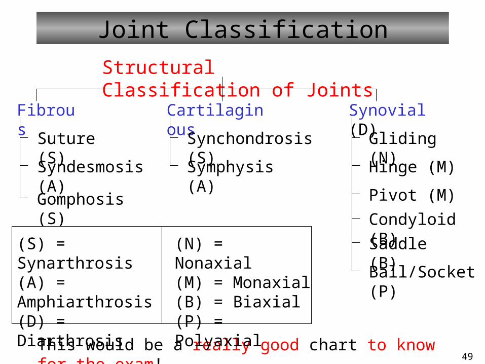
Joint Classification
Structural Classification of Joints
Fibrous Synovial (D)Cartilaginous
Suture (S)
Syndesmosis (A)
Gomphosis (S)
Synchondrosis (S)
Symphysis (A)
Gliding (N)
Hinge (M)
Pivot (M)
Condyloid (B)
Saddle (B)
Ball/Socket (P)
This would be a really good chart to know for the exam!
(S) = Synarthrosis(A) = Amphiarthrosis(D) = Diarthrosis
(N) = Nonaxial(M) = Monaxial(B) = Biaxial(P) = Polyaxial

50
Synovial Joint Movements Summary• Flexion – decrease in angle between bones
• Extension – increase in angle between bones
• ABduction – movement away from midline
• ADduction – movement toward midline
• Circumduction – Movement of the distal end of a limb in a circle
• Supination – palm facing anteriorly
• Pronation – palm facing posteriorly
• Protraction – anterior movement in transverse plane
• Retraction – posterior movement in transverse plane
• Dorsiflexion – Superior surface of foot moves superiorly
• Plantar flexion – Inferior surface of foot moves inferiorly

51
Know Actions of These MusclesMuscle Name Location Action
Masseter Cheek in front of ear Elevates mandible (raises lower jaw)Trapezius Upper shoulder Elevates clavicle;
Extends neck
Sternocleidomastoid Side of neck Rotates head; Flexes head toward shoulder
Deltoid Shoulder Abduction at shoulderBiceps brachii Front of upper arm Flexion at elbow and shoulderTriceps brachii Back of upper arm Extension at elbow
Abdominal muscles External oblique Internal oblique
Rectus abdominis
Front and side of abdomen Flex trunk (vertebral column); depress ribs (as in forced exhalation)
Pectoralis major Front of upper chest Flexion, adduction, and medial rotation of shoulderLatissimus dorsi Upper back Extension, adduction, and rotation of shoulderOrbicularis Oris Around mouth Compresses, purses lipsOrbicularis Oculi Around eye Closes eye
Temporalis Side of head (skull) Elevates mandibleGluteus maximus Buttocks Extension and lateral rotation at hipHamstring group Biceps femoris Semitendinosus
Semimembranosus
Back of thigh Lateral part of thigh Medial part of thigh Medial part of thigh
Flexes knee/extend thigh (all muscles in group)
Quadriceps group Rectus femoris Vastus lateralis Vastus medials
Vastus intermedius
Front of thigh Middle Lateral Medial Deep
Extends knee (all muscles in group)

52
Organization of Skeletal Muscle
• epimysium (around muscle)
• perimysium (around fascicles)
• endomysium (around fibers, or cells)
Alphabetical order largest to smallest: fascicle, fiber, fibril, and filament
Gross: Muscle (and fascia/epimysium), fascicle (and perimysium) Histological: Fiber (cell), endomysium Molecular: Myofibrils, sarcomere structure, actin/myosin arrangement

53
Skeletal Muscle Fiber (Cell)
Transverse tubules contain extracellular fluid ( [Na+], [K+])
Sarcoplasmic reticulum is like the ER of other cells; but it contains [Ca2+ ]
Fully differentiated, specialized cell – its structures are given special names
Figure from: Saladin, Anatomy & Physiology, McGraw Hill, 2007
• sarcolemma (plasma membrane)• sarcoplasm (cytoplasm)• sarcoplasmic reticulum (ER)
• transverse tubule• triad
• cisternae of sarcoplasmic reticulum (2)• transverse tubule
• myofibril (1-2 µm diam.)

54
Structure of the Sarcomere
‘A’ in A band stands for Anisotropic (dArk)
‘I’ in I band stands for Isotropic (LIght)
Zones of non-overlap: I band (thin filaments), and H zone (thick filaments)
A sarcomere runs from Z line (disk) to Z line (disk)
Figure from: Saladin, Anatomy & Physiology, McGraw Hill, 2007

55Latent period – time between motor nerve stimulation and contraction of skeletal muscle
Summary of Skeletal Muscle Contraction
Contraction Relaxation
- Bind (Ca, myosin) - Move- Detach- Reset

56
Modes of ATP Synthesis During Exercise
Continual shift from one energy source to another rather than an abrupt change
Muscle stores enough ATP for about 4-6 seconds worth of contraction, but is the only energy source used directly by muscle. So, how is energy provided for prolonged contraction?
Figures From: Marieb & Hoehn, Human Anatomy & Physiology, 9th ed., Pearson, 2013

57
Types of Contractions
• isotonic – muscle contracts and changes length
• concentric – shortening contraction
• isometric – muscle “contracts” but does not change length
• eccentric – lengthening contraction

5858
Motor Unit and Muscle Tone
• Motor unit - single motor neuron plus all muscle fibers controlled by that motor neuron
• recruitment - increase in the number of motor units activated to perform a task
• whole muscle composed of many motor units
• as intensity of stimulation increases, recruitment of motor units continues until all motor units are activated
• smaller motor units recruited first• larger motor units recruited later• produces smooth movements• muscle tone – continuous state of partial contraction

59
Table from: Martini & Ober, Visual A&P, 2011
*
*
*
*
*
*
*
*
**
*
*
*
*
*
*

60
Skeletal Muscle Actions
• origin – immovable* end• insertion – movable end
• agonist (prime mover ) – primarily responsible for movement• synergists – assist prime mover• antagonist – resist prime mover’s action and cause movement in the opposite direction
Understand these terms
Figure from: Saladin, Anatomy & Physiology, McGraw Hill, 2007