04 skeleton
62
Prof.Sunil Chavan Prin.K.M.Kundnani Pharmacy Polytechnic The Skeleton • The skeleton is the bony framework of the body. It forms the cavities and fossae that protect some structures, forms the joints and gives attachment to muscles. • The skeleton is described in two parts: axial and appendicular • The axial skeleton (axis of the body) consists of: • skull • vertebral column • sternum • ribs. • The appendicular skeleton (appendages attached to the axis of the body) consists of: bones of the upper limbs, the two clavicles and two scapulae, • the bones of the lower limbs and the two innominate bones of the pelvis.
-
Upload
prinkmkundnani-pharmacy-polytechnic -
Category
Education
-
view
144 -
download
0
Transcript of 04 skeleton

Prof.Sunil Chavan Prin.K.M.Kundnani Pharmacy Polytechnic
The Skeleton• The skeleton is the bony framework of the body.
It forms the cavities and fossae that protect some structures, forms the joints and gives attachment to muscles.
• The skeleton is described in two parts: axial and appendicular
• The axial skeleton (axis of the body) consists of: • skull • vertebral column • sternum • ribs.• The appendicular skeleton (appendages
attached to the axis of the body) consists of: bones of the upper limbs, the two clavicles and
two scapulae, • the bones of the lower limbs and the two innominate bones of the pelvis.

Prof.Sunil Chavan Prin.K.M.Kundnani Pharmacy Polytechnic
Terminology related to bones

Prof.Sunil Chavan Prin.K.M.Kundnani Pharmacy Polytechnic
A narrow slit
A hole in a structure
A hollow or depression
A tube-shaped cavity within a bone
A partition separating two cavities
Fissure or cleft
Foramen (plural: foramina)
Fossa (plural: fossae)
Meatus
Septum

Prof.Sunil Chavan Prin.K.M.Kundnani Pharmacy Polytechnic
AXIAL SKELETON
• This part consists of the skull, vertebral column, ribs and sternum.
• Together the bones forming these structures constitute the central bony core of the body, the axis.

Prof.Sunil Chavan Prin.K.M.Kundnani Pharmacy Polytechnic
Skull• The skull rests on the upper end of the vertebral
column• This bony structure is divided into two parts: the
cranium and the faceCranium:• The cranium is formed by a number of flat and
irregular bones• In the mature skull the joints (sutures) between
the bones are immovable (fibrous) • The bones have numerous perforations
(foramina, fissures) through which nerves, blood and lymph vessels pass.

Prof.Sunil Chavan Prin.K.M.Kundnani Pharmacy Polytechnic

Prof.Sunil Chavan Prin.K.M.Kundnani Pharmacy Polytechnic
The bones of the cranium are:
1 frontal bone 2 parietal bones 2 temporal bones 1 occipital bone 1 sphenoid bone 1 ethmoid bone.

Prof.Sunil Chavan Prin.K.M.Kundnani Pharmacy Polytechnic
Face• The skeleton of the face is formed by 13 bones
in addition to the frontal bone.• 2 zygomatic or cheek bones• 1 maxilla (originated as 2)
• 2 nasal bones• 2 lacrimal bones
• 1 vomer• 2 palatine bones
• 2 inferior conchae• 1 mandible (originated as 2)

Prof.Sunil Chavan Prin.K.M.Kundnani Pharmacy Polytechnic

Prof.Sunil Chavan Prin.K.M.Kundnani Pharmacy Polytechnic
Hyoid bone• This is an isolated horse-shoe-shaped
bone lying in the soft tissues of the neck just above the larynx and below the mandible.
• It does not articulate with any other bone but is attached to the styloid process of the temporal bone by ligaments.
• It gives attachment to the base of the tongue.

Prof.Sunil Chavan Prin.K.M.Kundnani Pharmacy Polytechnic

Prof.Sunil Chavan Prin.K.M.Kundnani Pharmacy Polytechnic
Sinuses• Sinuses containing air are present in the
sphenoid, ethmoid, maxillary and frontal bones. They all communicate with the nasal cavity and are lined with ciliated mucous membrane.
• Their functions are: - to give resonance to the voice - to lighten the bones of the face and
cranium, making it easier for the head to balance on top of the vertebral column.

Prof.Sunil Chavan Prin.K.M.Kundnani Pharmacy Polytechnic
Vertebral column• The vertebral column consists of 24
separate movable, irregular bones, the sacrum (five fused bones) and the coccyx (four fused bones).
• The 24 separate bones are in three groups: 7 cervical, 12 thoracic and 5 lumbar.

Prof.Sunil Chavan Prin.K.M.Kundnani Pharmacy Polytechnic
Characteristics of a typical vertebra

Prof.Sunil Chavan Prin.K.M.Kundnani Pharmacy Polytechnic
• The body: The body of each vertebra is situated anteriorly.
• The size of body varies with the site. They are smallest in the cervical region and become larger towards the lumbar region.
• The vertebral (neural) arch: encloses a large vertebral foramen. The ring of bone consists of two pedicles that project backwards from the body and two laminae

Prof.Sunil Chavan Prin.K.M.Kundnani Pharmacy Polytechnic
• Where the pedicles and laminae unite, transverse processes project laterally and where the two laminae meet in the midline posteriorly they form a spinous process.
• The neural arch has four articular surfaces: two articulate with the vertebra above and two with the one below.
• The vertebral foramina form the vertebral (neural) canal that contains the spinal cord.

Prof.Sunil Chavan Prin.K.M.Kundnani Pharmacy Polytechnic
Special features of vertebrae in different parts of the vertebral column• Cervical vertebrae

Prof.Sunil Chavan Prin.K.M.Kundnani Pharmacy Polytechnic
• The transverse processes have a foramen through which a vertebral artery passes upwards to the brain.
• The first two cervical vertebrae are atypical.• The atlas is the 1st cervical
vertebra and it consists simply of a ring of bone with two short transverse processes.
• The posterior part is the true vertebral foramen and is occupied by the spinal cord.
• On its superior surface the bone has two articular facets which form joints with the condyles of the occipital bone of the skull.
• The nodding movement of the head takes place at these joints.

Prof.Sunil Chavan Prin.K.M.Kundnani Pharmacy Polytechnic

Prof.Sunil Chavan Prin.K.M.Kundnani Pharmacy Polytechnic
• The axis is the 2nd cervical vertebra. The body is small and has the upward projecting odontoid process or dens that articulates with the first cervical vertebra, the atlas.
• The movement at this joint is turning the head from side to side.

Prof.Sunil Chavan Prin.K.M.Kundnani Pharmacy Polytechnic

Prof.Sunil Chavan Prin.K.M.Kundnani Pharmacy Polytechnic
Thoracic vertebrae• The bodies and
transverse processes have facets for articulation with the ribs.

Prof.Sunil Chavan Prin.K.M.Kundnani Pharmacy Polytechnic
Lumbar vertebrae• These have no special features.

Prof.Sunil Chavan Prin.K.M.Kundnani Pharmacy Polytechnic
Sacrum• Consists of five vertebrae fused to form a
triangular or wedge-shaped bone with a concave anterior surface.
• The upper part, or base, articulates with the 5th lumbar vertebra. On each side it articulates with the ilium to form a sacroiliac joint, and at its inferior tip it articulates with the coccyx.
• The anterior edge of the base, the promontory, protrudes into the pelvic cavity.
• The vertebral foramina are present, and on each side of the bone there is a series of foramina for the passage of nerves.

Prof.Sunil Chavan Prin.K.M.Kundnani Pharmacy Polytechnic

Prof.Sunil Chavan Prin.K.M.Kundnani Pharmacy Polytechnic
Coccyx• This consists of the four terminal
vertebrae fused to form a very small triangular bone, the broad base of which articulates with the tip of the sacrum.

Prof.Sunil Chavan Prin.K.M.Kundnani Pharmacy Polytechnic
Functions of the vertebral column• Collectively the vertebral foramina form the vertebral
canal which provides a strong bony protection for the spinal cord.
• The pedicles of adjacent vertebrae form intervertebral foramina, one on each side, providing access to the spinal cord for spinal nerves, blood vessels and lymph vessels.
• The numerous individual bones enable a certain amount of movement.
• It supports the skull.• The intervertebral discs act as shock absorbers,
protecting the brain.• It forms the axis of the trunk, giving attachment to the
ribs, shoulder girdle and upper limbs, and the pelvic girdle and lower limbs.

Prof.Sunil Chavan Prin.K.M.Kundnani Pharmacy Polytechnic
Thoracic cageThe bones of the
thorax or thoracic cage are:
• 1 sternum• 12 pairs of ribs• 12 thoracic vertebrae

Prof.Sunil Chavan Prin.K.M.Kundnani Pharmacy Polytechnic
Sternum or breast bone• This flat bone can be felt just under the skin in
the middle of the front of the chest.• The manubrium is the uppermost section and
articulates with the clavicles at the sternodavicular joints and with the first two pairs of ribs.
• The body or middle portion gives attachment to the ribs.
• The xiphoid process is the tip of the bone. It gives attachment to the diaphragm, muscles of the anterior abdominal wall and the linea alba.

Prof.Sunil Chavan Prin.K.M.Kundnani Pharmacy Polytechnic

Prof.Sunil Chavan Prin.K.M.Kundnani Pharmacy Polytechnic
Ribs• 2 pairs of ribs which form the bony lateral
walls of the thoracic cage and articulate posteriorly with the thoracic vertebrae.
• The first 10 pairs are attached anteriorly to the sternum by costal cartilages, some directly and some indirectly.
• The last two pairs (floating ribs] have no anterior attachment.

Prof.Sunil Chavan Prin.K.M.Kundnani Pharmacy Polytechnic
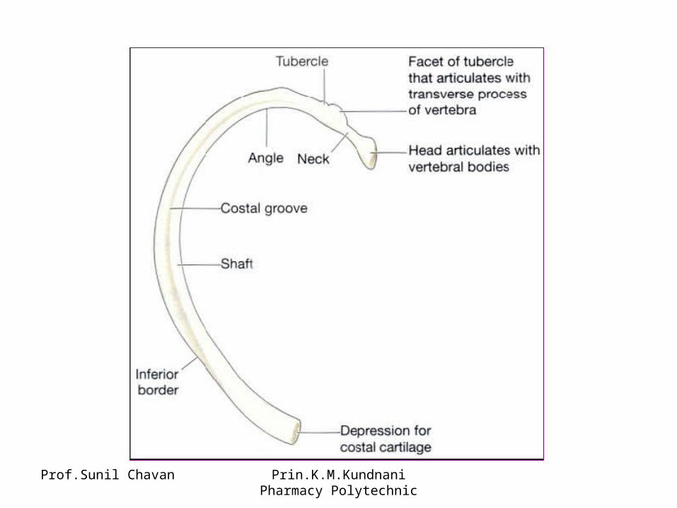
Characteristics of a rib• The head articulates posteriorly with the bodies of two
adjacent thoracic vertebrae and on the tubercle there is a facet that articulates with the transverse process of one.
• The sternal end is attached to the sternum by a costal cartilage, i.e. a band of hyaline cartilage.
• The superior border is rounded and smooth while the inferior border has a marked groove occupied by the intercostal blood vessels and nerves.
• The first rib does not move during respiration.• The spaces between the ribs are occupied by the
intercostal muscles. During inspiration, when these muscles contract, the ribs and sternum are lifted upwards and outwards, increasing the capacity of the thoracic cavity.
Prof.Sunil Chavan Prin.K.M.Kundnani Pharmacy Polytechnic
Prof.Sunil Chavan Prin.K.M.Kundnani Pharmacy Polytechnic
APPENDICULAR SKELETONThe appendicular skeleton consists of: • the shoulder girdle with the upper limbs
and• the pelvic girdle with the lower limbs

Prof.Sunil Chavan Prin.K.M.Kundnani Pharmacy Polytechnic
Shoulder girdle and upper limb• Each shoulder girdle consists of: • 1 clavicle • 1 scapula.• Each upper limb consists of the following bones: 1 humerus 1 radius 1 ulna 8 carpal bones 5 metacarpal bones 14 phalanges.

Prof.Sunil Chavan Prin.K.M.Kundnani Pharmacy Polytechnic
Clavicle or collar bone• The clavicle is a long bone which has a
double curve. It articulates with the manubrium of the sternum at the sternodavicular joint and forms the acromiodavicular joint with the acromion process of the scapula.
• The clavicle provides the only bony link between the upper limb and the axial skeleton.

Prof.Sunil Chavan Prin.K.M.Kundnani Pharmacy Polytechnic
Clavicle
Prof.Sunil Chavan Prin.K.M.Kundnani Pharmacy Polytechnic
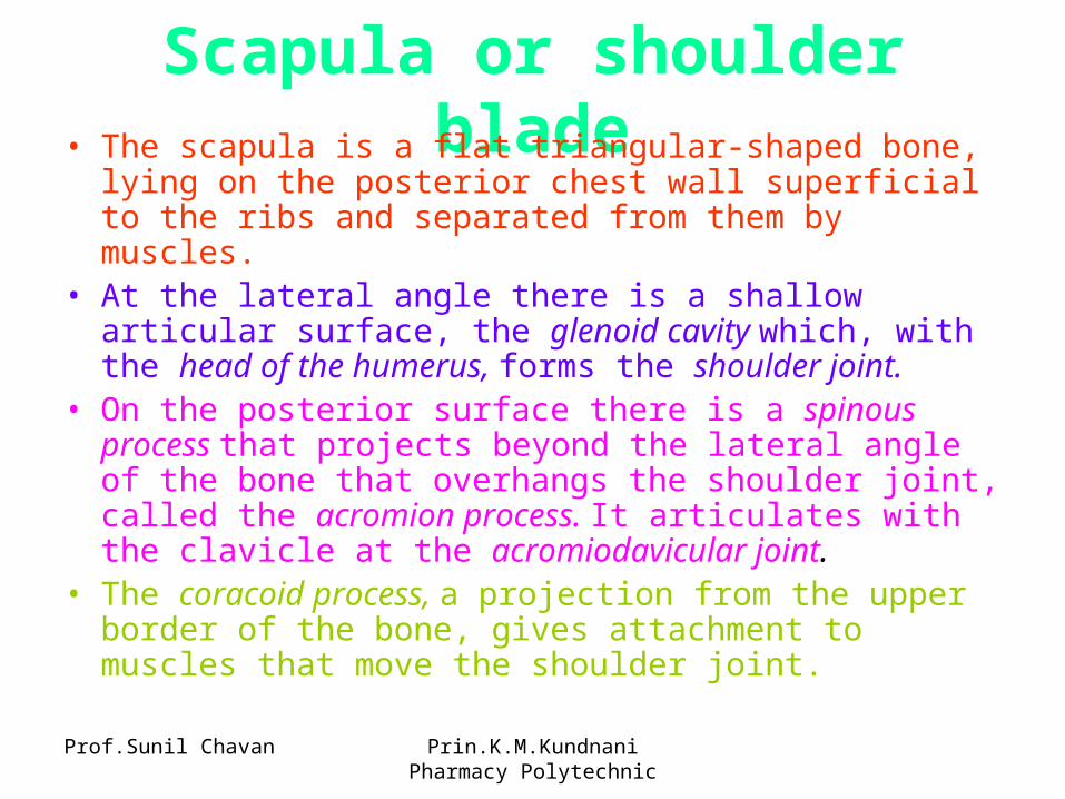
Scapula or shoulder blade• The scapula is a flat triangular-shaped bone, lying on the
posterior chest wall superficial to the ribs and separated from them by muscles.
• At the lateral angle there is a shallow articular surface, the glenoid cavity which, with the head of the humerus, forms the shoulder joint.
• On the posterior surface there is a spinous process that projects beyond the lateral angle of the bone that overhangs the shoulder joint, called the acromion process. It articulates with the clavicle at the acromiodavicular joint.
• The coracoid process, a projection from the upper border of the bone, gives attachment to muscles that move the shoulder joint.

Prof.Sunil Chavan Prin.K.M.Kundnani Pharmacy Polytechnic

Prof.Sunil Chavan Prin.K.M.Kundnani Pharmacy Polytechnic
Humerus• Bone of the upper arm. The head articulates with
the glenoid cavity of the scapula, forming the shoulder joint.
• Distal to the head there are two roughened projections of bone, the greater and lesser tubercles, and between them there is a deep groove, the bicipital groove or intertubercular sulcus, occupied by one of the tendons of the biceps muscle.
• The distal end of the bone presents two surfaces that articulate with the radius and ulna to form the elbow joint.

Prof.Sunil Chavan Prin.K.M.Kundnani Pharmacy Polytechnic

Prof.Sunil Chavan Prin.K.M.Kundnani Pharmacy Polytechnic
Ulna and radius• These are the two bones of the forearm. • The ulna is longer than and medial to the
radius. • They articulate with the humerus at the
elbow joint, the carpal bones at the wrist joint and with each other at the proximal and distal radioulnar joints.

Prof.Sunil Chavan Prin.K.M.Kundnani Pharmacy Polytechnic

Prof.Sunil Chavan Prin.K.M.Kundnani Pharmacy Polytechnic
Carpal or wrist bonesEight carpal bones arranged in two rows of four.
From outside inwards they are:• proximal row: scaphoid, lunate, triquetral,
pisiform• distal row: trapezium, trapezoid, capitate,
hamate.• These bones are closely fitted together and held
in position by ligaments which allow a certain amount of movement between them.
• The bones of the proximal row are associated with the wrist joint and those of the distal row form joints with the metacarpal bones.

Prof.Sunil Chavan Prin.K.M.Kundnani Pharmacy Polytechnic

Prof.Sunil Chavan Prin.K.M.Kundnani Pharmacy Polytechnic
Metacarpal bones or the bones of the hand• Five bones form the palm of the hand. They are
numbered from the thumb side inwards. The proximal ends articulate with the carpal bones and the distal ends with the phalanges.
Phalanges or finger bones• There are 14 phalanges, three in each finger
and two in the thumb• They articulate with the metacarpal bones and
with each other.

Prof.Sunil Chavan Prin.K.M.Kundnani Pharmacy Polytechnic
Pelvic girdle and lower limb• The bones of the pelvic girdle are: • 2 innominate bones • 1 sacrum.• The bones of the lower limb are: • 1 femur • 7 tarsal bones • 1 tibia • 5 metatarsal bones • 1 fibula • 14 phalanges. • 1 patella

Prof.Sunil Chavan Prin.K.M.Kundnani Pharmacy Polytechnic
Innominate or hip bones

Prof.Sunil Chavan Prin.K.M.Kundnani Pharmacy Polytechnic
• Each hip bone consists of three fused bones, the ilium, ischium and pubis. On its outer surface there is a deep depression, the acetabulum, which forms the hip joint with the almost-spherical head of femur.
• The ilium is the upper flattened part of the bone and it presents the iliac crest, the anterior point of which is called the anterior superior iliac spine.
• The pubis is the anterior part of the bone and it articulates with the pubis of the other hip bone at a cartilaginous joint, the symphysis pubis.
• The ischium is the inferior and posterior part.• The union of the three parts takes place in the
acetabulum.

Prof.Sunil Chavan Prin.K.M.Kundnani Pharmacy Polytechnic
The pelvisThe pelvis is formed by • two innominate bones which articulate
anteriorly at the symphysis pubis and • posteriorly with the sacrum at the
sacroiliac joints which are synovial joints.

Prof.Sunil Chavan Prin.K.M.Kundnani Pharmacy Polytechnic

Prof.Sunil Chavan Prin.K.M.Kundnani Pharmacy Polytechnic
Differences between male and female pelves :The shape of the female pelvis allows for the passage of the baby during childbirth. In comparison with the male pelvis, the female pelvis has lighter bones, is more shallow and rounded and is generally more roomy.

Prof.Sunil Chavan Prin.K.M.Kundnani Pharmacy Polytechnic
Femur or thigh bone• The longest and strongest bone of the body.• The head is almost spherical and fits into the
acetabulum of the hip bone to form the hip joint.• The neck extends outwards and slightly
downwards from the head to the shaft and most of it is within the capsule of the hip joint.
• The posterior surface of the lower third forms a flat triangular area called the popliteal surface. The distal extremity has two articular condyles which, with the tibia and patella, form the knee joint.

Prof.Sunil Chavan Prin.K.M.Kundnani Pharmacy Polytechnic

Prof.Sunil Chavan Prin.K.M.Kundnani Pharmacy Polytechnic
Tibia or shin bone• The tibia is the medial of the two bones of the
lower leg.• The proximal extremity is broad and flat and
presents two condoles for articulation with the femur at the knee joint.
• The head of the fibula articulates with the inferior aspect of the lateral condyle, forming the proximal tibiofibular joint.
• The distal extremity of the tibia forms the ankle joint. with the talus and the fibula. The medial malleolus is a downward projection of bone medial to the ankle joint.

Prof.Sunil Chavan Prin.K.M.Kundnani Pharmacy Polytechnic

Prof.Sunil Chavan Prin.K.M.Kundnani Pharmacy Polytechnic
Fibula• The fibula is the long slender lateral bone
in the leg. • The head or upper extremity articulates
with the lateral condyle of the tibia forming the proximal tibiofibularjoint
• The lower extremity articulates with the tibia then projects beyond it to form the lateral malleolus.

Prof.Sunil Chavan Prin.K.M.Kundnani Pharmacy Polytechnic
Patella or knee cap• This is a roughly triangular-shaped
sesamoid bone associated with the knee joint.
• Its posterior surface articulates with the patellar surface of the femur in the knee joint
• Its anterior surface is in the patellar tendon, i.e. the tendon of the quadriceps femoris muscle.

Prof.Sunil Chavan Prin.K.M.Kundnani Pharmacy Polytechnic
Tarsal or ankle bones• Seven tarsal bones which form the
posterior part of the foot. They are: 1 talus, 3 cuneiform,1 calcaneus, 1 cuboid, 1 navicular• The talus articulates with the tibia and
fibula at the ankle joint. • The calcaneus forms the heel of the foot. • The other bones articulate with each other
and with the metatarsal bones.

Prof.Sunil Chavan Prin.K.M.Kundnani Pharmacy Polytechnic

Prof.Sunil Chavan Prin.K.M.Kundnani Pharmacy Polytechnic
Metatarsal bones of the foot• These are five bones, numbered from
within outwards, which form the greater part of the dorsum of the foot.
• At their proximal ends they articulate with the tarsal bones.
• At their distal ends articulate with the phalanges.
• The enlarged distal head of the 1st metatarsal bone forms the 'ball' of the foot.

Prof.Sunil Chavan Prin.K.M.Kundnani Pharmacy Polytechnic
Phalanges of the toes• There are 14 phalanges arranged in a
similar manner to those in the fingers, i.e. two in the great toe and three in each of the other toes.