intranet.tdmu.edu.uaintranet.tdmu.edu.ua/data/kafedra/internal/anatomy/zbtest... · Web viewVessels...
207
Vessels and nerves of trunk and extremities Text tests questions 1. Atrioventricular node is supplied by: A. * Right coronary artery. B. Left coronary artery. C. Anterior coronary artery. D. Posterior coronary artery. E. Circumflex artery. 2. Boundary of what structures is coronary groove? A. * Between atriums and ventricles. B. Between atriums and heard apex. C. Between atriums and base of the heart. D. Between atriums and lateral surface of the heart. E. Between atriums and diaphragmatic surface of the heart. 3. Boundary of what structures on the heart surface is anterior and posterior interventricular grooves? A. * Right and left ventricles. B. Right and left atrium. C. Right atrium and right ventricle. D. Right atrium and left ventricle. E. Left atrium and right ventricle. 4. Bronchial veins of the right side open into: A. * Azygos vein. B. Superior vena cava. C. Hemiazygos vein. D. Brachiocephalic vein E. None of the above. 5. Conduction velocity is maximum in: A. SA node. B. AV node. C. * Bundle of His. D. Right ventricle. E. Left ventriclE. 6. Continuation of what structure is vena azygos? A. Posterior intercostalis. B. Left ascendens lumbalis. C. Inferior diaphragmatic. D. Superior diaphragmatic. E. * Right ascendens lumbalis. 7. Continuation of what structure is vena hemiaxygos? A. Left ascendens lumbalis. B. Right ascendens lumbalis. C. Superior diaphragmatic.
Transcript of intranet.tdmu.edu.uaintranet.tdmu.edu.ua/data/kafedra/internal/anatomy/zbtest... · Web viewVessels...
Vessels and nerves of trunk and extremities
Text tests questions
1. Atrioventricular node is supplied by: A. * Right coronary artery. B. Left coronary artery. C. Anterior coronary artery. D. Posterior coronary artery. E. Circumflex artery.
2. Boundary of what structures is coronary groove? A. * Between atriums and ventricles. B. Between atriums and heard apex. C. Between atriums and base of the heart. D. Between atriums and lateral surface of the heart. E. Between atriums and diaphragmatic surface of the heart.
3. Boundary of what structures on the heart surface is anterior and posterior interventricular grooves?
A. * Right and left ventricles. B. Right and left atrium. C. Right atrium and right ventricle. D. Right atrium and left ventricle. E. Left atrium and right ventricle.
4. Bronchial veins of the right side open into: A. * Azygos vein. B. Superior vena cava. C. Hemiazygos vein. D. Brachiocephalic vein E. None of the above.
5. Conduction velocity is maximum in: A. SA node. B. AV node. C. * Bundle of His. D. Right ventricle. E. Left ventriclE.
6. Continuation of what structure is vena azygos? A. Posterior intercostalis. B. Left ascendens lumbalis. C. Inferior diaphragmatic. D. Superior diaphragmatic. E. * Right ascendens lumbalis.
7. Continuation of what structure is vena hemiaxygos? A. Left ascendens lumbalis. B. Right ascendens lumbalis. C. Superior diaphragmatic. D. Inferior diaphragmatic. E. * Posterior intercostalis.
8. Coronary sinus opens into: A. * Right atrium. B. Inferior vena cava. C. Left atrium. D. Great cardiac vein. E. Lesser cardiac vein.
9. First heart sound is usually clearly heard on ventral surface of chest at A. 1st intercostals space to right of sternum. B. 2nd intercostals space to right of sternum. C. 2nd intercostals space toleft of sternum. D. * 5th intercostals space to left of sternum. E. 5th intercostals space to right of sternum.
10.From what structure fossa ovalis created? A. Venose sinus opening. B. Vena cava superior opening. C. Vena cava inferior opening. D. * Remnant of foramen ovale. E. Vena cava inferior and venose sinus openins.
11.From what structures tendinous cord starts? A. Interseptal papillar muscles. B. Frontal papillar muscles. C. Posterior papillar muscles. D. * From papillar muscles. E. Frontal and posterior papillar muscles.
12.How many aortic sinuses are? A. Four. B. Two. C. * Three. D. Two or three. E. FivE.
13.How many chambers heart have? A. * Four. B. Three. C. Five. D. Six. E. Two.
14.How many cuspes have left atrioventricular valve? A. Three or two. B. Three. C. Four. D. * Two. E. Five.
15.How many cusps have right atrioventricular valve? A. Two. B. * Three. C. Four. D. Two or three. E. Three or four.
16.How many openings have left atrium? A. Four. B. Two. C. Three. D. * Five. E. Two or three.
17.How many openings have left atrium? A. Four. B. Two. C. Three. D. * No right answer. E. Two or three.

18. In what part of mediastinum heart can be find ? A. Left. B. Right. C. Superior. D. * Middle-inferior E. Inferior.
19. Indicate a groove between the ventricles on the diaphragmatic surface of the heart.Mentioned above structure contains the middle cardiac vein.
A. Sulcus terminalis. B. * Posterior interventricular sulcus. C. Anterior interventricular sulcus. D. Oblique fissure. E. Horizontal fissure.
20. Indicate a ridgle of cardiac muscle separating the smooth sinus venarum posteriorly from the roughened wall of the primitive atrium anteriorly. The sinuatrial node lies within the superior end of the mentioned above structure.
A. Chordae tendineae. B. Cupula indicate. C. * Crista terminalis. D. Fossa ovaalis. E. Coronary sulcus.
21. Indicate a shallow depression in the left wall of the right atrium.Mentioned above structure is the remnant of some foramen which provided an open communication between the right atrium and left atrium in the fetus.
A. Coronary sulcus. B. * Fossa ovalis. C. Crista terminalis. D. Oblique fissure. E. Carina.
22. Indicate a small, nipple-like projection of cardiac muscle located within the ventricles.Mentioned structure attach to the cusps of the atrioventricular valves via chordaae tendinea and act to keep the valve cusps from prolapsing under systolic blood pressure.
A. Carina. B. Crista terminalis. C. * Pappilary muscles. D. Trabeculae carnae. E. Pectinate muscles.
23. Indicate part of the conduction system of the heart.This structure is located in the wall of the right atrium above the opening of the coronary sinus and the septal cusp of the tricuspid valve.
A. Fossa ovalis. B. Sinuatrial node. C. Chorda tendinae. D. Atrioventricular bundle. E. * Atrioventricular node.
24. Indicate small appendage that projects anteriorly from the atrium paired ,one on each atrium.
A. Cupula. B. Crista terminalis. C. * Auricle. D. Ligamentum arteriosum. E. Semilunal valves.
25. Indicate the superior aspect of heart.Mentioned above structure is where the aorta,pulmonary trunk and superior vena cava exit/enter the heart.
A. Apex. B. * Base. C. Fossa ovalis. D. Chorda tendinea. E. Crista terminalis.
26. Indicate thin connective tissue cords that attch the atrioventricular valve cusps to the papillary muscles.Mentioned above structures are found only in the ventricles,not in the atriums.
A. * Chorda tendinea. B. Crista terminalis. C. Cupula. D. Oblique fissure. E. Horizontal fissure.
27.Left and right parts of the heart connect together ? A. Partly. B. Yes. C. Yes and no. D. * No. E. Periodically.
28.Left coronary artery supplies all exept: A. SA node. B. Apex of heart. C. Anterior 1/3 of septum. D. Left ventriclE. E. * Posterior 1/3 of septum.
29.Name a fibrous cord of connective tissue that connects the left pulmonary artery near its origin with the undersurface of the aortic arch.Mentioned above structure is a remnant of the ductus arteriosus,the left recurrent laryngeal nerve passes beneath it.
A. Chorda tendinea. B. * Ligamentum arteriosum. C. Crista terminalis. D. Cupula. E. Horizontal fissure.
30.Name artery that supplies blood arterial part of the heart,2/3 of the interventricular septum.
A. * Left coronary artery. B. Right coronary artery. C. Interventricular artery. D. Circumflex artery. E. Posterior interventricular artery.
31.Name concavity or notch along anterior border of left lung that separate lingual below from upper portion of superior lobe of left lung.
A. Coronary sulcus. B. Pulmonary notch. C. Fossa ovalis. D. Cupula. E. * Cardiac notch.
32.Name groove between the ventricles on the front surface of the heart.Mentioned above structure contains the great cardiac vein.
A. Sulcus terminalis. B. Oblique fissure.

C. Horizontal fissurE. D. * Anterior interventricular sulcus. E. Posterior interventricular sulcus.
33.Name heart valve located between the left atrium and the left ventriclE. A. Aortic valve. B. * Mitral valve. C. Semilunar valve. D. Right atrioventricular valvE. E. Trabeculae carnae.
34.Name prominent ridges of myocardium located on the inner surface of the right atrium.Mentioned structures are very pronounced in the right atrium and in both auricles.
A. Crista terminalis. B. Carina. C. Trabeculae carnae. D. * Pectinate muscles. E. Pappilary muscles.
35.Name the chamber of the heart that receives deoxygenated blood from the systemic circulation (body).Mentioned above structure forms the right margin of the heart,it receives blood from the superior vena cava and coronary sinus.
A. Left atrium. B. * Right atrium. C. Auricle. D. Right ventricle. E. Left ventriclE.
36.Name the chamber of the heart that receives oxygenated blood from the pulmonic circulation (lungs) .Mentioned above structure is located on the posterior aspect of the heart,it receives blood from the pulmonary veins.
A. * Left atrium. B. Left ventricle. C. Right ventricle. D. Right atrium. E. Auricle.
37.Name the groove on the surface of the heart that separates atria from the ventricles.
A. Crista terminalis B. Horizontal fissure. C. * Coronary sulcus. D. Oblique fissure. E. Chorda tendinea
38.Name tip of the left ventricle of the heart.Mentioned structure of the heart is located 3" to the left of midline at the level of the 5th intercostals space.
A. Base. B. Ovalis. C. * Apex. D. Atrioventricular bundle. E. Atrioventricular node.
39.On the level of what cartilage vena cava superior fall into right atrium? A. Third left cartilage. B. Second right cartilage. C. First right cartilage. D. * Third right cartilage. E. Second left cartilage.
40.On what branches left coronary artery divide?

A. Posterior circumflex. B. Anterior interventricular. C. Posterior interventricular. D. Anterior circumflex. E. * Anterior interventricular and circumflex .
41.On what level thoracic part of aorta locate? A. 6-th thoracic vertebra. B. 5-th thoracic vertebra. C. * 4-th thoracic vertebra. D. 7-th cervical vertebra. E. 6-th cervicalvertebra.
42.Posterior interventricular artery is a branch the artery: A. Circumflex. B. Left coronary. C. * Right coronary. D. Anterior interventricular. E. None of the above.
43.The first branch from the aortic arch is the: A. Left common carotid artery. B. Right common carotid artery. C. Left subclavian artery. D. * Brachiocephalic trunk. E. Right subclavian artery.
44.The junction of what structures brachiocephalic veins are create? A. Internal jugular and groin. B. Internal and external jugular. C. Internal jugular and anterior jugular. D. * Internal jugular and subclavial. E. Subclavial and external jugular.
45.The junction of what structures vena cava superior is create? A. * Right and left brachiocephalic. B. Right brachiocephalic and internal jugular. C. Left brachiocephalic and internal jugular. D. Left brachiocephalic and subclavial. E. Internal jugular and subclavial.
46.The left coronary artery: A. Has a branch that commonly anastomoses with a branch of the right
coronary in the coronary sulcus. B. Has a branch that commonly anastomoses with the branch of the right
coronary in the interventricular sulcus. C. Is short in that soon after its origin it bifurcates into the anterior
interventricular and circumflex arteries. D. Passes anterior to the pulmonary trunk. E. * All correct answers.
47.The second branch from the aortic arch is the: A. Brachiocephalic trunk. B. * Left common carotid artery. C. Right common carotid artery. D. Left subclavian artery. E. Right subclavian artery.
48.The third branch of the aortic arch is the: A. * Left subclavian artery. B. Left common carotid artery. C. Right common carotid artery.

D. Brachiocephalic trunk. E. Right subclavian artery.
49.What chambers of the heart do you know? A. One ventricle and one atrium. B. One atrium and one ventricle. C. One atrium and two ventricles. D. * Two atriums and two ventricles. E. One ventricle and two atriums.
50.What closing opening to the thoracic trunk? A. Aortic valvE. B. * Pulmonary trunk valve. C. Left atrioventricular valve. D. Right atrioventricular valve. E. Conus arteriosus.
51.What connect together left and right inter ventricular grooves? A. * Cutting of heart apex. B. Heart apex. C. Base of the heart. D. Lateral surface. E. Paramedical surface.
52.What forms duplicate of endocardium? A. Flaps of coronary sinus. B. Valves. C. Flaps of vena cava superior. D. Flaps of vena cava inferior. E. * Valves,flaps of vena cava and coronary sinus.
53.What groove passing on the lower surface of the heart? A. Frontal interventricular. B. * Back interventricular. C. Lateral interventricular. D. Frontal interauricular. E. Middle interventricular.
54.What is approximate weight of the heart? A. 200-300. B. 200-250. C. * 250-300. D. 300-350. E. 350-400.
55.What is epicardium? A. Parietal and visceral plates of heart covering. B. Parietal plate of pericardium. C. * Visceral plate of pericardium. D. Fibrous plate of pericardium. E. Fibrous and serous part of pericardium.
56.What is the endocardium made of? A. Smooth musculature. B. Serous membrane. C. Mucous membrane. D. * Epithelial membrane. E. Skeletal musculature.
57.What is the epicardium made of? A. * Serous membrane. B. Smooth musculature. C. Striated musculature.

D. Epithelial membrane. E. Mucous membrane.
58.What is the function of parasymphatic innervation? A. Accelerate heart rhythm. B. Decelerate heart rhythm. C. Constrict coronary vessels. D. * Decelerate heart rhythm constrict coronary vessels. E. Dilate coronary vessels.
59.What is the function of symphatic innervations? A. * Accelerate heart rhythm,dilate coronary vessels. B. Accelerate heart rhythm. C. Dilate coronary vessels. D. Constrict coronary vessels. E. Decelerate heart rhythm.
60.What is the myocardium made of? A. Serous membrane. B. Smooth musculature. C. * Striated musculature. D. Epithelial membrane. E. Mucous membrane.
61.What is the name of endocardium fold beside coronary opening? A. Two. B. * Flip of coronary sinus C. Four. D. Two or three. E. Three or four.
62.What is the name of frontal surface of the heart? A. * Thoracicocostal. B. Thoracic. C. Pulmonary. D. Costal. E. Back.
63.What is the name of lower bigger part of interventricular septum? A. Frontal mammary muscles. B. * Muscular. C. Back mammary muscles. D. Septal mammary muscles. E. Frontal and back mammary muscles.
64.What is the name of lower part of the heart? A. Basck. B. Thoracic. C. Pulmonary. D. Costal. E. * Diaphragmatic.
65.What is the name of smaller upper part of interventricular septum? A. * Membranous. B. Aortic valve. C. Left atrioventricular valve. D. Right atrioventricular valve. E. Conus arteriosus.
66.What is the name of transversal groove on the heart surface? A. Lateral. B. Frontal. C. Back.

D. * Coronary. E. Posterior.
67.What layer does pericardium contain? A. * Serous membrane. B. Smooth musculature. C. Striated musculature. D. Epithelial membrane. E. Mucous membrane.
68.What pass along lower border of opening of vena cava inferior? A. On the external suface of the auricle. B. * Flap of the vena cava inferior. C. On the external surface ofleft auricle. D. On internal surface of left auricle. E. Beside sinus of vena cava.
69.What structure covers endocardium? A. Cavity of the left ventricle. B. Cavity of the right ventricle. C. Cavity of the right atrium. D. * Internal cavity of the heart. E. Cavity of the left atrium.
70.What structure covers epicardium? A. * Heart from outside,beginning parts of aorta and pulmonary trunk, terminal
parts of vena cava and pulmonary veins. B. AortA. C. Pulmonary trunk. D. Vena cava superior and inferior. E. Pulmonary veins.
71.What structure innervate heart? A. Anterior truncus of n.vagus. B. Nervus vagus. C. Truncus simpaticus. D. Posterior truncus of n.vagus. E. * Nervus vagus and truncus simpaticus.
72.What structure locate on interauricular septum? A. Oval chanal. B. * Oval fossA. C. Oval opening. D. Right auricle. E. Left auricle.
73.What structure separate atriums between one and another? A. Left auricle. B. Interventricular septum. C. * Interatrial septum. D. Left and right auricles. E. Right auricle.
74.What structure separate myocardium of atriums from myocardium of ventricles? A. * Fibrous ring. B. Right fibrous trigonous. C. Left fibrous trigonous. D. Septal part. E. Fibrous tisues.
75.What structures supply superior diaphragmatic arteries? A. Transversal part of diaphragm. B. Pleura.

C. Thoracic part of diaphragm. D. Costal part of diaphragm. E. * Lumbal part of diaphragm.
76.What vessel do not start from thoracic aorta? A. Posterior intercostals arteries. B. * Anterior intercostals arteries. C. Superior phrenic arteries. D. Mediastinal arteries. E. Bronchial arteries.
77.What vessel do not start from thoracic aorta? A. Posterior intercostals arteries. B. * Pulmonary arteries. C. Superior phrenic arteries. D. Mediastinal arteries. E. Bronchial arteries.
78.What vessel do not start from thoracic aorta? A. Posterior intercostals arteries. B. * Internal thoracic arteries. C. Superior phrenic arteries. D. Mediastinal arteries. E. Bronchial arteries.
79.What vessel do not start from thoracic aorta? A. Posterior intercostals arteries. B. * Brachiocephalic artery. C. Superior phrenic arteries. D. Mediastinal arteries. E. Bronchial arteries.
80.What vessel do not start from thoracic aorta? A. Posterior intercostals arteries. B. * Coronary arteries. C. Superior phrenic arteries. D. Mediastinal arteries. E. Bronchial arteries.
81.What vessel spring from thoracic aorta? A. * Oesophageal artery. B. Brachiocephalic trunk. C. Left common carotid artery. D. Right common carotid artery. E. Left subclavian artery.
82.What vessel spring from thoracic aorta? A. * Pericardial arteries. B. Brachiocephalic trunk. C. Left common carotid artery. D. Right common carotid artery. E. Left subclavian artery.
83.What vessel spring from thoracic aorta? A. * Bronchial arteries. B. Brachiocephalic trunk. C. Left common carotid artery. D. Right common carotid artery. E. Left subclavian artery.
84.What vessel spring from thoracic aorta? A. * Mediastinal arteries. B. Brachiocephalic trunk.

C. Left common carotid artery. D. Right common carotid artery. E. Left subclavian artery.
85.What vessel spring from thoracic aorta? A. * Posterior intercostals arteries. B. Brachiocephalic trunk. C. Left common carotid artery. D. Right common carotid artery. E. Left subclavian artery.
86.What vessel spring from thoracic aorta? A. * Superior phrenic arteries. B. Brachiocephalic trunk. C. Left common carotid artery. D. Right common carotid artery. E. Left subclavian artery.
87.What vessels does not start from thoracic aorta? A. * Inferior phrenic arteries. B. Posterior intercostals arteries. C. Superior phrenic arteries. D. Mediastinal arteries. E. Bronchial arteries.
88.Whatb is the name of lateral surface of the heart? A. Thoracic. B. * Pulmonary. C. Diaphragmatic. D. Costal. E. Posterior.
89.Where final branches of conductive system bundle of His finished? A. In myocardium of atriums. B. * In myocardium of ventricles. C. In myocardium of right ventricle. D. In myocardium of left ventricle E. In muscle part of interventricular septum.
90.Where in the left atrium comb-shaped muscles located? A. In the auricle of right atrium. B. In the auricles of right and left atriums. C. * In the auricle of left atrium. D. Beside openings of pulmonary veins. E. Beside valve of oval opening.
91.Where left coronary artery located? A. Right auricle. B. Between beginning of pulmonary trunk. C. Left auricle. D. * Between beginning of pulmonary trunk and left auricle. E. Between left and right auricles.
92.Where longitudinal arterial ring of the heart located? A. On the cutting of heart apex. B. In the coronary groove. C. In the back interventricular groove. D. In the front interventriculal groove. E. * In the interventricular grooves.
93.Where on the wall of right atrium comb-sharped muscles located? A. * On the internal part of auricle and adjoin area. B. Muscle and membraneous.

C. Membraneous. D. Tendineous. E. Vascular.
94.Where thoracic part of aorta located? A. In front mediastinum. B. * In back mediastinum. C. In middle- down mediastinum. D. In front-down mediastinum. E. Between internal and transversal muscles of the chest.
95.Where vena cava superior is formed? A. In front join cartilage of the first rib with sternum. B. * Behind join cartilage of the first rib with sternum. C. Behind join cartilage second rib with sternum. D. Behind join cartilage first rib with clavicle. E. Behind join of the clavicle and sternum.
96.Which does not drain into the coronary sinus? A. Great cardiac vein. B. * Anterior cardiac veins. C. Small cardiac vein. D. Middle cardiac vein. E. Posterior vein.
97.Which is true about coronary sinus? A. * Drains into right atrium. B. Developed from right anterior cardiac vein. C. Venae cardiae minimi drains into it. D. Drains into inferior vena cava E. Drains into superior vena cava.
98.Which of these veins drains blood from the posterior thoracic wall,and delivers blood to the superior vena cava?
A. Axillary vein. B. Basilic vein. C. * Azygos vein. D. Internal jugular vein. E. External jugular vein.
99.With what organ make crossing aorta in the chest? A. Bronchus. B. Trachea. C. * Esophagus. D. Vena hemiazygos. E. Vena azygos.
100. With what structure right coronary artery anastomosed? A. Left coronary artery. B. Back interventricular branch. C. Front interventricular branch. D. * Circumflex branch of the left coronary artery. E. Left coronary artery and front interventricular artery.
101. Which nodes belong to visceral thoracic group? A. Tracheobronchial nodes. B. Phrenic nodes. C. Prepericardial nodes. D. Prevertebral nodes. E. * Paraesophageal nodes.
102. From where go out middle suprarenal artery? A. Arteria renalis.
B. Superior mesenteric artry. C. Inferior mesenteric artery. D. * Abdominal part of aorta. E. Inferior diaphragmatic artery.
103. Between what muscles intercostals nerves locate? A. * Between external and internal intercostals. B. Between external and subcostal. C. Between external and transversal. D. Between internal and subcostal. E. Between internal and muscles levator ribs.
104. Branches of what arteries supply blood to greate omentum? A. A. gastroepiploica sinistra. B. A. gastroepiploica dextra. C. * Both of above. D. A. hepatica propria. E. A. hepatica communis.
105. Branches of what arteries supply blood to the pancreas gland? A. A. lienalis. B. Superior gastroduodenalis. C. * Superior gastroduodenalis and lienalis. D. Superior mesenteric. E. Hepatic communis.
106. From what lymphatic nodes lymph flow out of bronches and trachea? A. Right tracheobronchial. B. Superior trcheobronchial. C. Left tracheobronchial. D. Inferior tracheobronchial. E. * From all above.
107. From what nodes of truncus symphathicus go out branches of greater splanchnic nerve?
A. 5-6. B. 5-7. C. 5-8. D. * 5-9. E. 7-10.
108. From what nodes of truncus symphathicus go out branches of lesser splanchnic nerve?
A. 5-6. B. 5-7. C. 5-8. D. * 10-11. E. 7-10.
109. From what organ can flow lymph directly to the truncus lymphaticus? A. Heart. B. Thymus. C. * Esophagus. D. Pharynx. E. Kidney.
110. From what structures develop lymphatic system? A. Ectoderm. B. Mesoderm and endoderm. C. Endoderm. D. * MesenchimE. E. Ectoderm and endoderm.

111. From what trunks right ductus lymphaticus formed? A. Right and left subclavicular trunks. B. Right jugular,left bronchomediastinal trunks. C. * Right subclavicular and jugular,right bronchomediastinal trunks. D. Right and left lumbal trunks. E. Right bronchmediastinal,right subclavicular.
112. From where go nerves to the thymus gland? A. From thoracocervical node truncus symphaticus. B. From first thoracic node truncus symphaticus. C. From second thoracic node truncus symphaticus. D. * From left and right vagus nerves,superior thoracic nodes truncus
symphaticus. E. From right nervus vagus.
113. From where go out inferior suprarenalis artery? A. * Renalis artery. B. Superior mesenteric artery. C. Inferior mesenteric artery. D. Inferior diaphragmatic artery. E. Arteria testicularis.
114. From where go out right gastric artery? A. Hepatic communis. B. * Hepatic propria. C. Lienalis. D. Pancreatic. E. Left gastric.
115. From where go out superior suprarenalis artery? A. Superior diaphragmatic. B. * Inferior diaphragmatic. C. Visceral part of aorta. D. Renalis artery. E. Superior mesenteric artery.
116. How forme thoracocervicalis node? A. * Connection of inferior cervical node with superior thoracic node. B. Connection of inferior cervical node with superior cervical node. C. Connection of inferior cervical node with middle cervical node. D. Connection of inferior cervical node with second thoracic node. E. Connection of inferior cervical node with third thoracic node.
117. ?How many arteries supply the stomac? A. * 5. B. 1. C. 2. D. 3. E. 4.
118. How many lymphatic ducts are in the human body? A. * Two. B. Six. C. Eleven. D. Three. E. Seven.
119. How many nodes consist thoracic part truncus symphaticus? A. 11 pair. B. * 10-12 pair. C. 10 pair. D. 8-10 pair.
E. 9-10 pair. 120. In what direction flow lymph?
A. From heart to tissues. B. From lymphatic duct to tissues. C. * From tissues to heart. D. From lymphatic vessels to tissues. E. No special direction.
121. Indicate artery that gives off branches to liver and lesser curvature of the stomac?
A. * Proper hepatic. B. Common hepatic. C. Superior mesenteric. D. Right hepatic. E. Left hepatic.
122. Indicate artery that has following branches/:right gastric artery, right and left hepatic arteries?
A. * Proper hepatic. B. Common hepatic. C. Right hepatic. D. Left hepatic. E. Prevent breakdown of foreing antigens.
123. Indicate artery that supply blood to cecum,apeendix,terminal portion of the ileum?
A. Common iliac. B. * Ileocolic. C. Mesenteric. D. External iliac. E. Internal iliac.
124. Indicate artery that supply blood to right lobe of the liver and part of the caudate lobe of the liver?
A. Celiac trunk. B. Common hepatic. C. Proper hepatic. D. Left hepatic. E. * Right hepatic.
125. Indicate artery that supply blood to the left lobe of the liver, quadrate lobe of the liver, part of the caudate lobe of the liver?
A. Right hepatic. B. Proper hepatic. C. * Left hepatic. D. Common hepatic. E. Mesenteric.
126. Indicate artery that supply blood to the liver, upper parts of duodenum, upper parts of the pancreas, right side of the stomach?
A. Gastroduodenal. B. Left hepatic. C. Proper hepatic. D. * Common hepatic. E. Right hepatic.
127. Indicate lymph nodes that are from 2 to 5 in number and located along the course of the brachiocephalic vessels and aorta?
A. * Anterior mediastinal nodes. B. Anterior jugular nodes. C. Parasternal nodes.

D. Posterior mediastinal nodes. E. Prepericardial nodes.
128. Indicate lymph nodes that drain intercostals space and posteriolateral thoracic wall?
A. External jugular nodes. B. * Intercostals nodes. C. Hilar nodes. D. Infraclavicular nodes. E. Prepericardial nodes.
129. Lymph nodes located along azygos vein and esophagus; mentioned nodes drain to thethoracic duct?
A. Parasternal nodes. B. * Posterior mediastinal nodes. C. Anterior mediastinal nodes. D. Anterior jugular nodes. E. Prepericardial nodes.
130. Lymph nodes: A. * Have germinal centers where lymphocytes divide. B. Contain red and white pulp. C. Filter the blood. D. Are attached to blood vessels. E. Pump lymph toward the heart.
131. Name artery that supplies blood to the testis,epididimis,lower part of the ductus deferens,ureter?
A. Pudental. B. Umbilical. C. * Testicular. D. Sural. E. Genicular.
132. Name lymph nodesa that are located along the cephalic vein in the deltopectoral groove?
A. Prepericardial nodes. B. Internal iliac nodes. C. Intercostals nodes. D. * Infraclavicular nodes. E. Tracheobronchial nodes.
133. On what level arteria mesenterica inferior go out from abdominal part of aorta?
A. L2. B. L1. C. * L3. D. L4. E. TH12.
134. On what level arteria renalis go out from abdominal part of aorta? A. L2. B. L3. C. * L1. D. TH11. E. Th12.
135. On what level ductus thoracic start? A. C5-C7. B. TH5-TH7. C. * TH11-L2. D. L1-L2.

E. L3-L5. 136. On what level truncus celiacus go out from abdominal part of aorta?
A. TH11. B. L1. C. * TH12. D. L2. E. L3.
137. The artery that supply blood to the stomach,liverand spleen is the: A. Inferior phrenic. B. * Celiac trunk. C. Inferior mesenteric. D. Posterior intercostals. E. Superior mesenteric.
138. The artery that supplyes blood to the small intestine and upper portion of the colon is the:
A. * Superior mesenteric artery. B. Celiac trunk. C. Common iliac artery. D. Inferior mesenteric artery. E. Posterior intercostals artery.
139. The thymus: A. * Decreases in size in older adults. B. Produses neutrophils that move to other tissues. C. Responds to foreing substances in the blood. D. Produces a hormone called lymphopathin. E. All of these.
140. The tonsils that are reffered to as “the tonsils” are the: A. Lingual tonsils. B. Pyloric tonsils. C. * Palatine tonsils. D. Pharyngeal tonsils. E. Splenic tonsils.
141. Thoracic duct is formed with: A. Subclavian trunks. B. Bronchomediastinal trunks. C. Intestinal trunks. D. Jugular trunks. E. * Lumbal trunks.
142. To the structure of what plexus get into greater and lesser splanchnic nerves.?
A. Aotic. B. * Splanchnic. C. Renalis. D. Inferior mesenteric. E. Pelvicalis.
143. To what lymphatic nodes collecting lymph from esophagus and lungs? A. Superior esophageus. B. * Paraesophageus. C. Inferior esophageus. D. Lateral pericardialis. E. Pericardialis.
144. To what part of spleen comes arteria lienalis? A. * To the gate. B. To the visceral surface.

C. To diaphragmatic surface. D. To gastric surface. E. To renalis surface.
145. To what veins flow blood from thymus gland? A. To right brachiocephalic. B. * To brachiocephalic,internal thoracic veins. C. To internal thoracic veins. D. To left brachiocephalic vein. E. To frontal intercostals veins.
146. What arteries anastomose on the big curve of the stomac? A. * Right gastroepiploic and left gastroepiploic arteries. B. Right gastroepiploic and left gastric artery. C. Left gastroepiploic and a. lienalis. D. Left gastroepiploic and right gastric artery. E. Left and right gastric arteries.
147. What arteries anastomose on the small curve of the stomac? A. Short gastric artery and right gastric artery. B. * Short gastric artery and left gastric artery. C. Right and left gastric artery. D. Right gastric and A. cystic. E. Right gastric and A. lienalis.
148. What arteries give blood supply to the larynx? A. Superior laryngeal. B. * Superior and inferior laryngeal. C. Inferior laryngeal. D. Bronchial. E. Tracheal.
149. What artery supply blood to the big curve of stomac? A. Left gastroepiploica. B. * Right gastric. C. Left gastric. D. Pancreatic arteries. E. Short gastric arteries.
150. What artery supply blood to the duodenum? A. A. lienalis. B. * Superior gastroduodenalis artery. C. Common gastric artery. D. Right gastroepiploic artery. E. Left gastroepiploic artery.
151. What artery supply blood to the stomac? A. * Right gastric artery. B. Hepatic propria artery. C. A. lienalis. D. A. epiploica. E. A. esophageal.
152. What artery supply gallbladder? A. A. hepatica propria. B. A. hepatica communis. C. * A. cystica. D. A. lienalis. E. A. epiploica.
153. What blood supply have thymus gland? A. * Internal thoracic artery,truncus brachiocephalic,arch of aortA. B. Truncus brachiocephalic.
C. Arch of aorta. D. Internal thoracic artery. E. Anterior intercostals arteries.
154. What branches truncus celiacus divide on? A. A. gastrica sinistra. B. A. lienalis. C. A. hepatic communis. D. * AA. gastrica sinistra,lienalis,hepatic communis. E. Superior mesenteric.
155. What inervation have larynx? A. Pharingo-laringeal branches truncus symphaticus. B. Superior laryngeal nerve. C. Inferior laryngeal nerve. D. * Laryngeal brancher of nervus vagusand pharingo-laryngeal branches of
truncus symphaticus. E. Branches of nervus recurrent.
156. What innervations have pericard? A. Branches of diaphragmatic nerve. B. Branches of nervus vagus. C. * Branches of diaphragmatic and vagus nerves,branches of truncus
symphaticus. D. Cardiocervicalis nerves. E. Cardiothoracic nerves.
157. What is the name of anterior branche 12 thoracic nerve? A. * Subcostal nerve. B. Intercostals nerve. C. Lateral thoracic branch. D. Paramedical thoracic branch. E. Brachiointercostal nerve.
158. What lymphatic vessel locate in posterior mediastinum? A. Left jugular trunk. B. Right lymphatic duct. C. Suclavicular trunk. D. Right lumbal trunk. E. * Ductus thoracic.
159. What nerves make parasymphathetic and sensitive innervations of lungs? A. Pulmonal branches of nervus vagus. B. * Bronchial branches of nervus vagus. C. Esophageal branches of nervus vagus. D. Mediastinal branches of nervus vagus. E. Cardial branches of nervus vagus.
160. What nerves make symphathetic innervations of the lungs? A. Thoracobronchial branches of truncus symphaticus. B. * Thracopulmonal branches of truncus symphaticus. C. Thoracopulmonal branches of nervus vagus. D. Thoracobronchial branches of nervus vagus. E. Mediastinal branches of truncus symphticus.
161. What of lymphatic vessels is the biggest in the human body? A. Truncus jugularis. B. Bronchomediastinal trunk. C. * Ductus thoracic. D. Right lymphatic duct. E. Subclavicular duct.
162. Where brachiocephalic nodes locate?
A. * Near brachiocephalic veins. B. Near internal jugular vein. C. Near vena cava superior. D. Near subclavian vein. E. Near venous angle.
163. Where efferent vessels of thoracic lymphatic nodes open? A. Right bronchopulmonalis trunk. B. Left bronchopulmonalis trunk. C. Right ductus lymphaticus. D. * Left and right bronchopulmonalis trunk. E. Truncus lymphaticus.
164. Where lymph flow out from abdominal cavity? A. * Ductus thoracic. B. Right bronchmediastinal trunk. C. Left bronchomediastinal trunk. D. Right jugular trunk. E. Right lymphatic duct.
165. Where lymph flow out from head and neck? A. Right and left bronchomediastinal trunks. B. * Right and left jugular trunks. C. Right and left subclavicular trunks. D. Ductus lymphaticus. E. Right and left iliac trnks.
166. Where lymph flow out from lower limbs? A. * Ductus thoracic. B. Right lymphatic duct. C. Left lymphatic duct. D. Right jugular trunk. E. Left jugular trunk.
167. Where lymph flow out from thoracic cavity? A. Left and right jugular trunks. B. Left and right lumbal trunks. C. * Left and right bronchomediastinal trunks. D. Ductus thoracicand right jugular trunk E. None from these.
168. Where start and finish abdominal aorta? A. Th12-L3. B. * TH12-L4. C. TH12-L2. D. TH12-L1. E. TH11-L4.
169. Where superior diaphragmatic nodes locate? A. * Around vena cava ,aortic and esophageal opening. B. Around sternocostal trigonoum. C. Around aortic opening. D. Around esophageal opening.
170. Where thoracic nodes of truncus symphaticus locate? A. Beside from superior cardiocervicalis nervE. B. * In front of costal heads on lateral surface of vertebral bodies. C. Back from costal heads on lateral surface of vertebral bodies. D. Laterally from costal heads on lateral surface of vertebral bodies. E. Back from superior cardiocervicalis nerve.
171. Which branches do not belong to thoracic division of vagus nerve? A. Esophageal.
B. Posterior bronchial. C. Anterior bronchial. D. * Gray communicating. E. Inferior cardiac.
172. Which branches do not belong to thoracic division of vagus nerve? A. Esophageal. B. Posterior bronchial. C. Anterior bronchial. D. Inferior cardiac. E. * White communicant.
173. Which branches do not belong to thoracic division of vagus nerve? A. Esophageal. B. Posterior bronchial. C. Anterior bronchial. D. Inferior cardiac. E. * Pulmonary.
174. Which branches do not belong to thoracic division of vagus nerve? A. Esophageal. B. Posterior bronchial. C. Anterior bronchial. D. Inferior cardiac. E. * Aortic.
175. Which branches do not belong to thoracic division of vagus nerve? A. * Symphathetic. B. Posterior bronchial. C. Anterior bronchial. D. Inferior cardiac. E. Esophageal.
176. Which nerves decrease activity of the heart? A. Intercostal. B. * Parasymphathetic. C. Symphathetic. D. Lesser splanchnic. E. Greater splanchnic.
177. Which nerves decrease circulation in coronary arteries? A. Greater splanchnic. B. Lesser splanchnic. C. * Parasymphathetic. D. Intercostal. E. Symphathetic.
178. Which nerves decrease lumen of the bronchioli? A. Greater splanchnic. B. Lesser splanchnic. C. * Parasymphathetic. D. Intercostal. E. Symphathetic.
179. Which nerves do not belong to thoracic division of vagus nerve? A. Esophageal. B. Posterior bronchial. C. Anterior bronchial. D. * Greater splanchnic. E. Inferior cardiac.
180. Which nerves do not belong to thoracic division of vagus nerve? A. Esophageal.

B. Posterior bronchial. C. Anterior bronchial. D. * Lesser splanchnic. E. Inferior cardiac.
181. Which nerves increase activity of the heart? A. Splanchnic. B. Vagus nervE. C. Parasymphathetic. D. Intercostal. E. * Symphathetic.
182. Which nerves increase circulation in the coronary arteries? A. * Symphathetic. B. Intercostal. C. Parasymphathetic. D. Vagus nerve. E. Splanchnic.
183. Which nerves increase lumen of the bronchioli? A. Intercostal. B. * Symphathetic. C. Parasymphathetic. D. Vagus nerve. E. Splanchnic.
184. Which nerves supply muscles of anterior abdominal wall? A. Splanchnic. B. Symphathetic. C. * Intercostals. D. Vagus nerve. E. Parasymphathetic.
185. Which nerves supply muscles responsible for breathing? A. Splanchnic. B. Symphathetic. C. Vagus nerve. D. * Phrenic and intercostals. E. Parasymphathetic.
186. Which nodes belong to parietal thoracic group? A. Paratracheal nodes. B. Paraesophageal nodes. C. Bronchopulmonary nodes. D. Tracheobronchial nodes. E. * Parasternal nodes.
187. Which nodes belong to parietal thoracic group? A. * Paramammary nodes. B. Tracheobronchial nodes. C. Bronchopulmonary nodes. D. Paraesophageal nodes. E. Paratracheal nodes.
188. Which nodes belong to parietal thoracic group? A. * Intercostal nodes. B. Tracheobronchial nodes. C. Bronchopulmonary nodes. D. Paraesophageal nodes. E. Paratracheal nodes.
189. Which nodes belong to parietal thoracic group? A. Tracheobronchial nodes.
B. * Phrenic nodes. C. Tracheobronchial nodes. D. Bronchopulmonary nodes. E. Paraesophageal nodes.
190. Which nodes belong to parietal thoracic group? A. Tracheobronchial nodes. B. Tracheobronchial nodes. C. * Prepericardial nodes. D. Tracheobronchial nodes. E. Bronchopulmonary nodes.
191. Which nodes belong to parietal thoracic group? A. Tracheobronchial nodes. B. Tracheobronchial nodes. C. Prepericardial nodes. D. * Prevertebral nodes. E. Bronchopulmonary nodes.
192. Which nodes belong to visceral thoracic group? A. Tracheobronchial nodes. B. Phrenic nodes. C. Prepericardial nodes. D. Prevertebral nodes. E. * Paratracheal nodes.
193. Which nodes belong to visceral thoracic group? A. * Bronchopulmonary nodes. B. Phrenic nodes. C. Prepericardial nodes. D. Prevertebral nodes. E. Paraesophageal nodes.
194. Which nodes belong to visceral thoracic group? A. Bronchopulmonary nodes. B. * Tracheobronchial nodes. C. Prepericardial nodes. D. Prevertebral nodes. E. Paraesophageal nodes.
195. Which of these are NOT a location for aggregations of lymph nodes? A. * Around the heart. B. Axillary region. C. Neck region. D. Inguinal region. E. Around the intestines.
196. Which of these arteries are unpaired? A. Gonadial artery. B. Renal artery. C. * Hepatic artery. D. Suprarenal artery. E. Common iliac artery.
197. Which of these arteries carries blood to the diaphragm? A. Anterior intercostals. B. * Inferior phrenic. C. Inferior mesenteric. D. Common hepatic. E. Gastroduodenal.
198. Which of these statements about the lymphatic system is true?

A. * The spleen filteres blood,removing microorganisms and other foreing substances.
B. In the spleen ,reticular cells form a blood-splenic barrier. C. The thymus is an important filter for lymphatic fluiD. D. The tonsils are areas of diffuse lymphatic tissue that increase in size with
agE. E. All of these are true.
199. Which organs are not have lymphatic capillaries? A. In liver,lungs,heart. B. In the internal ear, dura mater. C. * In central and spinal brain, eyeball. D. In the skin,bones,muscles. E. Are inall organs.
200. With what artery anastomose inferior pancreatoduodenal artery? A. * Superior pancreatoduodenal. B. Inferior mesenteric. C. Superior mesenteric. D. Gastroduodenalis. E. Hepatic communis.
201. At what level arteria iliaca communis is forming? A. T12. B. L2. C. L3. D. L2-L3. E. * L4.
202. At what level vena cava inferior is forming? A. L2. B. * L4. C. L3. D. L2-L3. E. T12.
203. At what time umbilical vessels closing in the foetus? A. 8-12 days. B. 5-8 days. C. 6-9 days. D. * 2-6 days. E. 10-16 days.
204. Between what veins are forming portocaval anastomoses? A. Portal and cava superior. B. Portal and cava inferior. C. * Portal and caval veins. D. Portal and mesenteric superior. E. Portal and mesenteric inferior.
205. Between what veins areforming cavacaval anastomoses? A. Portal and cava inferior. B. * Superior and inferior cavA. C. Superior and inferior mesenteric. D. Portal and cavasuperior. E. Portal and mesenteric inferior.
206. Between what vessels locate ductus venosus in foetus? A. Portal and umbilical v. B. Umbilical and cava superior. C. Hepatic and cava inferior. D. * Umbilical and cava inferior.
E. Portal and hepatic. 207. Branches of what artery supplying parts of transversal,descendens,and
sigmoid colici intestines.? A. Truncus celiacus. B. Superior mesenteric. C. * Inferior mesenteric. D. Arteria lienalis. E. Arteria gastroduodenalis.
208. Continuation of what vessel is inferior mesenteric vein? A. * Superior rectalis. B. Inferior rectalis. C. Colica transversum. D. Ileocolica. E. Obturatorium.
209. From what organs collecting blood portal vein? A. * Pair organs of abdomen cavity. B. Non pair organs of abdomen cavity. C. Kidney. D. Testicles. E. Pancreatic gland.
210. From what organs collecting blood vena cava inferior? A. Non pair organs of abdominal cavity. B. * Pair organs of abdominal cavity. C. Pancreatic gland. D. Stomach. E. Liver.
211. From what part of stomach pass vena prepilorica? A. * From pylorus. B. From fundus of ventricle. C. From cardial part of ventricle. D. From body of ventricle. E. From frontal surface of ventricle.
212. From what veins and roots vena portae is forming? A. Hepatic and gastric dextra. B. Superior mesenteric and external iliac. C. * Superior and inferior mesenteric,lienalis. D. External and internal iliac. E. Superior and inferior rectalis.
213. From where collecting blood portal vein? A. * From non pair organs of abdomen cavity. B. From pair organs of abdomen cavity. C. From large bowel. D. From small bowel. E. From liver.
214. From where collecting blood vena lienalis? A. Spleen. B. Gastric fundus. C. Pancreatic gland. D. Big omentum. E. * All of above.
215. From where collecting blood vena mesenterica inferior? A. Sigmoid intestinum. B. Superior part of rectum. C. Middle part of rectum.

D. * Superior part of rectum,sigmoideum and colon descendens. E. Colon descendens.
216. From where flow out venous blood trough inferior diaphragmatic veins? A. From the stomach. B. From superior diaphragmatic surface. C. * From inferior diaphragmatic surface. D. From transversal part of mesenter. E. From the liver.
217. From where starts vena testicularis? A. Lateral surface of testis. B. Frontal border of testis. C. * Back border of testis. D. Low part of testis. E. Central part of testis.
218. In the composition of what ligament pass vena portae? A. Gastrohepatic. B. * Hepaticoduodenalis. C. Hepaticorenalis. D. Hepaticoesophagealis. E. Hepaticodiaphragmatic.
219. Indicate artery that has following branches:inferior epigastric artery,deep circumflex iliac artery?
A. * External iliac. B. Ileocolic. C. Internal iliac. D. Common iliac. E. Superficial circumflex iliac artery.
220. Indicate artery that is primary blood supply to the perineum.? A. Urethral artery. B. * Internal pudental artery. C. Perineal artery. D. Inferior rectal artery. E. Middle rectal zartery.
221. Indicate artery that supplies blood to anus,superficial and deep perineal muscles, clitoris/penis, posterior aspect of the scrotum/labium majus?
A. Urethral artery. B. Perineal artery. C. * Internal pudental artery. D. Inferior rectal artery. E. Deep clitoral artery.
222. Indicate artery that supply blood to pelvis.lover limb,mentioned artery bifurcates anterior to the sacroiliac articulations into its terminal branches?
A. Ileocolic. B. External iliac. C. Femoral. D. * Common iliac. E. Internal iliac.
223. Inferior vena cava receive tributaries from: A. Cecum. B. Duodenum. C. Jejunum. D. Ileum. E. * Liver.
224. Inferior vena cava receive tributaries from:
A. * Kidney. B. Duodenum. C. Jejunum. D. Ileum. E. Cecum.
225. Inferior vena cava receive tributaries from: A. * Abdominal wall. B. Duodenum. C. Jejunum. D. Ileum. E. Cecum.
226. Inferior vena cava receive tributaries from: A. Spleen B. * Testes. C. Jejunum. D. Ileum. E. Cecum.
227. Junction of what veins forming portal vein? A. Superior and inferior mesenteric. B. Inferior mesenteric and lienalis. C. * Superior mesenteric and lienalis. D. Hepatic and superior mesenteric. E. Renalis and superior mesenteric.
228. Junction of what veins forming vena iliaca communis? A. Iliaca interna and femoral. B. Iliaca interna andinternal pudental. C. * Internal and external iliac. D. External iliac and external pudental. E. External iliac and superior lumbal.
229. On what level vena renalis fall in vena cava inferior? A. L1. B. L2. C. * Between L1 and L2. D. L3. E. T12.
230. Portal vein drains venous blood from: A. Liver. B. Kidney. C. * Stomach. D. Adrenal gland E. Abdominal wall.
231. Portal vein drains venous blood from: A. Liver. B. Kidney. C. Adrenal gland. D. * Spleen. E. Abdominal wall.
232. Portal vein drains venous blood from: A. Liver. B. Kidney. C. Adrenal gland. D. Abdominal wall. E. * Small intestine.
233. Portal vein drains venous blood from:
A. * Large intestine. B. Kidney. C. Adrenal gland. D. Abdominal wall. E. Liver.
234. Portal vein drains venous blood from: A. Kidney. B. * Colon. C. Adrenal gland. D. Abdominal wall. E. Liver.
235. Portal vein drains venous blood from: A. Kidney. B. Adrenal glands. C. * Cecum. D. Abdominal wall. E. Liver.
236. Portal vein drains venous blood from: A. Kidney. B. Adrenal glands. C. Abdominal wall. D. * Duodenum. E. Liver.
237. Portal vein drains venous blood from: A. Kidney. B. Adrenal glands. C. Abdominal wall. D. Liver. E. * Ileum.
238. Portal vein drains venous blood from: A. * Jejunum. B. Adrenal glands. C. Abdominal wall. D. Liver. E. Kidney.
239. Regarding the left renal vein which is FALSE? A. Receives left testicular and left suprarenal veins. B. * Inferior mesenteric veins drains here. C. It drains into the inferior vena cava. D. Closes in front of the aorta. E. Carries more clear blood than artery.
240. Right testicular vein drains into: A. Common iliac vein. B. Splenic vein. C. * Inferior vena cava. D. Portal vein. E. Renal vein.
241. Structures related to the lesser omentum are: A. * C and E. B. Vagus nerve. C. Hepatic artery. D. Hepatic vein. E. Portal vein.
242. The inferior mesenteric vein enters the portal vein by:

A. Joining the superior mesenteric vein. B. * Joining the splenic vein. C. Directly entering the portal vein. D. Joining the right branch of portal vein. E. Joining the left branch of portal vein.
243. The left testicular vein drains into the: A. External iliac vein. B. Internal iliac vein. C. Right renal vein. D. * Left renal vein. E. Inferior vena cava.
244. The right suprarenal vein drains into the: A. Portal vein. B. Left renal vein. C. Lumbal veins. D. Right renal vein. E. * Inferior vena cava.
245. The venous drainage of liver into inferior vena cava is trough: A. * Hepatic veins. B. Portal vein. C. Azygos vein. D. Superior mesenteric vein. E. Inferior mesenteric vein.
246. There is deficiency of blood supply of pelvic organs.Which artery supplies to anal canal and anus?
A. Internal iliac a. B. * Internal pudentala. C. Middle rectala. D. Inferior vesical a. E. Obturator.a.
247. There is deficiency of blood supply of pelvic organs.Which artery supplies part of rectum above the anal canal?
A. Internal iliac a. B. Internal pudental a. C. * Middle rectal a. D. Inferior vesical a. E. Obturator.a.
248. There is deficiency of blood supply of pelvic organs.Which artery supplies to transition of sigmoid colon to rectum.
A. Internal iliac a. B. Internal pudental a. C. Middle rectal a. D. * Superior rectal a. E. Obturator.a.
249. To what branches divide arteria obturatoria? A. * Pudental. B. Anterior. C. Posterior. D. Pudental,anterior and posterior. E. Inferior.
250. To what branches divide internal iliac arteria? A. Parietal. B. Visceral. C. * Parietal and visceral.

D. Superior. E. Inferior.
251. To what branches divide portal vein in the liver lobes? A. Right. B. * Right and left. C. Left. D. Superior. E. Superior and inferior.
252. To what veins flow in blood from urinary bladder? A. * Superior ureterovesical. B. Inferior ureterovesical. C. Uteral. D. Lateral lumbalis. E. Superior and inferior ureterovesical.
253. To what vessels flow out venous blood from mail ureter? A. Inferior rectal v. B. Internal pudental v. C. Inferior ureterovesical v. D. Superior ureterovesical v. E. * Internal iliac v.
254. What does communicate portal and inferior vena cava in foetus? A. Atrioventricular ostium. B. Oval foramen. C. Arterial duct. D. Brachicephalic trunk. E. * Venous duct.
255. What does communicate pulmonary trunk and aorta in foetus? A. * Arterial duct. B. Brachiocephalic trunk. C. Venous duct. D. Oval foramen. E. Atrioventricular opening.
256. Where flow in blood from caecum and appendix? A. Vena cava superior. B. * Vena cava inferior. C. Portal vein. D. V.iliac communis. E. V.lumbalis
257. Where flow in hepatic veins? A. * V.cava inferior. B. V.mesenterica inferior. C. V.mesenterica superior. D. V.cava superior. E. V.portae.
258. Where flow in left ovarii vein? A. V.cava inferior. B. Right renal vein. C. * Left renal vein. D. Superior mesenteric v. E. Inferior mesenteric v.
259. Where flow in left suprarenal vein? A. V.mesenterica inferior. B. Right renal vein. C. V.portae.

D. V.cava inferior. E. * Left renal vein.
260. Where flow in right suprarenal vein? A. V.mesenterica inferior. B. Right renal vein. C. V.portae. D. * V.cava inferior. E. Left renal vein.
261. Where flow in right testicular vein? A. * V.cava inferior. B. Right renal vein. C. Left renal vein. D. Superior mesenteric v. E. Inferior mesenteric v.
262. Where flow out the blood from hepar? A. V.suprarenalis. B. V.portae. C. V.renalis. D. * V.hepatica. E. V.mesenteric superior.
263. Where locate superior mesenteric vein? A. In mesentery of appendix. B. In mesentery of transversal colic intestinum. C. In mesentery of ascendens part of colic intestinum. D. In mesentery of descendens part of colic intestinum. E. * In the root of mesentery small intestinum.
264. Where locate venacava inferior in the upper floor of abdominal cavity. A. * Back of the liver. B. Back of the stomach. C. Back of the root of mesentery of small intestinum. D. In front of the duodenum. E. In front of the head of pancreatic gland.
265. Where portal vein forming? A. Back of pancreatic tail. B. Back of pancreatic body. C. * Back of pancreatic head. D. Back of liver. E. Back of spleen.
266. Where starts vena ovarii? A. Middle surface of ovarium. B. Lateral surface of ovarium. C. * From gate of ovarium. D. From free border of ovarium. E. From mesenteric border of ovarium.
267. Where vena cava inferior flow in? A. Left ventricle. B. Left atrium. C. Right ventricle. D. * Right atrium. E. Brachiocephalic veins.
268. Where vena lienalis connect with superior mesenteric vein? A. Above head of pancreas. B. In front head of pancreas. C. * Back head of pancreas.
D. Down the head of pancreas. E. Back the root of small bowel.
269. Where vena lienalis forming? A. On renal surface. B. On visceral surface. C. On diaphragmatic surface. D. * In the gate of the spleen. E. On the stomach surface.
270. Which gap does inferior gluteal artery pass trough? A. Obturator foramen. B. * Infrapiriform foramen. C. Lacuna vasorum. D. Femoral canal. E. Obturator canal.
271. Which gap does internal pudental artery pass trough? A. Lacuna vasorum. B. Suprapiriform foramen. C. * Infrapiriform foramen. D. Femoral canal. E. Obturator canal.
272. Which gap does internal pudental artery pass trough? A. Lacuna vasorum. B. Suprapiriform foramen. C. * Lesser sciatic foramen. D. Femoral canal. E. Obturator canal.
273. Which gap does internal pudental artery pass trough? A. Lacuna vasorum. B. Suprapiriform foramen. C. * Ischiorectal fossa. D. Femoral canal. E. Obturator canal.
274. Which gap does superior gluteal artery pass trough? A. Lacuna vasorum. B. * Suprapiriform foramen. C. Ischiorectal fossa. D. Femoral canal. E. Obturator canal.
275. Which of the following does not take part in cava-caval anastomoses? A. * Esophageal veins. B. Vertebral plexus. C. Azygos vein. D. Hemiazygos vein. E. Lumbal veins.
276. Which of the following does not take part in cava-caval anastomoses? A. * Veins of the rectum. B. Vertebral plexus. C. Azygos vein. D. Hemiazygos vein. E. Lumbal veins.
277. Which of the following does not take part in cava-caval anastomoses? A. * Veins of the stomach. B. Vertebral plexus. C. Azygos vein.
D. Hemiazygos vein. E. Lumbal veins.
278. Which of the following does not take part in cava-caval anastomoses? A. * Mesenteric veins. B. Vertebral plexus. C. Azygos vein. D. Hemiazygos vein. E. Lumbal veins.
279. Which veins does not take part in porto-systemic anastomoses? A. * Vein of upper 1/3 of esophagus. B. Gastric veins. C. Veins around umbilicus. D. Lower part of rectum. E. Vertebral plexus.
280. Which veins does not take part in porto-systemic anastomoses? A. Gastric veins. B. * Ascendens lumbal veins. C. Veins around umbilicus. D. Lower part of rectum. E. Vertebral plexus.
281. Which veins does not take part in porto-systemic anastomoses? A. Gastric veins. B. Veins around umbilicus. C. * Azygos vein. D. Lower part of rectum. E. Portal veins.
282. Which veins does not take part in porto-systemic anastomoses? A. Gastric veins. B. Veins around umbilicus. C. * Hemiazygos vein. D. Lower part of rectum. E. Portal veins.
283. Which veins does not take part in porto-systemic anastomoses? A. Gastric veins. B. Veins around umbilicus. C. * Vertebral plexus. D. Lower part of rectum. E. Portal veins.
284. Which veins does not take part in venous anastomoses? A. Gastric veins. B. Veins around umbilicus. C. Vertebral plexus. D. Lower part of rectum. E. * Renal veins.
285. Which veins does not take part in venous anastomoses? A. Gastric veins. B. Veins around umbilicus. C. Vertebral plexus. D. Lower part of rectum. E. * Adrenal veins.
286. Which veins does not take part in venous anastomoses? A. Gastric veins. B. Veins around umbilicus. C. Vertebral plexus.
D. Lower part of rectum. E. * Veins of the heart.
287. Which veins does not take part in venous anastomoses? A. Veins of esophageus. B. Veins around umbilicus. C. Vertebral plexus. D. Lower part of rectum. E. * Veins of the heart.
288. Which vessels carry deoxybenated blood in foetus? A. * Umbilical artery. B. Carotid arteries. C. Brachiocephalic trunk. D. Aorta. E. Umbilical vein.
289. Which vessels carry oxybenated blood in foetus? A. Umbilical artery. B. Carotid arteries. C. Brachiocephalic trunk. D. Aorta. E. * Umbilical vein.
290. Which vessels pass in inguinal canal? A. * Testicular. B. Internal pudental. C. Middle rectal. D. Inferior vesical. E. Superior gluteal.
291. Which vessels pass in lesser sciatic foramen? A. Middle rectal. B. * Internal pudental. C. Inferior vesical. D. Testicular. E. Superior gluteal.
292. Which vessels pass in suprapiriform foramen? A. Middle rectal. B. Internal pudental. C. Inferior vesical. D. Testicular. E. * Superior gluteal.
293. Which vessels supply lower portion of anal canal? A. Middle rectal. B. * Internal pudental. C. Inferior vesical. D. Testicular. E. Superior gluteal.
294. Which vessels supply lower portion of uretra? A. Middle rectal. B. * Internal pudental. C. Inferior vesical. D. Testicular. E. Superior gluteal.
295. Which vessels supply muscles of pelvic outlet (perineum)? A. Middle rectal. B. * Internal pudental. C. Inferior vesical.
D. Testicular. E. Superior gluteal.
296. Which vessels supply the prostate gland? A. Middle rectal. B. Internal pudental. C. * Inferior vesical. D. Testicular. E. Superior gluteal.
297. Which vessels supply the seminal vesicles? A. Middle rectal. B. Internal pudental. C. * Inferior vesical. D. Testicular. E. Superior gluteal.
298. With what veins inferior diaphragmatic veins anastomosing? A. Renal veins. B. Superior suprarenalis veins. C. Middle suprarenalis veins. D. Lumbal veins. E. * Superior diaphragmatic veins.
299. With what veins left gastric vein anastomosing? A. Prepiloric. B. Vesical. C. * Esophageal. D. Pancreatic. E. Lienalis.
300. At what level in abdominal cavity forming truncus lymphaticus? A. T11-L1. B. * T12-L2. C. L2-L3. D. L3-L4. E. Non of these.
301. From what plexus of abdominal cavity take sympathetic innervation superior part of rectum?
A. Inferior hypogastric. B. Superior mesenteric. C. Superior hypogastric. D. Truncus celiacus. E. * Inferior mesenteric.
302. From what plexus of abdominal cavity take sympathetic innervation inferior part of rectum?
A. * Inferior hypogastric. B. Superior mesenteric. C. Superior hypogastric. D. Truncus celiacus. E. Inferior mesenteric.
303. From what plexus of abdominal cavity take sympathetic innervation colon descendens?
A. Inferior hypogastric. B. Superior mesenteric. C. Superior hypogastric. D. Truncus celiacus. E. * Inferior mesenteric.

304. From what plexus of abdominal cavity take sympathetic innervation sigmoid colon?
A. Inferior hypogastric. B. Superior mesenteric. C. Superior hypogastric. D. Truncus celiacus. E. * Inferior mesenteric.
305. From what segments of spinal cord comes white communicant branches? A. L2-L3. B. * L1-L2. C. L3-L4. D. L4-L5 E. None of these.
306. From what vegetative ganglions coming fibres major and minor splanchnic nerves?
A. * Inferior thoracic nodes truncus sympathicus. B. Intramural nodes. C. Celiac nodes. D. Inferior and superior mesenteric. E. Inferior and superior hypogastric.
307. In composition of what fibres nervus vagus coming to plexus celiacus? A. Anterior vagus trunk. B. * Posterior vagus trunk. C. Esophageal plexus. D. Fibres of left nerve. E. Fibres of right nerve.
308. In creating of what plexus take part sacral nerve ganglions of sympathetic trunk?
A. Inferior mesenteric. B. * Inferior hypogastric. C. Sacral plexus. D. Lumbal plexus. E. Vertebral.
309. In creating of what vegetative plexus take part major and minor splanchnic nerves?
A. Esophageal plexus. B. Pulmonary plexus. C. * Celiacus plexus. D. Inferior mesenteric plexus. E. Inferior hypogastric plexus.
310. In what place of diaphragm comes truncus lymphaticus? A. Esophageal opening. B. Venacava inferior opening. C. * Aortic opening. D. Between medial and lateral cruses of diaphragm. E. In lumbocostal triangle.
311. In what vegetative ganglions interrupted nervous impulses that going trough fibres of major and minor splanchnic nerves?
A. Inferior and superior hypogastric ganglions. B. Inferior and superior mesenteric ganglions. C. * Ganglions of truncus celiacus. D. Intramural ganglions. E. Ganglions of sympathic trunk.

312. Indicate lymph nodes that are located along the common iliac vessels; over the sacral promontory?
A. Celiac nodes. B. * Common iliac nodes. C. Middle colic nodes. D. Ileocolic nodes. E. Inguinal nodes.
313. Indicate nerves that innervate spleen? A. Branches of superior hypogastric plexus. B. Branches of inferior hypogastric plexus. C. * Branches of celiac plexus. D. Branches of major splanchnic nerve. E. Branches of minor splanchnic nerves.
314. Lymph nodes: A. Pump lymph toward the heart. B. Filter the blood. C. * Have germinal centers where lymphocytes divide. D. Are attached to blood vessels. E. Contain red and white pulp.
315. Lymphatic organs: A. All of these. B. Include the pancreas and adrenal glands. C. Contain dense collagen fibers. D. * May filter blood or lymph. E. Contain many neutrophils.
316. Name colic nodes that drain transverse colon? A. * Middle colic nodes. B. Right colic nodes. C. Common iliac nodes. D. Celiac nodes. E. Mesenteric nodes.
317. Name lymph nodes that are located along the course of the right colic vessels and drain ascending colon and cecum?
A. Common iliac nodes. B. Celiac nodes. C. Middle colic nodes. D. Mesenteric nodes. E. * Right colic nodes.
318. Parasympathetic preganglionic axons whose cell bodies are in the region of the spinal cord travel trough nerves that innervate the urinary bladder,lower colon, etc.?
A. Cranial. B. * Pelvic. C. Spinal. D. Splanchnic. E. Symphathetic.
319. Some sympathetic pregangliotic axons pass trough the chainganglia and collateral ganglion before synapsing with cells in the:
A. Blood vessels. B. Kidneys. C. Heart. D. * Adrenal medulla. E. Brain.

320. Some sympathetic pregangliotic axons synapse in collateral ganglia; the postgangliotic axon then passes through a:
A. Somatic motor nerve. B. Parasympathetic nerve. C. Sympathetic nerve. D. Spinal nerve. E. * Splanchnic nerve.
321. Sympatetic nerves innervate which of these structures? A. * All of these. B. Smooth muscle in the esophagus and lungs. C. Sweat glandsin the skin of the head and neck. D. Smooth muscle in the blood vessels of the head, the salivary E. Smooth muscle in the wall of the pancreas.
322. Sympathetic postgangliotic axons may pass trough a and then reenter a spinal nerve?
A. * Gray ramus communicans. B. Splanchnic nerve. C. Afferent neurons. D. Postgangliotic neuron. E. White ramus communicans.
323. Sympathetic pregangliotic axons enter the chin ganglia trough the: A. Postgangliotic neuron. B. * White ramus communicans. C. Splanchnic nerve. D. Afferent neurons. E. Gray ramus communicans.
324. The effect of sympathetic stimulation on the urinary bladder is to the wall and the sphincter:
A. Relax,relax. B. Contract,relax. C. * Relax,contract. D. Contract,contract. E. None of above.
325. The parasympathetic division: A. * Mediates many reflexes that regulate the digestive,urinary and reproductive
systems. B. Decreases blood flow to the skin. C. Is responsible for the fight. D. Causes an increase in metabolism. E. All of these.
326. The spleen : A. Has white pulp that is associated with the venous supply. B. Has red pulp that is associated with the arterial supply. C. Filters lymph and puts it back in the blood. D. * Acts as the blood reservoir. E. Destroys worn-out lymphocytes.
327. The tonsils that are referred to as “the tonsils” are the: A. Splenic tonsils. B. Pharyngeal tonsils. C. Pyloric tonsils. D. Lingual tonsils. E. * Palatine tonsils.
328. The walls of most blood vessels are innervated by: A. * Unmyelinated sympathetic neurons.
B. Myelinated motor neurons. C. Unmyelinated sensory neurons. D. Unmyelinated motor neurons. E. Unmyelinated parasympathetic neurons.
329. Thoracic duct starts at: A. * Continuation of cistern chili. B. Right lymphatic duct. C. Left lumbal trunk. D. None of above. E. Junction of azygos vein and inferior cava vein.
330. To what plexus comes sacral splanchnic nerves? A. Sacral. B. Superior hypogastric. C. Celiac. D. * Inferior hypogastric. E. Inferior mesenteric.
331. To what anatomic structure flow out lymph from ovaries? A. V.iliaca interna. B. * Truncus lumbalis. C. Truncus intestinalis. D. Plexus limphaticus. E. Ductus lymphaticus dexter.
332. To what anatomic structures ajoin left lumbal lymphatic nodes? A. Left kidney. B. * Abdominal aortae. C. Inferiof vena cava. D. Lien. E. Lateral crus of diaphragm.
333. To what anatomic structures ajoin right lumbal lymphatic nodes? A. Left kidney. B. Abdominal aortae. C. * Inferiof vena cava. D. Lien. E. Lateral crus of diaphragm.
334. To what departments divide right and left truncus sympathicus? A. Cervical,thoracic,lumbal,sacral. B. * Cervical,thoracic,lumbal,sacral,coccigea. C. Cervical,thoracic,abdominal,lumbal,coccigea, D. Cephalic,thoracic,abdominal,lumbal,sacral,coccigea. E. Cervical,thoracic,sacral,coccigea.
335. To what lymphatic nodes can come lymph from gastric fundus? A. Nodi gastric dextri. B. Nodi mesocolici. C. Nodi gastroomentales. D. * Annulus lymphaticus cardie. E. Nodi gastroomentales sinistri.
336. To what lymphatic nodes can come lymph from abdominal part of esophagus?
A. Nodi gastrici dextri. B. * Nodi gastrici sinistri. C. Nodi mesocolici. D. Nodi ileocolici. E. Nodi phrenici inferiores.
337. To what lymphatic nodes can come lymph from anterior abdominal wall?

A. Nodi gastrici dextri. B. * Nodi epigastrici inferiores. C. Nodi mesocolici. D. Nodi ileocolici. E. Nodi phrenici inferiores.
338. To what lymphatic nodes can come lymph from appendix? A. Nodi lumbales. B. Nodi mesocolici. C. * Nodi ileocolici. D. Nodi phrenici iferiores. E. Nodi gluteales.
339. To what lymphatic nodes can come lymph from colic colon? A. Nodi lumbales. B. * Nodi mesocolici C. Nodi retrocavales. D. Nodi phrenici inferiors. E. Nodi glutealis.
340. To what lymphatic nodes can come lymph from kidneys? A. Nodi retrocavales. B. Nodus foramonalis. C. Nodi mesentericus superior. D. Nodi lumbales. E. * Nodi mesentericus inferior.
341. To what lymphatic nodes can come lymph from liver? A. Nodi lumbales. B. Nodi precavalis. C. Nodi retrocavales. D. * Nodi phrenici inferiors. E. Nodi glutealis.
342. To what lymphatic nodes can come lymph from ovaries? A. Nodi gluteales B. Nodi precavalis. C. Nodi retrocavales. D. Nodi iliaci interni. E. * Nodi lumbales.
343. To what lymphatic nodes can come lymph from prostate gland? A. Nodi lumbales. B. Nodi precavalis. C. Nodi retrocavales. D. * Nodi iliaci interni. E. Nodi glutealis.
344. To what lymphatic nodes can come lymph from rectum? A. Nodi lumbales. B. Nodi precavalis. C. Nodi retrocavales. D. Nodi iliaci interni. E. * Nodi sacrales.
345. To what lymphatic nodes can come lymph from rectum? A. Nodi lumbales. B. Nodi precavalis. C. Nodi retrocavales. D. Nodi iliaci interni. E. * Nodi pararectales.
346. To what lymphatic nodes can come lymph from urinary bladder?

A. Nodi gluteales B. Nodi precavalis. C. Nodi retrocavales. D. * Nodi iliaci interni. E. Nodi pararectales.
347. To what lymphatic nodes can come lymph from uterus body? A. Nodi gluteales B. Nodi precavalis. C. Nodi retrocavales. D. * Nodi iliaci interni. E. Nodi lumbales.
348. To what lymphatic vessel flow out lymph from left kidney? A. V.lienalis. B. Left renal vein. C. V.portae. D. Right venose angle. E. * Left venose angle.
349. To what lymphatic vessel flow out lymph from organs of abdominal cavity? A. V.lienalis. B. Left renal vein. C. V.portae. D. Right venose angle. E. * Left venose angle.
350. To what nodes of truncus sympathicus comes white connective branches? A. * L1-L2. B. L2-L3. C. L3-L4. D. L4-L5. E. Non of these.
351. To what organs comes pregangliotic sympathic fibres? A. Kidneys. B. * Suprarenal glands. C. Ureters. D. Uterus. E. Urinar bladder.
352. To what parts divide nervus vagus? A. Cervical,thoracic,abdominal,iliac. B. Cervical,thoracic,diaphragmal,abdominal. C. Cephalic,thoracic,abdominal,lumbal,sacral. D. Intracranial,paracranial. E. * Cephalic,cervical,thoracic,abdominal.
353. What base function of the lymphatic system? A. Technical, barrier ,eritropoetic. B. Lymphatic,immune,eritropoetic. C. Haemopoetic,eritropoetic,transport. D. Immune,eritropoetic,transport. E. * Transport,barrier,filtration,immune,lymphpoetic.
354. What does form anterior vagal trunk? A. Right vagus nerve. B. * Left vagus nerve. C. Right greater splanchnic nerve. D. Left greater splanchnic nerve. E. Lesser splanchnic nerve.
355. What does form anterior vagal trunk?
A. * Right vagus nerve. B. Left vagus nerve. C. Right greater splanchnic nerve. D. Left greater splanchnic nerve. E. Lesser splanchnic nerve.
356. What innervate fibres of superior mesenteric plexus? A. Stomach. B. * Iliac colon. C. Hepar. D. Rectum. E. Kidney.
357. What innervate postgangliotic fibres of intramural parasympatehetic nervose system?
A. Muscle of pharynx. B. Muscle of laryngs. C. Muscle constrictor of uretra. D. * Muscle detrusor of urinar bladder. E. External muscle of anus.
358. What innervate postgangliotic fibres of intramural parasympatehetic nervose system?
A. Muscle of pharynx. B. Muscle of laryngs. C. Muscle constrictor of uretra. D. * Internal muscle of anus. E. External muscle of anus.
359. What innervate postgangliotic fibres of intramural parasympatehetic nervose system?
A. Muscle of pharynx. B. Muscle of laryngs. C. Muscle constrictor of uretra. D. * Muscle of pylorus. E. External muscle of anus.
360. What organs are innervate by celiac plexus? A. Cecum. B. * Liver. C. Ascending colon. D. Ileum. E. Kidney.
361. What organs are innervated by celiac plexus? A. Kidney. B. Ileum. C. * Spleen. D. Ascending colon. E. Cecum.
362. What organs are innervated by inferior hypogastric plexus? A. Transverse colon. B. Kidney. C. * Rectum. D. Gallbladder. E. Ascending colon.
363. What organs are innervated by inferior hypogastric plexus? A. Transverse colon. B. Kidney. C. * Urinary bladder.
D. Gallbladder. E. Ascending colon.
364. What organs are innervated by inferior hypogastric plexus? A. Transverse colon. B. Kidney. C. * Uterus. D. Gallbladder. E. Ascending colon.
365. What organs are innervated by inferior hypogastric plexus? A. Transverse colon. B. Kidney. C. * Vagina. D. Gallbladder. E. Ascending colon.
366. What organs are innervated by inferior hypogastric plexus? A. Transverse colon. B. Kidney. C. * Prostate. D. Gallbladder. E. Ascending colon.
367. What organs are innervated by inferior mesenteric plexus? A. * Sigmoid colon. B. Ascending colon. C. Rectum. D. Kidney. E. Urinary bladder.
368. What organs are innervated by inferior mesenteric plexus? A. * Descending colon. B. Ascending colon. C. Rectum. D. Kidney. E. Urinary bladder.
369. What organs are innervated by inferior mesenteric plexus? A. * Ampule of rectum. B. Ascending colon. C. Rectum. D. Kidney. E. Urinary bladder.
370. What organs are innervated by superior mesenteric plexus? A. Kidney. B. Adrenal glands. C. Descending colon. D. * Cecum. E. Spleen.
371. What organs are NOT innervate by vagus nerve? A. Cecum. B. Pancreas. C. Colon. D. Liver. E. * Adrenal medulla.
372. What organs are NOT innervated by splanchnic nerves,which start from sacral spinal segments?
A. * Transverse colon. B. Sigmoid colon.
C. Prostate. D. Urinary bladder. E. Rectum.
373. What organs are NOT innervated by splanchnic nerves,which start from sacral spinal segments?
A. * Cecum. B. Sigmoid colon. C. Prostate. D. Urinary bladder. E. Rectum.
374. What organs are NOT innervated by splanchnic nerves,which start from sacral spinal segments?
A. * Kidney. B. Sigmoid colon. C. Prostate. D. Urinary bladder. E. Rectum.
375. What organs innervateв by pelvic splanchnic nerves? A. Transverse colon. B. * Sigmoid colon. C. Transverse colon. D. Gallbladder. E. Kidney.
376. What organs innervateв by pelvic splanchnic nerves? A. Transverse colon. B. * Descending colon. C. Transverse colon. D. Gallbladder. E. Kidney.
377. What organs innervateв by pelvic splanchnic nerves? A. Transverse colon. B. * Rectum. C. Transverse colon. D. Gallbladder. E. Kidney.
378. What organs innervateв by pelvic splanchnic nerves? A. Transverse colon. B. * Urinary bladder. C. Transverse colon. D. Gallbladder. E. Kidney.
379. What organs innervateв by pelvic splanchnic nerves? A. Transverse colon. B. * Uterus. C. Transverse colon. D. Gallbladder. E. Kidney.
380. What organs innervateв by pelvic splanchnic nerves? A. Transverse colon. B. * Vagina. C. Transverse colon. D. Gallbladder. E. Kidney.
381. What organs innervateв by pelvic splanchnic nerves?

A. Transverse colon. B. * Prostate. C. Transverse colon. D. Gallbladder. E. Kidney.
382. What parts of intestine are NOT innervate by vagus nerve? A. * Sigmoid colon. B. Transverse colon. C. Cecum. D. Ascending colon. E. Jejunum.
383. Which anatomic structures has no lymphatic capillaries? A. In mucous membranes,gastric wall,spleen. B. In thymus,bone marrow, pancreatic gland. C. * In splenic parenchyma,bone marrow,spinal and cerebral cerebrum. D. In hepatic parenchyma,thymus,pancreatic gland. E. In bone marrow,bulbus oculi,large colon.
384. Which lymphatic nodes drain the lymph from ascending colon? A. Common iliac nodes. B. * Right colic nodes. C. Left colic nodes. D. Middle colic nodes. E. Mesenteric nodes.
385. Which lymphatic nodes drain the lymph from descending colon? A. Common iliac nodes. B. Right colic nodes. C. * Left colic nodes. D. Middle colic nodes. E. Mesenteric nodes.
386. Which lymphatic nodes drain the lymph from the small intestine? A. Common iliac nodes. B. Right colic nodes. C. Left colic nodes. D. Middle colic nodes. E. * Mesenteric nodes.
387. Which lymphatic nodes drain the lymph from transverse colon? A. Common iliac nodes. B. Right colic nodes. C. Left colic nodes. D. * Middle colic nodes. E. Mesenteric nodes.
388. Which lymphatic nodes drain to intestinal trunk? A. Common iliac nodes. B. Right colic nodes. C. Left colic nodes. D. Middle colic nodes. E. * Mesenteric nodes.
389. Which lymphatic nodes form the most numerous group? A. Common iliac nodes. B. Right colic nodes. C. Left colic nodes. D. Middle colic nodes. E. * Mesenteric nodes.
390. Which lymphatic nodes form the ring round the organ?
A. * Nodes of the gastric cardia. B. Nodes of the duodenojejunal junction. C. Nodes of the gastric pylorus. D. Nodes of the gastric body. E. Nodes of the duodenal bulb.
391. Which of these characteristics is correct for the parasymphathetic division of the ANS?
A. Chain ganglia and collateralganglia present. B. * Preganglionic cell bodies in the brainsteam and lateral parts of spinal cord
gray matter. C. Many postgangliotic neurons for each pregangliotic neuron. D. Short pregangliotic neurons,long postgangliotic neurons. E. All of these are correct.
392. Which of these effects results from the stimulation of the parasympathetic division?
A. Increased heart rate and force. B. * Increased contraction of smooth muscle in the wall of the digestive tract. C. Increased glucose in the blood. D. Increased blood flow to skeletal muscles. E. Contraction of erector pili muscles.
393. Which of these sentements about lymphatic system is true? A. In the spleen,reticular cells form a blood-splenic barrier. B. The tymus is an important filter for lymphatic fluid. C. * The spleen filters blood,removing microorganisms and other foreing
substances. D. The tonsils are areas of diffuse lymphatic tissue that increase with age. E. All of thes are true.
394. Which of these structures is innervated almost entirely by the sympathetic division?
A. Reproductive organs. B. Urinary bladder. C. * Blood vessels. D. Heart. E. Gastrointestinal tract.
395. Which of these structures is innervated entirely by the sympathetic division? A. Reproductive organs. B. Urinary bladder. C. * Adrenal medulla. D. Heart. E. Gastrointestinal tract.
396. Which vessels empty to cisterna chili? A. Right lymphatic duct. B. * Lumbal trunk. C. Bronchomediastinal trunk. D. Thoracic duct. E. Jugular trunk.
397. Which vessels empty to cisterna chili? A. Right lymphatic duct. B. Jugular trunk. C. * Intestinal trunk. D. Bronchomediastinal trunk. E. Thoracic duct.
398. Which vessels start from cisterna chili? A. Right lymphatic duct.
B. Lumbal trunk. C. Bronchomediastinal trunk. D. * Thoracic duct. E. Jugular trunk.
399. Branche of what artery is A.collateralis media? A. A.brachialis. B. A.interossea communis. C. A.ulnaris. D. A.radialis. E. * A.profunda brachii.
400. Branche of what artery is A.collateralis radialis? A. A.brachialis. B. A.interossea communis. C. A.ulnaris. D. A.radialis. E. * A.profunda brachii.
401. Branche of what artery is A.collateralis ulnaris inferior? A. * A.brachialis. B. A.interossea communis. C. A.ulnaris. D. A.radialis. E. A.profunda brachii.
402. Branche of what artery is A.collateralis ulnaris superior? A. * A.brachialis. B. A.interossea communis. C. A.ulnaris. D. A.radialis. E. A.profunda brachii.
403. Branche of what artery is A.reccurens interossea? A. A.interossea communis. B. A.profunda brachii. C. * A.interossea posterior. D. A.interossea anterior. E. A.radialis.
404. Branche of what artery is A.reccurens radialis? A. A.interossea communis. B. A.profunda brachii. C. A.interossea posterior. D. A.interossea anterior. E. * A.radialis.
405. Branche of what artery is A.reccurens ulnaris inferior? A. A.interossea communis. B. A.profunda brachii. C. A.interossea posterior. D. * A.ulnaris. E. A.radialis.
406. Branche of what artery is A.reccurens ulnaris superior? A. A.interossea communis. B. A.profunda brachii. C. A.interossea posterior. D. * A.ulnaris. E. A.radialis.
407. Continuation of what artery is A. brachialis? A. A. vertebralis.
B. A. subclavia. C. * A. axillaris. D. A. thoracodorsalis. E. Tr.brachiocephalicus.
408. Continuation of what vein is v.subclavia? A. Internal jugularis. B. * Axillary. C. Brachialis. D. External jugularis. E. Axygos.
409. From what artery go out A. circumflexa humeri anterior? A. A.radialis. B. * A.axillaris. C. A.brachialis. D. A.subscapularis. E. A.subclaviA.
410. From what artery go out A. circumflexa humeri posterior? A. A.radialis. B. * A.axillaris. C. A.brachialis. D. A.subscapularis. E. A.subclavia.
411. From what artery go out A. subscapularis? A. A.radialis. B. * A.axillaris. C. A.brachialis. D. A.subscapularis. E. A.subclaviA.
412. From what artery go out A. thoracica interna? A. A.radialis. B. A.axillaris. C. * A.brachialis. D. A.subscapularis. E. A.subclavia.
413. From what vessel go out left common carotid artery? A. A.radialis. B. A.axillaris. C. A.brachialis. D. * Arcus aortae. E. A.subclavia.
414. From what vessel go out right common carotid artery? A. * Tr.brachiocephalicus B. A.axillaris. C. A.brachialis. D. Arcus aortaE. E. A.subclavia.
415. How many layers forming the wall of A. subclavia? A. 1. B. 2. C. * 3. D. 4. E. 5.
416. How many parts has A. subclavia? A. OnE.

B. Two. C. * ThreE. D. Four. E. FivE.
417. In what anatomy region located vena subclavia? A. Scapulotracheal triangle. B. Interaponeurotic suprasternal space. C. * Prescalenus space. D. Interscalenus space. E. Carotid triangle.
418. In what place joining two brachial veins? A. Low border of minor pectoral muscle. B. Upper border of major pectoral muscle. C. Upper boder ofminor pectoral muscle. D. * Low border of major pectoral muscle. E. In elbow fossae.
419. Indicate artery that continuous with the axillary artery, the name changeoccurs at the lateral border of the first rib;anterior scalene muscle passes anterior to mentioned artery,dividing it into 3 parts?
A. * Subclavian. B. Aortic arch. C. Brachiocephalic. D. Vertebral. E. Internal thoracic artery.
420. Indicate lymph nodes that are located in the apex of the axill;6-12 in number;this is the highest group in the axilla?
A. Lateral axillary nodes. B. * Apical axxillary nodes. C. Subscapular axillary nodes. D. Infrascapulary nodes. E. Parasternal nodes.
421. Indicate the lymph nodes that are located in the cubital fossa of the upper limb and drain deep tissues of the forearm and hand?
A. Accessory nodes. B. Apical axxillary nodes. C. * Cubital nodes. D. Lateral axillary nodes. E. Production of T cells in the spleen.
422. Indicate the lymph nodes that are located in the fat of the axilla; are 4 or 5 in number;they are involved in cancer of the mammary gland?
A. Parasternal nodes. B. Apical axillary nodes. C. Lateral axillary nodes. D. * Central axillary nodes. E. Subscapular axillary nodes.
423. Indicate the lymphatic nodes that are situated along the distal axillary vein,they also known as brachial nodes.
A. Apcal axillary nodes. B. Subscapular axillary nodes. C. Infraclavicular nodes. D. Parasternal nodes. E. * Lateral axillary nodes.
424. It is necessary to examine subclavian artery.Where it is located? A. * Interscalenus space.
B. Anterscalenus space. C. Carotid trianglE. D. Omotrapezoid triangle. E. Omotracheal triangle.
425. It is necessary to put catheter into subclavian vein.Where is it located? A. Interscalenus space. B. * Anterscalenus space. C. Carotid trianglE. D. Omotrapezoid triangle. E. Omotracheal triangle.
426. Joining of what veins forming brachiocephalic veins? A. Jugularis anterior,subclavian. B. Jugularis externa,subclavian. C. * Ijugularis interna,subclavian. D. Anterior,external jugularis. E. Right ,left subclavian.
427. Lymphatic vessels of what group forming truncus subclavius? A. N.lymphatici brachialis. B. N.lymphtici cubitales. C. N.lymphatici profunda brachii. D. * N.lymphatici axillares. E. N. lymphatici interpectoralis.
428. Name artery that supplies blood to anconeus muscle and elbow joint.? A. Posterior interosseus artery. B. * Recurrent interosseus artery. C. Middle collateral artery. D. Common interosseus artery. E. Anterior interosseus artery.
429. Name artery that supplies blood to deep forearm flexor and deep forearm extensor muscle
A. Posterior interosseus artery. B. Recurrent interosseus artery. C. Middle collateral artery. D. * Common interosseus artery. E. Anterior interosseus artery.
430. Name artery that supplies blood to mediastinum,anterior thoracic wall,anterior abdominal wall,respiratory diaphragm,also known as internal mammary artery?
A. Posterior intercostals artery. B. Musculophrenic artery. C. * Internal thoracic artery. D. Anterior intercostals artery. E. Subclavian artery.
431. Name artery that supplies blood to the neck, brain, spinal cord, thyroid gland, larynx, sholder ,chest muscles and upper limB. ?
A. Brachiocephalic. B. Axillary. C. Aortic arch. D. Vertebral. E. * Subclavian.
432. Name continuation of inguinal vein? A. Superficial lateral. B. Superficial medial. C. * Subclavian.
D. Brachial. E. Ulnar vein.
433. On what veins going flow out of blood from upper limb? A. Lateral cephalic. B. Medial main. C. Internal ulnaris. D. Ulnaris,radialis,interossea,brachialis. E. * All abowe.
434. On what veins going flow out of blood from upper limb? A. Lateral cephalic. B. Medial main. C. Internal ulnaris. D. * All abowe. E. Ulnaris,radialis,interossea,brachialis
435. The internal thoracic veins are tributaries of the: A. Azygos. B. * Brachocephalic. C. Subclavian. D. Internal jugular. E. External jugular.
436. To what branches divide thyreocervical trunk? A. A.thyroid superior,A.ascendens cervical,A.subscapular. B. A.thyroid inferior,A.ascendens cervical,A.subscapular. C. A.thyroid inferior,A.descendens cervical,A.suprascapular. D. * A.thyroid inferior,A.ascendens cervical,A.suprascapular. E. A.thyroid superior,A.ascendens cervical,A.suprascapular.
437. To what down count veins fall in palmar digital veins? A. * Arcus venosus palmaris superficialis. B. Arcus venosus palmaris profundus. C. V.metacarpea palmares. D. V.metacarpae dorsales. E. Rete venosum carpea palmares.
438. To what down counted veins flow out blood from m. biceps brachii? A. V.interossea. B. * V.brachialis. C. V.profunda brachii. D. V.cephalica. E. V.basilica.
439. To what down counted veins flow out blood from m. biceps brachii? A. V.interossea. B. * V.collaterales ulnares superior and inferior. C. V.profunda brachii. D. V.cephalica. E. V.basilica.
440. To what down counted veins flow out blood from m. triceps brachii? A. V.interosseA. B. V.collaterales ulnares superior and inferior. C. * V.circumflexa humeri posterior. D. V.cephalica. E. V.basilica.
441. To what group divide lymphatic nodes of upper limb? A. Lateral,medial,superior. B. Parietal,visceral. C. * Superficial,deep.
D. External,internal. E. Pair and non pair.
442. To what group divide superficial lymphatic nodes of upper limb? A. Lateral,medial,superior. B. Medial,middle,inferior. C. * Lateral,medial,middlE. D. Medial,middle,superior.
443. To what group divide superficial lymphatic nodes of upper limb? A. Lateral,medial,superior. B. * Lateral,medial,middlE. C. Medial,middle,inferior. D. Medial,middle,superior.
444. To what group of lymphatic nodes come in lymph from nn.lymphatici interpectoralis?
A. Latral. B. * Apical. C. Posterior. D. Medial. E. Inferior.
445. To what lymphatic nodes flow out lymph from mammary gland? A. N.axillares. B. N.brachialis. C. * N.cubitales. D. N.profunda brachii. E. N.ulnaris.
446. To what vein flow in vv.thoracicae internae? A. Subclavian. B. * Brachiocephali. C. Cava superior. D. Cava inferior. E. Hemiazygos.
447. To what vessel can come whith blood flow thrombus forming in v.axillaris? A. V.brachialis. B. V.subscapularis. C. * V.subclavius. D. V.profunda brachii. E. Tr.brachiochephalicus.
448. To what vessel flow in right lymphatic duct? A. Thoracic duct. B. Brachiocephalic trunk. C. * Right venose anglE. D. Right jugular trunk. E. Right bronchomediastinal trunk.
449. To what vessels dividing A. brachialis? A. A.profunda brachii,A.ulnaris. B. A.profunda brachii,A.radialis. C. A.collateralis radialis,A.collateralis media. D. * A.ulnaris,A.radialis. E. A.collateralis radialis,A.collateralis ulnaris.
450. To what vessels dividing A.profunda brachii? A. A.collateralis ulnaris,A.collateralis mediA. B. A.collateralis ulnaris superior,inferior. C. * A.collateralis radialis,A.collateralis mediA. D. A.ulnaris,A.radialis.
E. A.collateralis radialis,A.collateralis ulnaris. 451. What artery directly supplies blood to the extensor muscles of shoulder and
forearm? A. A.ulnares. B. A.brachialis. C. * A.profunda brachii. D. A.axillaris. E. A.common interossea.
452. What artery directly supplies blood to the flexor muscles of shoulder and forearm?
A. A.ulnares. B. * A.brachialis. C. A.profunda brachii. D. A.axillaris. E. A.common interossea.
453. What artery supplies blood to the back group of shoulder muscles? A. A.radialis. B. A.axillaris. C. A.subscapularis. D. * A.profunda brachii. E. A.brachialis.
454. What artery supplies blood to the frontal group of shoulder muscles? A. A.radialis. B. A.axillaris. C. A.subscapularis. D. A.profunda brachii. E. * A.brachialis.
455. What is the name of external layer of the wall of axillary artery? A. Serouse. B. Fibrouse. C. Muscular. D. * Adventitium. E. Intima.
456. What is the name of internal layer of the wall of axillary artery? A. Serouse. B. Fibrouse. C. Muscular. D. Adventitium. E. * Intima.
457. What is the name of middle layer of the wall of axillary artery? A. Serouse. B. Fibrouse. C. * Muscular. D. Adventitium. E. Intima.
458. What is the position of A. brachialis to n.medianus on the level of upper third? A. Back to the nerve. B. * Medial to the nerve. C. Locate not beside. D. Lateral to nerve. E. In front to the nerve.
459. What is the position of A. brachialis to n.medianus on the level of lower third? A. Back to the nerve. B. Medial to the nerve.

C. Locate not beside. D. * Lateral to nerve. E. In front to the nerve.
460. What is the position of A. brachialis to n.medianus on the level of middle third?
A. * Back to the nerve. B. Medial to the nerve. C. Locate not beside. D. Lateral to nerve. E. In front to the nerve.
461. What is the position of A. brachialis to n.medianus on the level of upper third? A. Back to the nerve. B. Locate not beside. C. * Medial to the nerve D. Lateral to nerve. E. In front to the nerve.
462. What of down counted arteries supplying blood to m.biceps brachii? A. A.interosseA. B. * A.brachialis. C. A.interossea recurens. D. A.profunda brachii. E. A.circumflexa humeri posterior.
463. What of down counted arteries supplying blood to m.biceps brachii? A. A.interosseA. B. * A.recurens radialis. C. A.interossea recurens. D. A.profunda brachii. E. A.circumflexa humeri posterior.
464. What of down counted arteries supplying blood to m.brachioradialis? A. A.interossea,radialis,ulnaris. B. * A.recurens radialis,radialis,collateraels radialis. C. A.brachialis,recurens radialis,profunda brachii. D. A.interossea recurens,ulnares,circumflexa humeri. E. A.profunda brachii,interossea posterior,collaterals radialis.
465. What of down counted arteries supplying blood to m.flexor digitorum profundus?
A. A.interossea,radialis,ulnaris. B. A.recurens radialis,radialis,collateraels radialis. C. * A.radialis,interossea anterior. D. Interossea recurens,ulnares,circumflexa humeri. E. A.profunda brachii,interossea posterior,collaterals radialis.
466. What of down counted arteries supplying blood to m.flexor digitorum superficialis?
A. A.interossea,radialis,ulnares. B. A.recurens radialis,radialis,collateraels radialis. C. * A.radialis,ulnaris. D. Interossea recurens,ulnares,circumflexa humeri. E. A.profunda brachii,interossea posterior,collaterals radialis.
467. What of down counted arteries supplying blood to m.pronator teres? A. A.interossea,radialis,ulnares. B. A.recurens radialis,radialis,collateraels radialis. C. * A.brachialis,radialis,ulnares. D. Interossea recurens,ulnares,circumflexa humeri. E. A.profunda brachii,interossea posterior,collaterals radialis.
468. What of down counted arteries supplying blood to m.triceps brachii? A. A.interossea. B. A.brachialis. C. * A.profunda brachii. D. A.interossea recurens. E. A.circumflexa humeri anterior.
469. What of down counted arteries supplying blood to m.triceps brachii? A. A.interosseA. B. A.brachialis. C. * A.circumflexa humeri posterior. D. A.interossea recurens. E. A.circumflexa humeri anterior.
470. What vein forming after joining of vv. Brachialis? A. * V.axillaris. B. V.subclavia. C. Subcutaneous medial. D. Subcutaneous lateral. E. Intermediate cubiti.
471. Where does empty basilica vein? A. Axillary vein. B. Ulnar vein. C. * Brachial vein. D. Deep brachial vein. E. Cephalic vein.
472. Where flow out at the end lymph from first finger of right hand? A. Left brachiochephalic vein. B. Brachiocephalic trunk. C. Left brachiomediastinal trunk. D. Left venose angle. E. * Right subclavian vein.
473. Where flow out at the end lymph from first finger of left hand? A. Left brachiochephalic vein. B. Brachiocephalic trunk. C. * Left brachiomediastinal trunk. D. Left venose angle. E. Right subclavian vein.
474. Which artery anastomoses with circumflexa scapulae artery? A. * Subscapular artery. B. Anterior circumflexa humeri artery. C. Posterior circumflexa humeri artery. D. Axillary artery. E. Subscapular artery.
475. Which artery gives off anterior intercostal arteries? A. Vertebral artery. B. Deep cervical artery. C. * Internal thoracic artery. D. Ascending cervical artery. E. Posterior intercostals artery.
476. Which artery gives off suprascapular artery? A. Internal thoracic artery. B. Deep cervical artery. C. Ascending cervical artery. D. * Thyreocervical trunk. E. Vertebral artery.
477. Which artery located in quadrilateral foramen with axillary nerve? A. Subscapular artery. B. Suprascapular artery. C. Axillary artery. D. Circumfllexa scapulae artery. E. * Posterior circumflexa humeri artery.
478. Which artery located in trilaterum foramen? A. Subscapular artery. B. Suprascapular artery. C. Axillary artery. D. * Circumfllexa scapulae artery. E. Posterior circumflexa humeri artery.
479. Which artery pass trough the foramen magnum into cranial cavity? A. * Vertebral artery. B. Deep cervical artery. C. Ascending cervical artery. D. Subclavian artery. E. Internal thoracic artery.
480. Which artery passes trough the foramina of transverse processes? A. * Vertebral artery. B. Deep cervical artery. C. Ascending cervical artery. D. Subclavian artery. E. Internal thoracic artery.
481. Which artery supplies deltoid muscle? A. * Thoracoacromial artery. B. Vertebral artery. C. Suprascapular artery. D. Ascending cervical artery. E. Costocervical trunk.
482. Which artery supplies lower part of thyroid gland? A. * Thoracoacromial artery. B. Vertebral artery. C. Suprascapular artery. D. Ascending cervical artery. E. Costocervical trunk.
483. Which artery supplies the brain? A. Internal thoracic artery. B. Subclavian artery. C. Ascending cervical artery. D. * Vertebral artery. E. Deep cervical artery.
484. Which artery supplies the spinal cord,giving off anterior and posterior spinal arteries?
A. Internal thoracic artery. B. Subclavian artery. C. Ascending cervical artery. D. * Vertebral artery. E. Deep cervical artery.
485. Which artery supplies the the pons,forming the basilar artery? A. Internal thoracic artery. B. Subclavian artery. C. Ascending cervical artery. D. * Vertebral artery.

E. Deep cervical artery. 486. Which deep vein passes in medial bicipital groove?
A. * Brachial vein. B. Deep brachial vein. C. Cephalic vein. D. Bacilica vein. E. Ulnar vein.
487. Which of these veins is a deep vein? A. * Brachial vein. B. Deep brachial vein. C. Cephalic vein. D. Bacilica vein. E. Ulnar vein.
488. Which of these veins is frequently used as a site for draving blood from a patient?
A. Brachial vein. B. * Medialcubital vein. C. Axillary vein. D. Radial vein. E. Basilica vein.
489. Which paried veins accompany the brachial artery? A. Deep brachial vein. B. Cephalic vein. C. * Brachial vein. D. Basilica vein. E. Ulnar vein.
490. Which superficial vein passes in lateral bicipital groove? A. Deep brachial vein. B. * Cephalic vein. C. Brachial vein. D. Basilica vein. E. Ulnar vein.
491. Which superficial vein passes in lateral groove of forearm? A. Deep brachial vein. B. Cephalic vein. C. Brachial vein. D. * Radial vein. E. Ulnar vein.
492. Which superficial vein passes in medial bicipital groove? A. Deep brachial vein. B. Cephalic vein. C. * Brachial vein. D. Basilica vein. E. Ulnar vein.
493. Which superficial vein passes in medial groove of forearm? A. Deep brachial vein. B. Cephalic vein. C. Brachial vein. D. Radial vein. E. * Ulnar vein.
494. Which superficial vein passes on medial side of forearm? A. Deep brachial vein. B. Cephalic vein. C. * Brachial vein.
D. Basilica vein. E. Ulnar vein.
495. Which veins passes in radial canal? A. * Deep brachial vein. B. Cephalic vein. C. Brachial vein. D. Radial vein. E. Ulnar vein.
496. Which vessels are most numerous on anterior surface of forearm? A. * Intermediate anterbrachial. B. Ulnar veins. C. Basilica veins. D. Cephalic veins. E. Intermediate cubital veins.
497. With what vessel of forearm making anastomose A. collateralis ulnaris inferior?
A. * R.anterior A.recurrens ulnaris. B. A.recurrens ulnaris. C. A.recurrens interossea. D. R.posterior A.recurrens ulnaris. E. A.collateralis ulnaris superior.
498. With what vessel of forearm making anastomose A.collateralis media? A. R.anterior A.recurrens ulnaris. B. A.recurrens ulnaris. C. * A.recurrens interossea. D. R.posterior A.recurrens ulnaris. E. A.collateralis ulnaris superior.
499. Afetr trauma patient has violation of sensitivity on paramedical,dorsal and palmar surfaces of the hanD. Whaty nerve is damaged?
A. Radial. B. Median. C. Musculocutaneus. D. * Ulnar. E. Axillar.
500. Between what muscles passes trunks of brachial plexus on the neck? A. Medial and posterior trabecular musculi. B. Posterior trabecular and long muscles of the head. C. Sternocleidomastoideus and anterior trabecular muscles. D. * Anterior and medial trabecular muscles. E. Scapulohyoideus andmedial trabecular muscles.
501. Damage of what nerve can make violation of function of 1 and 2 musculi vermiformi?
A. Ulnar. B. Radial. C. * Median. D. Musculocutaneus. E. Cutaneus medial of forearm.
502. Damage of what nerve can make violation of function of 3 and 4 musculi vermiformi and all interosseus muscles?
A. * Ulnar. B. Radial. C. Median. D. Musculocutaneus. E. Cutaneus medial of forearm.

503. During manipulations at intertrabecular space must be remember about next structures
A. Diaphragmal nerve. B. Serratus symphatic nodlE C. * Trunks of brachial plexus. D. Bundles of brachial plexus. E. Nervous rami of cervical plexus.
504. How is forming brachial plexus? A. Posterior branches of 4-8 cervical spinal nerves and part of anterior branch
of 1-st thoracic nervE B. Anterior branches of 1-4 cervical spinal nerves and part of anterior branche
of 5-th cervical nervE C. * Anterior branches of 5-8 cervical spinal nerves,part of anterior branche of
4-th cervical and 1 thoracic nervE D. Posterior branches of 5-8 cervical spinal nerves and branches of cervical
part symphatic trunk. E. Anterior branches of 1-6 thoracic spinal nerves and part of anterior branche
of 8 cervical nervE 505. How situated arteria brachialis according to median nerve on the level of
down- third of brachii? A. Medialy. B. Back of the nerve. C. * Laterally. D. In front of the nerve. E. Locate separately.
506. How situated arteria brachialis according to median nerve on the level of middle- third of brachii?
A. Medialy. B. * Back of the nerve. C. Laterally. D. In front of the nerve. E. Locate separately.
507. Muscular fibres of what nerve innervate back group of forearm muscles? A. Ulnar. B. * Radial. C. Medial. D. Axillar. E. Musculocutaneus.
508. Name the long branches of brachial plexus going out from it lateral bundle? A. Radial,axillar. B. * Median,musculocutaneus. C. Median,ulnar,radial. D. Ulnar,cutaneus medial. E. Cutaneus medial nerves of arm and forearm,radial.
509. Sensory fibres of whar nerve innervate capsule of brachial joint? A. Axillar,subscapular. B. * Axillar,suprascapular. C. Posterior scapular,subscapular. D. Musculocutaneus,axillar. E. Subscapular,suprascapular.
510. Sensory fibres of what nerve innervate capsule of elbow joint? A. Ulnar,radial,axillar. B. * Radial,ulnar,median. C. Radial,median,musculocutaneus.

D. Musculocutaneus,radial. E. Axillar,ulnar.
511. Sensory fibres of what nerve innervate capsule of radiocarpal joint? A. Radial,median,musculocutaneus. B. Ulnar,radial,musculocutaneus. C. * Ulnar,radial,musculocutaneus. D. Musculocutaneus,radial. E. Axillar,ulnar.
512. Skin branches of what nerves of brachial plexus innervate skin of the arm? A. Radial,median,musculocutaneus. B. Ulnar,radial,musculocutaneus. C. Ulnar,radial,musculocutaneus. D. Musculocutaneus,radial. E. * Axillar,radiar.
513. Skin branches of what nerves of brachial plexus innervate skin of forearm? A. Radial,median,musculocutaneus. B. Ulnar,radial,musculocutaneus. C. Ulnar,radial,musculocutaneus. D. * Musculocutaneus,radial. E. Axillar,ulnar.
514. Trauma of what nerve causes to decrease of skin sensitivity in medial dorsal and palmar surfaces of the hand and of 4-th finger?
A. Radial. B. Median. C. * Ulnar. D. Musculocutaneus. E. Axillar.
515. What from down counted nerves apply to the short branches of brachial plexus?
A. N.subcostalis. B. N.musculocutaneus. C. Nn.supraclavicularis. D. N.occipitalis major. E. * N.subclavius.
516. What from down counted nerves apply to the short branches of brachial plexus?
A. N.subcostalis. B. Nn.supraclavicularis. C. N.suboccipitalis. D. * N.subscapularis. E. N.musculocutaneus.
517. What from down counted nerves apply to the short branches of brachial plexus?
A. * N.suprascapularis. B. Nn.clunium medii. C. N.subcostalis. D. N.suboccipitalis. E. N.occipitalis major.
518. What from down counted nerves apply to the short branches of brachial plexus?
A. * N.dorsalis scapula. B. N.subcostalis. C. N.musculocutaneus. D. Nn.clunium medii.

E. N.suboccipitalis. 519. What from down counted nerves apply to the short branches of brachial
plexus? A. N.musculocutaneus. B. Nn.clunium superior. C. * N.thoracicus longus. D. N.occipitalis. E. N. subcostalis.
520. What from down counted nerves apply to the short branches of brachial plexus?
A. N.musculocutaneus. B. * N.axillaris. C. N.ulnaris. D. N.occipitalis. E. N.subcostalis.
521. What from down counted nerves apply to the short branches of brachial plexus?
A. Nn.cluium medii. B. * Nn.pectoralis medialis et lateralis. C. N.suboccipitalis. D. N.subcostalis. E. N.musculocutaneus.
522. What from down counted nerves apply to the short branches of brachial plexus?
A. N.subcostalis. B. N.musculocutaneus. C. Nn.supraclavicularis. D. * N.subclavius. E. N.occipitalis major.
523. What from down counted nerves apply to the short branches of brachial plexus?
A. N.subcostalis. B. Nn.supraclavicularis. C. * N.subscapularis. D. N.suboccipitalis. E. N.musculocutaneus.
524. What from down counted nerves apply to the short branches of brachial plexus?
A. Nn.clunium medii. B. * N.suprascapularis. C. N.subcostalis. D. N.suboccipitalis. E. N.occipitalis major.
525. What from down counted nerves apply to the short branches of brachial plexus?
A. N.musculocutaneus. B. Nn.clunium superior. C. N.occipitalis. D. * N.thoracicus longus. E. N. subcostalis.
526. What from down counted nerves apply to the short branches of brachial plexus?
A. Nn.cluium medii. B. N.suboccipitalis.
C. * Nn.pectoralis medialis et lateralis. D. N.subcostalis. E. N.musculocutaneus.
527. What from down counted nerves apply to the short branches of brachial plexus?
A. * N.axillaris. B. N.musculocutaneus. C. N.ulnaris. D. N.occipitalis. E. N. subcostalis.
528. What from down counted nerves is part of brachial plexus? A. Subscapular. B. Axillar. C. Radial. D. Thoracospinalis. E. * All of the above.
529. What from down counted nerves is part of brachial plexus? A. Diaphragmal. B. * Axillar. C. Ichias. D. Supraclavicularis. E. Obturatorius.
530. What innervae motor fibres of ulnar nerve on the forearm? A. All muscles ofpalmar surface. B. * Ulnar flexor of the hand and paramedical part of deep flexor of fingers. C. Radial flexor of the hand and lateral part of superficial lflexor of fingers. D. Long palmar musclE E. Round and quadrant pronator.
531. What innervate motor fibres of suprascapular nerve? A. Major and minor round muscles. B. Musculus levator scapulae,rhomboid muscle. C. * Supraspinatal,infraspinatal muscle. D. Trapezium,minor round muscles. E. Infraspinatal,minor round muscles.
532. What innervate motor fibres of median nerve on forearm? A. All muscles of forearm. B. All flexors,exept radial flexor the hand and lateral head of superficial flexor of
digits. C. All flexors ,exept ulnar flexor of the hand and paramedical part of deep flexor
of the hanD. D. * All flexors,exept deep flexor of digits. E. All flexors,exept superficial flexor of digits.
533. What innervate motor fibres of subscapular nerve? A. Major and minor round muscles. B. Musculus levator scapulae,rhomboid muscle. C. Supraspinatal,infraspinatal muscle. D. Trapezium,minor round muscles. E. * Subscapular, major round muscles.
534. What long branches passes from medial bundle of brachial plexus? A. Radial,axillar. B. Cutaneus medialnerve of the arm and forearm,radial. C. * Cutaneus medial nerve of the arm and forearm,ulnar. D. Median,musculocutaneus. E. Ulnar,radial,median.

535. What muscles innervate motor fibres of posterior scapular nerve? A. Supraspinatal,infraspinatal. B. Major and minor round. C. Trapezial,supraspinal. D. Subscapular,major round. E. * Musculus levator scapulae,rhomboid muscle.
536. What nerve innervate 1 and 2 vermifofm muscles of the hand? A. Radial. B. * Median. C. Musculocutaneus. D. Ulnar. E. Medial cutaneus nerve of forearm.
537. What nerve innervate 3 and 4 vermifofm muscles of the hand? A. Radial. B. Median. C. Musculocutaneus. D. * Ulnar. E. Medial cutaneus nerve of forearm.
538. What nerve innervate anterior group of brachial muscles? A. Ulnar. B. Median. C. Radial. D. * Musculocutaneus. E. Axillar.
539. What nerve innervate anterior group of brachial muscles? A. Ulnar. B. Axillar. C. Radial. D. Medial. E. * Musculocutaneus.
540. What nerve innervate anterior group of brachial muscles? A. Ulnar. B. Median. C. Radial. D. Axillar. E. * Musculocutaneus.
541. What nerve innervate anterior serratus muscle? A. * Thoracic longus. B. Thoracicmedial. C. Anterior branches of thoracic spinal nerves. D. Thoracic lateral. E. Axillar.
542. What nerve innervate deep head of short flexor on 1-st finger on the hand? A. Radial. B. Median. C. Musculocutaneus. D. * Ulnar. E. Axillar.
543. What nerve innervate muscles of eminence of little finger? A. * Ulnar. B. Axillar. C. Radial. D. Medial. E. Musculocutaneus.

544. What nerve innervate muscles of hypotenar? A. Radial. B. Median. C. Musculocutaneus. D. * Ulnar. E. Medial cutaneus nerve of forearm.
545. What nerve innervate muscles of tenar? A. Radial,musculocutaneus. B. * Median,ulnar. C. Musculocutaneus,ulnar. D. Ulnar,radial. E. Medial cutaneus nerve of forearm.
546. What nerve innervate muscles of the hand? A. Axillar,radial. B. Musculocutaneus,radial. C. * Median,ulnar. D. Radial,median. E. Ulnar,radial.
547. What nerve innervate musculus abductor of 1-st digt on the hand A. Radial. B. Median. C. Musculocutaneus. D. * Ulnar. E. Axillar.
548. What nerve innervate musculus deltoideus and skin above it? A. Musculocutaneus. B. Ulnar. C. * Axillar. D. Radial. E. Median.
549. What nerve innervate musculus deltoideus and skin above it? A. Musculocutaneus. B. Ulnar. C. Radial. D. * Axillar. E. Median.
550. What nerve innervate musculus deltoideus? A. Medial thoracic nerve. B. Thoracic longus nerve. C. * Axillar nerve. D. Subscapular nerve. E. Suprascapular nerve.
551. What nerve innervate musculus deltoideus? A. Medial thoracic nerve. B. Thoracic longus nerve. C. Subscapular nerve. D. * Axillar nerve. E. Suprascapular nerve.
552. What nerve innervate posterior group of brachial muscles? A. Ulnar. B. * Radial. C. Median. D. Axillar. E. Musculocutaneus.
553. What nerve innervate posterior group of brachial muscles? A. Ulnar. B. Median. C. * Radial. D. Axillar. E. Musculocutaneus.
554. What nerve innervate skin of anterio- medial surface of forearm? A. Radial. B. Median. C. Musculocutaneus. D. Ulnar. E. * Medial cutaneus nerve of forearm.
555. What nerve innervate skin of back surface of forearm? A. * Radial. B. Median. C. Musculocutaneus. D. Ulnar. E. Axillar.
556. What nerve innervate skin of back surface of the arm? A. * Radial. B. Median. C. Musculocutaneus. D. Ulnar. E. Axillar.
557. What nerve innervate skin of dorsal surface 5,4 and ulnar side of 3-d fingers of the hand?
A. Radial. B. Median. C. Musculocutaneus. D. * Ulnar. E. Medial cutaneus nerve of forearm.
558. What nerve innervate skin of dorsal surface of 1,2,half of 3-d fingers? A. * Ulnar. Radial. B. Ulnar. C. Median. D. Musculocutaneus. E. Cutaneus medial nerve of forearm.
559. What nerve innervate skin of dorsal surface of 1,2,half of 3-d fingers? A. Ulnar. B. * Radial. C. Median. D. Musculocutaneus. E. Cutaneus medial nerve of forearm.
560. What nerve innervate skin of eminence of big finger and radial side of palm? A. Ulnar. B. Axillar. C. Radial. D. * Medial. E. Musculocutaneus.
561. What nerve innervate skin of lateral suface of forearm? A. Ulnar. B. Radial. C. * Musculocutaneus. D. Median.
E. Cutaneus medial nerve of forearm. 562. What nerve innervate skin of lateral suface of forearm?
A. Ulnar. B. Radial. C. Median. D. * Musculocutaneus. E. Cutaneus medial nerve of forearm.
563. What nerve innervate skin of medial surface of the arm? A. Radial. B. Median. C. Musculocutaneus. D. Ulnar. E. * Medial cutaneus nerve of the arm.
564. What nerve innervate skin of palmar surface of 1,2,3 and radial side of 4-th fingers of the hand?
A. Radial. B. * Median. C. Musculocutaneus. D. Ulnar. E. Axillar.
565. What nerve innervate skin of palmar surface of 5-th and half of 4-th digits and dorsal surface 5,4 and half of 3-d digits?
A. Radial. B. * Ulnar. C. Median. D. Musculocutaneus. E. Cutaneus medial nerve of forearm.
566. What nerve innervate skin of palmar surface of 5-th and ulnar side of 4-th fingers of the hand?
A. Radial. B. Median. C. Musculocutaneus. D. * Ulnar. E. Axillar.
567. What nerve innervate skin of palmar surface of 5-th and half of 4-th digits and dorsal surface 5,4 and half of 3-d digits?
A. * Ulnar. B. Radial. C. Median. D. Musculocutaneus. E. Cutaneus medial nerve of forearm.
568. What nerve innervate skin of palmar surface of both sides of 1,2,3 and radial side 0f 4-th digits?
A. Ulnar. B. Radial. C. Musculocutaneus. D. * Median. E. Cutaneus medial nerve of forearm.
569. What nerve innervate skin of palmar surface of both sides of 1,2,3 and radial side 0f 4-th digits?
A. Ulnar. B. Radial. C. * Median. D. Musculocutaneus.

E. Cutaneus medial nerve of forearm. 570. What nerve innervate skin on eminence of little finger
A. Radial. B. * Ulnar. C. Median. D. Musculocutaneus. E. Cutaneus medial nerve of forearm.
571. What nerve innervate skin on eminence of little finger A. * Ulnar. B. Radial. C. Median. D. Musculocutaneus. E. Cutaneus medial nerve of forearm.
572. What nerve passes with deep artery of brachii in brachio- muscular canal? A. Ulnar. B. Median. C. * Radial. D. Musculocutaneus. E. Cutaneus medial nerve of forearm.
573. What nerve passes with deep artery of brachii in brachio- muscular canal? A. Ulnar. B. * Radial. C. Median. D. Musculocutaneus. E. Cutaneus medial nerve of forearm.
574. What nerves innervate muscles extensors of forearm? A. Median. B. Ulnar. C. * Radial. D. Axillar. E. Musculocutaneus.
575. What nerves innervate muscles flexors of forearm? A. Axillar,radial. B. Musculocutaneus,radial. C. * Median,ulnar. D. Radial,median. E. Ulnar,radial.
576. What nerves passes from posterior bundle of brachial plexus? A. Median,radial. B. Median,ulnar. C. Radial,ulnar. D. * Radial,axillar. E. Radial,musculocutaneus.
577. What of down count concern to the axillary nerve? A. * Go out from posterior bundle subclavian part of brachial plexus. B. Concern to the long branches of brachial plexus. C. Contain only motor fibres and innervate only deltoid muscle. D. Pass through threeside opening. E. Go out from lateral bundle subclavian part of brachial plexus.
578. What of down count concern to the bundles of brachial plexus? A. Locatein trabecular space. B. Divide to lateral,anterior and posterior bundles. C. Surround axillar vein. D. * Surround axillar artery.

E. Locate in major supraclavicular fossae. 579. What of down counted nerves apply to the long branches of brachial plexus?
A. Thoracic longus,ulnar,radial. B. Thoracodorsalis,musculocutaneus,median. C. Subscapular,thoracic longus,ulnar. D. Posterior scapular,thoracic longus,musculocutaneus. E. * Musculocutaneus,cutaneus medialnerves of the arm and forearm.
580. What of down counted nerves go out from lateral bundle of subclavian part of brachial plexus?
A. * Musculocutaneus nerve. B. Ulnar nervE C. Medial ramus of median nerve. D. Medial thoracic nerve. E. Radial nerve.
581. What of down counted nerves go out from posterior bundle of brachial plexus?
A. Long thoracic nervE B. Musculocutaneus nervE C. Median nervE D. * Radial nervE E. Ulnar nervE
582. ?What vessel is surrounded by cords of brachial plexus? A. * Axillary artery. B. Carotid artery. C. Brachial artery. D. Brachial vein. E. Subclavian artery.
583. Where is brachial plexus located? A. Carotid triangle. B. * Axillary cavity. C. Anterscalenus space. D. Subpectoral triangle. E. Pectoral triangle.
584. Where pass posterior scapular nerve? A. Along lateral border of scapulae. B. On costal surface of scapulae. C. Along superior border of scapulae. D. * Along medial border of scapulae. E. On inferior angle of scapulae.
585. Which nerve arises from infrascapular part of brachial plexus? A. * Radial nerve. B. Thoracodorsal nerve. C. Subscapular nerve. D. Subclavius nerve. E. Long thoracic nerve.
586. Which nerve arises from infrascapular part of brachial plexus? A. * Median nerve. B. Thoracodorsal nerve. C. Subscapular nerve. D. Subclavius nerve. E. Long thoracic nerve.
587. Which nerve arises from lateral cord of brachial plexus? A. * N.musculocutaneus. B. N.cutaneus brachii laterallis.
C. N.radialis. D. N.ulnaris. E. N.axillaris.
588. Which nerve arises from medial cord of brachial plexus? A. N. musculocutaneus. B. N.cutaneus brachii laterallis. C. N.radialis. D. * N.ulnaris. E. N.axillaris.
589. Which nerve arises from posterior cord of brachial plexus? A. N. musculocutaneus. B. N.cutaneus brachii laterallis. C. * N.radialis. D. N.ulnaris. E. N.axillaris.
590. Which nerve passes near medial margin of scapula? A. Long thoracic nerve. B. Suprascapular nerve. C. Subscapular nerve. D. * Dorsal scapulae nerve. E. Thoracodorsal nerve.
591. Which nerve passes under transverse ligaments of scapula? A. Long thoracic nerve. B. * Suprascapular nerve. C. Subscapular nerve. D. Dorsal scapulae nerve. E. Thoracodorsal nerve.
592. Which nerve supply pronator quadratus muscle? A. Musculocutaneus nervE B. Axillar nerve. C. * Median nerve. D. Radial nerve. E. Ulnar nerve.
593. Which nerve supply pronator teres muscle? A. Musculocutaneus nerve. B. Axillary nerve. C. * Median nerve. D. Radial nerve. E. Ulnar nerve.
594. Which nerve supply supinator muscle? A. Musculocutaneus nerve. B. Axillary nerve. C. Median nerve. D. * Radial nerve. E. Ulnar nerve.
595. Which nerve supply teres major muscle? A. Musculocutaneus nerve. B. Axillary nerve. C. Median nerve. D. Radial nerve. E. * Subscapular nerve.
596. Which nerve supply teres minor muscle? A. Musculocutaneus nerve. B. * Axillary nerve.
C. Median nerve. D. Radial nerve. E. Subscapular nerve.
597. Which short branch aries from infrascapular part of brachial plexus? A. * Pectoral nerve. B. Thoracodorsal nerve. C. Subscapular nerve. D. Subclavius nerve. E. Long thoracic nerve.
598. Which short branch aries from infrascapular part of brachial plexus? A. * Axillary B. Thoracodorsal nerve. C. Subscapular nerve. D. Subclavius nerve. E. Long thoracic nerve.
599. Arteria dorsalis pedis is continuation of the next artery: A. Interossal. B. Posterior tibial. C. Posterior femoral. D. * Anterior tibial. E. Femoral.
600. At what place a. femoralis go out from inguinal ligament? A. From outside of muscular lacuna. B. Under lateral part of inguinal ligament. C. * At external part of lacuna vasorum. D. Under medial part of inguinal ligament. E. At internal part of lacuna vasorum.
601. At what region of tibia are more perforant veins? A. At upper part . B. At middle part. C. * At lover part. D. Evently on all tibia. E. No such veins on tibia.
602. At what topographic region of lower limb going a.femoralis. A. Inguinal canal. B. Obturatorial canal. C. Femoral canal. D. Popliteal canal. E. * Abductive canal.
603. By what line projecting femoral artery? A. Tuberculum pubicum and epicondylus medialis. B. Middle of tuberculum pubicum and epicondylus medialis. C. Middle of tuberculum pubicum and epicondylus lateralis. D. * Middle of ligament inguinalis and epicondylus medialis. E. pina iliaca anterior superior and epicondylus medialis.
604. By what veins going flow out of blood from lower limb? A. Vv.safena magna and parva. B. V.femoralis and poplitea. C. Vv.tibialis. D. * All above . E. Vv.fibularis.
605. By what veins going the most big part of blood flow from lower limb? A. Superficial veins. B. Equal on all veins.
C. V.safena magna. D. V.safena parva. E. * Deep veins.
606. Continuation of what artery is a. femoralis? A. Aortae. B. Iliaca communis. C. * Iliaca externa. D. Iliaca interna. E. Pudenta.
607. Continuation of what artery is a. tibialis posterior? A. Femoralis. B. * Poplitea. C. Fibularis. D. Anterior tibialis. E. Deep femoral artery.
608. Continuation of what vein is v.safena parva? A. Dorsal digital veins of the foot. B. Dorsal vein arch. C. * Lateral plantar vein. D. Calcaneus venose plexus. E. Dorsal venose plexus.
609. From what consist external layer of the vessel wall? A. Media. B. Intima. C. * Adventitia. D. Mesenchima. E. Stroma.
610. From what regions of human body go out lymph to inguinal lymphatic nodes? A. Lower limb,pelvic organs,seat region,external sex organs. B. Lower limb,pelvic organs,seat region,frontal abdominal wall,perineum. C. Sex organs,rectum,pelvic organs,. D. Lower limb,seat region,internal sex organs. E. * Lower limb,frontal abdominal wall,seat region,perineum,external sex
organs. 611. From what vessel go out deep femoral artery?
A. Iliac externa. B. Iliac interna. C. * Femoral. D. Poplitea. E. Iliac communis.
612. From what vessel go out middle poplitea artery? A. A.femoralis. B. Deep femoral artery. C. Anterior tibial artery. D. * A.poplitea. E. Posterior tibial artery.
613. From where collect blood deep veins of lower limb? A. Skin and subcutaneous tissues. B. Skin and muscles. C. Subcutaneous tissues and muscles. D. * Muscles and joints. E. Joints and subcutaneous tissues.
614. From where collect blood superficial veins of lower limb? A. * Skin and subcutaneous tissues.

B. Skin and muscles. C. Subcutaneous tissues and muscles. D. Muscles and joints. E. Joints and subcutaneous tissues.
615. How is ductus thoracicus forming? A. By confluence of right brachiocephalic and left subclavian,jugularis trunks. B. By confluence of left and rightnsubclavian trunks. C. By confluence of left jugular and right jugular and bronchomediastinal trunks. D. By confluence of left and right jugular trunks. E. * By confluence of right and left iliasc trunks.
616. How many arteries go out from a.poplitea? A. One. B. Two. C. Three. D. Four. E. * Five.
617. How vena poplitea is forming? A. Junction of lateral and medial plantar veins. B. Junction of fibular veins. C. * Junction of posterior and anterior tibial veins. D. unction of metatarsal dorsal veins. E. Junction of tarsal dorsal veins.
618. ?Indicate artery that has following branch:proper plantar digital artery to lateral side of 5-th finger.
A. * Lateral plantar artery. B. Medial plantar artery. C. Plantar metatarsal artery. D. Deep plantar artery. E. Plantar arterial arch.
619. Indicate artery that supplies blood to the deep foot,mentioned artery becomes continuous with
A. * Lateral plantar artery. B. Medial plantar artery. C. Plantar metatarsal artery. D. Deep plantar artery. E. Plantar arterial arch.
620. Name artery that supplies blood to the anterior side of the knee and adjacent muscules?
A. * Anterior tibial recurrent artery. B. A.ppoplitea. C. A.genicular. D. Posterior tibial recurrent artery. E. Anterior tibial artery.
621. Root of what vessel is vena femoralis? A. Inferior mesenteric. B. Internal iiliac. C. * External iliac. D. Poplitea. E. Obturatorii.
622. Small vessels that supply blood to the walls of veins and arteries are called: A. Glomus. B. * Vasa vasorum. C. Arteriovenus anastomosis. D. Venules.

E. Varicose veins. 623. To what vein flow in vena safena magna?
A. Anterior tibial. B. Posterior tibial. C. Poplitea. D. * Femoral. E. afena parva.
624. To what vein flow in vena safena parva? A. Anterior tibial. B. Posterior tibial. C. * Poplitea. D. Femoral. E. Safena parva.
625. Trough what anatomic structure going v.femoralis? A. Lacuna muscularis. B. Canalis femoralis. C. * Lacunavasorum. D. Canalis obturatorium. E. Foramen piriformis.
626. Venous drainage from the lower limbs is mainly due to: A. Venous valves. B. Arterial valves. C. * Muscle pump. D. Communication between superficial and deep veins. E. Valve at sapheno-femoral junction.
627. Whart arteries form net of genui ? A. * Descendens genui,anterior and posterior recurrent tibial,5 genual. B. 5 genual,fibular,recurrent tibial. C. Fibular,obturatorial,,rcurrent tibial. D. Femoral,deep femoral,obturatorial. E. Non of above.
628. What anatomical structures passes in tibiopopliteal canal? A. Poplitea vein. B. Poplitea artery. C. Poplitea artery and vein. D. Tibial and fibial nerves. E. * Posterior tibial artery and vein, tibial nerve.
629. What anatomical structures passes in tibiopopliteal canal? A. Poplitea vein. B. Poplitea artery. C. Poplitea artery and vein. D. Tibial and fibial nerves. E. * Posterior tibial artery and vein, tibial nerve.
630. What arteries go out from femoral artery? A. Deep femoral,medial and lateral circvumflex femoris,3 perforant. B. External pudental,poplitea,2 lateral and 2 medial poplitea,deep
femoral,superficial epigastrica. C. Poplitea,descendens genui ,medial genui,external pudental,deep
femoral,deep epigastric. D. * Superficial circumflex ilei, external pudental, descendens genui,deep
femoral,superficialepigastric. E. Deep circumflex femoral, internal pudental ,descendens genui,deep
femoral,superficial epigastric. 631. What arteries supplies anterior femoral muscle groupe?

A. Poplitea. B. Posterior tibial. C. * Anterior tibial. D. Perforant and inferior genui. E. Femoral and descendens genui.
632. What arteries supplies anterior femoral muscle groupe? A. Medial circumflex femoral and external pudental. B. * Lateral circumflex femoral and femoral. C. Perforant and poplitea. D. Descendens genuiand obturatoria. E. Femoral and medial circumflex femoral.
633. What arteries supplies posterior femoral muscle groupe? A. Femoral and descendens genui. B. * Perforant and lateral,medial genui. C. Obturatorii,medialcircumflex femori. D. Lateral circumflex femori,femoral. E. Medial circumflex femori,external pudental.
634. What arteries supplies posterior femoral muscle groupe and skin above them?
A. Superior gluteus. B. Inferior gluteus. C. Descendens genui. D. Poplitea artery. E. * A.perforant.
635. What artery locate in tibiopoplitea canal? A. A.femoral. B. A.poplitea. C. A.anterior tibial. D. A.fibular. E. * A.posterior tibial.
636. What artery passes in the inferior musculofibular canal? A. Posterior tibial. B. Anterior tibial. C. Descendens poplitea. D. * Fibular. E. Poplitea.
637. What artery supplies abductive muscles of femur and hip joint? A. * Superior iliac,medial circumflexum femoris. B. External pubis. C. Medial circumflexum femoris. D. Lateral circumflexum femoris. E. Descendens poplitea.
638. What artery supplies anterior group of tibial muscles and skin above them? A. Poplitea. B. Fibular. C. Posterior tibial. D. Anterior tibial. E. * Lateral circumflex femoral.
639. What artery supplies anterior tibial muscle group? A. A.descendens genii. B. A.posterior recurrens tibii. C. * A.anterior tibii. D. A.anterior recurrent tibii. E. A.fibular.
640. What artery supplies crossing ligaments of knee joint? A. Lateral superior. B. Lateral inferior. C. * Medial poplitea. D. Medial inferior. E. Medial superior.
641. What artery supplies lateral group of tibial muscles and skin above them? A. Poplitea. B. * Fibular. C. Posterior tibial. D. Anterior tibial. E. Dorsalis pedis.
642. What artery supplies posterior group of tibial muscles and skin above them? A. Poplitea. B. Fibular. C. * Posterior tibial. D. Anterior tibial. E. Dorsalis pedis.
643. What assertion about femoral artery is wrong? A. Deep femoral artery go out from femoral. B. * Femoral go out from deep femoral artery. C. Femoral artery continuing to poplitea. D. Femoral artery starts at lacuna vasorum. E. Superficial epigastric artery go out from femoral.
644. What deep vein passes posterior to knee joint? A. V.saphena magna. B. V.saphena parva. C. V.jugularis. D. * V.poplitea. E. V.femoralis.
645. What deep veins are tributaries of femoral vein? A. Superior and inferior gluteal. B. Posterior and anterior tibial. C. * Deep femoral vein. D. Fibular veins. E. V.safena magna.
646. What elements of neurovascular bundle locate in poplitea fossa? A. Femoral artery,femoral vein,femoral nerve. B. * Poplitea artery,poplitea vein,isciac nerve. C. Poplitesa artery,poplitea vein,poplitea nerve. D. Poplitea artery,vena safena magna. E. Poplitea artery,poplitea vein,femoral nerve.
647. What from down count is abnormal for superficial veins of lower limb? A. * Passes with the same arteries. B. Has lot of valves. C. Locate under the skin. D. Collected blood from skin and subcutaneous tissues. E. Has lot anastomoses with deep veins.
648. What from down count is not facilitate to out flow of blood from lower limb? A. * Vertical position. B. Horizontal position. C. Walking and running. D. Active life. E. Presence of valves.
649. What gap does femoral artery pass trough? A. * Lacuna vasorum. B. Obturator foramen. C. Obturator canal. D. Femoral canal. E. Lacuna musculorum.
650. What gap does femoral vein pass trough? A. * Lacuna vasorum. B. Obturator foramen. C. Obturator canal. D. Femoral canal. E. Lacuna musculorum.
651. What is composition of neurovessel bundle in the poplitea region.? A. * Arteria and vena poplitea,branches of n.isciaticus. B. N.isciaticus,v.safena magna,a.poplitea. C. A.and v. poplitea,n.obturatorius. D. N.isciaticus inferior,v.safena magna,a.poplitea. E. N.safenic,v.safena magna,a.poplitea.
652. What is consecution of lymph drainage? A. * Lymph capillaries,afferent vessels,lymph nodes,efferens vessels,trunks and
ducts. B. Lymph capillaries,efferent vessels,lymph nodes,afferent vessels,ducts and
trunks. C. Lymph capillaries,lymph nodes,afferent vessels,efferent vessels,ducts and
trunks. D. Lymph capillaries,vessels,nodes and ducts. E. Lymph capillaries,lymph nodes,vessels,ducts.
653. What is consecution of lymph drainage from thumb of left foot? A. Superficial nodes of medial and lateral group,superficial inguinal nodes,iliac
trunk,thoracic lymphatic duct,v.cava inferior. B. Superficial and deep lymphatic vessels,iliac nodes,iliac trunk,v.cava inferior. C. * Superficial and deep lymphatic vessels,inguinal,iliac nodes,iliac
trunk,thoracic trunk,left venous angle. D. Superficial and deep lymphatic vessels,inguinal nodes,iliac trunk,thoracic
duct,v.cava inferior. E. Superficial and deep lymphatic vessels,superficial inguinal nodes,iliac
trunk,v.cava inferior. 654. What is distribution of veins of lower limb?
A. Subcutaneous and intermuscular. B. Parasceletal. C. Supra and infrafascial. D. * Superficial and deep. E. Intercostalis.
655. What is location of poplitea vein to the same artery and tibial nerve? A. * In front artery,than vein and nerve back. B. Vein locate laterally from nerve and artery. C. Vein locate medialy from nerve and artery. D. Vein locate superficially from nerve and artery. E. In front vein ,than artery and nerve back.
656. What is the name of veins which are locate subcutaneously at the medial femoral and posterior medial tibial surface?
A. * V. safena magna and parva. B. V safena parva and deep femoral vein. C. V.poplitea and v.safena parva.

D. V.femoralis and posterior tibial. E. V.posterior tibial and v.safena magna.
657. What is topography o axillar nerve? A. * Passes through quadranse opening. B. Passes in brachiomuscular canal. C. Passes through triangual opening. D. Passes in canalis spiralis. E. Locate in premedial anterior ulnar sulcus.
658. What of down counted arteries supplies musculus tensor fascies latae? A. Superior iliac. B. Inferior iliac. C. Femoral. D. * R.ascendens lateral circumflexum ilii. E. R.descendens lateral circumflexum ilii.
659. What of down counted veins of lower limb are not doubling? A. Posterior and anterior tibial. B. Fibular,inferior gluteal. C. Medial and lateral plantar. D. Medial and lateral anterior maleolar. E. * Femoral,poplitea.
660. What of down counted veins of lower limb are not doubling? A. Posterior and anterior tibial. B. Fibular,inferior gluteal. C. Medial and lateral plantar. D. Medial and lateral anterior maleolar. E. * Femoral,poplitea.
661. What of superficial veins flow into femoral vein? A. V.obturatoria. B. V.femoralis. C. V.tibialis. D. * V.safena magna. E. V.safena parva.
662. What of superficial veins flow into femoral vein? A. V.obturatoria. B. V.femoralis. C. V.tibialis. D. * V.safena magna. E. V.safena parva.
663. What of superficial veins flow into vena poplitea? A. V.obturatoria. B. V.femoralis. C. V.tibialis. D. V.safena magna. E. * V.safena parva.
664. What of veins on lower limb is most haemodinamicaly important? A. Trunk of v.safena magna. B. * V.safena magna. C. Vv.perforans. D. Trunk of v.safena parva. E. Another vein.
665. What organs form lymphoid elements? A. Lymphoid follicles in mucous layers,nodles,spleen. B. Lymphoid organs of mucous layers,tissues,nodles,thymus. C. Spleen,hepar,thymus,lymphatic nodes.
D. * Spleen,lymphatic nodes,follicles,tonsils,thymus. E. Lymphoid follicles,bone marrow,lymphatic nodes,thymus.
666. What organs form lymphoid elements? A. Lymphoid follicles in mucous layers,nodles,spleen. B. Lymphoid organs of mucous layers,tissues,nodles,thymus. C. Spleen,hepar,thymus,lymphatic nodes. D. * Spleen,lymphatic nodes,follicles,tonsils,thymus. E. Lymphoid follicles,bone marrow,lymphatic nodes,thymus.
667. What structures locate in poplitea space? A. * Poplitea artery and vein,branches of isciac nerve. B. Inferior isciac nerve and poplitea artery. C. Poplitea artery,vein,inferior isciac nerve. D. Superior isciac nerve,poplitea artery and vein. E. V.safena magna,isciac nerve,femoral artery.
668. What structures passes trough the tibopoplitea canal? A. A.and v. poplitea,n.fibularis communis. B. A.andv. tibialis posterior,n.fibularis communis. C. A.andv. tibialis anterior ,n.fibularis superficialis. D. * A.andv. tibialis posterior,n.tibialis. E. A.andv.poplitea,n.tibialis.
669. What substances contain lymph? A. Fatsoluble acidizes,water,carbohydrates,proteins. B. Water,proteins,carbohydrates. C. * Colloide solutions of proteins,water,crystalloids,fats. D. Water,fats,carbohydrates. E. Fats,fatsoluble acidizes,water.
670. What tibial vein is most haemodinamicaly important vessel? A. V.safena magna. B. Vv.perforants. C. * V.poplitea. D. V.safena parva. E. Another vessel.
671. What vein applies to superficial veins of lower limb? A. V.safena magna. B. * V.safena parva. C. V.medial. D. V.posterior. E. V.lateral.
672. What vein collected blood from skin and subcutaneous tissues of dorsal surface of foot and medial surface of tibia?
A. V.obturatiria. B. * V.safena magna. C. V.safena parva. D. V.iliaca. E. V.poplitea.
673. What vein collected blood from skin and subcutaneous tssues of dorsal surface of foot and posterior surface of tibia?
A. V.obturatiria. B. V.safena magna. C. * V.safena parva. D. V.iliaca. E. V.poplitea.
674. What vein passes trough hiatus saphenus? A. .basilica.
B. .cephalica. C. .jugularis interna. D. .saphena parva. E. * .saphena magna.
675. What vein passes trough the adductorial canal? A. V.saphena magna. B. V.saphena parva. C. V.jugularis. D. V.poplitea. E. * V.femoralis.
676. What vein passes trough the crorupopliteal canal? A. V.saphena magna. B. V.saphena parva. C. V.jugularis. D. V.poplitea. E. * V.femoralis.
677. What vein passes trough the femoral triangle? A. V.saphena magna. B. V.saphena parva. C. V.jugularis. D. V.poplitea. E. * V.femoralis.
678. What vein passes trough the lacuna vasorum? A. V.saphena magna. B. V.saphena parva. C. V.jugularis. D. V.poplitea. E. * V.femoralis.
679. What vein passes under extensor muscles of leg? A. * V.tibialis anterior. B. V.femoralis. C. V.saphena magna. D. V.saphena parva. E. V.poplitea.
680. What vein passes under flexor muscles of leg? A. V.tibialis anterior. B. V.femoralis. C. V.saphena magna. D. V.saphena parva. E. * V.tibialis posterior.
681. What vein passes under triceps surae muscle? A. V.tibialis anterior. B. V.femoralis. C. V.saphena magna. D. V.saphena parva. E. * V.tibialis posterior.
682. What veins of lower limb have more valves than other? A. Superficialis. B. * Deep veins. C. Both of above. D. No special mark. E. No valves in those veins.
683. What vessel continued from femoral artery? A. * A.poplitea.
B. A.externa iliaca. C. Deep femoral artery. D. Inferior gluteal artery. E. Anterior tibial artery.
684. What vessel continued from femoral artery? A. * A.poplitea. B. A.externa iliaca. C. Deep femoral artery. D. Inferior gluteal artery. E. Anterior tibial artery.
685. What vessel is continuation of external iliac artery? A. Inferior epigastric artery. B. * Femoral artery. C. Deep circumflex iliac artery. D. Superficial epigastric artery. E. Inferior iliac artery.
686. What vessel is last branch of anterior tibial artery? A. Posterior recurrent tibial artery. B. Anterior recurrent tibial artery. C. * Arteria dorsalis pedis. D. Lateral anterior maleolar artery. E. Medial anterior maleolar artery.
687. What vessels are final branches of poplitea artery? A. Descendens artery of genui,perfarant arteries. B. Superior genui arteries,lateral and medial. C. Inferior genui arteries,lateral and medial. D. * Posterior and anterior tibal arteries. E. Middle genui artery,descendens genui artery.
688. What vessels form plantar arterial arch? A. Lateral and medial anterior maleolar arteries. B. * Deep plantar,dorsal plantar artery,lateral plantar artery. C. Posterior maleolararteries,metatarsal arteries. D. Arteria archuata,medial and lateral maleolar arteries. E. Middle genui artery,descendens genui artery.
689. Where can be palpate posterior tibial artery? A. * Back of premedial bone. B. In front of premedial bone. C. On dorsal surface of foot. D. In front of premedial bone. E. Back of lateral bone.
690. Where can palpate a. dorsalis pedis? A. Between 2 and 3 metatarsal bones. B. Between 3 and 4 metatarsal bones. C. Between 4 and 5 metatarsal bones. D. * Between 1 and 2 metatarsal bones. E. Near external bone.
691. Where you can palpate a.femoralis? A. At lower part of femur. B. At middle part of femur. C. Under lateral part of inguinal ligament. D. * Between middle and internal part of inguinal ligament. E. Between external and middle part of inguinal ligament.
692. Which lymph nodes locate in area of lacuna vasorum? A. Aortic.
B. Deep inguinal. C. Internal iliac. D. * Popliteal. E. Superficial inguinal.
693. Which lymph vessel drains the lymph from lower limbs? A. Right lymphatic duct. B. * Thoracic duct. C. Jugular trunk. D. Subclavian trunk. E. Mediastinal trunk.
694. Which lymph vessel drains the lymph from lower limbs? A. Right lymphatic duct. B. * Lumbal trunk. C. Jugular trunk. D. Subclavian trunk. E. Mediastinal trunk.
695. Which of these areas is drained by thoracic duct? A. Lower limbs. B. Abdomen. C. Left thorax. D. Left side of head E. * All of these.
696. Which of these vessels contain the most valves? A. * Middle veins in the legs. B. Elastic arteries near the heart. C. Small arteries in the upper extremities. D. Venules in the feet. E. Middle veins in the arm.
697. Which superficial vein start near medial maleolus and empty to femoral vein? A. V.saphena parva. B. * V.saphena magna. C. V.tibialis anterior. D. V.tibialis posterior. E. V.peronea.
698. Branche of what nerve is subcutaneous tibial nerve? A. Obturatorial. B. * Femoral. C. Gluteal. D. Tibial. E. Lateral cutaneous tibial nerve.
699. Branche of what nerve is superficial fibular nerve? A. N.iliacus. B. * Common fibular nerve. C. Tibial nerve. D. Deep fibular nerve. E. Femoral nerve.
700. Femoral nerve innervate next muscles: A. Qadriceps,major and longest adductional muscles. B. Musculus tenzor fascii,sartorius,external obturatorial muscles. C. * Quadriceps,majorand longus adductional muscles. D. Major.longus,and brevis adductional muscles. E. nternal and external obturatorial,Sartorius muscles.
701. How is created iliac plexus? A. Posterior branches of 12-th thoracic and 1-3 lumbal cerebrospinal nerves.
B. Connective branchesof 1-5 lumbal cerebrospinal nerves. C. * Anterior branchesof 12-th thoracic and 1-3 lumbal cerebrospinal nerves. D. Anterior branches of 1-5 lumbal cerebrospinal nerves. E. Posterior branches of 1-5 lumbal cerebrospinal nerves.
702. How is created sacral plexus? A. All sacral cerebrospinal nerves. B. Anterior branches of all lumbal cerebrospinal nerves and pelvic branches of
symphatetic trunk. C. * Anterior branches of 5-th lumbal,1-4 sacral,part of4-th lumbal cerebrospinal
nerves. D. Anterior branches of all lumbal and sacral cerebrospinal nerves. E. Anterior branches of 1-4 lumbal,part of 5-th lumbal cerebrospinal nerves.
703. Innervation of major gluteal nerve and hip joint making with the help of next nerve:
A. Sciatic. B. Obturator. C. * Inferior gluteal . D. Superior gluteal.. E. Muscle branches of lumbal plexus.
704. Lateral plantar nerve gives next branches: A. Medial and lateral. B. Superior and inferior. C. Medial and inferior. D. Superficial and lateral. E. * Superficial and deep.
705. Long branches of sacral plexus are: A. Pudental nerve. B. * Posterior cutaneus femoral nerve. C. Quadrates femoral nerve. D. Superior gluteal nerve. E. Inferior gluteal nerve.
706. Medial femoral muscle group innervte by next nerves: A. Femoral. B. * Obturator. C. Sciatic. D. Ileoepigastric. E. Muscle branches of lumbal plexus.
707. Medial plantar nerve locate at: A. * Lies in medial plantar sulcus. B. Passes on medial side of plant. C. Passes on medial dorsal surface of plante. D. Passes trough first interdigital interspace. E. Lies in lateral plantar sulcus.
708. Muscles of dorsal plantar part of foot innervate by next nerves: A. Tibial and subcutaneous. B. Fibular deep and superficial. C. * Deep fibular. D. Plantare medial and lateral. E. Obturatorial.
709. Musculus quadratus plantaris innervate by: A. Medial plantar nerve. B. * Lateral plantar nerve. C. Superficial fibular nerve. D. Common fibular nerve.
E. Deep fibular nerve. 710. Name the nerve which innervate skin of upper gluteal part?
A. Ileoinguinalis. B. Pudentofemoralis. C. Lateralcutaneous femoral. D. Obturator. E. * Ileoepigastric.
711. Plexus iliacus locate: A. In front of major lumbal muscle B. * In front of quadrant lumbal muscle. C. Back of quadrant iliac muscle. D. Back of transverse proceses of lumbal vertebres. E. In the small pelvis space.
712. Sciatic nerve is divided into following branches: A. Tibial nerves. B. Superior and inferior gluteal nerves. C. * Commmon peroneal and tibial nerves. D. Posterior cutanei femoral nerves. E. Tibial nerves and cutanei femoral nerves.
713. Short branches of sacral plexus are: A. Tibial nerves. B. * Superior and inferior gluteal nerves. C. Commmon peroneal and tibial nerves. D. Posterior cutanei femoral nerves. E. Tibial nerves and cutanei femoral nerves.
714. Superficial fibular nerve innervate: A. M.soleus. B. * M.fibularis longus and brevis. C. M.flexor halucis longus. D. M.tibialis posterior. E. M.tibialis anterior.
715. The adductional muscles innervate by next nerve: A. Sciatic. B. Obturator. C. * Femoral. D. Pudentofemoral. E. Muscle branches of lumbal plexus.
716. The biggest nerve of sacral plexus is: A. * Sciatic. B. Pudentofemoral. C. Obturator. D. Pudental. E. Femoral.
717. The skin of shin is innervate by: A. * Subcutaneous,medial and lateral suralis nerves. B. Femoral and obturatorial nerves. C. Lateral and posterior cutaneous femoral nerves. D. Non of those. E. All of above.
718. Trough what anatomical structure go out femoral nerve from pelvic cavity? A. Obturatorial canal. B. Lacuna vasorum. C. Inguinal canal. D. * Lacuna muscularis.
E. Minor obturatorial opening. 719. Trough what canal passes obturatorial nerve?
A. Femoral. B. Inguinal. C. Adductorial. D. Tibiopopliteal. E. * The same named.
720. What branches gives deep fibular nerve? A. Medial nerve of thumb. B. Medial nerve of 3-d finger. C. * Two dorsal digital nerves. D. Median dorsal cutaneous nerve. E. Medial dorsal cutaneous nerve.
721. What branches gives lateral plantar nerve? A. Medial and lateral. B. Superior and inferior. C. Medial and inferior. D. * Superficial and deep. E. Superficial and lateral.
722. What branches gives median dorsal cutaneous nerve? A. Calcaneus branches. B. Plantar digital nerves. C. * Dorsal digital plantar nerves. D. Medial dorsal cutaneous nerve. E. Median dorsal cutaneous nerve.
723. ?What does femoral branch of genitofemoral nerve innervate? A. * Skin of femoral triangle. B. Anterior muscles of thigh. C. Medial muscles of thigh. D. Posterior muscles of thigh. E. Skin on medial thigh area.
724. What does femoral nerve innervate? A. * Anterior muscles of thigh. B. Medial muscles of thigh. C. Posterior muscles of thigh. D. Skin of femoral triangle. E. Skin of back thigh area.
725. What innervate medial dorsal cutaneous nerve? A. Skin of anterior tibial surface. B. Skin of anterior femoral surface. C. * Skin of turned in surfaces of 1-4 foot digits. D. Skin of turned in surfaces of 1-4 palmar digits. E. Skin of medial side of foot and medial surface of digits.
726. What innervate medial plantar nerve? A. Musculus flexor digiti minimi. B. Skin of lateral surface of digiti minimi. C. Medial side and dorsal surface of plant. D. Skin of 1-4 digits. E. * Short flexor of foot digits and short flexor of thumb.
727. What innervate sensor branches of ileoinguinal nerve? A. Skin of superior surface of seats. B. Skin of medial surface of seats. C. Skin of inferior surface of seats. D. Skin of superior and anterior surface of femor.

E. * Skin of pubis and scrotum. 728. What innervate superficial branch of lateral plantar nerve?
A. * Skin on plantar surface of lateral side of 5-th digit. B. Skin on turned in surfaces of 1-2 digits of foot. C. Skin on turned in surfaces of 1-4 digits of foot. D. Medial side of foot. E. Medial side of thumb.
729. What is the name of nerve that is continuation of tibial nerve on the foot? A. * Lateral dorsal cutaneous nerve. B. Medial dorsal cutaneous nerve. C. Calcaneal medial branches. D. Calcaneal lateral branches. E. Median dorsal cutaneous nerve.
730. What muscle not innervate by deep fibular nerve? A. Anterior tibial. B. Long extensor of thumb. C. * Posterior tibial . D. Short extensor of foot digits. E. Long extensor of foot digits.
731. What muscles innervate femoral nerve? A. Major,longus and short adductional muscles. B. * Quadriceps femoral,Sartorius and pectineus muscles. C. Internal and external obturatorial,Sartorius muscles. D. Quadriceps femoral,major and longus adductional muscles. E. Musculus tensor fascii,Sartorius and gracilis muscles.
732. What muscles innervate superficial fibular nerve? A. Soleus. B. Posterior tibial. C. Anterior tibial. D. Long extensor of foot digits. E. * Long and short fibular.
733. What nerve innervate adductional muscles? A. Femoral. B. * Obturatorial. C. Gluteal. D. Ileoepigastric. E. Ileoinguinal.
734. What nerve innervate anterior femoral muscle group? A. Muscular branches of lumbal plexus. B. Genitofemoral. C. Obturatorial. D. luteal. E. * Femoral.
735. What nerve innervate anterior muscle group of femur? A. Ileoinguinal. B. Pudendofemoral. C. * Femoral. D. Iliac. E. Obturatorial.
736. What nerve innervate anterior tibial muscle group? A. Femoral. B. Tibial. C. * Deep fibular. D. Superficial fibular.
E. Obturatorial. 737. What nerve innervate anterior tibial muscle group?
A. Superficial fibular. B. Tibial. C. * Deep fibular. D. Common fibular. E. Gluteal.
738. What nerve innervate deep muscles of posterior tibial muscle group? A. Common fibular. B. Deep fibular. C. * Tibial. D. Gluteal. E. Superficial fibular.
739. What nerve innervate dorsal plantar muscles? A. * Deep fibular. B. Superficial fibular. C. Tibial. D. Common fibular. E. Gluteal.
740. What nerve innervate external obturatorial muscle? A. Femoral. B. * Obturatorial. C. Gluteal. D. Ileoepigastric. E. Pudendal.
741. What nerve innervate gracilis muscle? A. Femoral. B. * Obturatorial. C. Gluteal. D. Ileoepigastric. E. Muscle branches of lumbal plexus.
742. What nerve innervate lateral tibial muscle group? A. * Superficial fibular. B. Tibial. C. Deep fibular. D. Common fibular. E. Gluteal.
743. What nerve innervate major gluteal muscle? A. Gluteal. B. Superior gluteal. C. Femoral. D. * Inferior gluteal. E. Ileoinguinal.
744. What nerve innervate major gluteal muscle? A. Gluteal. B. Superior gluteal. C. Femoral. D. * Inferior gluteal. E. Ileoinguinal.
745. What nerve innervate major gluteus muscle and capsula of hip articulation? A. Pudendal. B. Superior sciatic nerve. C. Muscular branches of sacral plexus. D. Obturatorial.
E. * Inferior sciatic. 746. What nerve innervate medial femoral muscle group and skin above them?
A. Ileoepigastric. B. * Obturatorial. C. Genitofemoralis. D. Inferior gluteal. E. Femoral.
747. What nerve innervate medial femoral muscle group? A. Ileoepigastric. B. * Obturatorial. C. Genitofemoralis. D. Inferior gluteal. E. Femoral.
748. What nerve innervate medial femoral muscle group? A. Gluteal. B. Femoral. C. * Obturatorial. D. Ileoinguinal. E. Muscular branches of lumbal plexus.
749. What nerve innervate musculus levator ani? A. Superior gluteal. B. * Pudendaland branches of coccygea plexus. C. Inferior gluteal. D. Muscular branches of coccygea plexus. E. Obturatorial.
750. What nerve innervate musculus levator ani? A. Superior gluteal. B. * Pudendaland branches of coccygea plexus. C. Inferior gluteal. D. Muscular branches of coccygea plexus. E. Obturatorial.
751. What nerve innervate musculus pectineus? A. Femoral. B. * Obturatorial. C. Sciatic. D. Ileoepigastric. E. Muscular branches of lumbal plexus.
752. What nerve innervate musculus pectineus? A. Femoral. B. * Obturatorial. C. Sciatic. D. Ileoepigastric. E. Muscular branches of lumbal plexus.
753. What nerve innervate piriforme,obturatorial andqadrant muscles of femour? A. Superior gluteal nerve. B. Pudendal. C. Inferior gluteal. D. * Muscular branches of sacral plexus. E. Obturatorial.
754. What nerve innervate posterior femoral muscle group? A. Superior gluteal. B. Obturatorial. C. Femoral. D. Inferior gluteal.

E. * Tibiall. 755. What nerve innervate posterior femoral muscle group?
A. Femoral. B. Obturatorial. C. * Gluteal. D. Inferior gluteal. E. Pudendal.
756. What nerve innervate quadrant plantar muscle,short flexor digiti minimi,3,4 vermiform muscles?
A. Medial plantar. B. * Lateral plantar. C. Deep fibular. D. Superficial fibular. E. Common fibular.
757. What nerve innervate skin of anteriomedial surface of tibia and medial side of foot?
A. Ileoepigastric. B. Femoral branch of genitofemoral. C. * Safenus. D. Obturatorial. E. Gluteal.
758. What nerve innervate skin of anterior surface of femour? A. * Femoral. B. Obturatorial. C. Ileoepigastric. D. Lateral cutaneous. E. Safenus.
759. What nerve innervate skin of lateral femoral surface? A. Obturatorial. B. Femoral. C. Subcutaneous. D. * Lateral cutaneous femoral. E. emoral branch of genitofemoral.
760. What nerve innervate skin of medial surface of tibia and foot ? A. Obturatorial. B. Femoral. C. Tibial. D. * Subcutaneous tibial. E. Lateral cutaneous.
761. What nerve innervate skin of perineum? A. Posterior cutaneous femoral. B. * Pudendal. C. Gluteal. D. Superior gluteal. E. Inferior gluteal.
762. What nerve innervate testicular covers? A. Femoral. B. Pudendal. C. Obturatorial. D. Ileoepigastric. E. * Genitofemoralis.
763. What nerve innervate transversal and internal obliqual muscles of the abdomen?
A. Femoral,ileoepigastric.
B. Muscle branches of lumbal plexus. C. * Ileoepigastric ,ileoinguinal. D. Subcostal,femoral. E. Obturatorial.
764. What nerve is forming by connection of premedian cutaneus tibial nerve with fibular connective branch?
A. Deep fibular. B. Lateral cutaneous tibial. C. * Tibial. D. Safenus. E. Common fibular.
765. What nerve is responsible for abduction of the foot? A. Cutaneous. B. * Tibial. C. Superficial fibular. D. Deep fibular. E. Femoral.
766. What nerve is the branch of lumbal plexus? A. * Ileoepigastric. B. Superior iliac. C. Pudendal. D. Posterior cutaneous femoral. E. Iliac.
767. What nerve is the branch of lumbal plexus? A. Superior iliac. B. * Lateral cutaneous femoral. C. Pudendal. D. Posterior cutaneous femoral. E. Femoral branches.
768. What nerve is the branch of lumbal plexus? A. Superior iliac. B. * Ileoinguinal. C. Pudendal. D. Posterior cutaneous femoral. E. Femoral branches.
769. What nerve is the branch of lumbal plexus? A. Superior iliac. B. * Femoral. C. Pudendal. D. Posterior cutaneous femoral. E. Femoral branches.
770. What nerve is the branch of lumbal plexus? A. Superior iliac. B. * Pudendofemoral. C. Pudendal. D. Posterior cutaneous femoral. E. emoral branches.
771. What nerve pass from common fibular nerve? A. Medial plantar. B. Lateral plantar. C. Median dorsal cutaneous. D. Mdial cutaneous tibial. E. * Lateral cutaneous tibial.
772. What nerve passes trough lacuna muscularis?

A. * Femoral. B. Obturatorial. C. Ileoepigastric. D. Lateral cutaneous. E. Safenus.
773. What nerve passes trough adductorial canal? A. * Safenus. B. Femoral branch of genitofemoral n. C. Ileoinguinal. D. Obturatorial. E. Femoral.
774. What nerve passes trough inguinal canal? A. Genital branch of genitofemoral. B. Femoral branch of genitofemoral. C. * Ileoinguinal. D. Obturatorial. E. Femoral.
775. What nerve passes trough inguinal canal? A. Genital branch of genitofemoral. B. Femoral branch of genitofemoral. C. * Ileoinguinal. D. Obturatorial. E. Femoral.
776. What nerve passes trough lacuna muscularis? A. Safenus. B. Obturatorial. C. Femoral. D. Ileoinguinal. E. * Femoral branch of genitofemoral n.
777. What nerve passes trough obturatorial canal? A. Femoral. B. * Obturatorial. C. Ileoepigastric. D. Lateral cutaneous. E. Safenus.
778. What nerve passes trough obturatorial canal? A. Femoral. B. * Obturatorial. C. Ileoepigastric. D. Lateral cutaneous. E. Safenus.
779. What nerve responsible for abduction of the foot? A. Subcutaneous. B. Femoral. C. * Tibial. D. Deep fibular. E. Superficial fibular.
780. What nerves innervate skin of anterior abdominal wall,pubis and inguinal region?
A. Intercostals. B. Genitofemoral. C. Subcostal. D. Obturatorial. E. * Ileoepigastric.
781. What nerves take part in the innervation of the foot muscles? A. Tibial and subcutaneous. B. Fibular (deep and superficial). C. Plantar (medial and lateral). D. * Plantar (medial and lateral),deep fibular. E. Gluteal and obturatorial.
782. What nerves take part in the innervation of the skin of plantar surface of the foot?
A. * Plantar medial and lateral. B. Superficial and deep fibular. C. Tibial and deep fibular. D. Femoral,cutaneous tibial,subcutaneous. E. Plantar medial,deep fibular.
783. What nerves take part in the innervation of the tibial skin? A. Lateral and posterior cutaneous femoral. B. Obturatorial and femoral. C. * Subcutaneous,cutaneous medial and lateral tibial. D. All of above. E. Non of above.
784. What nerves take part in the innervations of dorsal plantar muscles? A. Tibial and subcutaneous. B. Fibular (deep and superficial). C. Plantar (medial and lateral). D. * Deep fibular. E. Gluteal and obturatorial.
785. What nerves take part in the innervations of plantar skin? A. Superficial and deep fibular. B. * Plantar medial and lateral. C. Femoral,cutaneous tibial subcutaneous. D. Plantar medial and deep fibular. E. Plantar medial ,fmoral.
786. What of down counted muscles innervate by tibial nerve? A. Anterior tibial. B. Long extensor of thumb. C. * Posterior tibial . D. Short extensor of foot digits. E. Long extensor of foot digits.
787. What of down counted muscles is not innervate by muscular fibres of lateral plantar nerve?
A. Quadrant plantar muscle. B. * Short flexor of foot digits. C. Short flexor of minimum digit.musculus adductor digiti minimi. D. Interosseous plantar muscles.
788. Where locate medial plantar nerve? A. Pass on the medial side of foot. B. Lies down in lateral plantar sulcus. C. * Lies down in medial plantar sulcus. D. Passes on medial side ofdorsal surface of plante. E. Passes trough first interdigital interspace.
789. Where lumbal plexus is locate? A. In front of major lumbal muscle. B. * In front of lumbal quadratus muscle. C. Back of lumbal quadrates muscle. D. Back of transversal proceseces of lumbal vertebres.

E. In the cavity of small pelvis. 790. Which gap does femoral branch of genitofemoral nerve pass through?
A. * Lacuna vasorum. B. Obturator foramen. C. Obturator canal. D. Femoral canal. E. Lacuna musculorum.
791. Which gap does femoral nerve pass through? A. * Lacuna vasorum. B. Obturator foramen. C. Obturator canal. D. Femoral canal. E. Lacuna musculorum.
792. Which gap does obturator nerve pass through? A. Lacuna vasorum. B. Obturator foramen. C. * Obturator canal. D. Femoral canal. E. Lacuna musculorum.
793. Which gap does pudendus nerve pass through? A. Obturator foramen. B. * Lesser sciatic foramen. C. Obturator canal. D. Femoral canal. E. Suprapiriform foramen.
794. Which gap does pudental nerve pass trough? A. * Infrapiriform foramen. B. Suprapiriform foramen. C. Obturator foramen. D. Obturator canal. E. Femoral canal.
795. Which gap pudental nerve pass through? A. * Ischiorectal fossa. B. Suprapiriform foramen. C. Obturator foramen. D. Obturator canal. E. Femoral canal.
796. Which nerves innervate posterior femoral muscle group? A. * Sciatic. B. Obturator. C. Inferior sciatic. D. Superior sciatic. E. Muscle branches of lumbal plexus.
Situational Tasks
1. Doctor analised X-ray picture of the heart. He found pathology on the right contour of the cardio-vessel shadow. What parts of the heart formed it?
A. Right atrium and right ventricle. B. Pulmonary trunk and right atrium. C. Pulmonary trunk and right ventricle.
D. * Ascending aorta and right atrium. E. Ascending aorta and right ventricle.
2. During auscultation of the heart it was diagnosed doublening of the first tone on the level of cartilage of 5-th right rib. Pathology of what vale listen physician?
A. * Tricuspid valve. B. Mitral valve. C. Aortic valve. D. Pulmonary trunk valve. E. Vena cava superior valve
3. During auscultation of the heart it was diagnosed diastolic sound in the second intercostal space on the right parasternal line. Pathology of what vale listen physician?
A. * Tricuspid valve. B. Mitral valve. C. Aortic valve. D. Pulmonary trunk valve. E. Vena cava superior valve
4. During auscultation of the heart it was diagnosed diastolic sound in the second intercostal space on the left parasternal line. Pathology of what vale listen physician?
A. Tricuspid valve. B. Mitral valve. C. Aortic valve. D. * Pulmonary trunk valve. E. Vena cava superior valve
5. During auscultation of the patient physician listen sound in the proection of the heart apex. Insufficiency of what valve can be suspect?
A. Aortic valve. B. Tricuspid valve. C. Pulmonary trunk valve. D. * Mitral valve. E. Valve of vena cava inferior.
6. During investigation of 45-year old patient it was diagnosed insufficiency of tricuspid valve. Where is it location?
A. Between left atrium and left ventricle. B. * Between right atrium and right ventricle. C. Aorta ostium. D. Pulmonary trunk ostium. E. Coronary sinus ostium.
7. During ultrasound investigation of 1,5 year child it was found nonclosure of the oval opening, which was confirmed clinically. In what part of the heart can be find this defect?
A. * Interventricular septum. B. Interatrial septum. C. Right atrioventricular septum. D. Left atrioventricular septum. E. Apex of the heart.
8. On X-ray picture of the chest in the frontal direct position doctor diagnosed dilation of the left contour of middle shadow in it low part. What part of the heart is increased?
A. Auricle of the left atrium. B. Right ventricle. C. Left atrium. D. Right atrium.

E. * Left ventricle. 9. Pathology of what vale listen physician?
A. Pulmonary trunk valve. B. Tricuspid valve. C. * Mitral valve D. Aortic valve. E. Vena cava superior valve.
10.Physician inspects the patient with insufficiency of systemic circulation. Which chamber of the heart receives blood from systemic circulation?
A. * Right atrium. B. Left atrium. C. Coronary sinus. D. Right ventricle. E. Left ventricle.
11.Physician inspects the patient with insufficiency of systemic circulation. Which chamber of the heart sends the blood to systemic circulation?
A. Left atrium. B. * Left ventricle. C. Right atrium. D. Right ventricle. E. Coronary sinus.
12.Physician inspects the patient with insufficiency of pulmonary circulation. Which chamber of the heart sends the blood to pulmonary circulation?
A. Right atrium. B. * Right ventricle. C. Left atrium. D. Left ventricle. E. Coronary sinus.
13.Physician inspects the patient with insufficiency of circulation. Which vessel carries the oxygenated blood?
A. Pulmonary trunk. B. Coronary sinus. C. Venule. D. * Brachiocephalic trunk. E. Brachiocephalic vein.
14.The investigation of patient show violation in blood supply of frontal part interventricular septum of the heart. What artery has bad function?
A. * R. interventriculares anterior. B. R. interventriculares posterior. C. Right coronary artery. D. Left coronary artery. E. R. circumflex.
15.The patient has myocardial infarction back wall of the right ventricle. Branches of what artery are thrombosed?
A. Left subclavian artery. B. Left coronary artery. C. Left and right coronary artery. D. Right subclavian artery. E. * Right coronary artery.
16.The patient has myocardial infarction frontal wall of the left ventricle. Branches of what artery are thrombosed?
A. Anterior ventricular branches of right coronary artery. B. * Anterior interventricular branche of left coronary artery. C. Circumflex branche of left coronary artery.
D. Left marginal branche of the left coronary artery. E. Atrioventricular branches of the left coronary artery.
17.The physician inspected the patient with insufficiency of pulmonary circulation. Which chamber of the heart receives blood from pulmonary circulation?
A. Right atrium. B. Left ventricle. C. Right ventricle. D. * Left atrium. E. Coronary sinus.
18.The physician listening to the heart tones and discovered a violation of the tricuspid valve. At what place did he auscultate it?
A. At the apex of the heart. B. * Near the xiphoid processus. C. Near the join of the 5th costal cartilages with the sternum to the right. D. In the second intercostal space to the left of the sternum. E. In the second intercostals space to the right of the sternum.
19.The physitian listening to the heart tones and discovered a violation of the pulmonary trunk valve. At what place did he auscultate it?
A. At the apex of the heart. B. Near the xiphoid processus. C. Near the join of the 5th costal cartilages with the sternum. D. * In the second intercostals space to the left of the sternum. E. In the second intercostals space to the right of the sternum.
20.To the cardiologic department come patient with violation of venous drainage from back walls of both ventricles. Violation of blood flow in what vein is it?
A. V. cordis parva. B. V. cordis magna. C. Vv. cordis minimae. D. V. obliqua atria sinistri. E. * V. cordis media. v. obliqua atria sinistri.
21.To the cardiologic department come patient with violation of venous drainage from back walls of both ventricles. Violation of blood flow in what vein is it?
A. V. cordis parva. B. V. cordis magna. C. Vv. cordis minimae. D. * V. cordis media. v. obliqua atria sinistri. E. V. obliqua atria sinistri.
22.To the cardiologic department come patient with violation of blood supply of the left atrium. Changes in perfusion of what artery bring to this pathology?
A. A. coronaria dextra. B. R. interventriculares anterior. C. R. circumflexus. D. R. interventriculares posterior. E. * Rr. Interventriculares.
23.To the hospital come 63-year old patient for coronarografy (investigation of blood flow in heart arteries). Radiologist must know on what branches divided left coronary artery?
A. * R. interventriculares anterior et r. ascendens. B. R. interventriculares anterior et r. circumflexus. C. R. interventriculares posterior et r. descendens. D. R. interventriculares anterior et posterior. E. R. ventriculi dexter et sinister.
24.?When physician listening to the heart tones he discovered a violation of bicuspid(mitral) valve. At what place did he auscultate it?
A. * At the apex of the heart. B. Near the xiphoid processus. C. Near the join of the 5th costal cartilages with the sternum. D. In the second intercostal space to the left of the sternum. E. In the second intercostal space to the right of the sternum.
25.When the physitian listening to the heart tones he discovered a violation of the aortic valve. At what place he auscultate it?
A. Near the xiphoid processus. B. At the apex of the heart. C. * In the second intercostals space to the right of the sternum. D. Near the join of the 5th costal cartilages with the sternum. E. In the second intercostals space to the left of the sternum.
26.17-th year old patient has abscess at the medial corner of the eye.After two days she was delivered to the neuro-surgical department with thrombose of cavernose sinus.Infection come through:
A. V.maxillaris. B. * V.angularis. C. V.profunda faciei. D. V.transversa faciei. E. V.diploicae frontalis.
27.After course of chemotherapy patient has decrease of all blood cells (pancitopenia). What organ was damaged?
A. Yellow bone marrow. B. Spleen. C. Liver. D. * Red bone marrow. E. Thymus.
28.During investigation of the patient on CT picture thrombosis of celiac trunk was diagnosted. What organ will get decreased blood supplying?
A. Sigmoid colon. B. Transverse colon. C. Cecum. D. * Stomach. E. Ileum.
29.During investigation of the patient on CT picture thrombosis of celiac trunk was diagnosteD. What organ will get decreased blood supplying?
A. Sigmoid colon. B. Transverse colon. C. Cecum. D. Ileum. E. * Gallbladder.
30.During investigation of the patient on CT picture thrombosis of celiac trunk was diagnosted. What organ will get decreased blood supplying?
A. * Liver. B. Transverse colon. C. Cecum. D. Ileum. E. Sigmoid colon.
31.During investigation of the patient on CT picture thrombosis of inferior mesenteric artery was diagnosted. What organ will get decreased blood supplying?
A. Stomach. B. * Colon. C. Duodenum. D. Ileum.

E. Appendix. 32.During investigation of the patient on CT picture thrombosis of inferior mesenteric
artery was diagnosted What organ will get decreased blood supplying? A. Stomach. B. Transverse colon. C. * Sigmoid colon. D. Spleen. E. Liver.
33.During investigation of the patient on CT picture thrombosis of superior mesenteric artery was diagnosted. What organ will get decreased blood supplying?
A. Stomach, B. Duodenum. C. Sigmoid colon. D. * Ileum. E. Liver.
34.During investigation of the patient on CT picture thrombosis of superior mesenteric artery was diagnosted. What organ will get decreased blood supplying?
A. Stomach. B. Duodenum. C. Sigmoid colon. D. Spleen. E. * Transverse colon.
35.During operation of extraction of the tumor in front of the tragus of auricle surgeon found vein.It was:
A. V.facialis. B. V.jugularis interna. C. V.jugularis externa. D. V.auricularis posterior. E. * V.retromandibularis.
36.During operation on the thyroid glande after opening of interaponeurotic suprasternal space some vessel can be damag.eIt is:
A. A. thyroid inferior. B. V.jugularis externa. C. V.subclavia. D. Jugular venous arch. E. A. thyroid superior.
37.For extraction of 27- th tooth stomatologist made tuberal anaesthesiA. Next day patient has haematoma of soft tissues at paraauricular region. What vessels were damaged?
A. V.facialis dextra. B. V.facialis sinistra. C. V.retromandibularis. D. Plexus pterygoideus dexter. E. * Plexus pterygoideus sinister.
38.Physician inspects the patient with insufficiency of circulation.Which vessels carries the oxygenated blood?
A. Brachiocephalic vein. B. Pulmonary trunk. C. * Celiac trunk. D. Capillary. E. Venule.
39.The 45 years old patient has liver cirrhosis. System of what vein was damaged? A. V.cavae inferioris. B. V.cavae superioris.
C. * V.portae hepatis. D. V.azygos E. V.hemiaxygos.
40.The man 70 years old during shaving cut small abscess of the skin in the mastoid region. After two days he was deliverd to the hospital with inflammation of the layers of the brain. Infection come to the skull cavity through:
A. V.stylomastoideae. B. V.labirinthi. C. V.tympanici. D. * V.emissaria mastoideae. E. V.auriculares.
41.The patiens has varicose veins on the left leg.Which superficial veins make drainage of this region?
A. V.safena parva and v.femoralis profunda. B. * V.safena magna and v. safena parva. C. V.poplitea and v.supervicialis subcutaneus. D. V.tibialis and v.safena magna. E. V. tibialis posterior and v.safena parva.
42.The patient undergo operation of spleenectomy.What consequences can be after this operation?
A. * Diseases of blood system. B. Frequent stool. C. Frequent uresis. D. Diseases of kidney. E. Fractures of the bones.
43.The patient 45 years old has varicose veins on the anteriomedial surface of the left leg.Which vein drainage blood from this region?
A. * Safena magna. B. Safena parva. C. Lateral subcutaneous. D. Medial subcutaneous. E. Medial.
44.The patient 67 years old has an inflammation of adipose body of the orbit, thromboflebitis of v.ophtalmicae was developed and sinus cavernosus was damaged. Through what opening v.ophtalmicae come to this sinus.
A. Fissure orbitalis inferior. B. * Fissure orbitalis superior. C. Canalis opticum. D. Foramen ovale. E. Foramen rotundum.
45.The patient 68 years old after extraction of second low big tooth, meningitis was developed. Phisician ascertain that infection come to dura mater through:
A. * Plexux pterygoideus. B. V.jugularis externA. C. V.jugularis anterior. D. V.facialis. E. V.retromandibularis.
46.The patient has cancer of some organ in thoracic cavity.Tumor developes very quckly,because lymph from this organ get out directly to the thoracic duct.It is:
A. * Esophagus. B. Lung. C. Trachea. D. Heart. E. Thymus.

47.The patient has inflammation in the hip region (postinjection abscess).What lymphatic nodes will be enlarge?
A. Paratracheal. B. Ander knee. C. Posterior cervical. D. * Superficial and profound inguinal. E. Submandibular.
48.?The patient was delivered to neuro-surgeon with trauma of spinal cord.Which nervous rami form intercostal nerves?
A. * T1-T11. B. C1-C4. C. C5-T1. D. T12-S4. E. S5-Co1.
49.The patient was delivered to neuro-surgeon with trauma of spinal cord.Which nervous rami form subcostal nerves?
A. C1-C4. B. * T12. C. C5-T1. D. T1—T11. E. T12-S4.
50.The patient with trauma of carotid triangle and massive bleeding of dark colour was delivered to the surgical department.What vessel damaged?
A. * V.jugularis interna. B. V.jugularis externa. C. A. carotis interna. D. A. facialis. E. V.facialis.
51.The 50 years old patient has dilated paraumbilical veins .Which of the following does not take part in porto-systemic anastomoses?
A. Gastric veins. B. Vertebral plexus. C. Veins around umbilicus. D. Lower part of rectum. E. * Vein at upper 1/3 of esophagus.
52.The 50 years old patient has dilated veins of esophagus.Which of the following does not take part in cava-caval anastomoses?
A. * Mesenteric veins. B. Vertebral plexus. C. Azygos vein. D. Hemiazygos vein. E. Lumbal veins.
53.The 54 years old patient has dilated veins of esophagus.Which of the following does not take part in cava-caval anastomoses?
A. * Veins of the stomach. B. Vertebral plexus. C. Azygos vein. D. Hemiazygos vein. E. Lumbal veins.
54.?The 56 years old patient has deficiency of blood supply of pelvic organs.Which artery supplies to anal canal and anus?
A. * Internal pudental . B. Internal iliaC. C. Middle rectal .
D. Inferior vecical. E. Obturator A.
55.The 60 years old patient has dilated paraumbilical veins .Which of the following does not take part in porto-systemic anastomoses?
A. Gastric veins. B. Portal veins. C. Veins around umbilicus. D. Lower part of rectum. E. * Ascending lumbal veins.
56.The 62 years old patient has dilated veins of esophagus.Which of the following does not take part in cava-caval anastomoses?
A. * Veins of the rectum. B. Vertebral plexus. C. Azygos vein. D. Hemiazygos vein. E. Lumbal veins.
57.The 63 years old patient has deficiency of blood supply of pelvic organs.Which artery supplies to transition of sigmoid colon to rectum?
A. A. obturatoria. B. Inferior vesical. C. Middle rectal. D. Inferior rectal. E. * Superior rectal.
58.The 66 years old patient has dilated paraumbilical veins .Which of the following does not take part in porto-systemic anastomoses?
A. Gastric veins. B. Portal veins. C. Veins around umbilicus. D. Lower part of rectum. E. * Azygos vein.
59.The 67 years old patient has dilated veins of esophagus.Which of the following does not take part in cava-caval anastomoses?
A. * Esophageal veins. B. Vertebral plexus. C. Azygos vein. D. Hemiazygos vein. E. Lumbal veins.
60.The 70 years old patient has dilated paraumbilical veins .Which of the following does not take part in porto-systemic anastomoses?
A. Gastric veins. B. Portal veins. C. Veins around umbilicus. D. Lower part of rectum. E. * Hemiazygos vein.
61.The 70 years old patient has dilated paraumbilical veins .Which of the following does not take part in porto-systemic anastomoses?
A. Gastric veins. B. Portal veins. C. Veins around umbilicus. D. Lower part of rectum. E. * Vertebral plexus.
62.The CT picture show deficiency of blood supply of pelvic organs.Which artery supplies part of rectum above the anal canal?
A. Internal pudental.
B. * Middle rectal. C. Inferior rectal. D. Inferior vesical. E. Obturator A.
63.The CT picture show thrombosis in the joining of the inferior mesenteric and portal vein. This veins joints by:
A. * Joining the splenic vein. B. Joining the superior mesenteric vein. C. Directly entering the portal vein. D. Joining thу left branch of portal vein. E. Joining the right branch of portal vein.
64.The CT picture show thrombosis of the right suprarenal vein.Where drains into this vessel?
A. Right renal vein. B. Portal vein. C. Lumbal veins. D. * Inferior vena cava. E. Left renal vein.
65.The patient has deficiency of primary blood supply to perineum. Which artery supplies this structure?
A. * Internal pudental artery. B. Urethral artery . C. Perineal artery. D. Inferior rectal artery. E. Middle rectal artery.
66.The patient has liver cirrhosis .Venous drainage of liver into vena cava inferior is trough:
A. Azygos vein. B. Inferior mesenteric vein. C. Superior mesenteric vein. D. Portal vein. E. * Hepatic veins
67.The physician diagnosed that patient has blocking in system of portal vein.From what organs drains the venous blood this vein?
A. * Stomach. B. Liver. C. Kidney. D. Adrenal gland. E. Abdominal wall.
68.The physician diagnosed that patient has blocking in system of portal vein.From what organs drains the venous blood this vein?
A. * Spleen. B. Liver. C. Kidney. D. Adrenal gland. E. Abdominal wall.
69.The physician diagnosed that patient has blocking in system of portal vein.From what organs drains the venous blood this vein?
A. * Small intestinum. B. Liver. C. Kidney. D. Adrenal gland. E. Abdominal wall.

70.The physician diagnosed that patient has blocking in system of portal vein.From what organs drains the venous blood this vein?
A. * Large intestinum. B. Liver. C. Kidney. D. Adrenal gland. E. Abdominal wall.
71.The physician diagnosed that patient has blocking in system of portal vein.From what organs drains the venous blood this vein?
A. * Colon. B. Liver. C. Kidney. D. Adrenal gland. E. Abdominal wall.
72.The physician diagnosed that patient has blocking in system of portal vein.From what organs drains the venous blood this vein?
A. * Caecum. B. Liver. C. Kidney. D. Adrenal gland. E. Abdominal wall.
73.The physician diagnosed that patient has blocking in system of portal vein.From what organs drains the venous blood this vein?
A. * Duodenum. B. Liver. C. Kidney. D. Adrenal gland. E. Abdominal wall.
74.The physician diagnosed that patient has blocking in system of portal vein.From what organs drains the venous blood this vein?
A. * Ileum. B. Liver. C. Kidney. D. Adrenal gland. E. Abdominal wall.
75.The physician diagnosed that patient has blocking in system of portal vein.From what organs drains the venous blood this vein?
A. * Jejunum. B. Liver. C. Kidney. D. Adrenal gland. E. Abdominal wall.
76.During operation surgeon found inflammation of appendix.To what lymphatic nodes flow out lymph from this organ?
A. Nodi lumbales. B. Nodi mesocolici. C. * Nodi iliocolici. D. Nodi phrenici inferiors. E. Nodi glutealis.
77.During operation surgeon found some lymphatic nodes that form ring round the organ.Which nodes they are?
A. * Gastric cardia. B. Duodenojejunal junction. C. Gastric pylorus.
D. Gastric body. E. Duodenal bulb.
78.During operation surgeon found tumor of ovarium.To what anatomic structure flow out lymph from ovarii?
A. V.iliaca interna. B. * Truncus llumbales. C. Truncus intestinales. D. Plexus limphaticus. E. Ductus limphaticus dexter.
79.During operation surgeon found tumor of stomach.To what lymphatic nodes flow out lymph from fundus of stomach.?
A. Nodi gastric dextri. B. Nodi mesocolici. C. Nodi gastroomentalis. D. Annulus limphaticus cardie. E. Nodi gastroomentales sinistri.
80.On the CT picture surgeon found tumor of abdominal part of esophagus. To what lymphatic nodes can flow lymph from this structure?
A. Nodi igastrici dextri. B. Nodi mesocolici. C. Nodi ileocolici. D. Nodi phrenici inferiors. E. * Nodi gastric sinistri.
81.Surgeon making operation and cut tissues near cisterna chili.What vessels empty to this structure?
A. Jugular trunk. B. Thoracic duct. C. Bronchomediastinal trunk. D. * Lumbal trunk. E. Right lymphatic duct.
82.Surgeon making operation and cut tissues near cisterna chili.What vessels empty to this structure?
A. Jugular trunk. B. Thoracic duct. C. Bronchomediastinal trunk. D. * Intestinal trunk. E. Right lymphatic duct.
83.Surgeon making operation and cut tissues near cisterna chili.What vessels start from this structure?
A. Jugular trunk. B. * Thoracic duct. C. Bronchomediastinal trunk. D. Lumbal trunk. E. Right lymphatic duct.
84.The patient has abscess on the lateral surface of abdomen wall.To what lymphatic nodes flow out lymph from aabdominal wall?
A. * Nodi epigastrici inferiors. B. Nodi retrocavales. C. Nodi sacrales. D. Nodi obturatorii. E. Nodi glutealis.
85.The patient has chronic cholecyscitis. To what lymphatic nodes come lymph from this organ?
A. Nodi paravesicales.
B. Nodi retrocavales. C. * Nodus foraminalis. D. Nodi mesenterici superior. E. Nodi mesenterici inferior.
86.The patient has chronic inflammation of kidney.To what lymphatic nodes come lymph from this organ?
A. Nodi paravesicales. B. Nodi retrocavales. C. Nodus foraminalis. D. Nodi mesenterici superior. E. * Nodi mesenterici inferior.
87.?The patient has chronic pain in the epigastric region.What organsare innervated by celiac plexus?
A. * Liver. B. Cecum. C. Ascending colon. D. Ileum. E. Kidney.
88.The patient has chronic pain in the epigastric region.What organsare innervated by celiac plexus?
A. Cecum. B. * Spleen. C. Ascending colon. D. Ileum. E. Kidney.
89.The patient has chronic pain in the epigastric region.What organsare innervated by superior mesenteric plexus?
A. * Cecum. B. Spleen. C. Ascending colon. D. Adrenal gland. E. Kidney.
90.The patient suffering from pain in left part of abdomen region .What organs are innervated by inferior mesenteric plexus?
A. Sigmoid colon. B. Transverse colon. C. Ascending colon. D. Rectum. E. * Descendens colon.
91.The patient suffering from pain in lower part of abdomen region .What organs are innervated by pelvic splanchnic nerves?
A. Urinary bladder. B. Transverse colon. C. Ascending colon. D. Kidney. E. * Uterus.
92.The patient suffering from pain in lower part of abdomen region .What organs are innervated by pelvic splanchnic nerves?
A. * Urinary bladder. B. Transverse colon. C. Ascending colon. D. Kidney. E. Gallbladder.

93.The patient suffering from pain in lower part of abdomen region .What organs are innervated by inferior hypogastric plexus?
A. * Urinary bladder. B. Transverse colon. C. Ascending colon. D. Kidney. E. Gallbladder.
94.The patient suffering from pain in lower part of abdomen region .What organs are innervated by inferior hypogastric plexus?
A. * Uterus. B. Transverse colon. C. Ascending colon. D. Kidney. E. Gallbladder.
95.The patient suffering from pain in lower part of abdomen region .What organs are innervated by inferior hypogastric plexus?
A. * Vagina. B. Transverse colon. C. Ascending colon. D. Kidney. E. Gallbladder.
96.The patient suffering from pain in lower part of abdomen region .What organs are innervated by inferior hypogastric plexus?
A. Sigmoid colon. B. Transverse colon. C. Ascending colon. D. * Prostate. E. Gallbladder.
97.The patient suffering from pain in lower part of abdomen region .What organs are innervated by inferior mesenteric plexus?
A. * Sigmoid colon. B. Transverse colon. C. Ascending colon. D. Rectum. E. Gallbladder.
98.The patient suffering from pain in lower part of abdomen region .What organs are innervated by inferior mesenteric plexus?
A. Sigmoid colon. B. Transverse colon. C. Ascending colon. D. * Ampuleof rectum. E. Gallbladder.
99.The patient suffering from pain in lower part of abdomen region .What organs are innervated by pelvic splanchnic nerves?
A. Sigmoid colon. B. Transverse colon. C. Ascending colon. D. * Rectum. E. Gallbladder.
100. The patient suffering from pain in the anal region .What organs are innervated by inferior hypogastric plexus?
A. * Rectum. B. Transverse colon. C. Ascending colon.
D. Kidney. E. Gallbladder.
101. Physician checks the pulse rate on lower lateral portion of forearm. Which artery passes there?
A. Brachial artery. B. * Radial artery. C. Axillary artery. D. Ulnar artery. E. Anterior interosseus artery.
102. Physician inspects forearm of the patient.Which vessels are most numerous on anterior surface of forearm?
A. Ulnar veins. B. * Intermediate antebrachial veins. C. Basilica veins. D. Cephalic veins. E. Intermediate cubital veins.
103. Physician inspects the hand. Which arteries anastomose with common digital arteries?
A. Dorsal carpal arteries. B. Dorsal metacarpal arteries. C. * Palmar metacarpal arteries. D. Proper digital arteries. E. Carpal arteries.
104. Physician inspects the hand. Which arteries branch off from deep palmar arch??
A. Dorsal carpal arteries. B. Dorsal metacarpal arteries. C. * Palmar metacarpal arteries. D. Proper digital arteries. E. Carpal arteries.
105. Physician inspects the hand. Which arteries branch off from superficial palmar arch??
A. Dorsal carpal arteries. B. Dorsal metacarpal arteries. C. Palmar metacarpal arteries. D. * Common digital arteries. E. Carpal arteries.
106. Physician inspects the hand. Which arteries branch off from dorsal carpal arch??
A. Dorsal carpal arteries. B. Carpal arteries C. Palmar metacarpal arteries. D. Common digital arteries. E. * Dorsal metacarpal arteries.
107. Physician inspects the hand. Which arteries branch off from common digital arteries??
A. * Proper digital arteries. B. Carpal arteries. C. Palmar metacarpal arteries. D. Common digital arteries. E. Dorsal metacarpal arteries.
108. Physician inspects the hand. Which arteries branch off from dorsal metacarpal arteries??
A. * Proper digital arteries.
B. Carpal arteries. C. Palmar metacarpal arteries. D. Common digital arteries. E. Dorsal metacarpal arteries.
109. Physician inspects the palmar surface of the hand. Which artery forms main part of superficial palmar arch?
A. * Ulnar artery. B. Metacarpal arteries. C. Radial artery. D. Anterior interosseus artery. E. Posterior interosseua artery.
110. Physician inspects the palmar surface of the hand. Which artery forms main part of deep palmar arch?
A. Ulnar artery. B. Metacarpal arteries. C. * Radial artery. D. Anterior interosseus artery. E. Posterior interosseua artery.
111. Physician inspects the patient with insufficiency of circulation.Which vessels carries the oxygenated blood?
A. Pulmonary trunk. B. Pulmonary artery. C. Capillary. D. Venule. E. * Thyreocervical trunk.
112. Physician inspects the patient with insufficiency of circulation. Which vessels carries the oxygenated blood?
A. Pulmonary trunk. B. Pulmonary artery. C. Capillary. D. Venule. E. * Costocervical trunk.
113. Physician inspects the wrist. Which artery passes in “anatomical snuff-box”? A. * Radial artery. B. Brachial artery. C. Axillary artery. D. Ulnar artery. E. Anterior interosseus artery.
114. Surgeon performing operation of the fracture of olecranon scapulae. Which artery anastomose with circumflexa scapulae artery?
A. Anterior circumflexa humeri artery. B. Posterior circumflexa humeri artery. C. * Suprascapular artery. D. Axillary artery. E. Subscapular artery.
115. Surgeon performing operation of the fracture of olecranon scapulae. Which artery gives off suprascapular artery?
A. Anterior circumflexa humeri artery. B. Posterior circumflexa humeri artery. C. Suprascapular artery. D. * Subscapular artery. E. Thyreocervical trunk.
116. The patient delivered to the hospital with the cut trauma of medial part of arm, with the venous bleeding.What vessels are probably damaged?

A. Cephalic vein. B. Deep brachial vein. C. * Basilica vein. D. Subscapular vein. E. Brachial vein.
117. The patient delivered to the hospital with the cut trauma of lateral part of arm, with the venous bleeding.What vessels are probably damaged?
A. * Cephalic vein. B. Deep brachial vein. C. Basilica vein. D. Subscapular vein. E. Brachial vein.
118. The patient delivered to the hospital with the cut trauma of cubital fossa, with the venous bleeding.What vessels are probably damaged?
A. Cephalic vein. B. * Intermediate cubital vein. C. Basilica vein. D. Subscapular vein. E. Brachial vein.
119. ?The patient with wound of elbow presented to the surgeon. Which artery anastomoses with superior collateral ulnal artery?
A. * Anterior recureent ulnar artery. B. Interosseus recurrent artery. C. Posterior recurrent ulnar artery. D. Collateral media artery. E. Collateral radial artery.
120. The patient with wound of elbow presented to the surgeon .Which artery anastomoses with inferior collateral ulnar artery?
A. Anterior recureent ulnar artery. B. Interosseus recurrent artery. C. * Posterior recurrent ulnar artery. D. Collateral media artery. E. Collateral radial artery.
121. The patient with wound of elbow presented to the surgeon. Which artery anastomoses with collateral media artery?
A. Anterior recureent ulnar artery. B. * Interosseus recurrent artery. C. Posterior recurrent ulnar artery. D. Collateral media artery. E. Collateral radial artery.
122. The patient with wound of elbow presented to the surgeon. Which artery anastomoses with collateral radial artery?
A. Anterior recureent ulnar artery. B. Interosseus recurrent artery. C. Posterior recurrent ulnar artery. D. * Radial recurrent artery. E. Collateral radial artery.
123. The patient with wound of elbow presented to the surgeon. Which artery anastomoses with recurrent radial artery?
A. Anterior recureent ulnar artery. B. Interosseus recurrent artery. C. Posterior recurrent ulnar artery. D. Radial recurrent artery. E. * Collateral radial artery.

124. The patient with wound of elbow presented to the surgeon. Which artery anastomoses with recurrent interosseus artery?
A. * Collateral media artery. B. Interosseus recurrent artery. C. Posterior recurrent ulnar artery. D. Radial recurrent artery. E. Collateral radial artery.
125. The patient with wound of elbow presented to the surgeon. Which artery anastomoses with recurrent ulnar artery?
A. * Collateral media artery. B. Inferior collateral ulnar artery. C. Posterior recurrent ulnar artery. D. Radial recurrent artery. E. Collateral radial artery.
126. ?40 years old patient of trauma department has fracture of distal part of humerus and separation of a medial epicondyle. At examination is detected delicacy in flection of a hand and medial adduction.What nerve is damaged?
A. * Ulnar nerve. B. Median nerve. C. Radial nerve. D. Musculocutaneus nerve. E. Axillar nerve.
127. After fracture of surgical neck of brachial bone the patient can”t adduct arm.What nerve is damaged?
A. Radial nerve. B. Median nerve. C. Musculocutaneus nerve. D. * Axillary nerve. E. Ulnar nerve.
128. After the inflammatory process ill began to mark delicacy at flexion of the 1,2,3 and 4 digits of the hand,decreasing of a volume of a muscle,increase of the thenar. At examination: 3 and radial surfaces of the fouth digits are detected pain disturbance and thermoesthesia in a palm surface of 1,2 digits.What nerve is damaged?
A. Radial nerve. B. * Median nerve. C. Musculocutaneus nerve. D. Axillary nerve. E. Ulnar nerve.
129. After trauma patient complains for difficulties of abduction of arm.Which nerve supply the supraspinatus muscle?
A. * Suprascapular nerve. B. Dorsal scapulae nerve. C. Subscapular nerve. D. Long thoracic nerve. E. Thoracodorsal nerve.
130. After trauma patient complains for difficulties of adduction of arm.Which nerve supply the latissimus dorsi muscle?
A. Suprascapular nerve. B. Dorsal scapulae nerve. C. Subscapular nerve. D. Long thoracic nerve. E. * Thoracodorsal nerve.
131. After trauma patient complains for difficulties with abduction of arm.Which nerve can be damaged?
A. Pectoral nerves. B. Thoracodorsal nerve. C. * Axillary nervE. D. Subscapular nerve. E. Long thoracic nerve.
132. After trauma patient complains for pain during deep inspiration.Which nerve supply the serratus anterior muscle?
A. Suprascapular nerve. B. Dorsal scapulae nerve. C. Subscapular nerve. D. * Long thoracic nerve. E. Thoracodorsal nerve.
133. After trauma the patient can”t flexe forearm in the elbow joint.What nerve is damaged?
A. Radial nerve. B. Median nerve. C. * Musculocutaneus nerve. D. Axillary nerve. E. Ulnar nerve.
134. At an inflammation of deep lymph nodes of an inguinal region the surgeon was compelled to open deeply located gathering. Neverless, after operation in patient there was a complication: was lostflexion of forearm in a elbow joint and was broke dermal sensitivity of the antero-lateral surface of a forearm.What of brachial plexus nerve was damaged at operation?
A. Radial nerve. B. * Musculocutaneus nerve. C. Ulnar nerve. D. Median nerve. E. Cutaneus medial antebrachii nerve.
135. In ill after a trauma in a region of an ulnar join the handicapping has appeared at a bending 4 and 5 fingers,has decreased in the sizes protrusion of a little finger.At the review is noticed also decrease pain and thermoesthesia in a 1,5 fingers of a palmar surface and 2,5 digits on a dorsal surface of a little finger.What nerve is damaged?
A. * Ulnar nerve. B. Radial nerve. C. Median nerve. D. Musculocutaneus nerve. E. Medial cutaneus nerve of a forearm.
136. In patient after fracture of the upper third of humerus was advanced the paralis of posterior group of muscles of an arm and forearm.What nerve is damaged?
A. Ulnar nerve. B. * Radial nerve. C. Median nerve. D. Musculocutaneus nerve. E. Medial cutaneus nerve of a forearm.
137. Patient after trauma of anterior surface of the upper third of forearm have affected pronation loosened palmar flexion of the hand,and affected sensitivity of a skin of the1,2,3 and radialside of 4th digits,and of dorsal surface of distal and medial phalanxes of a 2,3,4 digits.What nerve is struck at disturbance of the indicated functions?
A. * Median nerve in the upper third of forearm. B. Radial nerve. C. Median nerve. D. Musculocutaneus nerve. E. Medial cutaneus nerve of a forearm.
138. Patient complains for violation of skin sensitivity on medial palmar part of hand. What nerve is damaged?
A. Radial nerve. B. Median nerve. C. Musculocutaneus nerve. D. Medial cutaneus nerve of a forearm. E. * Ulnar nerve.
139. Patient of 30 years old with a cut wound of a forearm has appeared the disturbance of fingers extension. It testifies about damage of:
A. * Radial nerve. B. Median nerve. C. Musculocutaneus nerve. D. Medial cutaneus nerve of a forearm. E. Ulnar nerve.
140. Patient present to neuro-surgeon with trauma of spinal cord. Which nervous rami form brachial plexus?
A. * Ventral rami 5-T1. B. Ventral rami C1-4. C. Ventral rami T12-L4. D. Ventral rami L5-S4. E. Ventral rami S5-Co1.
141. Patient present to neuro-surgeon with trauma of spinal cord. Which nervous rami form cervical plexus?
A. Ventral rami 5-T1. B. * Ventral rami C1-4. C. Ventral rami T12-L4. D. Ventral rami L5-S4. E. Ventral rami S5-Co1.
142. The girl complains of the hindered and morbid extension of a digits and a hand,anesthesia of the skin of posterior surface of arm and forearm and 1-3 fingers.What nerve is struck?
A. * Radial nerve. B. Median nerve. C. Musculocutaneus nerve. D. Medial cutaneus nerve of a forearm. E. Ulnar nervE.
143. The patient can”t adduct right hand after transferred traumA. At examination passive motions are not limiteD. Detected atrophy of deltoid muscle. What nerve is damaged?
A. Radial nerve. B. Median nerve. C. Musculocutaneus nerve. D. * Axillary nerve. E. Ulnar nerve.
144. The patient complains for violation of skin sensitivity on dorsal lateral part of hand. What nerve is damaged?
A. N. medianus. B. N.ulnaris. C. * N.radialis.
D. N.musculocutaneus. E. N.cutaneus antebrahii medialis.
145. The patient complains for violation of skin sensitivity on dorsal medial part of hand. What nerve is damaged?
A. N. medianus. B. * N.ulnaris. C. N.radialis. D. N.musculocutaneus. E. N.cutaneus antebrahii medialis.
146. The patient complains for violation of skin sensitivity on lateral part of forearm.What nerve is damaged?
A. N. medianus. B. N.ulnaris. C. N.radialis. D. * N.musculocutaneus. E. N.cutaneus antebrahii medialis.
147. The patient complains for violation of skin sensitivity on dorsal part of arm .What nerve is damaged?
A. N.medianus. B. N.ulnaris. C. * N.radialis. D. N.musculocutaneus. E. N.cutaneus antebrahii medialis.
148. The patient complains for violation of skin sensitivity on dorsal part of forearm .What nerve is damaged?
A. N.medianus. B. N.ulnaris. C. * N.radialis. D. N.musculocutaneus. E. N.cutaneus antebrahii medialis.
149. The patient complains for violation of skin sensitivity on upper lateral part of arm .What nerves damaged?
A. N. medianus. B. N.ulnaris. C. N.radialis. D. N.musculocutaneus. E. * N.axillaris.
150. The patient complains for violation of skin sensitivity on lateral palmar part of hand. What nerve is damaged?
A. * N. medianus. B. N.ulnaris. C. N.radialis. D. N.musculocutaneus. E. N.cutaneus antebrahii medialis.
151. 19.The patient has swelling and pain on the way of vessels on medial surface of femur.What veins are damaged?
A. Femoral artery. B. V.safena parva. C. * V.safena magna. D. Anterior tibial artery. E. Posterior tibial artery
152. After trauma patient came to physician with complains of the pain,blue color and brashing on the premedial surface of the tibyA. What vessel is damaged?
A. Femoral artery.
B. V.safena parva. C. * V.safena magnA. D. Anterior tibial artery. E. Posterior tibial artery
153. After wound in the region of genu articulation surgeon put some stitches on the vessels.What elements of neuro- vascular bundle locallised in poplitea fossa?
A. Femoral artery,femoral vein,femoral nerve. B. * Poplitea artery,poplitea vein,ischiatic nerve. C. Poplitea artery,poplitea vein,poplitea nerve. D. Poplitea artery,vsafena magna,subcutaneous nerve. E. Poplitea artery,poplitea vein,femoral nerve.
154. ?Man 47-years old complains for deficiency of blood supply of leg. Which artery pases in lacuna vasorum?
A. * Femoral artery. B. Arteria mediana plantaris. C. Poplitea artery. D. Plantar metatarsal artery. E. All of these.
155. Man 47-years old complains for deficiency of blood supply of leg. Which artery pases in adductorial canal?
A. * Femoral artery. B. Arteria mediana plantaris. C. Poplitea artery. D. Plantar metatarsal artery. E. All of these.
156. The patient has the slight swelling on medial surface of thigh ,increased of sizes vein.What vein is pathology marked?
A. * V.safena magnA. B. V.tibialis anterior. C. V.safena parva. D. V.cephalica. E. V.basilica.
157. The patient has the slight swelling on medial surface of thigh ,increased of sizes vein.What vein is pathology marked?
A. * V.safena magnA. B. V.tibialis anterior. C. V.safena parva. D. V.cephalica. E. V.basilica.
158. The patient has the slight swelling on medial surface of leg,increased of sizes vein.What vein is pathology marked?
A. * V.safena magna. B. V.tibialis anterior. C. V.safena parva. D. V.cephalic.a E. V.basilica.
159. The patient has the slight swelling on the posterior surface of leg ,increased of sises vein.What vein is pathology marked?
A. V.safena magnA. B. * V.safena parvA. C. V.jugularis internA. D. V.cephalicA. E. V.basilicA.

160. The patient has wound on the plantar surface of foot.What vessels formed plantar arterial arche?
A. Lateral and medial anterior maleolar arteries. B. * Deep plantar,dorsal arteria pedis,lateral plantar artery. C. Posterior maleolar arteries,medial and lateral. D. Lateral and medial metatarsal arteries. E. Arteria arcuata,medial ,lateral maleolar arteries.
161. The patient suffers from deficiency of blood supply of leg.Which artery supplies frontal femoral muscles?
A. Obturatory artery. B. * Femoral artery. C. Deep femoral artery. D. Perforating artery. E. Poplitea artery.
162. The patient suffers from deficiency of blood supply of leg.Which artery pulsation physician palpate between medial malleolus and heel?
A. Femoral artery. B. Fibular artery. C. Poplitea artery. D. Anterior tibial artery. E. * Posterior tibial artery.
163. The patient suffers from deficiency of blood supply of kneE. Which artery does not take part in articular rete of knee joint?
A. * Peroneal artery. B. Lateral superior genicular artery. C. Descending genicular artery. D. Lateral inferior genicular artery. E. Mediall superior genicular artery.
164. The patient suffers from deficiency of blood supply of kneE. Which artery does not take part in articular rete of knee joint?
A. * Femoral artery. B. Lateral superior genicular artery. C. Descending genicular artery. D. Lateral inferior genicular artery. E. Mediall superior genicular artery.
165. The patient suffers from deficiency of blood supply of kneE. Which artery does not take part in articular rete of knee joint?
A. * Deep femoral artery. B. Lateral superior genicular artery. C. Descending genicular artery. D. Lateral inferior genicular artery. E. Mediall superior genicular artery.
166. The patient suffers from deficiency of blood supply of leg.Which artery pulsation physician palpate near extensor hallucis in region of ankle joint?
A. * Dorsalis pedis artery. B. Fibular artery. C. Poplitea artery. D. Anterior tibial artery. E. Posterior tibial artery.
167. The patient suffers from deficiency of blood supply of leg.Which artery pulsation physician palpate between medial malleolus and heel?
A. Femoral artery. B. Fibular artery. C. Poplitea artery.
D. Anterior tibial artery. E. * Posterior tibial artery.
168. The patient suffers from deficiency of blood supply of leg.Which artery passes in musculofibular canal?
A. Femoral artery. B. * Fibular artery. C. Poplitea artery. D. Anterior tibial artery. E. Posterior tibial artery.
169. The patient suffers from deficiency of blood supply of leg.Which artery supplies back femoral muscles?
A. Obturatory artery. B. Femoral artery. C. Deep femoral artery. D. * Perforating artery. E. Poplitea artery.
170. The patient suffers from deficiency of blood supply of leg.Which artery supplies frontal femoral muscles?
A. Obturatory artery. B. * Femoral artery. C. Deep femoral artery. D. Perforating artery. E. Poplitea artery.
171. The patient suffers from deficiency of blood supply of leg.Which artery supplies femoral adductors muscles?
A. * Obturatory artery. B. Femoral artery. C. Deep femoral artery. D. Perforating artery. E. Poplitea artery.
172. The patient,female 55-years old suffers from femoral herniA. Which vessels form lateral wall of femoral canal?
A. * Femoral vein. B. Femoral artery. C. External iliak artery. D. External iliac vein. E. Internal iliac artery.
173. The patient,male 45 years old complains for deficiency of blood supply of leg.Which artery passes in superior musculofibular canal?
A. * No one artery. B. Popliteal artery. C. Fibular artery. D. Anterior tibial artery. E. Posterior tibial artery.
174. The patient,male 45 years old complains for deficiency of blood supply of leg.Which artery passes in superior musculofibular canal?
A. * No one artery. B. Popliteal artery. C. Fibular artery. D. Anterior tibial artery. E. Posterior tibial artery.
175. The patient,male 45 years old complains for deficiency of blood supply of leg.Which artery passes in femoral triangle?
A. * Femoral artery.
B. Popliteal artery. C. Common iliac artery. D. Internal iliac artery. E. Obturatory artery.
176. A 65-year old patient got inflammation of ischiorectal fossa and needs the operation.What nerves are locate here?
A. * Pudendal. B. Obturatory. C. Inferior gluteal. D. Superior gluteal. E. Sciatic.
177. A man got deep knife wound of anterior leg muscles.What nerve can be damaged?
A. * Peroneus profundus. B. Cutaneus surae lateralis. C. Peroneus superficialis. D. Tibialis. E. Peroneus communis.
178. A man got deep knife wound of lateral leg muscles.What nerve can be damaged?
A. Peroneus profundus. B. Cutaneus surae lateralis. C. * Peroneus superficialis. D. Tibialis. E. Peroneus communis.
179. In trauma department was delivered patient with a wound of right lumbal region.It was detected wound of psoas major muscle,he lost capability of leg extension in a knee joint.What nerve is damaged?
A. * Femoral. B. Ilioepigastric. C. Ilioinguinal. D. Genitofemoral. E. Obturatorial.
180. Patient complains for sensitivity and motion deficiency in lower limB. What nerve supplies cremaster muscle?
A. * Genitofemoral. B. Anterior cutaneus femoral. C. Lateral femoral cutaneus. D. Femoral. E. Ilioinguinal.
181. The patient complains for absence of sensitiveness of skin on plantar foot. What nerves needs to be checked up?
A. * Plantar nerves. B. Sural nerves. C. Tibial nerves. D. Common peroneal nerves. E. Deep peroneal nerves.
182. The patient complains for absence of sensitiveness of skin in perineal region.Which nerve is branching off there?
A. Obturator nerve. B. * Pudendal nerve. C. Femoral nerve. D. Inferior gluteal. E. Superior gluteal.

183. The patient complains for difficulties with abduction of the thigh after traumA. Which nerve can be damaged?
A. * Superior gluteus. B. Femoral. C. Sciatic. D. Obturator. E. Inferior gluteus.
184. The patient complains for difficulties with adduction of the thigh after traumA. Which nerve can be damaged?
A. Superior gluteus. B. Femoral. C. Sciatic. D. * Obturator. E. Inferior gluteus.
185. The patient complains for difficulties with eversion of the foot after traumA. He can not lift lateral edge of foot.Which nerve can be damaged?
A. * Superficial peroneal. B. Sciatic. C. Tibial. D. Deep peroneal. E. Common peroneal.
186. The patient complains for difficulties with extension of the thigh after traumA. Which nerve can be damaged?
A. Femoral. B. * SciatiC. C. Obturator. D. Ilioinguinal. E. Pudendal.
187. The patient complains for difficulties with extension of the leg in knee joint after traumA. Which nerve can be damaged?
A. Obturator. B. * Femoral. C. SciatiC. D. Superior gluteus. E. Inferior gluteus.
188. The patient complains for difficulties with extension of the foot in ankle joint after traumA. Which nerve can be damaged?
A. Obturator. B. Femoral. C. Deep peroneal. D. * Superior gluteus. E. Inferior gluteus.
189. The patient complains for difficulties with flexion of the thigh after traumA. Which nerve can be damaged?
A. * Femoral. B. Sciatic. C. Obturator. D. Ilioinguinal. E. Pudendal.
190. The patient complains for difficulties with flexion of the leg in knee joint after trauma. Which nerve can be damaged?
A. Obturator. B. * Tibial. C. Femoral.

D. Superior gluteus. E. Inferior gluteus.
191. The patient complains for difficulties with flexion of the foot in ankle joint after trauma. Which nerve can be damaged?
A. Obturator. B. Femoral. C. * Tibial. D. Superior gluteus. E. Inferior gluteus.
192. The patient complains for difficulties with inversion of the foot after traumA. He can not lift lateral edge of foot.Which nerve can be damaged?
A. Superficial peroneal. B. Sciatic. C. * Tibial. D. Deep peroneal. E. Common peroneal.
193. The patient complains for sensitivity and motion deficiency in lower limB. Which nerve innervate the skin on medial leg region?
A. * Saphenus nervE. B. Anterior femoral cutaneus nerve. C. Cutaneus medial surae nerve. D. Femoral nerve. E. Tibial nerve.
194. The patient complains for sensitivity deficiency in lower limb. Which nerve passes trough the lacuna vasorum?
A. Genital branch. B. * Femoral branch. C. Femoral nervE. D. Ilioinguinal nerve. E. Pudendus nerve.
195. The patient complains for sensitivity deficiency in lower limb. Which nerve passes trough the lacuna musculorum?
A. Genital branch. B. Femoral branch. C. * Femoral nerve. D. Ilioinguinal nerve. E. Pudendus nerve.
196. The patient complains for sensitivity deficiency in lower limb. Which nerve passes below the anterior superior iliac spine?
A. * Lateral femoral cutaneus nerve. B. Anterior femoral cutaneus nerve. posterior femoral cutaneus nerve. C. Posterior femoral cutaneus nerve. D. Ilioinguinal nerve. E. Pudendus nerve.
197. The patient presented to neuro-surgeon with trauma of spinal cord. Which nervous rami form the lumbal plexus?
A. * Ventral rami L5-S4. B. Ventral rami C1-C4. C. Ventral rami C5-T1. D. Ventral rami T12-L4. E. Ventral rami S5-Co1.
198. The patient presented to neuro-surgeon with trauma of spinal cord. Which nervous rami form the cocygeal plexus?
A. Ventral rami L5-S4.

B. Ventral rami C1-C4. C. Ventral rami C5-T1. D. Ventral rami T12-L4. E. * Ventral rami S5-Co1.
199. The patient presented to neuro-surgeon with trauma of spinal cord. Which nervous rami form the sacral plexus?
A. * Ventral rami L5-S4. B. Ventral rami C1-C4. C. Ventral rami C5-T1. D. Ventral rami T12-L4. E. Ventral rami S5-Co1.
200. The patient suffers from sensitivity deficiency in lower limb. Which nerve passes trough inguinal canal?
A. Femoral branch. B. * Genital branch. C. Obturator nervE. D. Ilioinguinal nerve. E. Pudendus nerve.
Text questions for pictures
1. What is indicated on figure 19 with number 1?A. а. pulmonalis sinistraB. conus arteriosusC. pars ascendens aortaeD. truncus pulmonalisE. *arcus aortae
2. What is indicated on figure 19 with number 2?A. conus arteriosusB. pars ascendens aortaeC. truncus pulmonalisD. v. cava superiorE. *а. pulmonalis sinistra
3. What is indicated on figure 19 with number 3?A. pars ascendens aortaeB. а. pulmonalis sinistraC. conus arteriosusD. v. cava superiorE. *truncus pulmonalis
4. What is indicated on figure 19 with number 4?A. pars ascendens aortaeB. а. pulmonalis sinistraC. conus arteriosusD. v. cava superiorE. *auricula sinistra
5. What is indicated on figure 19 with number 5?A. pars ascendens aortaeB. а. pulmonalis sinistra

C. auricula sinistraD. v. cava superiorE. *conus arteriosus
6. What is indicated on figure 19 with number 6?A. sulcus interventricularis inferior B. sulcus interventricularis sinistraC. v. cava superior D. auricula sinistraE. *sulcus interventricularis anterior
7. What is indicated on figure 19 with number 7?A. sulcus interventricularis inferior B. sulcus interventricularis sinistraC. v. cava superiorD. auricula sinistraE. *ventriculus sinister
8. What is indicated on figure 19 with number 8?A. sulcus interventricularis inferior B. sulcus interventricularis sinistraC. v. cava superiorD. auricula sinistraE. * apex cordis
9. What is indicated on figure 19 with number 9?A. sulcus interventricularis inferior B. sulcus interventricularis sinistraC. apex cordisD. auricula sinistraE. *incisura apicis cordis;
10.What is indicated on figure 19 with number 10?A. sulcus interventricularis inferior B. sulcus interventricularis sinistraC. apex cordisD. auricula sinistraE. *ventriculus dexter
11.What is indicated on figure 19 with number 11?A. ventriculus dexterB. arcus aortaeC. apex cordisD. auricula sinistraE. *auricula dextra
12.What is indicated on figure 19 with number 12?A. ventriculus dexterB. auricula dextraC. apex cordisD. auricula sinistraE. *pars ascendens aortae
13.What is indicated on figure 19 with number 13?

A. pars ascendens aortaeB. auricula dextra C. apex cordisD. auricula sinistraE. *v. cava superior
14.What is indicated on figure 20 with number 1?A. pars ascendens aortaeB. auricula dextra C. apex cordisD. auricula sinistraE. *v. cava superior
15.What is indicated on figure 20 with number 2?A. pars ascendens aortaeB. auricula dextra C. v. cava superiorD. auricula sinistraE. *а. pulmonalis dextra
16.What is indicated on figure 20 with number 3?A. pars ascendens aortaeB. а. pulmonalis dextraC. v. cava superiorD. auricula sinistraE. *vv. pulmonales dextrae (superior et inferior)
17.What is indicated on figure 20 with number 4?A. pars ascendens aortaeB. а. pulmonalis dextraC. v. cava superiorD. auricula sinistraE. *atrium dextrum
18.What is indicated on figure 20 with number 5?A. pars ascendens aortaeB. а. pulmonalis dextraC. v. cava superiorD. auricula sinistraE. *v. cava inferior
19.What is indicated on figure 20 with number 6?A. pars ascendens aortaeB. а. pulmonalis dextraC. v. cava inferiorD. auricula sinistraE. *sulcus coronarius
20.What is indicated on figure 20 with number 7?A. pars ascendens aortaeB. а. pulmonalis dextraC. v. cava inferiorD. auricula sinistraE. *ventriculus dexter
21.What is indicated on figure 20 with number 8?A. pars ascendens aortaeB. а. pulmonalis dextraC. v. cava inferior D. sulcus coronariusE. *sulcus interventricularis posterior
22.What is indicated on figure 20 with number 9?A. pars ascendens aortaeB. а. pulmonalis dextraC. v. cava inferior D. sulcus coronariusE. *apex cordis
23.What is indicated on figure 20 with number 10?A. pars ascendens aortaeB. а. pulmonalis dextraC. v. cava inferior D. sulcus coronariusE. *ventriculus sinister
24.What is indicated on figure 20 with number 11?A. pars ascendens aortaeB. а. pulmonalis dextraC. v. cava inferior D. sulcus coronariusE. *sinus coronarius cordis
25.What is indicated on figure 20 with number 12?A. pars ascendens aortaeB. а. pulmonalis dextraC. v. cava inferior D. sulcus coronariusE. *atrium sinistrum
26.What is indicated on figure 20 with number 13?A. pars ascendens aortaeB. а. pulmonalis dextraC. v. cava inferior D. sulcus coronariusE. *vv. pulmonales sinistrae
27.What is indicated on figure 20 with number 14?A. pars ascendens aortaeB. а. pulmonalis dextraC. v. cava inferior D. vv. pulmonales sinistraeE. *a. pulmonalis sinistra
28.What is indicated on figure 20 with number 15?A. pars ascendens aortaeB. а. pulmonalis dextraC. v. cava inferior

D. vv. pulmonales sinistraeE. *lig. arteriosum
29.What is indicated on figure 20 with number 16?A. pars ascendens aortaeB. а. pulmonalis dextraC. v. cava inferior D. vv. pulmonales sinistraeE. *aorta
30.What is indicated on figure 21 with number 1?A. pars ascendens aortaeB. а. pulmonalis dextraC. v. cava inferior D. vv. pulmonales sinistraeE. *ostium v. cavae superioris
31.What is indicated on figure 21 with number 2?A. pars ascendens aortaeB. а. pulmonalis dextraC. v. cava inferior D. vv. pulmonales sinistraeE. *aorta
32.What is indicated on figure 21 with number 3?A. pars ascendens aortaeB. а. pulmonalis dextraC. v. cava inferior D. vv. pulmonales sinistraeE. *limbus fossae ovalis
33.What is indicated on figure 21 with number 4?A. pars ascendens aortaeB. а. pulmonalis dextraC. v. cava inferior D. vv. pulmonales sinistraeE. *foramina venarum minimarum
34.What is indicated on figure 21 with number 5?A. foramina venarum minimarumB. pars ascendens aortaeC. mm. papillares D. vv. pulmonales sinistraeE. *mm. pectinati
35.What is indicated on figure 21 with number 6?A. mm. pectinatiB. pars ascendens aortaeC. mm. papillares D. vv. pulmonales sinistraeE. *valva atrioventricularis dextra
36.What is indicated on figure 21 with number 7?A. mm pectinati
B. mm ascendensC. m.atrioventricularis dextraD. vv. pulmonales sinistraeE. *mm. papillares
37.What is indicated on figure 21 with number 8?A. mm. pectinatiB. mm. papillaresC. m.atrioventricularis dextraD. vv. pulmonales sinistraeE. *apex cordis
38.What is indicated on figure 21 with number 9?A. mm. pectinatiB. mm. papillaresC. m.atrioventricularis dextraD. vv. pulmonales sinistraeE. *trabeculae carneae
39.What is indicated on figure 21 with number 10?A. mm. pectinatiB. mm. papillaresC. m.atrioventricularis dextraD. trabeculae carneaeE. *chordae tendineae
40.What is indicated on figure 21 with number 11?A. mm. pectinatiB. chordae tendineaeC. m.atrioventricularis dextraD. trabeculae carneaeE. *ostium sinus coronarii cordis
41.What is indicated on figure 21 with number 12?A. mm. pectinatiB. chordae tendineaeC. ostium sinus coronarii cordisD. trabeculae carneaeE. *valvula sinus coronarii cordis
42.What is indicated on figure 21 with number 13?A. valvula sinus coronarii cordisB. chordae tendineaeC. ostium sinus coronarii cordisD. trabeculae carneaeE. *valvula v. cavae inferioris
43.What is indicated on figure 21 with number 14?A. valvula sinus coronarii cordisB. chordae tendineaeC. ostium sinus coronarii cordisD. trabeculae carneaeE. *v. cava inferior
44.What is indicated on figure 21 with number 15?A. valvula sinus coronarii cordisB. chordae tendineaeC. ostium sinus coronarii cordisD. trabeculae carneaeE. *fossa ovalis
45.What is indicated on figure 23 with number 1?A. valvula sinus coronarii cordisB. chordae tendineaeC. ostium sinus coronarii cordisD. trabeculae carneaeE. *v. cava superior
46.What is indicated on figure 23 with number 2?A. valvula sinus coronarii cordisB. v. cava superiorC. ostium sinus coronarii cordisD. trabeculae carneaeE. *vv. pulmonales dextrae
47.What is indicated on figure 23 with number 3?A. valvula sinus coronarii cordisB. v. cava superiorC. ostium sinus coronarii cordisD. vv. pulmonales dextraeE. *vv. pulmonales sinistrae
48.What is indicated on figure 23 with number 4?A. valvula sinus coronarii cordisB. v. cava superiorC. ostium sinus coronarii cordisD. vv. pulmonales dextraeE. *valva atrioventricularis sinistra
49.What is indicated on figure 23 with number 5?A. valvula sinus coronarii cordisB. v. cava superiorC. ostium sinus coronarii cordisD. vv. pulmonales dextraeE. *mm. papillares
50.What is indicated on figure 23 with number 6?A. valvula sinus coronarii cordisB. v. cava superiorC. mm. papillaresD. vv. pulmonales dextraeE. *trabeculae carneae
51.What is indicated on figure 23 with number 7?A. valvula sinus coronarii cordisB. trabeculae carneaeC. mm. papillaresD. vv. pulmonales dextrae

E. *epicardium
52.What is indicated on figure 23 with number 8?A. valvula sinus coronarii cordisB. trabeculae carneaeC. mm. papillaresD. epicardiumE. *myocardium
53.What is indicated on figure 23 with number 9?A. valvula sinus coronarii cordisB. trabeculae carneaeC. mm. papillaresD. epicardiumE. *chordae tendineae
54.What is indicated on figure 23 with number 10?A. valvula sinus coronarii cordisB. trabeculae carneaeC. mm. papillaresD. chordae tendineaeE. *mm. pectinati
55.What is indicated on figure 23 with number 11?A. valvula sinus coronarii cordisB. trabeculae carneaeC. mm. papillares D. chordae tendineaeE. *fossa ovalis
56.What is indicated on figure 23 with number 12?A. valvula sinus coronarii cordisB. vv. pulmonales dextraeC. mm. papillares D. chordae tendineaeE. *truncus pulmonalis
57.What is indicated on figure 23 with number 13?A. valvula sinus coronarii cordis B. vv. pulmonales dextrae C. mm. papillares D. chordae tendineaeE. *aorta
58.What is indicated on figure 24 with number 1?A. valvula sinus coronarii cordis B. vv. pulmonales dextrae C. mm. papillares D. а. coronaria dextraE. *aorta
59.What is indicated on figure 24 with number 2?A. valvula sinus coronarii cordis B. vv. pulmonales dextrae
C. а. coronaria dextraD. chordae tendineaeE. *а. coronaria sinistra
60.What is indicated on figure 24 with number 3?A. valvula sinus coronarii cordis B. vv. pulmonales dextrae C. а. coronaria dextraD. chordae tendineaeE. *valvula semilunaris sinistra aortae
61.What is indicated on figure 24 with number 4?A. valvula sinus coronarii cordis B. vv. pulmonales dextrae C. valvula semilunaris sinistra aortaeD. chordae tendineaeE. *valva atrioventricularis sinistra
62.What is indicated on figure 24 with number 5?A. valvula sinus coronarii cordis B. valva atrioventricularis sinistraC. valvula semilunaris sinistra aortaeD. chordae tendineae E. *valvula semilunaris posterior aortae
63.What is indicated on figure 24 with number 7?A. valvula sinus coronarii cordis B. valva atrioventricularis sinistraC. valvula semilunaris sinistra aortaeD. chordae tendineae E. *valvula semilunaris dextra aortae
64.What is indicated on figure 24 with number 8?A. valvula sinus coronarii cordis B. valva atrioventricularis sinistraC. valvula semilunaris sinistra aortaeD. chordae tendineae E. *nodulus valvulae semilunaris aortae
65.What is indicated on figure 24 with number 9?A. valvula sinus coronarii cordis B. valva atrioventricularis sinistraC. valvula semilunaris sinistra aortaeD. а. coronaria sinistraE. *а. coronaria dextra
66.What is indicated on figure 24 with number 10?A. valvula sinus coronarii cordis B. valva atrioventricularis sinistraC. valvula semilunaris sinistra aortaeD. а. coronaria sinistraE. * sinus aortae
67.What is indicated on figure 25 with number 1?

A. valvula sinus coronarii cordis B. valva atrioventricularis sinistraC. valvula semilunaris sinistra aortaeD. а. coronaria sinistraE. *ostium v. pulmonalis dextra
68.What is indicated on figure 25 with number 2?A. valvula sinus coronarii cordis B. valva atrioventricularis sinistraC. ostium v. pulmonalis dextraD. а. coronaria sinistraE. *v. pulmonalis sinistra
69.What is indicated on figure 25 with number 3?A. valvula sinus coronarii cordis B. valva atrioventricularis sinistraC. ostium v. pulmonalis dextraD. а. coronaria sinistraE. *septum interatriale
70.What is indicated on figure 25 with number 4?A. valvula sinus coronarii cordis B. valva atrioventricularis sinistraC. ostium v. pulmonalis dextraD. septum interatrialeE. *pars membranacea septi interventricularis
71.What is indicated on figure 25 with number 5?A. valvula sinus coronarii cordis B. valva atrioventricularis sinistraC. pars membranacea septi interventricularisD. septum interatrialeE. *valva atrioventricularis sinistra
72.What is indicated on figure 25 with number 6?A. valvula sinus coronarii cordis B. valva atrioventricularis sinistraC. pars membranacea septi interventricularisD. septum interatrialeE. *chordae tendineae
73.What is indicated on figure 25 with number 15?A. valvula sinus coronarii cordis B. valva atrioventricularis sinistraC. pars membranacea septi interventricularisD. septum interatrialeE. *chordae tendineae
74.What is indicated on figure 25 with number 7?A. valvula sinus coronarii cordis B. valva atrioventricularis sinistraC. pars membranacea septi interventricularisD. mm. pectinatiE. *mm. papillares

75.What is indicated on figure 25 with number 14?A. valvula sinus coronarii cordis B. valva atrioventricularis sinistraC. pars membranacea septi interventricularisD. mm. pectinatiE. *mm. papillares
76.What is indicated on figure 25 with number 8?A. mm. papillaresB. valva atrioventricularis sinistraC. pars membranacea septi interventricularisD. mm. pectinatiE. *myocardium
77.What is indicated on figure 25 with number 9?A. mm. papillaresB. valva atrioventricularis sinistraC. pars membranacea septi interventricularisD. mm. pectinatiE. *pars muscularis septi interventricularis
78.What is indicated on figure 25 with number 10?A. mm. papillaresB. valva atrioventricularis sinistra C. pars membranacea septi interventricularisD. mm. pectinatiE. *trabeculae carneae
79.What is indicated on figure 25 with number 12?A. mm. papillaresB. valva atrioventricularis sinistra C. pars membranacea septi interventricularisD. mm. pectinatiE. *trabeculae carneae
80.What is indicated on figure 25 with number 11?A. mm. papillaresB. myocardium;C. endocardiumD. mm. pectinatiE. *epicardium
81.What is indicated on figure 25 with number 16?A. mm. papillaresB. valva atrioventricularis sinistra C. pars membranacea septi interventricularisD. mm. pectinatiE. *valva atrioventricularis dextra
82.What is indicated on figure 25 with number 17?A. valvula sinus coronarii cordis B. valva atrioventricularis sinistraC. ostium v. pulmonalis dextra

D. septum interatrialeE. *ostium sinus coronarii
83.What is indicated on figure 25 with number 18?A. valvula sinus coronarii cordis B. valva atrioventricularis sinistraC. ostium v. pulmonalis dextraD. septum interatrialeE. *mm. pectinati
84.What is indicated on figure 25 with number 19?A. valvula sinus coronarii cordis B. valva atrioventricularis sinistraC. ostium v. pulmonalis dextraD. septum interatrialeE. *auricula dextra
85.What is indicated on figure 25 with number 20?A. valvula sinus coronarii cordis B. valva atrioventricularis sinistraC. ostium v. pulmonalis dextraD. septum interatrialeE. *ostium v. cavae inferioris
86.What is indicated on figure 26 with number 1?A. valvula sinus coronarii cordis B. valva atrioventricularis sinistraC. ostium v. pulmonalis dextraD. ostium v. cavae inferiorisE. *ostium atrioventriculare dextrum
87.What is indicated on figure 26 with number 2?A. valvula sinus coronarii cordis B. valva atrioventricularis sinistraC. ostium v. pulmonalis dextraD. ostium v. cavae inferiorisE. *anulus fibrosus dexter
88.What is indicated on figure 26 with number 3?A. valvula sinus coronarii cordis B. anulus fibrosus dexterC. ventriculus sinisterD. ostium v. cavae inferiorisE. *ventriculus dexter
89.What is indicated on figure 26 with number 4?A. valvula sinus coronarii cordis B. anulus fibrosus dexterC. ostium v. pulmonalis dextraD. ostium v. cavae inferiorisE. *trigonum fibrosum dextrum
90.What is indicated on figure 26 with number 5?A. valvula sinus coronarii cordis
B. anulus fibrosus dexterC. ostium v. pulmonalis dextraD. ostium v. cavae inferiorisE. *ostium atrioventriculare sinistrum
91.What is indicated on figure 26 with number 6?A. valvula sinus coronarii cordis B. anulus fibrosus dexterC. ventriculus dexterD. ostium v. cavae inferiorisE. *ventriculus sinister
92.What is indicated on figure 26 with number 7?A. valvula sinus coronarii cordis B. anulus fibrosus dexterC. ventriculus dexterD. ostium v. cavae inferiorisE. *anulus fibrosus sinister
93.What is indicated on figure 26 with number 8?A. valvula sinus coronarii cordis B. anulus fibrosus dexterC. ventriculus dexterD. ostium v. cavae inferiorisE. *trigonum fibrosum sinistrum
94.What is indicated on figure 26 with number 9?A. valvula sinus coronarii cordis B. anulus fibrosus dexterC. ventriculus dexterD. ostium v. cavae inferiorisE. *ostium aortae
95.What is indicated on figure 26 with number 10?A. ostium aortaeB. anulus fibrosus dexterC. ventriculus dexterD. ostium v. cavae inferiorisE. *ostium trunci pulmonalis.
96.What is indicated on figure 27 with number 4?A. crus sinistrumB. nodus atrioventricularisC. nodus sinuatrialis.D. atrium sinistrumE. *crus dextrum
97.What is indicated on figure 27 with number 7?A. crus dextrumB. nodus atrioventricularisC. nodus sinuatrialis.D. atrium sinistrumE. *crus sinistrum

98.What is indicated on figure 27 with number 8?A. crus dextrumB. nodus atrioventricularisC. nodus sinuatrialis.D. crus sinistrumE. *fasciculus atrioventricularis
99.What is indicated on figure 27 with number 10?A. fasciculus atrioventricularisB. crus dextrumC. nodus sinuatrialis.D. crus sinistrumE. *nodus atrioventricularis
100. What is indicated on figure 27 with number 12?A. fasciculus atrioventricularisB. crus dextrumC. nodus atrioventricularisD. crus sinistrumE. *nodus sinuatrialis
101. What is indicated on figure 28 with number 1?A. а. pulmonalis sinistraB. а. coronaria sinistraC. r. circumflexusD. r. interventricularis anteriorE. *arcus aortae
102. What is indicated on figure 28 with number 2?A. arcus aortaeB. а. coronaria sinistraC. r. circumflexusD. r. interventricularis anteriorE. *а. pulmonalis sinistra
103. What is indicated on figure 28 with number 3?A. а. pulmonalis sinistraB. а. coronaria sinistraC. r. circumflexusD. r. interventricularis anteriorE. *truncus pulmonalis
104. What is indicated on figure 28 with number 4?A. truncus pulmonalisB. а. pulmonalis sinistraC. r. circumflexusD. r. interventricularis anteriorE. *а. coronaria sinistra
105. What is indicated on figure 28 with number 5?A. а. coronaria sinistraB. truncus pulmonalisC. а. pulmonalis sinistraD. r. interventricularis anterior
E. *r. circumflexus
106. What is indicated on figure 28 with number 6?A. r. circumflexusB. а. coronaria sinistraC. truncus pulmonalisD. а. pulmonalis sinistraE. *r. interventricularis anterior
107. What is indicated on figure 28 with number 7?A. r. circumflexusB. а. coronaria sinistraC. truncus pulmonalisD. а. pulmonalis sinistraE. *v. cordis magna
108. What is indicated on figure 28 with number 12?A. r. circumflexusB. а. coronaria sinistraC. truncus pulmonalisD. а. pulmonalis sinistraE. *a. coronaria dextra
109. What is indicated on figure 28 with number 13?A. r. circumflexusB. а. coronaria sinistraC. truncus pulmonalisD. а. pulmonalis sinistraE. *v. cava superior
110. What is indicated on figure 29 with number 1?A. r. circumflexusB. а. coronaria sinistraC. truncus pulmonalisD. а. pulmonalis sinistraE. *arcus aortae
111. What is indicated on figure 29 with number 2?A. arcus aortaeB. а. coronaria sinistraC. truncus pulmonalisD. а. pulmonalis sinistraE. *v. cava superior
112. What is indicated on figure 29 with number 4?A. arcus aortaeB. v. cava superiorC. truncus pulmonalisD. а. pulmonalis sinistraE. *v. cava inferior
113. What is indicated on figure 29 with number 5?A. arcus aortaeB. v. cava superior

C. v. cava inferiorD. а. pulmonalis sinistraE. *v. cordis parva
114. What is indicated on figure 29 with number 6?A. arcus aortaeB. v. cava superiorC. v. cava inferiorD. а. pulmonalis sinistraE. *а. coronaria dextra
115. What is indicated on figure 29 with number 7?A. arcus aortaeB. v. cava superiorC. v. cava inferiorD. а. pulmonalis sinistraE. *sinus coronarius cordis
116. What is indicated on figure 29 with number 9?A. arcus aortaeB. v. cava superiorC. v. cava inferiorD. а. pulmonalis sinistraE. *v. cordis media
117. What is indicated on figure 29 with number 10?A. arcus aortaeB. v. cava superiorC. v. cava inferiorD. а. pulmonalis sinistraE. *r. interventricularis posterior
118. What is indicated on figure 29 with number 12?A. arcus aortaeB. v. cava superiorC. v. cava inferiorD. а. pulmonalis sinistraE. *v. posterior ventriculi sinistri
119. What is indicated on figure 29 with number 13?A. arcus aortaeB. v. cava superiorC. v. cava inferiorD. а. pulmonalis sinistraE. *a. coronaria sinistra
120. What is indicated on figure 29 with number 14?A. arcus aortaeB. v. cava superiorC. v. cava inferiorD. а. pulmonalis sinistraE. *v. cordis magna
121. What is indicated on figure 29 with number 15?
A. v. cordis magnaB. v. cava superiorC. v. cava inferiorD. а. pulmonalis sinistraE. *v. obliqua atrii sinistri
122. What is indicated on figure 33 with number 1?A. а. subclavia sinistraB. а. intercostalis supremaC. rr. oesophagealesD. pars ascendens aortaeE. *а.carotis communis sinistra
123. What is indicated on figure 33 with number 2?A. а.carotis communis sinistraB. а. intercostalis supremaC. rr. oesophagealesD. pars ascendens aortaeE. *а. subclavia sinistra
124. What is indicated on figure 33 with number 3?A. а. subclavia sinistraB. а.carotis communis sinistraC. rr. oesophagealesD. pars ascendens aortaeE. *а. intercostalis suprema
125. What is indicated on figure 33 with number 25?A. а. subclavia sinistraB. а.carotis communis sinistraC. rr. oesophagealesD. а. intercostalis supremaE. *truncus brachiocephalicus
126. What is indicated on figure 33 with number 23?A. truncus brachiocephalicusB. а. subclavia sinistraC. а.carotis communis sinistraD. а. intercostalis supremaE. *rr. oesophageales
127. What is indicated on figure 33 with number 24?A. rr. oesophagealesB. truncus brachiocephalicusC. а. subclavia sinistraD. а.carotis communis sinistraE. *pars ascendens aortae
128. What is indicated on figure 33 with number 5?A. pars ascendens aortaeB. truncus brachiocephalicusC. а. subclavia sinistraD. а.carotis communis sinistraE. *pars thoracica aortae
129. What is indicated on figure 33 with number 21?A. pars ascendens aortaeB. truncus brachiocephalicusC. а. subclavia sinistraD. а.carotis communis sinistraE. *a. intercostalis posterior
130. What is indicated on figure 39 with number 1?A. truncus coeliacusB. a. gastrica sinistraC. а. splenica (lienalis)D. cauda pancreaticE. *colon transversum
131. What is indicated on figure 39 with number 2?A. a. gastrica sinistraB. а. splenica (lienalis)C. cauda pancreaticD. v. lienalis E. *truncus coeliacus
132. What is indicated on figure 39 with number 3?A. а. splenica (lienalis)B. cauda pancreaticC. v. lienalis D. а. mesenterica superiorE. *a.gastrica sinistra
133. What is indicated on figure 39 with number 4?A. cauda pancreaticB. v. lienalis C. а. mesenterica superiorD. aa. jejunales et ilealesE. *а. splenica (lienalis)
134. What is indicated on figure 39 with number 5?A. v. lienalis B. а. mesenterica superiorC. aa. jejunales et ilealesD. а. colica sinistra E. *cauda pancreatic
135. What is indicated on figure 39 with number 6?A. а. mesenterica superiorB. aa. jejunales et ilealesC. а. colica sinistra D. a. mesenterica inferiorE. *v. lienalis
136. What is indicated on figure 39 with number 7?A. aa. jejunales et ilealesB. а. colica sinistra C. a. mesenterica inferior
D. a. sigmoideaE. *а. mesenterica superior
137. What is indicated on figure 39 with number 8?A. а. colica sinistra B. a. mesenterica inferiorC. a. sigmoideaD. a. iliaca communisE. *aa. jejunales et ileales
138. What is indicated on figure 39 with number 9?A. a. mesenterica inferiorB. a. sigmoideaC. a. iliaca communisD. a. rectalis superiorE. *а. colica sinistra
139. What is indicated on figure 39 with number 10?A. a. sigmoideaB. a. iliaca communisC. a. rectalis superiorD. rectumE. *a. mesenterica inferior
140. What is indicated on figure 39 with number 11?A. a. iliaca communisB. a. rectalis superiorC. rectumD. colon sigmoideumE. *a. sigmoidea
141. What is indicated on figure 39 with number 12?A. a. rectalis superiorB. rectumC. colon sigmoideumD. a. appendicularisE. *a. iliaca communis
142. What is indicated on figure 39 with number 13?A. rectumB. colon sigmoideumC. a. appendicularisD. caecumE. *a. rectalis superior
143. What is indicated on figure 39 with number 14?A. colon sigmoideumB. a. appendicularisC. caecumD. a. ileocaecalisE. *rectum
144. What is indicated on figure 39 with number 15?A. a. appendicularis

B. caecumC. a. ileocaecalisD. a. colica dextraE. *colon sigmoideum
145. What is indicated on figure 39 with number 16?A. caecumB. a. ileocaecalisC. a. colica dextraD. a. colica mediaE. *a. appendicularis
146. What is indicated on figure 39 with number 17?A. a. ileocaecalisB. a. colica dextraC. a. colica mediaD. v. mesenterica superiorE. *caecum
147. What is indicated on figure 39 with number 18?A. a. colica dextraB. a. colica mediaC. v. mesenterica superiorD. v. mesenterica inferiorE. *a. ileocaecalis
148. What is indicated on figure 39 with number 19?A. a. colica mediaB. v. mesenterica superiorC. v. mesenterica inferiorD. v. porta hepatisE. *a. colica dextra
149. What is indicated on figure 39 with number 20?A. v. mesenterica superiorB. v. mesenterica inferiorC. v. porta hepatisD. caput pancreaticE. *a. colica media
150. What is indicated on figure 39 with number 21?A. v. mesenterica inferiorB. v. porta hepatisC. caput pancreaticD. duodenumE. *v. mesenterica superior
151. What is indicated on figure 39 with number 22?A. v. porta hepatisB. caput pancreaticC. duodenumD. heparE. *v. mesenterica inferior
152. What is indicated on figure 39 with number 23?A. caput pancreaticB. duodenumC. heparD. vesica felaeE. *v. porta hepatis
153. What is indicated on figure 39 with number 24?A. duodenumB. heparC. vesica felaeD. a. hepatica communis.E. *caput pancreatic
154. What is indicated on figure 39 with number 25?A. heparB. vesica felaeC. a. hepatica communis.D. truncus coeliacusE. *duodenum
155. What is indicated on figure 39 with number 26?A. vesica felaeB. a. hepatica communis.C. colon transversumD. truncus coeliacusE. *hepar
156. What is indicated on figure 39 with number 27?A. a. hepatica communis.B. truncus coeliacusC. a. gastrica sinistraD. а. splenica (lienalis)E. *vesica felae
157. What is indicated on figure 39 with number 28?A. colon transversumB. truncus coeliacusC. a. gastrica sinistraD. а. splenica (lienalis)E. *a. hepatica communis.
158. What is indicated on figure 42 with number 1?A. а. mesenterica inferiorB. а. colica sinistraC. а. sigmoideaD. а. rectalis superiorE. *а. colica media
159. What is indicated on figure 42 with number 4?A. а. colica sinistraB. а. sigmoideaC. а. rectalis superiorD. v. iliaca communis

E. *а. mesenterica inferior
160. What is indicated on figure 42 with number 5?A. а. sigmoideaB. а. rectalis superiorC. v. iliaca communisD. bifurcatio aortaeE. *а. colica sinistra
161. What is indicated on figure 42 with number 6?A. а. rectalis superiorB. v. iliaca communisC. bifurcatio aortaeD. a. iliaca communisE. *а. sigmoidea
162. What is indicated on figure 42 with number 7?A. v. iliaca communisB. bifurcatio aortaeC. a. iliaca communisD. v. cava inferiorE. *а. rectalis superior
163. What is indicated on figure 42 with number 8?A. bifurcatio aortaeB. a. iliaca communisC. v. cava inferiorD. pars abdominalis aortaeE. *v. iliaca communis
164. What is indicated on figure 42 with number 9?A. a. iliaca communisB. v. cava inferiorC. pars abdominalis aortaeD. colon transversumE. *bifurcatio aortae
165. What is indicated on figure 42 with number 10?A. v. cava inferiorB. pars abdominalis aortaeC. colon transversumD. omentum majus.E. *a. iliaca communis
166. What is indicated on figure 42 with number 11?A. pars abdominalis aortaeB. colon transversumC. omentum majus.D. а. sigmoideaE. *v. cava inferior
167. What is indicated on figure 42 with number 12?A. colon transversumB. omentum majus.
C. а. colica sinistraD. а. sigmoideaE. *pars abdominalis aortae
168. What is indicated on figure 42 with number 13?A. omentum majus.B. а. mesenterica inferiorC. а. colica sinistraD. а. sigmoideaE. *colon transversum
169. What is indicated on figure 42 with number 14?A. а. colica mediaB. а. mesenterica inferiorC. а. colica sinistraD. а. sigmoideaE. *omentum majus.
170. What is indicated on figure 43 with number 1?A. а. iliaca communis sinistraB. v. iliaca communis sinistraC. а. sacralis mediaD. a. et v. iliaca externa sinistraE. *pars abdominalis aortae
171. What is indicated on figure 43 with number 2?A. v. iliaca communis sinistraB. а. sacralis mediaC. a. et v. iliaca externa sinistraD. a. et v. iliaca interna sinistraE. *а. iliaca communis sinistra
172. What is indicated on figure 43 with number 3?A. а. sacralis mediaB. a. et v. iliaca externa sinistraC. a. et v. iliaca interna sinistraD. а. iliolumbalisE. *v. iliaca communis sinistra
173. What is indicated on figure 43 with number 4?A. a. et v. iliaca externa sinistraB. a. et v. iliaca interna sinistraC. а. iliolumbalisD. а. glutea superior sinistraE. *а. sacralis media
174. What is indicated on figure 43 with number 5?A. a. et v. iliaca interna sinistraB. а. iliolumbalisC. а. glutea superior sinistraD. а. sacralis lateralisE. *a. et v. iliaca externa sinistra
175. What is indicated on figure 43 with number 6?

A. а. iliolumbalisB. а. glutea superior sinistraC. а. sacralis lateralisD. a. obturatoria sinistraE. *a. et v. iliaca interna sinistra
176. What is indicated on figure 43 with number 7?A. а. glutea superior sinistraB. а. sacralis lateralisC. a. obturatoria sinistraD. a. umbilicalis sinistraE. *а. iliolumbalis
177. What is indicated on figure 43 with number 8?A. а. sacralis lateralisB. a. obturatoria sinistraC. a. umbilicalis sinistraD. a. glutea superior sinistraE. *а. glutea superior sinistra
178. What is indicated on figure 43 with number 9?A. a. obturatoria sinistraB. a. umbilicalis sinistraC. a. glutea superior sinistraD. a. vesicalis inferiorE. *а. sacralis lateralis
179. What is indicated on figure 43 with number 10?A. a. umbilicalis sinistraB. a. glutea superior sinistraC. a. vesicalis inferiorD. a. rectalis mediaE. *a. obturatoria sinistra
180. What is indicated on figure 43 with number 11?A. a. glutea superior sinistraB. a. vesicalis inferiorC. a. rectalis mediaD. a. pudenda internaE. *a. umbilicalis sinistra
181. What is indicated on figure 43 with number 12?A. a. vesicalis inferiorB. a. rectalis mediaC. a. pudenda internaD. rr. prostaticae a. vesicalis inferiorE. *a. glutea superior sinistra
182. What is indicated on figure 43 with number 13?A. a. rectalis mediaB. a. pudenda internaC. rr. prostaticae a. vesicalis inferiorD. a. rectalis inferiorE. *a. vesicalis inferior

183. What is indicated on figure 43 with number 14?A. a. pudenda internaB. rr. prostaticae a. vesicalis inferiorC. a. rectalis inferiorD. a. perinelisE. *a. rectalis media
184. What is indicated on figure 43 with number 15?A. rr. prostaticae a. vesicalis inferiorB. a. rectalis inferiorC. a. perinelisD. rr. scrotales posteriores a. perinealisE. *a. pudenda interna
185. What is indicated on figure 43 with number 16?A. a. rectalis inferiorB. a. perinelisC. rr. scrotales posteriores a. pernealisD. a. ductus deferentisE. *rr. prostaticae a. vesicalis inferior
186. What is indicated on figure 43 with number 17?A. a. perinelisB. rr. scrotales posteriores a. perinealisC. a. ductus deferentisD. plexus pampiniformisE. *a. rectalis inferior
187. What is indicated on figure 43 with number 18?A. rr. scrotales posteriores a. perinealisB. a. ductus deferentisC. plexus pampiniformisD. a. testicularisE. *a. perinelis
188. What is indicated on figure 43 with number 19?A. a. ductus deferentisB. plexus pampiniformisC. a. testicularisD. a. dorsalis et v. dorsalis profunda penisE. *rr. scrotales posteriores a. perinealis
189. What is indicated on figure 43 with number 20?A. plexus pampiniformisB. a. testicularisC. a. dorsalis et v. dorsalis profunda penisD. a. umbilicalis sinistraE. *a. ductus deferentis
190. What is indicated on figure 43 with number 21?A. a. testicularisB. a. dorsalis et v. dorsalis profunda penisC. a. umbilicalis sinistra
D. a. vesicalis inferiorE. *plexus pampiniformis
191. What is indicated on figure 43 with number 22?A. a. dorsalis et v. dorsalis profunda penisB. a. umbilicalis sinistraC. a. vesicalis inferiorD. lig. umbilicale medianumE. *a. testicularis
192. What is indicated on figure 43 with number 23?A. a. umbilicalis sinistraB. a. vesicalis inferiorC. lig. umbilicale medianumD. lig. umbilicale mediaE. *a. dorsalis et v. dorsalis profunda penis
193. What is indicated on figure 43 with number 24?A. a. vesicalis inferiorB. lig. umbilicale medianumC. lig. umbilicale mediaD. ductus deferensE. *a. umbilicalis sinistra
194. What is indicated on figure 43 with number 25?A. lig. umbilicale medianumB. lig. umbilicale mediaC. ductus deferensD. a. epigasrica inferiorE. *a. vesicalis inferior
195. What is indicated on figure 43 with number 26?A. lig. umbilicale mediaB. ductus deferensC. a. epigasrica inferiorD. a. circumflexa ilium profundaE. *lig. umbilicale medianum
196. What is indicated on figure 43 with number 27?A. ductus deferensB. a. epigasrica inferiorC. a. circumflexa ilium profundaD. a. vesicalis superior dextraE. *lig. umbilicale media
197. What is indicated on figure 43 with number 28?A. a. epigasrica inferiorB. a. circumflexa ilium profundaC. a. vesicalis superior dextraD. a. et v. obturatoria dextraE. *ductus deferens
198. What is indicated on figure 43 with number 29?A. a. circumflexa ilium profunda
B. a. vesicalis superior dextraC. a. et v. obturatoria dextraD. a. iliaca communis dextraE. *a. epigasrica inferior
199. What is indicated on figure 43 with number 30?A. a. vesicalis superior dextraB. a. et v. obturatoria dextraC. a. iliaca communis dextraD. v. iliaca communis dextraE. *a. circumflexa ilium profunda
200. What is indicated on figure 43 with number 31?A. a. et v. obturatoria dextraB. a. iliaca communis dextraC. v. iliaca communis dextraD. v. cava inferior.E. *a. vesicalis superior dextra
201. What is indicated on figure 43 with number 32?A. a. iliaca communis dextraB. v. iliaca communis dextraC. v. cava inferior.D. а. iliaca communis sinistraE. *a. et v. obturatoria dextra
202. What is indicated on figure 43 with number 33?A. v. iliaca communis dextraB. v. cava inferior.C. а. iliaca communis sinistraD. v. iliaca communis sinistraE. *a. iliaca communis dextra
203. What is indicated on figure 43 with number 34?A. v. cava inferiorB. а. iliaca communis sinistraC. v. iliaca communis sinistraD. а. sacralis mediaE. *v. iliaca communis dextra
204. What is indicated on figure 43 with number 35?A. а. iliaca communis sinistraB. v. iliaca communis sinistraC. а. sacralis mediaD. a. et v. iliaca externa sinistraE. *v. cava inferior.
205. What is indicated on figure 44 with number 3?A. а. iliaca externaB. а. glutea superiorC. а. circumflexa ilii superficialisD. а. circumflexa femoris medialisE. *v. iliaca externa

206. What is indicated on figure 44 with number 4?A. а. glutea superiorB. а. circumflexa ilii superficialisC. а. circumflexa femoris medialisD. aa. pudendae externaeE. *а. iliaca externa
207. What is indicated on figure 44 with number 5?A. а. circumflexa ilii superficialisB. а. circumflexa femoris medialisC. aa. pudendae externaeD. а. femoralisE. *а. glutea superior
208. What is indicated on figure 44 with number 6?A. а. circumflexa femoris medialisB. aa. pudendae externaeC. а. femoralisD. aa. genu descendensE. *а. circumflexa ilii superficialis
209. What is indicated on figure 44 with number 7?A. aa. pudendae externaeB. а. femoralisC. aa. genu descendensD. rete ariculare genusE. *а. circumflexa femoris medialis
210. What is indicated on figure 44 with number 8?A. а. femoralisB. aa. genu descendensC. rete ariculare genusD. lamina vastoadductoriaE. *aa. pudendae externae
211. What is indicated on figure 44 with number 9?A. aa. genu descendensB. rete ariculare genusC. lamina vas-toadductoriaD. v. femoralis E. *а. femoralis
212. What is indicated on figure 44 with number 10?A. rete ariculare genusB. lamina vas-toadductoriaC. v. femoralisD. r. descendens a. circumflexae femoris lateralisE. *aa. genu descendens
213. What is indicated on figure 44 with number 11?A. lamina vas-toadductoriaB. v. femoralisC. r. descendens a. circumflexae femoris lateralisD. a.circumflexa femoris lateralis

E. *rete ariculare genus
214. What is indicated on figure 44 with number 12?A. v. femoralisB. r. descendens a. circumflexae femoris lateralisC. a.circumflexa femoris lateralisD. r. ascendens a. circumflexae femoris lateralisE. *lamina vastoadductoria
215. What is indicated on figure 44 with number 13?A. r. descendens a. circumflexae femoris lateralisB. a.circumflexa femoris lateralisC. r. ascendens a. circumflexae femoris lateralisD. lig. inguinale a. circumflexa ilii profundaE. *v. femoralis
216. What is indicated on figure 44 with number 14?A. a.circumflexa femoris lateralisB. r. ascendens a. circumflexae femoris lateralisC. lig. inguinale a. circumflexa ilii profundaD. n. cutaneus femoris lateralis.E. *r. descendens a. circumflexae femoris lateralis
217. What is indicated on figure 44 with number 15?A. r. ascendens a. circumflexae femoris lateralisB. lig. inguinale a. circumflexa ilii profundaC. n. cutaneus femoris lateralis.D. v. iliaca externaE. *a.circumflexa femoris lateralis
218. What is indicated on figure 44 with number 16?A. lig. inguinale a. circumflexa ilii profundaB. n. cutaneus femoris lateralis.C. v. iliaca externaD. а. iliaca externaE. *r. ascendens a. circumflexae femoris lateralis
219. What is indicated on figure 44 with number 17?A. n. cutaneus femoris lateralis.B. v. iliaca externaC. а. iliaca externaD. а. glutea superiorE. *lig. inguinale a. circumflexa ilii profunda
220. What is indicated on figure 44 with number 19?A. v. iliaca externaB. а. iliaca externaC. а. glutea superiorD. а. circumflexa ilii superficialisE. *n. cutaneus femoris lateralis.
221. What is indicated on figure 53 with number 1?A. vv. phrenicae inferiorsB. oesophagus
C. v. suprarenalis sinistraD. v. renalis sinistraE. *vv. hepaticae
222. What is indicated on figure 53 with number 2?A. oesophagusB. v. suprarenalis sinistraC. v. renalis sinistraD. v. ovarica sinistraE. *vv. phrenicae inferiors
223. What is indicated on figure 53 with number 3?A. v. suprarenalis sinistraB. v. renalis sinistraC. v. ovarica sinistraD. ureterE. *oesophagus
224. What is indicated on figure 53 with number 4?A. v. renalis sinistraB. v. ovarica sinistraC. ureterD. v. iliaca communisE. *v. suprarenalis sinistra
225. What is indicated on figure 53 with number 5?A. v. ovarica sinistraB. ureterC. v. iliaca communisD. v. glutea superiorE. *v. renalis sinistra
226. What is indicated on figure 53 with number 6?A. ureterB. v. iliaca communisC. v. glutea superiorD. v. iliaca externaE. *v. ovarica sinistra
227. What is indicated on figure 53 with number 7?A. v. iliaca communisB. v. glutea superiorC. v. iliaca externaD. v. circumflexa ilium superficialisE. *ureter
228. What is indicated on figure 53 with number 8?A. v. glutea superiorB. v. iliaca externaC. v. circumflexa ilium superficialisD. v. pudenda internaE. *v. iliaca communis
229. What is indicated on figure 53 with number 9?

A. v. iliaca externaB. v. circumflexa ilium superficialisC. v. pudenda internaD. v. epigastrica inferiorE. *v. glutea superior
230. What is indicated on figure 53 with number 10?A. v. circumflexa ilium superficialisB. v. pudenda internaC. v. epigastrica inferiorD. plexus venosus uterinus et vaginalisE. *v. iliaca externa
231. What is indicated on figure 53 with number 11?A. v. pudenda internaB. v. epigastrica inferiorC. plexus venosus uterinus et vaginalisD. v. dorsalis profunda clitoridisE. *v. circumflexa ilium superficialis
232. What is indicated on figure 53 with number 12?A. v. epigastrica inferiorB. plexus venosus uterinus et vaginalisC. v. dorsalis profunda clitoridisD. v. saphena magnaE. *v. pudenda interna
233. What is indicated on figure 53 with number 13?A. plexus venosus uterinus et vaginalisB. v. dorsalis profunda clitoridisC. v. saphena magnaD. v. femoralisE. *v. epigastrica inferior
234. What is indicated on figure 53 with number 14?A. v. dorsalis profunda clitoridisB. v. saphena magnaC. v. femoralisD. v. epigastrica superficialisE. *plexus venosus uterinus et vaginalis
235. What is indicated on figure 53 with number 15?A. v. saphena magnaB. v. femoralisC. v. epigastrica superficialisD. v. sacralis medianaE. *v. dorsalis profunda clitoridis
236. What is indicated on figure 53 with number 16?A. v. femoralisB. v. epigastrica superficialisC. v. sacralis medianaD. v. iliaca internaE. *v. saphena magna

237. What is indicated on figure 53 with number 17?A. v. epigastrica superficialisB. v. sacralis medianaC. v. iliaca internaD. w. lumbalesE. *v. femoralis
238. What is indicated on figure 53 with number 18?A. v. sacralis medianaB. v. iliaca internaC. w. lumbalesD. vv. lumbales ascendensE. *v. epigastrica superficialis
239. What is indicated on figure 53 with number 19?A. v. iliaca internaB. w. lumbalesC. vv. lumbales ascendensD. v. ovarica dextraE. *v. sacralis mediana
240. What is indicated on figure 53 with number 20?A. w. lumbalesB. vv. lumbales ascendensC. v. ovarica dextraD. v. renalis dextraE. *v. iliaca interna
241. What is indicated on figure 53 with number 21?A. vv. lumbales ascendensB. v. ovarica dextraC. v. renalis dextraD. v. suprarenalis dextraE. *vv. lumbales
242. What is indicated on figure 53 with number 22?A. v. ovarica dextraB. v. renalis dextraC. v. suprarenalis dextraD. v. cava inferior.E. *vv. lumbales ascendens
243. What is indicated on figure 53 with number 23?A. v. renalis dextraB. v. suprarenalis dextraC. v. cava inferior.D. v. iliaca communisE. *v. ovarica dextra
244. What is indicated on figure 53 with number 24?A. v. suprarenalis dextraB. v. cava inferior.C. v. iliaca communis

D. v. glutea superiorE. *v. renalis dextra
245. What is indicated on figure 53 with number 25?A. v. cava inferior.B. v. iliaca communisC. v. glutea superiorD. v. iliaca externaE. *v. suprarenalis dextra
246. What is indicated on figure 53 with number 26?A. v. iliaca communisB. v. glutea superiorC. v. iliaca externaD. v. circumflexa ilium superficialisE. *v. cava inferior.
247. What is indicated on figure 54 with number 1?A. v. gastrica sinistraB. vv. gastricae brevesC. v. gastroomentalis sinistraD. v. splenicaE. *vv. oesophageales
248. What is indicated on figure 54 with number 2?A. vv. gastricae brevesB. v. gastroomentalis sinistraC. v. splenicaD. v. gastroomentalis dextraE. *v. gastrica sinistra
249. What is indicated on figure 54 with number 3?A. v. gastroomentalis sinistraB. v. splenicaC. v. gastroomentalis dextraD. v. mesenterica inferiorE. *vv. gastricae breves
250. What is indicated on figure 54 with number 4?A. v. splenicaB. v. gastroomentalis dextraC. v. mesenterica inferiorD. v. colica sinistraE. *v. gastroomentalis sinistra
251. What is indicated on figure 54 with number 5?A. v. gastroomentalis dextraB. v. mesenterica inferiorC. v. colica sinistraD. vv. sigmoideaeE. *v. splenica
252. What is indicated on figure 54 with number 6?A. v. mesenterica inferior
B. v. colica sinistraC. vv. sigmoideaeD. v. rectalis superiorE. *v. gastroomentalis dextra
253. What is indicated on figure 54 with number 7?A. v. colica sinistraB. vv. sigmoideaeC. v. rectalis superiorD. vv. rectales mediaeE. *v. mesenterica inferior
254. What is indicated on figure 54 with number 8?A. vv. sigmoideaeB. v. rectalis superiorC. vv. rectales mediaeD. vv. rectales inferiorsE. *v. colica sinistra
255. What is indicated on figure 54 with number 9?A. v. rectalis superiorB. vv. rectales mediaeC. vv. rectales inferiorsD. v. appendicularisE. *vv. sigmoideae
256. What is indicated on figure 54 with number 10?A. vv. rectales mediaeB. vv. rectales inferiorsC. v. appendicularisD. v. ileocolicaE. *v. rectalis superior
257. What is indicated on figure 54 with number 11?A. vv. rectales inferiorsB. v. appendicularisC. v. ileocolicaD. v. colica dextraE. *vv. rectales mediae
258. What is indicated on figure 54 with number 12?A. v. appendicularisB. v. ileocolicaC. v. colica dextraD. v. colica mediaE. *vv. rectales inferiors
259. What is indicated on figure 54 with number 13?A. v. ileocolicaB. v. colica dextraC. v. colica mediaD. v. mesenterica inferiorE. *v. appendicularis

260. What is indicated on figure 54 with number 14?A. v. colica dextraB. v. colica mediaC. v. mesenterica inferiorD. v. porta hepatisE. *v. ileocolica
261. What is indicated on figure 54 with number 15?A. v. colica mediaB. v. mesenterica inferiorC. v. porta hepatisD. v gastrica dextraE. *v. colica dextra
262. What is indicated on figure 54 with number 16?A. v. mesenterica inferiorB. v. porta hepatisC. v gastrica dextraD. vv. paraumbilicalesE. *v. colica media
263. What is indicated on figure 54 with number 17?A. v. porta hepatisB. v gastrica dextraC. vv. paraumbilicalesD. lig. falciforme hepatisE. *v. mesenterica inferior
264. What is indicated on figure 54 with number 18?A. v gastrica dextraB. vv. paraumbilicalesC. lig. falciforme hepatisD. v. gastrica sinistraE. *v. porta hepatis
265. What is indicated on figure 54 with number 19?A. vv. paraumbilicalesB. lig. falciforme hepatisC. vv. oesophagealesD. v. gastrica sinistraE. *v. gastrica dextra
266. What is indicated on figure 54 with number 20?A. lig. falciforme hepatisB. vv. oesophagealesC. v. gastrica sinistraD. vv. gastricae brevesE. *vv. paraumbilicales
267. What is indicated on figure 56 with number 1?A. v. cava superiorB. v. azygosC. vv. oesophagealesD. v. gastrica sinistra

E. *v. brachiocephalica
268. What is indicated on figure 56 with number 2?A. v. azygosB. vv. oesophagealesC. v. gastrica sinistraD. v. gastrica dextraE. *v. cava superior
269. What is indicated on figure 56 with number 3?A. vv. oesophagealesB. v. gastrica sinistraC. v. gastrica dextraD. v. lienalisE. *v. azygos
270. What is indicated on figure 56 with number 4?A. v. gastrica sinistraB. v. gastrica dextraC. v. lienalisD. v. mesenterica superiorE. *vv. oesophageales
271. What is indicated on figure 56 with number 5?A. v. gastrica dextraB. v. lienalisC. v. mesenterica superiorD. v. mesenterica inferiorE. *v. gastrica sinistra
272. What is indicated on figure 56 with number 6?A. v. lienalisB. v. mesenterica superiorC. v. mesenterica inferiorD. v. cava inferiorE. *v. gastrica dextra
273. What is indicated on figure 56 with number 7?A. v. mesenterica superiorB. v. mesenterica inferiorC. v. cava inferiorD. v. rectalis superiorE. *v. lienalis
274. What is indicated on figure 56 with number 8?A. v. mesenterica inferiorB. v. cava inferiorC. v. rectalis superiorD. plexus venosus rectalisE. *v. mesenterica superior
275. What is indicated on figure 56 with number 9?A. v. cava inferiorB. v. rectalis superior
C. plexus venosus rectalisD. vv. rectales media et inferiorE. *v. mesenterica inferior
276. What is indicated on figure 56 with number 10?A. v. rectalis superiorB. plexus venosus rectalisC. vv. rectales media et inferiorD. v. femoralisE. *v. cava inferior
277. What is indicated on figure 56 with number 11?A. plexus venosus rectalisB. vv. rectales media et inferiorC. v. femoralisD. v. iliaca communisE. *v. rectalis superior
278. What is indicated on figure 56 with number 12?A. vv. rectales media et inferiorB. v. femoralisC. v. iliaca communisD. v. epigastrica superficialisE. *plexus venosus rectalis
279. What is indicated on figure 56 with number 13?A. v. femoralisB. v. iliaca communisC. v. epigastrica superficialisD. v. epigastrica inferiorE. *vv. rectales media et inferior
280. What is indicated on figure 56 with number 14?A. v. iliaca communisB. v. epigastrica superficialisC. v. epigastrica inferiorD. vv. paraumbilicalE. *v. femoralis
281. What is indicated on figure 56 with number 15?A. v. epigastrica superficialisB. v. epigastrica inferiorC. vv. paraumbilicalD. v. porta hepatisE. *v. iliaca communis
282. What is indicated on figure 56 with number 16?A. v. epigastrica inferiorB. vv. paraumbilicalC. v. porta hepatisD. v. cava inferiorE. *v. epigastrica superficialis
283. What is indicated on figure 56 with number 17?
A. vv. paraumbilicalB. v. porta hepatisC. v. cava inferiorD. v. thoracoe-pigastricaE. *v. epigastrica inferior
284. What is indicated on figure 56 with number 18?A. v. porta hepatisB. v. cava inferiorC. v. thoracoe-pigastricaD. vv. epigastricae superiorsE. *vv. paraumbilical
285. What is indicated on figure 56 with number 19?A. v. cava inferiorB. v. thoracoe-pigastricaC. vv. epigastricae superiorsD. v. axillarisE. *v. porta hepatis
286. What is indicated on figure 56 with number 20?A. v. thoracoe-pigastricaB. vv. epigastricae superiorsC. v. axillarisD. v. subclavia.E. *v. cava inferior
287. What is indicated on figure 56 with number 21?A. vv. epigastricae superiorsB. v. axillarisC. v. subclavia.D. v. brachiocephalicaE. *v. thoracoe-pigastrica
288. What is indicated on figure 56 with number 22?A. v. axillarisB. v. subclavia.C. v. brachiocephalicaD. v. cava superiorE. *vv. epigastricae superiors
289. What is indicated on figure 56 with number 23?A. v. subclavia.B. v. brachiocephalicaC. v. cava superiorD. v. azygosE. *v. axillaris
290. What is indicated on figure 56 with number 24?A. v. brachiocephalicaB. v. cava superiorC. v. azygosD. vv. oesophagealesE. *v. subclavia.
291. What is indicated on figure 44 with number 3?A. а. iliaca externaB. а. glutea superiorC. а. circumflexa ilii superficialisD. а. circumflexa femoris medialisE. *v. iliaca externa
292. What is indicated on figure 44 with number 4?A. а. glutea superiorB. а. circumflexa ilii superficialisC. а. circumflexa femoris medialisD. aa. pudendae externaeE. *а. iliaca externa
293. What is indicated on figure 44 with number 5?A. а. circumflexa ilii superficialisB. а. circumflexa femoris medialisC. aa. pudendae externaeD. а. FemoralisE. *а. glutea superior
294. What is indicated on figure 44 with number 6?A. а. circumflexa femoris medialisB. aa. pudendae externaeC. а. FemoralisD. aa. genu descendensE. *а. circumflexa ilii superficialis
295. What is indicated on figure 44 with number 7?A. aa. pudendae externaeB. а. FemoralisC. aa. genu descendensD. rete ariculare genusE. *а. circumflexa femoris medialis
296. What is indicated on figure 44 with number 8?A. а. FemoralisB. aa. genu descendensC. rete ariculare genusD. v. femoralisE. *aa. pudendae externae
297. What is indicated on figure 44 with number 9?A. aa. genu descendensB. rete ariculare genusC. v. femoralisD. r. descendens a. circumflexae femoris lateralisE. *а. Femoralis
298. What is indicated on figure 44 with number 10?A. rete ariculare genusB. v. femoralisC. r. descendens a. circumflexae femoris lateralis
D. a. circumflexa femoris lateralisE. *aa. genu descendens
299. What is indicated on figure 44 with number 11?A. v. femoralisB. r. descendens a. circumflexae femoris lateralisC. a. circumflexa femoris lateralisD. r. ascendens a. circumflexae femoris lateralisE. *rete ariculare genus
300. What is indicated on figure 44 with number 13?A. r. descendens a. circumflexae femoris lateralisB. a. circumflexa femoris lateralisC. r. ascendens a. circumflexae femoris lateralisD. a. circumflexa ilii profundaE. *v. femoralis
301. What is indicated on figure 44 with number 14?A. a. circumflexa femoris lateralisB. r. ascendens a. circumflexae femoris lateralisC. a. circumflexa ilii profundaD. v. iliaca externaE. *r. descendens a. circumflexae femoris lateralis
302. What is indicated on figure 44 with number 15?A. r. ascendens a. circumflexae femoris lateralisB. a. circumflexa ilii profundaC. v. iliaca externaD. а. iliaca externaE. *a. circumflexa femoris lateralis
303. What is indicated on figure 44 with number 16?A. a. circumflexa ilii profundaB. v. iliaca externaC. а. iliaca externaD. а. glutea superiorE. *r. ascendens a. circumflexae femoris lateralis
304. What is indicated on figure 44 with number 18?A. v. iliaca externaB. а. iliaca externaC. а. glutea superiorD. а. circumflexa ilii superficialisE. *a. circumflexa ilii profunda
305. What is indicated on figure 45 with number 1?A. а. superior lateralis genusB. a. inferior medialis genusC. а. tibialis anteriorD. а. PeroneaE. *а. Poplitea
306. What is indicated on figure 45 with number 2?A. a. inferior medialis genus
B. а. tibialis anteriorC. а. PeroneaD. r. perforansE. *а. superior lateralis genus
307. What is indicated on figure 45 with number 3?A. а. tibialis anteriorB. а. PeroneaC. r. perforansD. rr. malleolares lateralesE. *a. inferior lateralis genus
308. What is indicated on figure 45 with number 4?A. а. PeroneaB. r. perforansC. rr. malleolares lateralesD. r. communicansE. *а. tibialis anterior
309. What is indicated on figure 45 with number 5?A. r. perforansB. rr. malleolares lateralesC. r. communicansD. rr. calcanei a. peroneaeE. *а. Peronea
310. What is indicated on figure 45 with number 6?A. rr. malleolares lateralesB. r. communicansC. rr. calcanei a. peroneaeD. rete calcaneiE. *r. perforans
311. What is indicated on figure 45 with number 7?A. r. communicansB. rr. calcanei a. peroneaeC. rete calcaneiD. rr. calcanei a. tibialis posteriorE. *rr. malleolares laterales
312. What is indicated on figure 45 with number 8?A. rr. calcanei a. peroneaeB. rete calcaneiC. rr. calcanei a. tibialis posteriorD. rr. malleolares medialesE. *r. communicans
313. What is indicated on figure 45 with number 9?A. rete calcaneiB. rr. calcanei a. tibialis posteriorC. rr. malleolares medialesD. rr. muscularesE. *rr. calcanei a. peroneae
314. What is indicated on figure 45 with number 10?A. rr. calcanei a. tibialis posteriorB. rr. malleolares medialesC. rr. muscularesD. a. tibialis posteriorE. *rete calcanei
315. What is indicated on figure 45 with number 11?A. rr. malleolares medialesB. rr. muscularesC. a. tibialis posteriorD. a. inferior medialis genusE. *rr. calcanei a. tibialis posterior
316. What is indicated on figure 45 with number 12?A. rr. muscularesB. a. tibialis posteriorC. a. inferior medialis genusD. a. media genusE. *rr. malleolares mediales
317. What is indicated on figure 45 with number 13?A. a. tibialis posteriorB. a. inferior medialis genusC. a. media genusD. a. superior lateralis genus.E. *rr. musculares
318. What is indicated on figure 45 with number 14?A. a. inferior medialis genusB. a. media genusC. a. superior lateralis genus.D. а. superior lateralis genusE. *a. tibialis posterior
319. What is indicated on figure 45 with number 15?A. a. media genusB. a. superior lateralis genus.C. а. superior lateralis genusD. a. inferior medialis genusE. *a. inferior medialis genus
320. What is indicated on figure 45 with number 16?A. a. superior lateralis genus.B. а. superior lateralis genusC. a. inferior medialis genusD. а. tibialis anteriorE. *a. media genus
321. What is indicated on figure 45 with number 17?A. а. PopliteaB. а. superior lateralis genusC. a. inferior medialis genusD. а. tibialis anterior
E. *a. superior lateralis genus.
322. What is indicated on figure 46 with number 1?A. a. tibialis anteriorB. а. malleolaris anterior medialisC. а. tarsalis medialisD. aa. metatarsales dorsalesE. *rete patellare
323. What is indicated on figure 46 with number 2?A. а. malleolaris anterior medialisB. а. tarsalis medialisC. aa. metatarsales dorsalesD. aa. digitales dorsalesE. *a. tibialis anterior
324. What is indicated on figure 46 with number 4?A. а. tarsalis medialisB. aa. metatarsales dorsalesC. aa. digitales dorsalesD. а. tarsalis lateralisE. *а. malleolaris anterior medialis
325. What is indicated on figure 46 with number 4?A. aa. metatarsales dorsalesB. aa. digitales dorsalesC. а. tarsalis lateralisD. a. malleolaris anterior lateralisE. *а. tarsalis medialis
326. What is indicated on figure 46 with number 5?A. aa. digitales dorsalesB. а. tarsalis lateralisC. a. malleolaris anterior lateralisD. a. reccurens tibialis anterior.E. *aa. metatarsales dorsales
327. What is indicated on figure 46 with number 6?A. а. tarsalis lateralisB. a. malleolaris anterior lateralisC. a. reccurens tibialis anterior.D. a. tibialis anteriorE. *aa. digitales dorsales
328. What is indicated on figure 46 with number 7?A. a. malleolaris anterior lateralisB. a. reccurens tibialis anterior.C. a. tibialis anteriorD. а. malleolaris anterior medialisE. *а. tarsalis lateralis
329. What is indicated on figure 46 with number 8?A. a. reccurens tibialis anterior.B. a. tibialis anterior
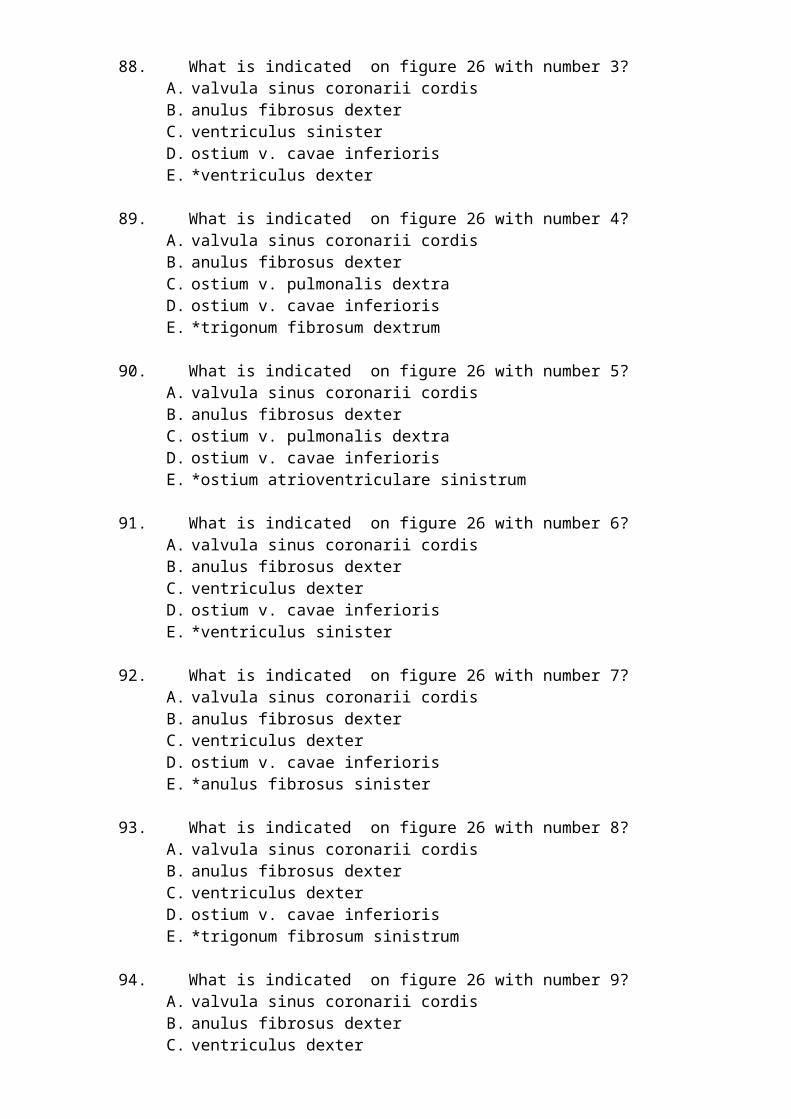
C. а. malleolaris anterior medialisD. а. tarsalis medialisE. *a. malleolaris anterior lateralis
330. What is indicated on figure 46 with number 1?A. a. tibialis anteriorB. а. malleolaris anterior medialisC. а. tarsalis medialisD. aa. metatarsales dorsalesE. *a. reccurens tibialis anterior.
331. What is indicated on figure 47 with number 1?A. а. tibialis anteriorB. rete malleolare medialeC. retinaculum mm. extensorum inferiorD. а. tarsalis medialisE. *а. malleolaris anterior medialis
332. What is indicated on figure 47 with number 2?A. rete malleolare medialeB. retinaculum mm. extensorum inferiorC. а. tarsalis medialisD. aa. metatarsals dorsalesE. *а. tibialis anterior
333. What is indicated on figure 47 with number 3?A. retinaculum mm. extensorum inferiorB. а. tarsalis medialisC. aa. metatarsals dorsalesD. aa. digitales dorsalesE. *rete malleolare mediale
334. What is indicated on figure 47 with number 4?A. а. tarsalis medialisB. aa. metatarsals dorsalesC. aa. digitales dorsalesD. rr. perforantesE. *retinaculum mm. extensorum inferior
335. What is indicated on figure 47 with number 5?A. aa. metatarsals dorsalesB. aa. digitales dorsalesC. rr. perforantesD. а. ArcuataE. *а. tarsalis medialis
336. What is indicated on figure 47 with number 6?A. aa. digitales dorsalesB. rr. perforantesC. а. ArcuataD. a. tarsalis lateralisE. *aa. metatarsals dorsales
337. What is indicated on figure 47 with number 7?

A. rr. perforantesB. а. ArcuataC. a. tarsalis lateralisD. a. dorsalis pedisE. *aa. digitales dorsales
338. What is indicated on figure 47 with number 8?A. а. ArcuataB. a. tarsalis lateralisC. a. dorsalis pedisD. rete malleolare lateraleE. *rr. perforantes
339. What is indicated on figure 47 with number 9?A. a. tarsalis lateralisB. a. dorsalis pedisC. rete malleolare lateraleD. а. malleolaris anterior lateralisE. *а. Arcuata
340. What is indicated on figure 47 with number 10?A. a. dorsalis pedisB. rete malleolare lateraleC. а. malleolaris anterior lateralisD. retinaculum mm. extensorum superiorE. *a. tarsalis lateralis
341. What is indicated on figure 47 with number 11?A. rete malleolare lateraleB. а. malleolaris anterior lateralisC. retinaculum mm. extensorum superiorD. а. malleolaris anterior medialisE. *a. dorsalis pedis
342. What is indicated on figure 47 with number 12?A. а. malleolaris anterior lateralisB. retinaculum mm. extensorum superiorC. а. malleolaris anterior medialisD. а. tibialis anteriorE. *rete malleolare laterale
343. What is indicated on figure 47 with number 13?A. retinaculum mm. extensorum superiorB. а. malleolaris anterior medialisC. а. tibialis anteriorD. rete malleolare medialeE. *а. malleolaris anterior lateralis
344. What is indicated on figure 47 with number 14?A. а. malleolaris anterior medialisB. а. tibialis anteriorC. rete malleolare medialeD. retinaculum mm. extensorum inferiorE. *retinaculum mm. extensorum superior
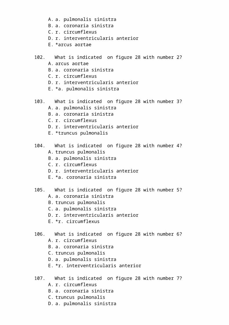
345. What is indicated on figure 48 with number 1?A. r. calcaneus anterior a. plantartis lateralisB. a. plantarius lateralisC. aa. metatarsals plantaresD. aa. digitales plantares communesE. *rete calcanei
346. What is indicated on figure 48 with number 2?A. a. plantarius lateralisB. aa. metatarsals plantaresC. aa. digitales plantares communesD. aa. digitales plantares propriaeE. *r. calcaneus anterior a. plantartis lateralis
347. What is indicated on figure 48 with number 3?A. aa. metatarsals plantaresB. aa. digitales plantares communesC. aa. digitales plantares propriaeD. r. perforansE. *a. plantarius lateralis
348. What is indicated on figure 48 with number 6?A. aa. digitales plantares communesB. aa. digitales plantares propriaeC. r. perforansD. arcus plantaris profundusE. *aa. metatarsals plantares
349. What is indicated on figure 48 with number 7?A. aa. digitales plantares propriaeB. r. perforansC. arcus plantaris profundusD. r. profundus a. plantaris medialisE. *aa. digitales plantares communes
350. What is indicated on figure 48 with number 8?A. r. perforansB. arcus plantaris profundusC. r. profundus a. plantaris medialisD. a. plantaris medialisE. *aa. digitales plantares propriae
351. What is indicated on figure 48 with number 9?A. arcus plantaris profundusB. r. profundus a. plantaris medialisC. a. plantaris medialisD. r. superficialis a. plantaris medialisE. *r. perforans
352. What is indicated on figure 48 with number 10?A. r. profundus a. plantaris medialisB. a. plantaris medialisC. r. superficialis a. plantaris medialis
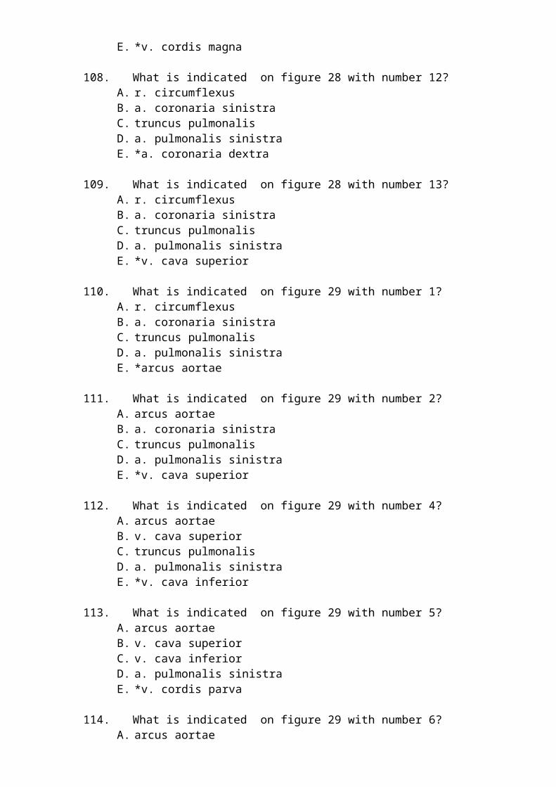
D. a. tibialis posterior.E. *arcus plantaris profundus
353. What is indicated on figure 48 with number 11?A. a. plantaris medialisB. r. superficialis a. plantaris medialisC. a. tibialis posterior.D. a. plantarius lateralisE. *r. profundus a. plantaris medialis
354. What is indicated on figure 48 with number 12?A. r. superficialis a. plantaris medialisB. a. tibialis posterior.C. a. plantarius lateralisD. aa. metatarsals plantaresE. *a. plantaris medialis
355. What is indicated on figure 48 with number 13?A. a. tibialis posterior.B. r. calcaneus anterior a. plantartis lateralisC. a. plantarius lateralisD. aa. metatarsals plantaresE. *r. superficialis a. plantaris medialis
356. What is indicated on figure 48 with number 14?A. rete calcaneiB. r. calcaneus anterior a. plantartis lateralisC. a. plantarius lateralisD. aa. metatarsals plantaresE. *a. tibialis posterior.
357. What is indicated on figure 55 with number 1?A. v. epigastrica superficialisB. vv. pudendae externaeC. v. saphena accessoriaD. v. saphena magnaE. *v. femoralis
358. What is indicated on figure 55 with number 2?A. vv. pudendae externaeB. v. saphena accessoriaC. v. saphena magnaD. rr. cutanei (n. obturatorius)E. *v. epigastrica superficialis
359. What is indicated on figure 55 with number 3?A. v. saphena accessoriaB. v. saphena magnaC. rr. cutanei (n. obturatorius)D. r.InfrapatellarisE. *vv. pudendae externae
360. What is indicated on figure 55 with number 4?A. v. saphena magna

B. rr. cutanei (n. obturatorius)C. r.InfrapatellarisD. arcus venosus dorsalis pedis; E. *v. saphena accessoria
361. What is indicated on figure 55 with number 5?A. rr. cutanei (n. obturatorius)B. r.InfrapatellarisC. arcus venosus dorsalis pedis; D. vv. metatarsales dorsales E. *v. saphena magna
362. What is indicated on figure 55 with number 6?A. r.InfrapatellarisB. arcus venosus dorsalis pedis; C. vv. metatarsales dorsales D. v. circumflexa iliaca superficialisE. *rr. cutanei (n. obturatorius)
363. What is indicated on figure 55 with number 7?A. arcus venosus dorsalis pedis; B. vv. metatarsales dorsales C. v. circumflexa iliaca superficialisD. v. femoralisE. *r.Infrapatellaris
364. What is indicated on figure 55 with number 9?A. vv. metatarsales dorsales B. v. circumflexa iliaca superficialisC. v. femoralisD. v. epigastrica superficialisE. *arcus venosus dorsalis pedis;
365. What is indicated on figure 55 with number 10?A. v. circumflexa iliaca superficialisB. v. femoralisC. v. epigastrica superficialisD. vv. pudendae externaeE. *vv. metatarsales dorsales
366. What is indicated on figure 55 with number 17?A. v. femoralisB. v. epigastrica superficialisC. vv. pudendae externaeD. v. saphena accessoriaE. *v. circumflexa iliaca superficialis
367. What is indicated on figure 65 with number 1?A. nodi lymphoidei inguinales superficiales (inferiores)B. fascia crurisC. vasa lymphatica superficiales pedisD. vasa lymphatica superficiales crurisE. *nodi lymphoidei inguinales superficiales (superomediales)

368. What is indicated on figure 65 with number 3?A. fascia crurisB. vasa lymphatica superficiales pedisC. vasa lymphatica superficiales crurisD. vasa lymphatica superficiales femorisE. *nodi lymphoidei inguinales superficiales (inferiores)
369. What is indicated on figure 65 with number 4?A. vasa lymphatica superficiales pedisB. vasa lymphatica superficiales crurisC. vasa lymphatica superficiales femorisD. v. saphena magnaE. *fascia cruris
370. What is indicated on figure 65 with number 5?A. vasa lymphatica superficiales crurisB. vasa lymphatica superficiales femorisC. v. saphena magnaD. nodi lymphoidei inguinales superficiales (superolaterales)E. *vasa lymphatica superficiales pedis
371. What is indicated on figure 65 with number 6?A. vasa lymphatica superficiales femorisB. v. saphena magnaC. nodi lymphoidei inguinales superficiales (superolaterales)D. fascia lataE. *vasa lymphatica superficiales cruris
372. What is indicated on figure 65 with number 7?A. v. saphena magnaB. nodi lymphoidei inguinales superficiales (superolaterales)C. fascia lataD. nodi lymphoidei inguinales superficiales (superomediales)E. *vasa lymphatica superficiales femoris
373. What is indicated on figure 65 with number 8?A. nodi lymphoidei inguinales superficiales (superolaterales)B. fascia lataC. nodi lymphoidei inguinales superficiales (superomediales)D. nodi lymphoidei inguinales superficiales (inferiores)E. *v. saphena magna
374. What is indicated on figure 65 with number 9?A. fascia lataB. nodi lymphoidei inguinales superficiales (superomediales)C. nodi lymphoidei inguinales superficiales (inferiores)D. fascia crurisE. *nodi lymphoidei inguinales superficiales (superolaterales)
375. What is indicated on figure 65 with number 9?A. fascia lataB. nodi lymphoidei inguinales superficiales (inferiores)C. fascia crurisD. vasa lymphatica superficiales pedis

E. *nodi lymphoidei inguinales superficiales (superomediales)
376. What is indicated on figure 71 with number 1?A. vas lymphaticum afferentiaB. capsulaC. trabeculaeD. sinus marginalisE. *cortex lymphonodi
377. What is indicated on figure 71 with number 2?A. capsulaB. trabeculaeC. sinus marginalisD. hilum lymphonodiE. *vas lymphaticum afferentia
378. What is indicated on figure 71 with number 3?A. trabeculaeB. sinus marginalisC. hilum lymphonodiD. vasa lymphatica efferentiaE. *capsula
379. What is indicated on figure 71 with number 4?A. sinus marginalisB. hilum lymphonodiC. vasa lymphatica efferentiaD. sinus terminalisE. *trabeculae
380. What is indicated on figure 71 with number 5?A. hilum lymphonodiB. vasa lymphatica efferentiaC. sinus terminalisD. medulla lymphonodi.E. *sinus marginalis
381. What is indicated on figure 71 with number 6?A. vasa lymphatica efferentiaB. sinus terminalisC. medulla lymphonodi.D. cortex lymphonodiE. *hilum lymphonodi
382. What is indicated on figure 71 with number 7?A. sinus terminalisB. medulla lymphonodi.C. cortex lymphonodiD. vas lymphaticum afferentiaE. *vasa lymphatica efferentia
383. What is indicated on figure 71 with number 8?A. medulla lymphonodi.B. cortex lymphonodi

C. vas lymphaticum afferentiaD. capsulaE. *sinus terminalis
384. What is indicated on figure 71 with number 9?A. cortex lymphonodiB. vas lymphaticum afferentiaC. capsulaD. trabeculaeE. *medulla lymphonodi.