
spiral.imperial.ac.uk · Web viewThe IASLC Lung Cancer Staging Project: Proposals for the Revision...
39
The IASLC Lung Cancer Staging Project: Proposals for the Revision of the TNM Stage Groupings in the Forthcoming (Eighth) Edition of the TNM Classification for Lung Cancer Peter Goldstraw, FRCS 1 , Kari Chansky, MS, 2 John Crowley, PhD, 2 Ramon Rami-Porta, MD 3 , Hisao Asamura, MD 4 , Wilfried EE Eberhardt, MD 5 , Andrew G Nicholson, DM FRCP 6 , Patti Groome, PhD 7 ,Alan Mitchell, MS, 2 and Vanessa Bolejack, MPH 2 on behalf of the International Association for the Study of Lung Cancer Staging and Prognostic Factors Committee, Advisory Boards and Participating Institutions 8 1: Department of Thoracic Surgery, Royal Brompton and Harefield NHS Foundation Trust and Imperial College, London, United Kingdom 2: Cancer Research And Biostatistics, Seattle, WA, United States of America 3: Department of Thoracic Surgery, Hospital Universitari Mutua Terrassa, University of Barcelona, and CIBERES Lung Cancer Group, Terrassa, Barcelona, Spain 4: Division of Thoracic Surgery, Keio University School of Medicine, Tokyo, Japan 1
Transcript of spiral.imperial.ac.uk · Web viewThe IASLC Lung Cancer Staging Project: Proposals for the Revision...

The IASLC Lung Cancer Staging Project: Proposals for the Revision of the TNM Stage Groupings in the Forthcoming (Eighth) Edition of the TNM Classification for Lung Cancer
Peter Goldstraw, FRCS 1, Kari Chansky, MS,2 John Crowley, PhD,2 Ramon Rami-Porta, MD 3 , Hisao
Asamura, MD 4 , Wilfried EE Eberhardt, MD 5 , Andrew G Nicholson, DMFRCP 6 , Patti Groome, PhD 7 ,Alan Mitchell, MS, 2 and Vanessa Bolejack, MPH 2 on behalf of the International Association for the
Study of Lung Cancer Staging and Prognostic Factors Committee, Advisory Boards and Participating
Institutions 8
1: Department of Thoracic Surgery, Royal Brompton and Harefield NHS Foundation Trust and
Imperial College, London, United Kingdom
2: Cancer Research And Biostatistics, Seattle, WA, United States of America
3: Department of Thoracic Surgery, Hospital Universitari Mutua Terrassa, University of Barcelona,
and CIBERES Lung Cancer Group, Terrassa, Barcelona, Spain
4: Division of Thoracic Surgery, Keio University School of Medicine, Tokyo, Japan
5: West German Cancer Centre, University Hospital, Ruhrlandklinik, University Duisburg-Essen,
Essen, Germany
6: Andrew G. Nicholson, Department of Pathology, Royal Brompton and Harefield NHS Foundation
Trust and Imperial College, London, United Kingdom
7: Queen’s Cancer Research Institute, Kingston, Ontario, Canada
8: See appendix
1

Abstract
Introduction
The 8th edition of the TNM Classification for Lung Cancer will be published late in 2016 and enacted
in January 2017. In preparation for this the International Association for the Study of Lung Cancer
(IASLC) Lung Cancer Staging Project established a new data set, which lead to proposals for changes
to the T and M descriptors and confirmation of the existing N descriptors for the next edition. Here
we report the recommendations for the resultant stage groupings.
Methods
The database collected 94,708 new cases of lung cancer treated between 1999 and 2010, donated
by 35 sources from 16 countries. After exclusions 77,156 cases of non-small cell lung cancer were
available for various sub-analyses and 33,441 cases were available for this analysis. All T, N and M
subsets were analysed by recursive partitioning and amalgamation and the resultant terminal nodes
were grouped according to the power of the statistical value and their clinical utility.
Results
A new stage classification for lung cancer is proposed, the result of changes to the assignment of
some descriptors within T and M categories, the re-allocation of some TNM subsets, and the
creation of a small number of new stage groupings.
Conclusions
The publication of these proposals will allow the lung cancer community to engage in an informed
debate about the new proposals and prepare clinicians for the changes, which if implemented, will
affect many aspects of their practice. It is hoped that aligning prognosis more accurately with stage
will benefit patients around the globe.
Word count for Abstract: 250
Keywords: Lung cancer, Lung cancer staging, Stage grouping, TNM classification
2

The 7th Edition of the tumour (T), node (N) and metastasis (M) (TNM) Classification for Lung Cancer
was published in September 2009 (1, 2) and enacted in January 2010 (3, 4). This revision was novel in
that the changes were entirely based upon the proposals of the International Association for the
Study of Lung Cancer (IASLC) International Staging Project (5-13). This project was organised and
funded by the IASLC and collected and analysed over 100,000 cases contributed by colleagues in 46
centres from over 19 countries around the world. Data entry and analysis were undertaken by
Cancer Research And Biostatistics (CRAB), a not-for-profit organisation based in Seattle, USA.
Validation, both internal and external, was more rigorous than that undertaken in any previous
revision (14). The success of this project led the IASLC to expand the remit of its Staging and
Prognostic Factors Committee, and with other collaborating organisations, to develop proposals for
the 8th edition of the TNM classification for other thoracic malignancies in addition to lung cancer.
In preparation for the impending 8th edition of TNM for lung cancer the committee and partners in
CRAB developed a new database. The characteristics of the new database (15) and the committee's
proposals for changes to the T, N and M descriptors (16-18) have been published and here we
present the proposals for the resultant TNM stage groupings. All these proposals will be submitted
to the Union for International Cancer Control (UICC) and the American Joint Committee on Cancer
(AJCC), for inclusion in the 8th edition of TNM for Lung Cancer due to be published late in 2016 and
enacted in January 2017.
Methods
During the transition from adopting the 7th edition to working toward the 8th edition, a new data
dictionary was developed, in conjunction with a new Electronic Data Capture system (EDC). Housed
at CRAB, the EDC has provided 4,667 cases overall that were used in this latest revision, and another
90,041 cases have been contributed by individual sites in retrospective fashion, and mapped to be
compatible with the EDC data fields (15). The database contains cases treated by all modalities of
care, including multi-modality treatment, diagnosed between 1999 and 2010. For the analyses of
TNM categories presented here, only cases with non-small cell lung cancer histology and complete
staging information were included. For cases where chemotherapy was received prior to surgery (yp
cases), only clinical stage was considered.
Candidate proposals for overall TNM stage groups were developed in conjunction with proposed
changes to the T and M categories (16, 17). These proposed changes are highlighted in the full list
of T, N and M descriptors shown in Table 1, which also incorporate the subsequent
3

recommendations of the IASLC on the classification of minimally invasive adenocarcinoma (18). The
existing N descriptors were validated and no changes were proposed for the 8th edition (19).
Applying the new T and M proposals to the training dataset, the resultant TNM subsets and the
numbers of cases in each subset by clinical stage and pathological stage are shown in Table 2. A
small number of candidate stage grouping schemes were developed initially on the M0 cases, using a
recursive partitioning and amalgamation (RPA) algorithm (20). The analysis was applied using the
statistical package R, Version 3.1.0. The algorithm generates tree-based model for the survival data
using logrank test statistics for recursive partitioning and, for selection of the important groupings,
bootstrap resampling to correct for the adaptive nature of the splitting algorithm (Figure 1). The
tree-based analysis was stratified on type of data submission: registry versus all others. The analysis
grouped cases based on best stage (pathological if available, otherwise clinical) after determination
of best-split points based on overall survival on an ordered variable for the newly proposed T
categories and for current N categories, excluding NX cases. This analysis was performed on a
randomly selected training set comprising 2/3 of the available data that met the requirements for
conversion to newly proposed T and M categories (N=25,911 M0 cases plus 599 M1 cases), reserving
12,931 cases for subsequent internal validation. The random selection process was stratified by type
of database submission and time period of case entry (1999-2004 vs 2005-2010). M1 cases were also
split but were not the focus of the tree-based analysis.
An ordered list of groupings was constructed from the terminal nodes of the survival tree. With this
as a guide, several proposed stage groupings were created by combining adjacent groups. Selection
of a final stage grouping proposal from among the candidate schemes was based upon its statistical
properties in the training set and its relevance to clinical practice, and was arrived at by consensus.
Candidate TNM stage grouping schemes were evaluated in part by assessing overall survival in
clinical, pathologic, and best stage. Survival was measured from the date of diagnosis for clinically
staged tumours and the date of surgery for pathologically staged tumours, and was calculated by the
Kaplan-Meier method. Adjusted survival curves (21, 22) were drawn using inverse probability
weights applied to the survival calculations, based on the proportion of cases that were from registry
databases (vs others) in each stage category. This method was utilized in light of the different overall
survival prognosis in registry databases in general, combined with the disproportionate
representation of registry cases in some of the stage groups. Contrasts between adjacent stage
groups were evaluated by Cox regression analysis, adjusted for baseline factors (age, performance
status, cell type) and type of database submission, using the SAS System for Windows Version 9.4
PHREG procedure.
4

Results.
Figure 1 shows the survival tree based on best stage in the training set, M0 cases only (N=25,911).
The ordered list of terminal nodes, with stratified hazard ratios relative to the best prognosis node
(T1aN0), is shown in Table 3. Proposed 8th edition TNM stage groupings are summarized in Table 4,
in which are highlighted those TNM subsets that it is proposed should move from their present stage
grouping. The moving of some cases from within a descriptor in the present staging system to
another in the proposals for the 8th edition of the TNM Classification and the creation of new
descriptors have led to the migration of certain TNM subsets between stage groups. For example, an
N0 case with either diaphragm invasion or 7 cm size, T3 by 7th edition definitions, moves from stage
category IIB to IIIA, by virtue of being re-defined as T4 by the 8 th edition. A new category, IIIC, is
composed of T4N3M0 cases, which showed a worse prognosis than the others. The number of
available cases (training and validation set combined) occupying each of these T, N, and M
categories by best stage is shown in Table 5.
Figures 2 and 3 show weighted survival by stage according to the 7 th Edition of TNM and by the
newly proposed TNM stage based on the entire set of cases available for reclassification, including
the M1 cases. There were 17,477 cases clinically staged I-IV, and 31,836 cases with pathologic stage.
Tables 6 and 7 show the statistics from adjusted Cox proportional hazards regression modelling of
the 7th Edition of TNM and the proposed new system for clinical and pathological stage, respectively,
using the new database collected for the 8th edition. The hazard ratios between adjacent stage
group categories are uniformly significant for both 7th edition stage and the more finely parsed
proposed 8th edition. The additional stage categories within the stage I and the stage III in the
proposed system are sufficiently distinct from one another. The weighted clinical survival estimates
for the proposed 8th edition (Figure 2B) show overlap between stages IIIC (T4N3) and IVA (M1a cases
with metastases restricted to the thoracic cavity and M1b cases with single metastasis outside the
thoracic cavity). This overlap is not evident in the multivariate analyses, where the hazard ratio for
the comparison between stage IVA and stage IIIC is 1.75 (P<.0001). The overlap in the survival
curves may partly be a result of the distribution of registry and non-registry cases, despite the
attempt to correct for this with weighting. There are no registry cases in the stage IV groups, which
had to have a sufficient description of metastatic disease in order to be classified as M1a, M1b, or
M1c.
5

For both the clinical and pathological stage models, there is a slight increase in the value for R2, an
estimate of the percent variance explained (PVE) (23) by the models described above, for the
proposed 8th edition staging scheme. For clinical stage, the R2 value for the 7th edition staging scheme
in the complete dataset is 67.5. The R2 value for the proposed 8th edition is 68.3. Likewise for
pathologic stage, the R2 value for the 7th edition classification is 45.7, versus 46.9 for the proposal.
The proposed stage groupings are summarized in Table 8.
Validation
The proposals derived from the training set were internally validated against the validation set of
12,931 cases (5,785 classifiable by clinical stage and 10,558 classifiable by pathologic stage). The
validation set generated survival curves which were generally similar to those in the training set and
Cox proportional hazards regression analyses that calculated the hazard ratios between each pair of
adjacent stage groups while controlling for cell type, sex, age and database type were all statistically
significant for the clinical and the pathologic staging data, with one exception. The comparison of
clinical IIA versus IB disease was not significant, with a hazard ratio of 1.35 and a p-value of 0.18. The
HR is slightly higher than that which is found when analyzing the combined training and validation
sets; however the p-value is reduced, possibly because there are only 183 cases in the clinical IIA
category, in the validation set.
External validation against an outside database is desirable. The North American
Surveillance, Epidemiology and End Results Registries (SEER) database was a valuable tool in this
regard for the development of the 7th edition. For the current proposal, certain cases in the SEER
data cannot be classified. Although additional site-specific factors were collected during recent
years (2010-2012), it is, for example, impossible to identify and reclassify cases that were classified
as T3 in the 7th edition with diaphragm invasion. In any case, given the fact that the additional site-
specific factors were not in effect for earlier years, too few cases from the appropriate time frame
are available that can be classified. The SEER data have been classified according to the 7 th edition,
however, and so a comparison of the IASLC database to SEER, with respect to overall survival across
7th edition stage categories, was performed to assess the validity of the IASLC database.
Figure S1 shows survival according to 7th edition stage for the SEER database and the IASLC
database. Stage for stage, the median survival time is consistently higher in the IASLC database,
which is not unexpected given the various data sources, only a proportion of which originated from a
national registry. However, when exploring the difference in survival between the SEER data and the
IASLC data via Cox regression analysis with stage and member database as factors, adjusting for
6

surgical management diminishes the difference between the two databases. The adjusted hazard
ratio for overall survival is 0.91 in favor of the IASLC database (95% CI .85-.96, p<.0001). The analysis
dataset for TNM stage from the IASLC database is 87% surgically managed, versus 31% of the SEER
cases used for this comparison. The unadjusted hazard ratio is 0.73 (95% C.I. .68-.77).
Discussion.
This publication sets out the IASLC proposals for Stage Groupings in the 8th edition of the TNM
Classification for Lung Cancer. The database used in this analysis consisted entirely of cases of
NSCLC, and a separate publication will cover the recommendations for small-cell lung cancer (24).
The IASLC Staging and Prognostic Factors Committee remains committed to the accumulation of
prospectively acquired data using the data set specifically designed to inform revisions to the TNM
classification (25). However, the added complexity of such data accrual has resulted in our continued
dependence to a large degree on retrospective accrual of data mostly collected for other purposes.
The committee accepted the advice of its statisticians and epidemiologists that new cases be kept
separate from the data accrued for the 7th edition. This decision has been vindicated by the
improved survival by stage for the newly acquired data set. This may reflect improvements in
diagnosis, such as the increasing vogue for CT screening, improvements in the staging algorithm with
the widespread use of PET scanning and less invasive mediastinal staging by EBUS and EUS and
improvements in treatment such as the use of adjuvant therapy after complete resection, the
availability of radical treatment options in less fit individuals with SABR and minimally invasive
surgical options, and improved results in stage IV disease with targeted agents, the toxicity profile of
which allows such treatment to be considered in patients with worse performance levels. However,
there are also differences in the data set when compared to that developed for the revisions leading
to the 7th edition of TNM for lung cancer. The new data set had a higher proportion of cases from
Asia, mostly from Japan which contributed 41% of the total, and this resulted in the proportion of
cases having surgery as a component of their treatment rising from 53% in the previous data set to
85% in the new one. In addition there was an increase in the proportion of cases derived from
registry data and a lack of cases from clinical trials. The net effect of all these variations was that
whilst survival stage for stage was increased in all stages, there was a relative worsening of survival
for advanced stages, especially stage IIIB. We attempted to correct these biases by performing the
tree-based analysis stratified by type of database and adjusting the survival curves by inverse
probability weighting.
Changes to some T and M descriptors will result in these cases being assigned to a different stage
category than that to which they would have been assigned in the 7th edition. In addition some TNM
7

subsets have been moved to a new stage grouping. Whilst such changes might raise the issue as to
whether there should be consequent changes to treatment algorithms, it is important to remind
ourselves that stage does not dictate treatment. Stage is one, perhaps the single most important
one, of several prognostic factors that guide the appropriate treatment option(s) to offer the
patient. Any change to established treatment algorithms should be based upon clinical judgement
informed by prospective trials.
The 7th Edition of TNM for lung cancer placed additional emphasis on tumour size, and as could
have been anticipated, size cut points have further proliferated in the proposals for the 8th Edition,
such that size will now be a descriptor in all T categories.
Some new stage groupings are proposed for the 8th edition of TNM for lung cancer. The division of
the T1 category into T1a, T1b and T1c, based on new size cut points of 1cm and 2cm, has resulted in
these cases, when associated with N0 and M0 categories, being assigned to Stage IA1, IA2 and IA3
respectively, reflecting the statistically different prognosis of such cases. These new cut points and
the stage groupings should be used in any trials of novel therapies such as sub-lobar resection and
non-surgical treatment options. They should not, in themselves, be taken to be a constraint on the
use of structured surveillance in studies of CT screening as the proportion of screen-detected
tumours in our data set is unknown.
A new stage grouping has also been created for the most advanced local disease categories, T3 and
T4 associated with N3 disease but M0. These will now be classified as stage IIIC, reflecting their
worse outcome when compared to those tumours which remain in stage IIIB. The prognosis of IIIC
cases is similar to that of stage IVA cases but the separation is justified by the different treatment
approaches used in such cases.
Finally there are changes proposed to stage IV disease. Cases with intrathoracic metastatic disease,
to contralateral lung or with pleural/pericardial dissemination will remain as M1a disease. The
category of M1b will now be assigned to cases in which there is a single metastatic deposit (in one
organ), and M1a and M1b cases will be entered into a new stage grouping of stage IVA. Whilst the
survival on these 2 M1 categories is sufficiently similar to justify their inclusion in a single stage
grouping the committee felt that it would be useful to retain the separate M categories of M1a and
M1b for future data collection and analysis as some cases with oligometastatic disease are now
receiving more aggressive local therapy, in addition to systemic treatment. The commoner situation
in which there are multiple metastatic deposits, usually in more than one organ, will now be
classified as M1c and staged as IVB.
There are other changes to stage groupings proposed. In some cases this will result from a T
descriptor being allocated to a higher stage in the 8th edition, such as has occurred with T3 tumours,
8

so classified due to invasion of the chest wall or mediastinal structures, when associated with N2
disease moving from stage IIIA in the 7th edition to IIIB in the 8th, and when associated with N3
disease moving from stage IIIB to IIIC. Similarly all subdivisions of the T1 category now associated
with N1 disease will move from stage IIA to IIB. In other situations tumours may be allocated to a
different T category in the 8th edition, which results in a change of stage, as for the re-classification
of tumours associated with diaphragmatic invasion to T4, and hence when associated with N0
disease moving from stage IIB to IIIA. For some T and N categories both influences may affect the
stage grouping assigned to a case. For example, the shift of tumours larger than 5 cm in size from
T2b to T3 will lead them to be assigned to a higher stage grouping whatever the N category. When
associated with N0 and N1 disease such tumours move from IIA to IIB and from IIB to IIIA,
respectively, because of the change in T category, whilst when associated with N2 or N3 disease the
shift from IIIA to IIIB and from IIIB to IIIC is the result of the change of stage proposed to that TNM
subset in the 8th edition.
As happened in the 7th edition there are instances in which the power of the data requires changes
that at first sight might appear counter-intuitive. The transfer of tumours greater than 5 cm in size to
the T3 category, leaving T2a for tumours >3cm but not larger than 4cm and T2b for tumours >4cm
but not larger than 5cm should, if a variant of stage migration were acting, result in a lower stage
being assigned to these tumours for all N categories. In reality the new data set showed that these
cases should remain in the same stage grouping as assigned in the 7th edition, except when T2a
cases are associated with N1 disease when a higher stage grouping was appropriate. Will this change
encourage the wider consideration of adjuvant chemotherapy after the complete resection of such
small tumours, 4cm and less in size? Such questions challenge us all to reconsider treatment
algorithms and test new options with appropriate clinical trials.
Central to the greater emphasis on tumour size, now proposed as a descriptor in all T categories, is
the need to study the appropriate way in which size is measured. This is especially important when
dealing with mixed attenuation tumours, increasingly found as CT screening for lung cancer,
becomes more widely adopted, and with the recently revised classification for adenocarcinoma. A
discussion document on this topic has been published by the IASLC Staging and Prognostic Factors
Committee (18).
When presenting its proposals for the 7th edition of TNM for lung cancer we expressed the hope
that the IASLC through the support of the pharmaceutical industry, by the cooperation of its
members and the generous donation of hard-earned data could go on to develop validated
proposals for the 8th edition and beyond. This paper is an important step in realising that ambition.
9

The Staging and Prognostic Factors Committee of the IASLC presents these recommendations to
inform the discussions leading to the 8th edition of TNM for Lung Cancer. We hope that the thoracic
oncology community find the proposals of value and that, when accepted, they will have a positive
impact on the effectiveness of treatment for lung cancer which will benefit patients around the
globe.
10

APPENDIX
IASLC Staging and Prognostic Factors Committee
Peter Goldstraw, Past Chair, Royal Brompton Hospital and Imperial College, London, United
Kingdom; Ramón Rami-Porta, Chair, Hospital Universitari Mutua Terrassa, Terrassa, Spain; Hisao
Asamura, Chair Elect, Keio University, Tokyo, Japan; David Ball, Peter MacCallum Cancer Centre,
Melbourne, Australia; David Beer, University of Michigan, Ann Arbor, MI, United States of America
(USA); Ricardo Beyruti, University of Sao Paulo, Brazil; Vanessa Bolejack, Cancer Research And
Biostatistics, Seattle, WA, USA; Kari Chansky, Cancer Research And Biostatistics, Seattle, WA, USA;
John Crowley, Cancer Research And Biostatistics, Seattle, WA, USA; Frank Detterbeck, Yale
University, New Haven, CT, USA; Wilfried Ernst Erich Eberhardt, West German Cancer Centre,
University Hospital, Ruhrlandklinik, University Duisburg-Essen, Essen, Germany; John Edwards,
Northern General Hospital, Sheffield, United Kingdom; Françoise Galateau-Sallé, Centre Hospitalier
Universitaire, Caen, France; Dorothy Giroux, Cancer Research And Biostatistics, Seattle, WA, USA;
Fergus Gleeson, Churchill Hospital, Oxford, United Kingdom; Patti Groome, Queen’s Cancer Research
Institute, Kingston, Ontario, Canada; James Huang, Memorial Sloan-Kettering Cancer Center, New
York, NY, USA; Catherine Kennedy, University of Sydney, Sydney, Australia; Jhingook Kim, Samsung
Medical Center, Seoul, Korea; Young Tae Kim, Seoul National University, Seoul, South Korea; Laura
Kingsbury, Cancer Research And Biostatistics, Seattle, WA, USA; Haruhiko Kondo, Kyorin University
Hospital, Tokyo, Japan; Mark Krasnik, Gentofte Hospital, Copenhagen, Denmark; Kaoru Kubota,
Nippon Medical School Hospital, Tokyo, Japan; Antoon Lerut, University Hospitals, Leuven, Belgium;
Gustavo Lyons, British Hospital, Buenos Aires, Argentina; Mirella Marino, Regina Elena National
Cancer Institute, Rome, Italy; Edith M. Marom, MD Anderson Cancer Center, Houston, TX, USA; Jan
van Meerbeeck, Antwerp University Hospital, Edegem (Antwerp), Belgium; Alan Mitchell, Cancer
Research And Biostatistics, Seattle, WA, USA; Takashi Nakano, Hyogo College of Medicine, Hyogo,
Japan; Andrew G. Nicholson, Royal Brompton and Harefield NHS Foundation Trust and Imperial
College, London, United Kingdom; Anna Nowak, University of Western Australia, Perth, Australia;
Michael Peake, Glenfield Hospital, Leicester, United Kingdom; Thomas Rice, Cleveland Clinic,
Cleveland, OH, USA; Kenneth Rosenzweig, Mount Sinai Hospital, New York, NY, USA; Enrico Ruffini,
University of Torino, Torino, Italy; Valerie Rusch, Memorial Sloan-Kettering Cancer Center, New York,
NY, USA; Nagahiro Saijo, National Cancer Center Hospital East, Chiba, Japan; Paul Van Schil, Antwerp
University Hospital, Edegem (Antwerp), Belgium; Jean-Paul Sculier, Institut Jules Bordet, Brussels,
Belgium; Lynn Shemanski, Cancer Research And Biostatistics, Seattle, WA, USA; Kelly Stratton,
Cancer Research And Biostatistics, Seattle, WA, USA; Kenji Suzuki, Juntendo University, Tokyo, Japan;
11
Yuji Tachimori, National Cancer Center, Tokyo, Japan; Charles F. Thomas Jr, Mayo Clinic, Rochester,
MN, USA; William Travis, Memorial Sloan-Kettering Cancer Center, New York, NY, USA; Ming S. Tsao,
The Princess Margaret Cancer Centre, Toronto, Ontario, Canada; Andrew Turrisi, Sinai Grace
Hospital, Detroit, MI, USA; Johan Vansteenkiste, University Hospitals, Leuven, Belgium; Hirokazu
Watanabe, National Cancer Center Hospital, Tokyo, Japan; Yi-Long Wu, Guangdong General Hospital,
Guangzhou, People’s Republic of China.
Advisory Board of the IASLC Mesothelioma Domain
Paul Baas, The Netherlands Cancer Institute, Amsterdam, The Netherlands; Jeremy Erasmus, MD
Anderson Cancer Center, Houston, TX, USA; Seiki Hasegawa, Hyogo College of Medicine, Hyogo,
Japan; Kouki Inai, Hiroshima University Postgraduate School, Hiroshima, Japan; Kemp Kernstine, City
of Hope, Duarte, CA, USA; Hedy Kindler, The University of Chicago Medical Center, Chicago, IL, USA;
Lee Krug, Memorial Sloan-Kettering Cancer Center, New York, NY, USA; Kristiaan Nackaerts,
University Hospitals, Leuven, Belgium; Harvey Pass, New York University, NY, USA; David Rice, MD
Anderson Cancer Center, Houston, TX, USA.
Advisory Board of the IASLC Thymic Malignancies Domain
Conrad Falkson, Queen’s University, Ontario, Canada; Pier Luigi Filosso, University of Torino, Italy;
Giuseppe Giaccone, Georgetown University, Washington, DC, USA; Kazuya Kondo, University of
Tokushima, Tokushima, Japan; Marco Lucchi, University of Pisa, Pisa, Italy; Meinoshin Okumura,
Osaka University, Osaka, Japan.
Advisory Board of the IASLC Esophageal Cancer Domain
Eugene Blackstone, Cleveland Clinic, OH, USA.
Participating Institutions in the new IASLC Lung Cancer Staging Project
F. Abad Cavaco and E. Ansótegui Barrera, Hospital La Fe, Valencia, Spain; J. Abal Arca and I. Parente
Lamelas, Complejo Hospitalario de Ourense, Ourense, Spain; A. Arnau Obrer and R. Guijarro Jorge,
Hospital General Universitario de Valencia, Valencia, Spain; D. Ball, Peter MacCallum Cancer Centre,
Melbourne, Australia; G. K. Bascom, Good Samaritan Hospital, Kearney, NE, USA; A. I. Blanco Orozco
and M. A. González Castro, Hospital Virgen del Rocío, Sevilla, Spain; M. G. Blum, Penrose Cancer
Center, Colorado Springs, USA; D. Chimondeguy, Hospital Universitario Austral, Argentina; V.
Cvijanovic, Military Medical Academy, Belgrade, Serbia; S. Defranchi, Hospital Universitario-
Fundacion Favaloro, Buenos Aires, Argentina; B. de Olaiz Navarro, Hospital de Getafe, Getafe, Spain;
I. Escobar Campuzano and I. Macía Vidueira, Hospital de Bellvitge, L’Hospitalet de Llobregat, Spain;
12
E. Fernández Araujo and F. Andreo García, Hospital Universitari Germans Trias i Pujol, Badalona,
Spain; K. M. Fong, Prince Charles Hospital, Brisbane, Australia; G. Francisco Corral and S. Cerezo
González, Hospital La Mancha Centro, Ciudad Real, Spain; J. Freixinet Gilart, Hospital Universitario
‘Dr. Negrín’, Las Palmas de Gran Canaria, Spain; L. García Arangüena, Hospital Sierrallana,
Torrelavega, Spain; S. García Barajas, Hospital Infanta Cristina, Badajoz, Spain; P. Girard, L'Institut
Mutualiste Montsouris, Paris, France; T. Goksel, Turkish Thoracic Society, Turkey; M. T. González
Budiño, Hospital General Universitario de Oviedo, Oviedo, Spain; G. González Casaurrán, Hospital
Gregorio Marañón, Madrid, Spain; J. A. Gullón Blanco, Hospital San Agustín, Avilés, Spain; J.
Hernández Hernández, Hospital de Ávila, Avila, Spain; H. Hernández Rodríguez, Hospital
Universitario de Tenerife, Santa Cruz de Tenerife, Spain; J. Herrero Collantes, Hospital Universitario
Nuestra Señora de la Candelaria, Santa Cruz de Tenerife, Spain; M. Iglesias Heras, Hospital de Ávila,
Ávila, Spain; J. M. Izquierdo Elena, Hospital Nuestra Señora de Aránzazu, Donostia, Spain; E.
Jakobsen, Danish Lung Cancer Registry, Denmark; S. Kostas, Athens School of Medicine, Athens,
Greece; P. León Atance and A. Núñez Ares, Complejo Hospitalario de Albacete, Albacete, Spain; M.
Liao, Shanghai Lung Tumor Clinical Medical Center, Shanghai, China; M. Losanovscky, Clinica y
Maternidad Suizo Argentina, Buenos Aires, Argentina; G. Lyons, Hospital Britanico de Buenos Aires,
Buenos Aires, Argentina; R. Magaroles and L. De Esteban Júlvez, Hospital Joan XXIII, Tarragona.
Spain; M. Mariñán Gorospe, Hospital de San Pedro de Logroño, Logroño, Spain; B. McCaughan and C.
Kennedy, University of Sydney, Sydney, Australia; R. Melchor Íñiguez, Fundación Jiménez Díaz,
Madrid, Spain; L. Miravet Sorribes, Hospital La Plana, Castellón, Spain; S. Naranjo Gozalo and C.
Álvarez de Arriba, Hospital Universitario Marqués de Valdecilla, Santander, Spain; M. Núñez
Delgado, Hospital de Meixoeiro, Vigo, Spain; J. Padilla Alarcón and J. C. Peñalver Cuesta, Instituto
Valenciano de Oncología, Valencia, Spain; J. S. Park, Samsung Medical Center, Seoul, South Korea; H.
Pass, New York University Langone Medical Center and Cancer Center, New York, USA; M. J. Pavón
Fernández, Hospital ‘Severo Ochoa’, Leganés, Spain; M. Rosenberg, Alexander Fleming Institute and
Hospital de Rehabilitación Respiratoria, Buenos Aires, Argentina; E. Ruffini, University of Torino,
Torino , Italy; V. Rusch, Memorial Sloan-Kettering Cancer Center, New York, USA; J. Sánchez de Cos
Escuín, Hospital de Cáceres, Cáceres, Spain; A. Saura Vinuesa, Hospital de Sagunto, Sagunto, Spain;
M. Serra Mitjans, Hospital Universitari Mutua Terrassa, Terrassa, Spain; T.E. Strand, Cancer Registry
of Norway, Norway; D. Subotic, Clinical Centre of Serbia, Belgrade, Serbia; S. Swisher, M.D. Anderson
Cancer Center (MDACC), Houston, USA; R. Terra, University of Sao Paulo Medical Center, Sao Paulo,
Brazil; C. Thomas, Mayo Clinic Rochester, Rochester, MN, USA; K. Tournoy, University Hospital
Ghent, Belgium; P. Van Schil, Antwerp University Hospital, Edegem (Antwerp), Belgium; M.
13

Velasquez, Fundacion Clinica Valle del Lili, Cali, Colombia; Y. L. Wu, Guangdong General Hospital,
Guangzhou, China; K. Yokoi, Japanese Joint Committee for Lung Cancer Registry, Osaka, Japan.
14
Table 1. Proposed T, N and M descriptors for the 8th edition of TNM for Lung Cancer (changes to the 7th edition are highlighted in bold and underlined).
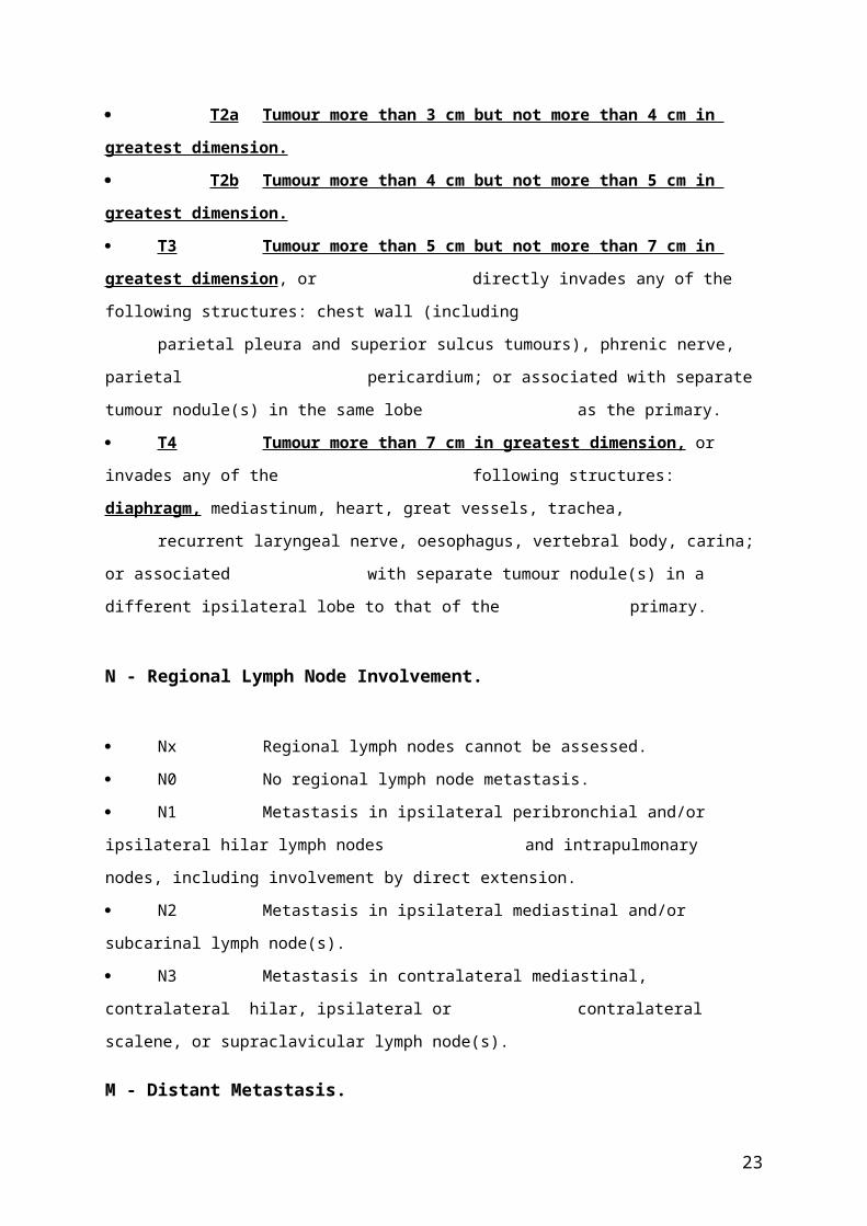
T - Primary Tumour
Tx Primary tumour cannot be assessed, or tumour proven by the presence of
malignant cells in sputum or bronchial washings but not visualized by
imaging or bronchoscopy.
T0 No evidence of primary tumour.
Tis Carcinoma in situ
T1 Tumour 3 cm or less in greatest dimension, surrounded by lung or visceral
pleura, without bronchoscopic evidence of invasion more proximal than the
lobar bronchus (i.e. not in the main bronchus)1.
T1a(mi) Minimally invasive adenocarcinoma 2 .
T1a Tumour 1 cm or less in greatest dimension 1 .
T1b Tumour more than 1 cm but not more than 2 cm in greatest dimension 1 .
T1c Tumour more than 2 cm but not more than 3 cm in greatest dimension 1 .
T2 Tumour more than 3cm but not more than 5 cm; or tumour with any of the
following features3:
- Involves main bronchus regardless of distance from the carina, but
without involvement of the carina.
- Invades visceral pleura.
- Associated with atelectasis or obstructive pneumonitis that extends to
the hilar region, involving part or all of the lung.
T2a Tumour more than 3 cm but not more than 4 cm in greatest dimension.
T2b Tumour more than 4 cm but not more than 5 cm in greatest dimension.
T3 Tumour more than 5 cm but not more than 7 cm in greatest dimension, or
directly invades any of the following structures: chest wall (including
parietal pleura and superior sulcus tumours), phrenic nerve, parietal
pericardium; or associated with separate tumour nodule(s) in the same lobe
as the primary.
T4 Tumour more than 7 cm in greatest dimension, or invades any of the
following structures: diaphragm, mediastinum, heart, great vessels, trachea,
recurrent laryngeal nerve, oesophagus, vertebral body, carina; or associated
15

with separate tumour nodule(s) in a different ipsilateral lobe to that of the
primary.
N - Regional Lymph Node Involvement.
Nx Regional lymph nodes cannot be assessed.
N0 No regional lymph node metastasis.
N1 Metastasis in ipsilateral peribronchial and/or ipsilateral hilar lymph nodes
and intrapulmonary nodes, including involvement by direct extension.
N2 Metastasis in ipsilateral mediastinal and/or subcarinal lymph node(s).
N3 Metastasis in contralateral mediastinal, contralateral hilar, ipsilateral or
contralateral scalene, or supraclavicular lymph node(s).
M - Distant Metastasis.
M0 No distant metastasis.
M1 Distant metastasis present.
M1a Separate tumour nodule(s) in a contralateral lobe; tumour with pleural or
pericardial nodule(s) or malignant pleural or pericardial effusion4.
M1b Single extrathoracic metastasis 5 .
M1c Multiple extrathoracic metastases in one or several organs.
Notes:
1. The uncommon superficial spreading tumour of any size with its invasive component limited to the bronchial wall, which may extend proximal to the main bronchus, is also classified as T1a.
2. Solitary adenocarcinoma, ≤ 3 cm in size, with a predominately lepidic pattern and ≤ 5mm invasion in any one focus.
3. T2 tumours with these features are classified T2a if 4 cm or less in greatest dimension or if size cannot be determined, and T2b if greater than 4 cm but not larger than 5 cm in greatest dimension.
4. Most pleural (pericardial) effusions with lung cancer are due to tumour. In a few patients, however, multiple microscopic examinations of pleural (pericardial) fluid are negative for tumour, and the fluid is non-bloody and is not an exudate. Where these elements and clinical judgement dictate that the effusion is not related to the tumour, the effusion should be excluded as a staging descriptor.
16

5. This includes involvement of a single distant (non-regional) lymph node.
17

Table 2a. Distribution of T, N, and M categories in the training set (Clinical classification.)
Proposed T/M
Categories
N Category
TotalN0 N1 N2 N3
T1a 781 7 19 6 813
T1b 3105 68 124 30 3327
T1c 2417 142 208 32 2799
T2a 1928 268 372 50 2618
T2b 585 131 183 36 935
T3 837 191 344 77 1449
T4 1711 392 1642 909 4654
M1a 64 9 77 127 277
M1b 39 16 67 85 207
M1c 67 15 120 196 398
Total 11534 1239 3156 1548 17477
Table 2b. Distribution of T, N, and M categories in the training set (Pathologic classification.)
Proposed T/M
Categories
N Categories
TotalN0 N1 N2 N3
T1a 1390 45 49 2 1486
T1b 5638 311 392 7 6348
T1c 4403 484 515 13 5415
T2a 6102 1223 1526 55 8906
T2b 1640 485 490 16 2631
T3 2683 795 1025 39 4542
T4 1447 546 613 30 2636
Total 23303 3889 4610 162 31964
18

Table 3. Terminal nodes defined based on best stage, from a stratified tree-based analysis (RPA) of the training dataset. Hazard ratios are relative to the best prognosis group (T1aN0) and are stratified on type of database submission: Registry versus others.
Terminal NodeSample Size
(Training Set) Hazard Ratio
T1a N0 1223 1.00
T1b N0 4140 1.55
T1c N0 3098 2.07
T2a N0 4316 2.83
T2b N0 1139 3.89
T3 N0 1965 5.08
T1a-T2b N1 1755 5.09
T3 N1 558 7.56
T1a-T2b N2 2226 7.82
T4 N0-N1 2459 10.10
T1a-T2b N3 127 14.08
T3-T4 N2 2229 14.34
T3-T4 N3 676 21.73
19

Table 4. Descriptors and T and M categories in the 7th edition and as proposed for the 8th edition. Resultant stage groupings proposed for the 8th edition are highlighted in bold where there is a change and the 7th edition stage is given in parenthesis.
N-Categories
Overall Stage
Seventh Edition Descriptor Proposed T/M N0 N1 N2 N3
T1 <=1cm T1a IA1 (IA) IIB (IIA) IIIA IIIB
T1 >1-2cm T1b IA2 (IA) IIB (IIA) IIIA IIIB
T1 >2-3cm T1c IA3 (IA) IIB (IIA) IIIA IIIB
T2 >3-4cm T2a IB IIB (IIA) IIIA IIIB
T2 >4-5cm T2b IIA (IB) IIB (IIA) IIIA IIIB
T2 >5-7cm T3 IIB (IIA) IIIA (IIB) IIIB (IIIA) IIIC (IIIB)
T3 structures T3 IIB IIIA IIIB (IIIA) IIIC (IIIB)
T3 >7cm T4 IIIA (IIB) IIIA IIIB (IIIA) IIIC (IIIB)
T3 Diaphragm T4 IIIA (IIB) IIIA IIIB (IIIA) IIIC (IIIB)
T3 endobronch: location/atelectasis 3-4cm T2a IB (IIB) IIB (IIIA) IIIA IIIB
T3 endobronch: location/atelectasis 4-5cm T2b IIA (IIB) IIB (IIIA) IIIA IIIB
T4 T4 IIIA IIIA IIIB IIIC (IIIB)
M1a M1a IVA (IV) IVA (IV) IVA (IV) IVA (IV)
M1b single lesion M1b IVA (IV) IVA (IV) IVA (IV) IVA (IV)
M1c multiple lesions M1c IVB (IV) IVB (IV) IVB (IV) IVB (IV)
20

Table 5. Sample sizes for TNM subsets providing the basis for proposed changes, best stage.
N0 N1 N2 N3
Seventh Edition Descriptor
Proposed T/M
Overall Stage
Sample Size Overall Stage
Sample Size
Overall Stage
Sample Size Overall Stage
Sample Size
T1 <=1cm T1a IA ->IA1 1765 IIA ->IIB 47 IIIA 59 IIIB 4
T1 >1-2cm T1b IA ->IA2 6127 IIA ->IIB 321 IIIA 444 IIIB 20
T1 >2-3cm T1c IA ->IA3 4606 IIA ->IIB 492 IIIA 596 IIIB 37
T2 >3-4cm T2a IB 6382 IIA ->IIB 1250 IIIA 1666 IIIB 89
T2 >4-5cm T2b IB ->IIA 1689 IIA ->IIB 497 IIIA 559 IIIB 35
T2 >5-7cm T3 IIA ->IIB 1244 IIB ->IIIA 418 IIIA->IIIB 455 IIIB->IIIC 45
T3 structures T3 IIB 1666 IIIA 432 IIIA->IIIB 736 IIIB->IIIC 55
T3 >7cm T4 IIB ->IIIA 870 IIIA 316 IIIA->IIIB 320 IIIB->IIIC 33
T3 Diaphragm T4 IIB ->IIIA 47 IIIA 16 IIIA->IIIB 22 IIIB->IIIC 0
T3 endobronchial location/atelect >3-4 cm T2a IIB ->IB 18 IIIA->IIB 18 IIIA 10 IIIB 1
>4-5cm T2b IIB ->IIA 11 IIIA->IIB 2 IIIA 9 IIIB 1
T4 T4 IIIA 1862 IIIA 538 IIIB 1770 IIIB->IIIC 893
M1a M1a IV ->IVA 62 IV ->IVA 11 IV ->IVA 100 IV ->IVA 145
M1b single lesion M1b IV ->IVA 38 IV ->IVA 13 IV ->IVA 68 IV ->IVA 74
M1b multiple lesions M1c IV ->IVB 59 IV ->IVB 18 IV ->IVB 128 IV ->IVB 191
21

Table 6. Cox proportional hazards regression model output for the seventh edition of TNM, and proposed 8th edition clinical stage groupings using the entire database available for the 8th edition. Adjusted for age (≥70), sex, and histology (adenocarcinoma vs. others). Stratified by type of database submission (registry vs. others).
Comparison
Hazard Ratio P
7th Edition Proposed
8th Edition 7th Edition Proposed
8th Edition
IA2 vs IA1 - 1.82 - <.0001
IA3 vs IA2 - 1.40 - <.0001
IB vs IA 1.75 - <.0001 -
IB vs IA3 - 1.29 - <.0001
IIA vs IB 1.57 1.30 <.0001 0.0012
IIB vs IIA 1.22 1.30 0.0046 0.0008
IIIA vs IIB 1.28 1.48 <.0001 <.0001
IIIB vs IIIA 1.57 1.38 <.0001 <.0001
IIIC vs IIIB - 1.36 - <.0001
IVA vs IIIC - 1.75 - <.0001
IVB vs IVA - 1.91 - <.0001
IV vs IIIB 2.61 - <.0001 -
Table 7. Cox proportional hazards regression model output for the seventh edition of TNM, and proposed 8th edition pathologic stage groupings using the entire database available for the 8th edition. Adjusted for age (≥70), sex, histology (adenocarcinoma vs. others), and type of database submission (registry vs. others).
Comparison
Hazard Ratio P
7th Edition Proposed
8th Edition 7th Edition Proposed
8th Edition
IA2 vs IA1 - 1.44 - <.0001
IA3 vs IA2 - 1.31 - <.0001
IB vs IA 1.68 - <.0001 -
IB vs IA3 - 1.32 - <.0001
IIA vs IB 1.66 1.29 <.0001 <.0001
IIB vs IIA 1.22 1.40 <.0001 <.0001
IIIA vs IIB 1.61 1.66 <.0001 <.0001
22
Comparison
Hazard Ratio P
7th Edition Proposed
8th Edition 7th Edition Proposed
8th Edition
IIIB vs IIIA 1.58 1.67 <.0001 <.0001
IIIC vs IIIB - 1.85 - <.0001
23

Table 8. Proposed Stage Groupings for 8 th edition of TNM for Lung Cancer (changes to the 7th edition are highlighted in bold and underlined).
Occult Carcinoma Tx N0 M0
Stage 0 Tis N0 M0
Stage IA1 Tia(mi) N0 M0
T1a N0 M0
Stage IA2 T1b N0 M0
Stage IA3 T1c N0 M0
Stage IB T2a N0 M0
Stage IIA T2b N0 M0
Stage IIB T1a-c N1 M0
T2a N1 M0
T2b N1 M0
T3 N0 M0
Stage IIIA T1a-c N2 M0
T2a-b N2 M0
T3 N1 M0
T4 N0 M0
T4 N1 M0
Stage IIIB T1a-c N3 M0
T2a-b N3 M0
T3 N2 M0
T4 N2 M0
Stage IIIC T3 N3 M0
T4 N3 M0
Stage IVA Any T Any N M1a
Any T Any N M1b
Stage IVB Any T Any N M1c
24

Figure 1. Recursive partitioning and amalgamation-generated survival tree based on best stage for 25,911 M0 training set cases. T and N categories are modelled as order variables. Stratified hazard ratios are given relative to the left-most terminal node, T1aN0.
25

A.
B.
Figure 2. Overall survival by clinical stage according to the 7th edition (A) and the proposed 8th edition (B) groupings using the entire database available for the 8th edition. MST = Median Survival Time. Survival is weighted by type of database submission: Registry versus Other.
Events / N MST 24 Month 60 MonthIA 1119 / 6303 NR 93% 82%IB 768 / 2492 NR 85% 66%IIA 424 / 1008 66.0 74% 52%IIB 382 / 824 49.0 64% 47%IIIA 2139 / 3344 29.0 55% 36%IIIB 2101 / 2624 14.1 34% 19%IV 664 / 882 8.8 17% 6%
Events / N MST24
Month60
MonthIA1 68 / 781 NR 97% 92%IA2 505 / 3105 NR 94% 83%IA3 546 / 2417 NR 90% 77%IB 560 / 1928 NR 87% 68%IIA 215 / 585 NR 79% 60%IIB 605 / 1453 66.0 72% 53%IIIA 2052 / 3200 29.3 55% 36%IIIB 1551 / 2140 19.0 44% 26%IIIC 831 / 986 12.6 24% 13%IVA 336 / 484 11.5 23% 10%IVB 328 / 398 6.0 10% 0%
26

A.
B.
Figure 3. Overall survival by pathologic stage according to the 7th edition (A) and the proposed 8th edition (B) groupings using the entire database available for the 8th edition. MST = Median Survival time. Survival is weighted by type of database submission: Registry versus Other.
Events / N MST24
Month60
MonthIA 1837 / 11423 NR 94% 83%IB 2168 / 7711 NR 87% 71%IIA 1514 / 3702 NR 77% 57%IIB 1325 / 2776 58.9 70% 49%IIIA 3467 / 5818 35.0 61% 36%IIIB 364 / 506 20.0 45% 23%
Events / N MST24
Month60
MonthIA1 139 / 1389 NR 97% 90%IA2 823 / 5633 NR 94% 85%IA3 875 / 4401 NR 92% 80%IB 1618 / 6095 NR 89% 73%IIA 556 / 1638 NR 82% 65%IIB 2175 / 5226 NR 76% 56%IIIA 3219 / 5756 41.9 65% 41%IIIB 1215 / 1729 22.0 47% 24%IIIC 55 / 69 11.0 30% 12%
27

A.
B.
Figure Supp. 1. Survival by 7th edition TNM “best” stage applied to a) the North American Surveillance, Epidemiology, and End Results (SEER) registry, and b) the IASLC database collected to inform the 8th edition recommendations. Survival for the IASLC database is weighted by type of database submission: Registry versus Other.
7th Ed. TNM Stage Events / N MST
24 Month
IA 77 / 433 NR 84%IB 79 / 248 NR 73%IIA 59 / 141 NR 63%IIB 82 / 158 28.0 54%IIIA 253 / 403 18.0 41%IIIB 130 / 171 13.0 29%IV 1246 / 1408 6.0 14%
7th Ed. TNM Stage Events / N MST
24 Month
IA 2030 / 12520 NR 93%IB 2304 / 8069 NR 86%IIA 1581 / 3853 NR 76%IIB 1455 / 3057 55.9 68%IIIA 5025 / 7957 30.0 57%IIIB 2377 / 2957 14.0 34%IV 665 / 883 8.7 17%
28
0 24Months
0%
20%
40%
60%
80%
100%
0 24Months
0%
20%
40%
60%
80%
100%

Reference List
(1) Goldstraw P. IASLC Staging Handbook in Thoracic Oncology. 1st Ed. Orange Park, FL: EditorialRx Press, 2009.
(2) Goldstraw P. IASLC Staging Manual in Thoracic Oncology. 1st Ed. Orange Park, FL: EditorialRx Press, 2009.
(3) Sobin LH, Gospodarowicz MK, Wittekind C. UICC TNM Classification of Malignant Tumours. 7th Ed. Oxford, England: Wiley-Blackwell, 2009
(4) Edge SB, Byrd DR, Compton CC, Fritz AG, Greene FL, Trotti A III (Eds.). AJCC Cancer Staging Handbook. 7th Ed. New York, Springer, 2010.
(5) Goldstraw P, Crowley J, IASLC International Staging Project. The IASLC international staging project on lung cancer. J Thorac Oncol 2006; 1: 281-286.
(6) Rami-Porta R, Ball D, Crowley JJ, et al. The IASLC Lung Cancer Staging Project: proposals for the revision of the T descriptors in the forthcoming (seventh) edition of the TNM classification for lung cancer. J Thorac Oncol 2007; 2: 593-602.
(7) Rusch VR, Crowley JJ, Giroux DJ, et al. The IASLC Lung Cancer Staging Project: proposals for revision of the N descriptors in the forthcoming (seventh) edition of the TNM classification for lung cancer. J Thorac Oncol 2007; 2: 603-612.
(8) Postmus PE, Brambilla E, Chansky K, et al. The IASLC Lung Cancer Staging Project: proposals for revision of the M descriptors in the forthcoming (seventh) edition of the TNM classification for lung cancer. J Thorac Oncol 2007; 2: 686-693.
(9) Goldstraw P, Crowley JJ, Chansky K, et al. The IASLC Lung Cancer Staging Project: proposals for revision of the stage groupings in the forthcoming (seventh) edition of the TNM classification for lung cancer. J Thorac Oncol 2007; 2: 706-714.
(10) Shepherd FA, Crowley J, Van Houtte P, et al. The IASLC Lung Cancer Staging Project: proposals regarding the clinical staging of small-cell lung cancer in the forthcoming (seventh) edition of the TNM classification for lung cancer. J Thorac Oncol 2007; 2: 1067-1077.
(11) Vallières E, Shepherd FA, Crowley J, et al. The IASLC Lung Cancer Staging Project: proposals regarding the relevance of TNM in the pathological staging of small-cell lung cancer in the forthcoming (seventh) edition of the TNM classification for lung cancer. J Thorac Oncol 2009;
29

4: 1049-1059.
(12) Travis WD, Giroux DJ, Chansky K, et al. The IASLC Lung Cancer Staging Project: proposals for the inclusion of carcinoid tumours in the forthcoming (seventh) edition of the TNM classification for lung cancer. J Thorac Oncol 2008; 3: 1213-1223.
(13) Travis WD, Brambilla E, Rami-Porta R, et al. Visceral pleural invasion: pathologic criteria and use of elastic stains: proposals for the 7th edition of the TNM classification for lung cancer. J Thorac Oncol 2008; 3: 1384-1390.
(14) Groome PA, Bolejack V, Crowley JJ, et al. The IASLC Lung Cancer Staging Project: validation of the proposals for revision of the T, N and M descriptors and consequent stage groupings in the forthcoming (seventh) TNM classification for lung cancer. J Thorac Oncol 2007; 2: 694-705.
(15) Rami-Porta R, Bolejack V, Giroux DJ, et al. The IASLC Lung Cancer Staging Project: the new database to inform the eighth edition of the TNM classification of lung cancer. J Thorac Oncol 2014; 9: 1618-1624.
(16) Rami-Porta R, Bolejack V, Crowley J, et al. The IASLC Lung Cancer Staging Project: proposals for the revisions of the T descriptors in the forthcoming eighth edition of the TNM classification for lung cancer. J Thorac Oncol 2015; 10: 990-1003.
(17) Eberhardt WEE, Mitchell A, Crowley J, et al. The IASLC Lung Cancer Staging Project: proposals for the revision of the M descriptors in the forthcoming (8th) edition of the TNM classification of lung cancer. J Thorac Oncol 2015. In press.
(18) Travis WD, Asamura H, Bankier A, et al. The IASLC Lung Cancer Staging Project: Proposals for Coding T Categories for Subsolid Nodules and Assessment of Tumor Size in Part-Solid Tumors in the Forthcoming Eighth Edition of the TNM Classification of Lung Cancer. J Thorac Oncol 2015. In press
(19) Asamura H, Chansky K, Crowley J, et al. The IASLC Lung Cancer Staging Project: Proposals for the revision of the N descriptors in the forthcoming eighth edition of the TNM classification for Lung Cancer. J Thorac Oncol 2015. In press.
(20) LeBlanc M, Crowley J. Survival trees by goodness of split. J Am Stat Assoc 1993; 88: 457-467.
(21) Cole SR, Hernan MA. Adjusted survival curves with inverse probability weights. Computer Methods and Programs in Biomedicine 2004; 75: 45-49.
(22) Cole SR, Hernan MA. Constructing inverse probability weights for marginal structural models. Am J Epidemiology 2008; 168: 656-664.
30

(23) O'Quigley J, Xu R. Explained variation in proportional hazards regression. In: Crowley J, Ankerst D, editors. Handbook of Statistics in Clinical Oncology. 2nd ed. New York: Chapman and Hall/CRC Press; 2006. 347-363.
(24) Nicholson AG, Chansky K, Crowley J, et al. The IASLC Lung Cancer Staging Project: Proposals for the Revision of the Clinical and Pathological Staging of Small Cell Lung Cancer in the Forthcoming Eighth Edition of the TNM Classification for Lung Cancer. J Thorac Oncol 2015, in press.
(25) Giroux DJ, Rami-Porta R, Chansky K, et al. The IASLC Lung Cancer Staging Project: data elements for the prospective project. J Thorac Oncol 2009; 4: 679-683.
31