#-toxin Facilitates the Targeting of Perfringolysin O to ... · Biochemistry is published by the...
45
Subscriber access provided by University of Massachusetts Amherst Libraries Biochemistry is published by the American Chemical Society. 1155 Sixteenth Street N.W., Washington, DC 20036 Published by American Chemical Society. Copyright © American Chemical Society. However, no copyright claim is made to original U.S. Government works, or works produced by employees of any Commonwealth realm Crown government in the course of their duties. Article Phospholipid Hydrolysis Caused by <i>Clostridium perfringens</i> #-toxin Facilitates the Targeting of Perfringolysin O to Membrane Bilayers Paul C Moe, and Alejandro P Heuck Biochemistry, Just Accepted Manuscript • Publication Date (Web): 01 October 2010 Downloaded from http://pubs.acs.org on October 1, 2010 Just Accepted “Just Accepted” manuscripts have been peer-reviewed and accepted for publication. They are posted online prior to technical editing, formatting for publication and author proofing. The American Chemical Society provides “Just Accepted” as a free service to the research community to expedite the dissemination of scientific material as soon as possible after acceptance. “Just Accepted” manuscripts appear in full in PDF format accompanied by an HTML abstract. “Just Accepted” manuscripts have been fully peer reviewed, but should not be considered the official version of record. They are accessible to all readers and citable by the Digital Object Identifier (DOI®). “Just Accepted” is an optional service offered to authors. Therefore, the “Just Accepted” Web site may not include all articles that will be published in the journal. After a manuscript is technically edited and formatted, it will be removed from the “Just Accepted” Web site and published as an ASAP article. Note that technical editing may introduce minor changes to the manuscript text and/or graphics which could affect content, and all legal disclaimers and ethical guidelines that apply to the journal pertain. ACS cannot be held responsible for errors or consequences arising from the use of information contained in these “Just Accepted” manuscripts.
-
Upload
trinhxuyen -
Category
Documents
-
view
213 -
download
0
Transcript of #-toxin Facilitates the Targeting of Perfringolysin O to ... · Biochemistry is published by the...
Subscriber access provided by University of Massachusetts Amherst Libraries
Biochemistry is published by the American Chemical Society. 1155 Sixteenth StreetN.W., Washington, DC 20036Published by American Chemical Society. Copyright © American Chemical Society.However, no copyright claim is made to original U.S. Government works, or worksproduced by employees of any Commonwealth realm Crown government in the courseof their duties.
Article
Phospholipid Hydrolysis Caused by <i>Clostridium perfringens</i>#-toxin Facilitates the Targeting of Perfringolysin O to Membrane Bilayers
Paul C Moe, and Alejandro P HeuckBiochemistry, Just Accepted Manuscript • Publication Date (Web): 01 October 2010
Downloaded from http://pubs.acs.org on October 1, 2010
Just Accepted
“Just Accepted” manuscripts have been peer-reviewed and accepted for publication. They are postedonline prior to technical editing, formatting for publication and author proofing. The American ChemicalSociety provides “Just Accepted” as a free service to the research community to expedite thedissemination of scientific material as soon as possible after acceptance. “Just Accepted” manuscriptsappear in full in PDF format accompanied by an HTML abstract. “Just Accepted” manuscripts have beenfully peer reviewed, but should not be considered the official version of record. They are accessible to allreaders and citable by the Digital Object Identifier (DOI®). “Just Accepted” is an optional service offeredto authors. Therefore, the “Just Accepted” Web site may not include all articles that will be publishedin the journal. After a manuscript is technically edited and formatted, it will be removed from the “JustAccepted” Web site and published as an ASAP article. Note that technical editing may introduce minorchanges to the manuscript text and/or graphics which could affect content, and all legal disclaimersand ethical guidelines that apply to the journal pertain. ACS cannot be held responsible for errorsor consequences arising from the use of information contained in these “Just Accepted” manuscripts.
1
Phospholipid Hydrolysis Caused by Clostridium perfringens α-Toxin
Facilitates the Targeting of Perfringolysin O to Membrane Bilayers†
Paul C. Moe and Alejandro P. Heuck‡
Department of Biochemistry and Molecular Biology, University of Massachusetts, Amherst, MA 01003, USA.
Running title: Alpha-toxin activity facilitates PFO cytolysis
‡Corresponding Author:
Dr. Alejandro P. Heuck 710 N. Pleasant St. Lederle GRT Rm 816 University of Massachusetts Amherst, MA 01003 Phone: (413) 545-2497, Fax: (413) 545-3291 Email: [email protected]
† This work was supported in part by an award from the American Heart Association to A.P.H..
Page 1 of 44
ACS Paragon Plus Environment
Biochemistry
123456789101112131415161718192021222324252627282930313233343536373839404142434445464748495051525354555657585960

2
1 Abbreviations: PFO, perfringolysin O; CDCs, cholesterol-dependent cytolysins; LLO,
listeriolysin O; POPC, 1-palmitoyl-2-oleoyl-sn-glycero-3-phosphocholine; POPE, 1-palmitoyl-2-
oleoyl-sn-glycero-3-phosphoethanolamine; DAG, 1-palmitoyl-2-oleoyl-sn-glycerol; GSH,
reduced L-glutathione; OG, Oregon Green-488X; Fl, fluorescein; PC, phosphatidylcholine; PI,
phosphatidylinositol; SM sphingomyelin; DTT, (2S,3S)-1,4-bis-sulfanylbutane-2,3-diol; EDTA,
ethylenedinitrilotetraacetic acid; D4, domain 4; DPA, dipicolinic acid.
Page 2 of 44
ACS Paragon Plus Environment
Biochemistry
123456789101112131415161718192021222324252627282930313233343536373839404142434445464748495051525354555657585960

3
ABSTRACT
Clostridium perfringens causes gas gangrene and gastrointestinal disease in humans. These
pathologies are mediated by potent extracellular protein toxins, particularly α-toxin and
perfringolysin O (PFO). While α-toxin hydrolyzes phosphatidylcholine and sphingomyelin, PFO
forms large transmembrane pores on cholesterol-containing membranes. It has been suggested
that the ability of PFO to perforate the membrane of target cells is dictated by how much free-
cholesterol molecules are present. Given that C. perfringens α-toxin cleaves the phosphocholine
head group of phosphatidylcholine, we reasoned that α-toxin may increase the number of free-
cholesterol molecules in the membrane. Our present studies reveal that α-toxin action on
membrane bilayers facilitates the PFO-cholesterol interaction as evidenced by a reduction in the
amount of cholesterol required in the membrane for PFO binding and pore-formation. These
studies suggest a mechanism for the concerted action of α-toxin and PFO during C. perfringens
pathogenesis.
Page 3 of 44
ACS Paragon Plus Environment
Biochemistry
123456789101112131415161718192021222324252627282930313233343536373839404142434445464748495051525354555657585960

4
Clostridial myonecrosis or gas gangrene, a fulminant human infection inflicted by several
Gram-positive Clostridium species, promotes a painful and fast destruction of healthy tissue if
not properly treated with antibiotics. Clostridium perfringens type A is the most common
bacterium isolated from patients presenting trauma-induced gas gangrene. Analysis of infected
tissues shows edema, thrombosis, and restriction of leukocyte infiltration to the perivascular
regions in the infected site. This complex pathology is mediated by potent extracellular protein
toxins, especially α-toxin (a phospholipase C) and θ-toxin (perfringolysin O or PFO) (1). While
α-toxin hydrolyzes phosphatidylcholine (PC) and sphingomyelin (SM), PFO forms large
transmembrane pores on cholesterol-containing membranes. Of the several exotoxins produced
by C. perfringens, only α-toxin and PFO have been implicated in pathogenesis (2).
α-Toxin is essential for growth and spread of infection in the host (3) and it helps C.
perfringens avoid the host defense mechanism by altering the normal traffic of the host
phagocytes (4, 5). The role played by α-toxin in pathogenesis is dictated by its ability to interact
with membranes, whether from outside the cell or while inside the phagosomes (6).
PFO is the prototypic member of the cholesterol-dependent cytolysin (CDC) family that
includes listeriolysin O (LLO), streptolysin O (SLO), pneumolysin, and others (7-9). The CDC
are β-barrel pore-forming toxins that are secreted by the bacterium as monomeric water-soluble
proteins (10). Upon encountering a cholesterol containing membrane (11-14), PFO monomers
bind (15, 16), oligomerize and form ring-like structures (17, 18), that ultimately insert a large
β-barrel into the membrane (19-22). Despite the progress made in understanding the molecular
mechanism of PFO cytolysis (14, 15, 23, 24), the importance of PFO in the development and
progression of C. perfringens gas gangrene is less well understood. Interestingly, it has been
Page 4 of 44
ACS Paragon Plus Environment
Biochemistry
123456789101112131415161718192021222324252627282930313233343536373839404142434445464748495051525354555657585960
5
shown that PFO and α-toxin exhibit a synergic effect in the establishment of the infection and
development of gangrene (1, 3, 25).
We have shown that PFO binding to model membranes requires a relatively high
concentration of cholesterol (16, 26), and the ability of PFO to puncture the membrane seems to
be dictated by how much free cholesterol is present in the lipid bilayer (11, 27-30). Similar
effects were observed for the activity of cholesterol oxidase (31-33), the rate of sterol transfer by
β-methyl-cyclodextrin (34-37), and the activation of SREBP-2 on the endoplasmic reticulum
(38).
The enzymatic activity of C. perfringens α-toxin generates diacylglycerol by releasing the
phosphocholine head group of PC. Therefore, we reasoned that α-toxin activity may increase the
amount of free cholesterol in the membrane and assist the interaction of PFO with the cell
membranes (see Fig. 4 below) (31). This is note-worthy because certain CDCs have been
reported to act on intracellular membranes which may not ordinarily contain enough cholesterol
to trigger toxin binding (39-42)
Our present studies revealed that α-toxin action on membrane bilayers triggers PFO binding
even in membranes containing low cholesterol levels. These studies suggest a mechanism for the
concerted action of α-toxin and PFO during C. perfringens pathogenesis: the α-toxin activity
facilitates the exposure of free cholesterol molecules, sensitizing the cell membrane for PFO
binding and cytolysis.
Page 5 of 44
ACS Paragon Plus Environment
Biochemistry
123456789101112131415161718192021222324252627282930313233343536373839404142434445464748495051525354555657585960
6
EXPERIMENTAL PROCEDURES
Preparation of PFO derivatives. The expression and purification of the PFO derivatives
was done as described previously (11, 15, 20, 29). The PFO derivative containing the native
sequence (amino acids 29-500) plus the polyhistidine tag that came from the pRSETB vector
(Invitrogen) is named nPFO. The Cys-less derivative of nPFO (where Cys459 was replaced by
Ala) is named rPFO. Since no significant functional or structural differences were found between
PFO derivatives bearing or lacking the polyhistidine tag, the nPFO and rPFO derivatives were
used in this study directly as purified (11).
Preparation of lipids and liposomes. Non-sterol lipids were obtained from Avanti Polar
Lipids (Alabaster, AL) and cholesterol from Steraloids (Newport, RI). Large unilamellar vesicles
were prepared with mixtures of 1-palmitoyl-2-oleoyl-sn-glycero-3-phosphocholine (POPC), 1-
palmitoyl-2-oleoyl-sn-glycero-3-phosphoethanolamine (POPE), 1-palmitoyl-2-oleoyl-sn-
glycerol (DAG), or cholesterol (5-cholesten-3β-ol), and were generated as described previously
(43). Briefly, chloroform solutions of the lipids were combined, shell-dried under nitrogen,
rehydrated at ~21-23 °C in buffer A (50mM HEPES, 100mM NaCl, pH 7.5) to 5-30 mM final
concentration of total lipids (final volume 0.5 mL), and extruded through 0.1 µm polycarbonate
filters (Avanti Polar Lipids) (44). Reduced L-glutathione (GSH) was labeled with 5-
iodoacetamidofluorescein (Fl, Molecular Probes, Invitrogen) by incubating a 1:1 molar ratio
mixture in buffer A for 18 h and stored at -20ºC until use. β-amylase was labeled with the
succinimidyl ester of Oregon Green-488X, 6-isomer (OG, Molecular Probes, Invitrogen) by
incubating a 1:1 molar ration mixture in sodium bicarbonate 100 mM pH 8.3. Liposomes
encapsulating terbium-dipicolinic acid [Tb(DPA)33-], GSH-Fl, or β-amylase OG, were prepared
as described previously (43).
Page 6 of 44
ACS Paragon Plus Environment
Biochemistry
123456789101112131415161718192021222324252627282930313233343536373839404142434445464748495051525354555657585960

7
Assay for PFO binding. For the spectroscopic analysis of PFO-membrane interactions,
288 µl aliquots of 200 nM purified PFO in buffer A were distributed into quartz microcells.
After measuring the initial net (after blank subtraction) fluorescence intensity (Fsol) of each
sample, each cuvette received 12 µl of 5 mM liposomes (total lipid) to a final concentration of
200 µM. The PFO-liposome samples were mixed and incubated at 37ºC for 30 min. After
equilibration to 25ºC, the net (after blank subtraction and dilution correction) emission intensity
(Fmemb) of each sample was measured. The change in the Trp emission intensity produced by the
binding of PFO to cholesterol-containing membranes was expressed as Fmemb/Fsol (11, 16, 29,
43).
Assay for pore formation. Pore formation was determined using liposomes loaded with
GSH-Fl, or β-amylase-OG, and an anti-fluorescein antibody added to the external buffer solution
as a quencher (43). Liposomes (50 µM total lipids) were suspended in buffer A containing 5 mM
CaCl2 and 10 µl of a 1:10-diluted (in buffer A) solution of anti-fluorescein antibody (0.5 µl of
this rabbit polyclonal IgG fraction quenches ~95% of the emission intensity of 0.8 fmol of GSH-
Fl in buffer A; lot 84B1, Molecular Probes, Invitrogen). After thermal equilibration of the
liposomes at 37ºC, a background signal was recorded for 5 min and C. perfringens α-toxin
(Type XIV, Sigma, MO) was added to a final concentration of 0.5 units/ml (1.4 mg/ml, final
volume 1.6 ml). The signal was continuously recorded and after 15 min of incubation, PFO and
ethylenedinitrilotetraacetic acid (EDTA) in buffer A were added to a final concentration of 300
nM and 10 mM, respectively. The PFO dependent release of the encapsulated GSH-Fl was
recorded for an additional 10 min. An identical sample was analyzed but an equivalent volume
of buffer A was added instead of α-toxin (control). Emission intensities were integrated for 5 sec
at intervals of 30 sec and recorded. The net emission intensity (F) of the sample at each time
Page 7 of 44
ACS Paragon Plus Environment
Biochemistry
123456789101112131415161718192021222324252627282930313233343536373839404142434445464748495051525354555657585960

8
point was determined after dilution correction. The GSH-Fl quenched by the antibody was
plotted as (F/F0), where F is the intensity at any time t, and F0 is the initial intensity of the
liposomes. The maximal quenching (F/F0) for GSH-Fl under these experimental conditions is
typically 0.12 (43). Blank measurements were made using an otherwise identical sample that
lacked α-toxin and PFO.
Steady-state fluorescence spectroscopy. Intensity measurements were performed using a
SLM-8100 spectrofluorimeter as described earlier (20) or a Fluorolog 3-21 spectrofluorimeter
equipped with a 450 W xenon arc lamp, a double excitation monochromator, a single emission
monochromator, and a cooled PMT. Unless otherwise indicated, samples were equilibrated to
25ºC before fluorescence determinations. The excitation wavelength and band-pass, and the
emission wavelength and band-pass, respectively, were: 295, 2, 348, and 4 nm for Trp; 495, 2,
520, and 2 nm for fluorescein; 492, 2, 518, and 4 for OG. End-point measurements were done in
4 × 4 mm quartz microcells (43). When additions were made to microcells, the contents were
mixed thoroughly with a 2 × 2 mm magnetic stirring bar as described previously (45). Kinetics
measurements were done using 1 × 1 cm quartz cells, and the samples were continuously stirred
using a magnetic stirring bar (1.5 × 8 mm) (43).
For emission spectra determination, aliquots of 2 mL of each PFO derivative were dialyzed
simultaneously against 4 L of buffer A supplemented with 1 mM (2S,3S)-1,4-bis-sulfanylbutane-
2,3-diol (DTT) and 0.5 mM EDTA at 4ºC for 10 hr, and clarified by centrifugation at 21000 x g
for 10 min at 4ºC. After dialysis samples were diluted in quartz microcells with dialysis buffer to
a final concentration of 0.5 µM (final volume 300 µL). Spectra for each of these samples were
recorded at 25ºC with the excitation wavelength fixed at 270 nm and a bandpass of 2 nm. The
emitted light was collected through a vertically oriented Glan-Thompson polarizer (to account
Page 8 of 44
ACS Paragon Plus Environment
Biochemistry
123456789101112131415161718192021222324252627282930313233343536373839404142434445464748495051525354555657585960
9
for polarization effects in the emission monochromator) (46) and wavelengths scanned from 280
nm to 450 nm using a bandpass of 4 nm. The signal was integrated for 1 sec at intervals of 1 nm.
Three independent spectra were recorded for the sample containing the toxin and for the control
sample (dialysis buffer). The emission spectrum of the control sample was subtracted from the
spectrum of the equivalent sample containing the toxin, and the blank corrected spectrum
smoothed using a Savitzky-Golay smoothing filter (of degree 2) using a window of 11 points
(OriginLab software). Fluorescence emission spectra for Trp residues were recorded similarly,
except that the excitation wavelength was set at 297 nm and the emission wavelength scanned
from 305 to 450 nm. Total intensity for each sample was calculated as the sum of the intensities
obtained at each wavelength of the scanned spectrum.
Urea unfolding-refolding equilibrium studies. Unfolding was done by mixing the
concentrated protein solutions (9-23 µL) into the urea solution (final volume 300 µL, protein
concentration 0.5 µM). Urea concentration was determined by measuring the refractive index of
the urea stock solution as described by Pace & Scholtz (47). Both solutions contained buffer A
supplemented with 1 mM DTT and 0.5 mM EDTA. The proteins were incubated for 12 h at
23oC-25oC to reach equilibrium. nPFO refolding was done similarly using a stock solution of the
protein in 6M urea. Trp fluorescence emission spectra were measured at 25 oC in the wavelength
range 310-450 nm with an excitation wavelength of 297 nm and excitation and emission band-
pass widths of 4 nm and 8 nm, respectively. Total intrinsic fluorescence emission spectra were
measured at 25ºC in the wavelength range 287-450 nm with an excitation wavelength of 274 nm
and excitation and emission band-pass widths of 2 nm and 8 nm, respectively. The signal was
integrated for 1 sec at intervals of 1 nm. Two independent spectra were averaged to reduce
background noise. The magic angle configuration was used (Glan-Thompson prism polarizers in
Page 9 of 44
ACS Paragon Plus Environment
Biochemistry
123456789101112131415161718192021222324252627282930313233343536373839404142434445464748495051525354555657585960

10
both the excitation, 54.7º, and emission, 0º, beams) to assure that the intensity was proportional
to the total light intensity of the sample and to correct for spectral distortions caused by the
monochromators (46). All spectra were corrected by subtraction of a spectrum of an equivalent
buffer solution at the given urea concentration. The observed average energy of emission (or
spectral center of mass <υp>) was calculated according to: <υp> = ∑υi F i/∑F where Fi stands for
the fluorescence emitted at wavenumber υi (48). The corresponding wavelength values in nm
(1/<υp>) were used to estimate the conformational stability of the proteins [∆GU-Fwater], assuming
a two-state unfolding model for the PFO monomers (49, 50).
Page 10 of 44
ACS Paragon Plus Environment
Biochemistry
123456789101112131415161718192021222324252627282930313233343536373839404142434445464748495051525354555657585960

11
RESULTS
Assessing PFO wild-type binding to membranes using intrinsic fluorescence. When PFO
is secreted from C. perfringens, the first 28 amino acids required for protein secretion are
cleaved (51). Recombinant PFO proteins typically carry a modified N-terminus that contains
additional amino acids including a polyhistidine tag and antibody epitope, which extends the
native mature sequence by ~30 amino acids. To analyze how the spectroscopic properties of
PFO are affected by amino acid modifications we characterized four commonly used PFO
derivatives (Table 1, see supporting information): i) the Cys-less original variant of PFO (named
pRT20) (20, 52) which contains 36 additional amino acids at the N-terminus, two of which are
aromatic and contribute to the spectroscopic signals of the protein (one Trp and one Tyr) (see 11,
for sequence details); ii) the PFO construct without the non-native Trp residue in the N-terminus
(named rPFO or pAH21) (11, 15); iii) the His-tag minus derivative generated by enterokinase
cleavage of the first thirty-one amino acids of rPFO, generating a protein that contains aromatic
residues identical to the wild-type protein, and only six extra amino acids on its N-terminus
(pAH21His) (11) and iv) the native-like PFO derivative originated by reintroduction of the
Cys459 residue into the rPFO construct (named nPFO or pAH11) (11, 15).
The emission spectrum for the Cys-less rPFO revealed a small red shift of the emission
maximum and an increase in the total fluorescence intensity when compared with nPFO (Table
1). Examination of the three dimensional structure of the PFO monomer (53) showed that the
thiol group of Cys459 lies close to the aromatic ring of Trp467. This suggests that the red shift in
the emission spectrum and higher fluorescence emission intensity observed for rPFO result
mainly from the absence of Cys459 and its thiol group which quenches Trp467. The pRT20
derivative showed an additional red shift in the maximum of the emission spectrum, suggesting
Page 11 of 44
ACS Paragon Plus Environment
Biochemistry
123456789101112131415161718192021222324252627282930313233343536373839404142434445464748495051525354555657585960
12
that the extra Trp in the N-terminus of this derivative is located in a polar environment,
presumably exposed to an aqueous environment as expected for a non-structured segment (53).
We have shown previously that the Trp fluorescence change that follows PFO/membrane
incubations can be used to quantify toxin binding (11). Since the water-soluble forms of PFO
employed in our studies differ slightly in their intrinsic fluorescence properties, it is expected
that the relative emission intensity change observed upon membrane binding, (Fmemb/Fsol), will
differ for different PFO derivatives. Therefore, fluorescence intensities were normalized to
directly compare the binding isotherms for different PFO derivatives (see below).
PFO binding and pore-formation have similar cholesterol dependence. Studies using
Intermedilysin, a CDC that requires cholesterol for pore-formation but not for binding, revealed
that cholesterol plays multiple roles in the CDC mechanism (14). In addition to modulating the
binding of PFO, cholesterol also seems to be required for the insertion of the amphipathic
β-hairpins. Does PFO pore-formation have a different cholesterol threshold than the binding to
membranes? Liposomes formed with different mixtures of POPC and cholesterol were used to
follow nPFO binding and pore-formation as a function of cholesterol content. Binding was
followed using the intrinsic Trp fluorescence increment that results from the interaction of PFO
domain 4 (D4) with the membrane (Fig 1B, filled symbols) (11). For nPFO, membrane binding
increases sharply above 35 mol% cholesterol with apparent saturation near 45 mol%. PFO pore-
formation was detected by the decrease in the fluorescence signal (fraction of GSH-Fl quenched)
caused by the specific binding of the antibody to the fluorescein dye (43). In good agreement
with the binding data, pore formation was minimal at cholesterol levels below 35 mol%. The
mid transition to maximal pore-formation occurs at 42 mol% cholesterol, just 2 mol% above the
mid transition for the binding process. This small effect could be explained by the non-
Page 12 of 44
ACS Paragon Plus Environment
Biochemistry
123456789101112131415161718192021222324252627282930313233343536373839404142434445464748495051525354555657585960
13
homogeneous distribution of PFO molecules on liposomes containing slightly different amounts
of cholesterol. While some vesicles are punctured by more than one pore, others may not bind
enough monomers to trigger the insertion of the β-hairpins (21). As observed for the binding
measurements, maximal pore-formation was reached as cholesterol approaches 50 mol%. We
conclude from these data that if there is enough cholesterol to trigger PFO binding, under
conditions that do not preclude pore-formation (like for example low temperature or when using
non-lytic PFO variants, 16, 18), pore-formation will occur. Thus, the PFO cytolytic mechanism
is modulated by cholesterol at the initial binding step.
Enhancing free cholesterol molecules promotes PFO binding: effect of POPE. The
appearance of free cholesterol in the membrane is affected by both, the total amount of
cholesterol present in the membrane and the overall phospholipid composition of the lipid
bilayer (54). Hence, PFO binding can be modulated by changes in the phospholipid composition,
like the length of the phospholipid acyl chains and the degree of acyl chain saturation (11, 30,
55).
Appearance of free cholesterol molecules in the membrane is also affected by the head
group of the phospholipids (56). We therefore quantified how much the change of a choline
group, for a smaller ethanolamine group, affected PFO binding at a fixed cholesterol
concentration. We prepared membranes containing increasing amounts of POPE (by replacing
an equal amount of POPC) at a cholesterol concentration fixed at 35 mol% of the total lipids,
and determined the binding of nPFO. When only POPC is present in membranes containing 35
mol% cholesterol nPFO does not bind (Fig. 1). However, nPFO started to bind when 30 % of
POPC was replaced by POPE (Fig. 1B, POPE 19.5 mol% of the total lipids). Maximal binding
was obtained with an equimolar mixture of POPE/POPC. Based on these observations, one
Page 13 of 44
ACS Paragon Plus Environment
Biochemistry
123456789101112131415161718192021222324252627282930313233343536373839404142434445464748495051525354555657585960
14
would predict that the smaller the head group of the phospholipid, the less cholesterol will be
required in the membrane to trigger PFO binding. Does the addition of POPE have any effect on
pore formation? As observed for just POPC, binding of PFO to POPC/POPE membranes dictates
how much pore formation occurs, as determined by the quenching of the encapsulated marker
(Fig. 1B, bars). No pore formation was detected on membranes containing 5 % POPE in the non-
sterol lipid fraction, but pore-formation paralleled PFO binding to membranes above 30 %
POPE.
Enhanced free cholesterol exposure promotes PFO binding: effect of DAG. Elimination
of the phosphocholine group in POPC, which leaves the glycerol backbone plus both acyl-
chains, should have a maximal affect on the exposure of free cholesterol molecules and
consequent PFO binding. Using a similar approach to that outlined in the previous section we
analyzed the binding of nPFO to liposomes prepared with different amounts of POPC and DAG
(Fig. 1C, open circles).
Surprisingly, the threshold for PFO binding was abruptly achieved as the membrane
population of DAG composes only 5 mol% of the total lipids, with saturation levels reached at
approximately 13 mol% DAG. The replacement of small amounts of POPC by DAG was enough
to enhance the exposure of free cholesterol molecules and trigger PFO binding. As observed for
the analysis of POPE, pore formation paralleled the binding of nPFO at different DAG
concentrations (Fig. 1C, bars).
The above results confirmed that at a fixed cholesterol concentration, the smaller the
phospholipid head of the added glycerolipid, the lower the threshold for PFO binding. Binding
of PFO to membrane bilayers can therefore, be modulated by the overall lipid composition rather
than solely by the total cholesterol content.
Page 14 of 44
ACS Paragon Plus Environment
Biochemistry
123456789101112131415161718192021222324252627282930313233343536373839404142434445464748495051525354555657585960
15
Cholesterol dependence of the Cys-less PFO mutant rPFO. Most of the structural and
mechanistic studies of PFO have been done with a Cys-less derivative rPFO, where the unique
and conserved Cys459 was replaced by Ala (11, 15-17, 19-22, 29, 43). Both nPFO and rPFO
have similar hemolytic activity and efficiently bind to liposomes containing high cholesterol
(more than 50 mol% cholesterol) (20). Based on these observations, it has been commonly
assumed that the binding properties of the Cys less derivatives for some CDCs were the same.
However, we noticed that the cholesterol-dependent binding of these two PFO derivatives has
different sterol concentration threshold (Fig 2A and Fig. 3). The C459A mutant rPFO required
~5 mol% more cholesterol to trigger binding to POPC:cholesterol liposomes as compared with
nPFO.
We therefore asked if the C459A mutation affected the conformational stability of the
toxin. It was found that, despite causing a small but significant change in the cholesterol-
dependent binding properties of PFO, the C459A mutation did not alter the stability of the toxin,
as determined by the equilibrium urea denaturation of the monomeric protein (Fig. 2C) (49, 50,
57). nPFO unfolding by urea was reversible, with a ∆GU-Fwater = 11 ± 3 kcal mol-1 (Fig. 2B). The
data were fitted assuming a two state model, and similar results were obtained whether we
measured the changes in the average energy of emission (Fig. 2B and 2C) or the total
fluorescence intensity under the spectrum (data not shown) (49). The whole PFO molecule
unfolded cooperatively as indicated by the similar concentration of urea required for 50%
unfolding when only Trp were excited at 297 nm (Fig. 2B), or when all aromatic amino acids
were excited at 274 nm (data not shown). While Trp fluorescence reports mostly the unfolding
of D4 (six of the seven Trp residues are located in this domain), excitation at 274 nm reports the
Page 15 of 44
ACS Paragon Plus Environment
Biochemistry
123456789101112131415161718192021222324252627282930313233343536373839404142434445464748495051525354555657585960
16
unfolding of the overall molecule (23 Tyr residues are distributed all over the molecule, Table
S1 supporting information).
The change of the conserved Cys459 to Ala neither affected the stability of the PFO
molecule nor the activity of PFO at high cholesterol concentration. However, it is clear that this
residue contributes to the ability of PFO to interact with cholesterol when the membrane
contains lower cholesterol levels.
C. perfringens α-toxin facilitates PFO membrane interaction. α-Toxin alters the
properties of the target cell membrane by removing the phosphocholine moieties from PC and
SM. As a consequence of the head-group removal, more free cholesterol molecules appear in the
membrane. Does the α-toxin modification of the membrane facilitate PFO binding and pore-
formation as a result of the increase in the number of free cholesterol?
PFO binding changes abruptly from no binding to complete binding in a very narrow
range of cholesterol concentrations (only a 10 mol % cholesterol increment). To evaluate how
α-toxin activity affects the number of free cholesterol molecules present in the membrane, we
measured the affect of α-toxin on PFO activity using membranes containing cholesterol
concentrations just below the binding threshold. We measured the kinetics of pore formation for
both nPFO and rPFO derivatives, on membranes treated, or non-treated, with α-toxin. The extent
of PFO pore-formation on untreated liposomes (Fig. 3, filled symbols) correlated well with the
extent of binding observed for these PFO derivatives (Fig. 2A).
A baseline signal was measured during 5 min before the addition of α-toxin (Fig. 3 open
circles), or the addition of an equivalent amount of buffer A (control, Fig. 3 filled circles). Some
quenching of GSH-Fl was observed during α-toxin incubations. The origin for the GSH-Fl
leakage is not known, but it may be caused by the fusion of some vesicles (58, 59), or by
Page 16 of 44
ACS Paragon Plus Environment
Biochemistry
123456789101112131415161718192021222324252627282930313233343536373839404142434445464748495051525354555657585960

17
nonspecific GSH-Fl leakage through transient disruptions caused by α-toxin. The amount and
rate of leakage of GSH-Fl was independent of the cholesterol concentration (Fig. 3, open circles
between 5 and 20 min), but dependent on the concentration of α-toxin (Fig. 2S, supporting
information). Similar leakage was observed when a larger reporter, β-amylase-OG (ca. 100Å vs.
ca. 10Å for GSH-Fl), was encapsulated in the liposomes, indicating that both small and large
molecules were released and quenched by the anti-Fl/OG-antibody (ca. ~120 Å) (43) as a
consequence of the α-toxin activity (data not shown).
After 15 min of incubation with α-toxin, PFO and EDTA were simultaneously added
(the latter to chelate Ca2+ ions and thus inhibit α-toxin) (60) and pore formation followed for 10
additional minutes. An increase in the extent of pore formation was observed for both PFO
derivatives when membranes containing more than 30 mol% cholesterol were previously
exposed to α-toxin (compare open vs. filled circles in Fig. 3). When only EDTA was added
(control with no PFO), no significant change was observed in GSH-Fl quenching (Fig. 3E, solid
line). The closer the cholesterol concentration approached the threshold required to trigger PFO
binding, the more prominent was the effect of α-toxin. In particular for rPFO, note that the
extent of pore formation increased more than 3-fold when liposomes containing 40 mol%
cholesterol were pre-incubated with α-toxin (Fig. 3F).
In agreement with the data obtained with liposomes prepared with POPE or DAG (Fig
1B and 1C), hydrolysis of the phosphocholine group from POPC by α-toxin increased the
amount of free cholesterol molecules in the membrane and as a consequence facilitated PFO
binding and pore formation.
Page 17 of 44
ACS Paragon Plus Environment
Biochemistry
123456789101112131415161718192021222324252627282930313233343536373839404142434445464748495051525354555657585960

18
DISCUSSION
Our examination of the cholesterol dependence of PFO binding and pore-formation has
provided four primary insights into the mechanism of PFO interaction with cholesterol in
membrane bilayers. First, cholesterol-dependent PFO pore formation is regulated at the initial
binding step of the cytolytic mechanism. Second, the amount of cholesterol required to trigger
PFO binding to membranes is reduced when phospholipids with smaller head groups are present
(e.g., DAG or POPE, compared to POPC). Third, the conserved Cys459 is important for
cholesterol recognition in membranes containing low cholesterol levels. Four, α-toxin activity
(i.e., hydrolysis and release of phospholipid head-groups) facilitates PFO cytolytic activity in
membranes containing low cholesterol content. In addition, we have determined that the
chemical denaturation and refolding of the PFO monomer by urea is a reversible process, with a
∆GU-Fwater = 11 ± 3 kcal mol-1.
Cholesterol is essential for PFO cytolysis; however the exact mechanism for this specific
protein-lipid interaction remains elusive (9, 61, 62). It has become clear that the organization of
the cholesterol molecules in the membrane bilayer (11, 30, 56, 63) and the conformation of the
loops located in the tip of PFO D4 play an important role on this interaction (12, 13, 23).
PFO binds directly to pure cholesterol aggregates in aqueous solutions, but not when
cholesterol is complexed with phospholipids or shielded from the membrane surface (Fig. 4A)
(16, 29, 56). The minimal amount of cholesterol required to trigger PFO membrane binding is
determined by the interactions between the sterol molecules and the phospholipids (11, 30). Less
cholesterol is required in the membrane when the liposomes contain unsaturated phospholipids
rather than phospholipids with saturated acyl chains. The kinks introduced by the double bonds
reduce the area of interaction between the acyl chains and the sterol. Consequently, free
Page 18 of 44
ACS Paragon Plus Environment
Biochemistry
123456789101112131415161718192021222324252627282930313233343536373839404142434445464748495051525354555657585960
19
cholesterol molecules become more readily available to interact with PFO. In accordance with
these observations, molecules that intercalate with the phospholipids but do not interact with
PFO move the cholesterol binding threshold to lower cholesterol concentrations (11, 64). A
typical binding isotherm for nPFO to POPC membranes prepared with increasing amounts of
cholesterol is shown in Fig. 1A. Under these conditions, PFO requires at least 35 mol%
cholesterol in the membrane before any binding is detectable (16). Once the binding threshold is
achieved there is a relatively narrow range required to reach saturation. This cooperative binding
behavior is characteristic of several CDCs like tetanolysin (65), SLO (66), and LLO (67).
Binding of individual PFO monomers to the membrane surface is followed by
oligomerization and the formation of the pre-pore complex (18, 43). The final step of the
cytolytic mechanism is the cooperative insertion of two amphipathic β-hairpins per monomer to
form a transmembrane β-barrel (19, 68). A comparative analysis between PFO and
intermedilysin (a CDC secreted by Streptococcus intermedius), showed that the initial and final
steps of the cytolytic mechanism are sensitive to the total cholesterol content in the bilayer (14).
To investigate how cholesterol affects each of these steps, both binding and pore-formation were
measured using membranes with identical lipid composition. As shown in Fig. 1, no significant
differences were observed for the binding and the pore formation activity of PFO. The small
difference observed between binding and pore formation can be explained by a non-
heterogeneous distribution of PFO among the vesicles, or alternatively, by the presence of a
small number of non-inserted incomplete oligomers. Hence, we concluded that the initial step of
the cytolytic mechanism (i.e., cholesterol-dependent binding) determines the ability of PFO to
form pores in membranes. What membrane factors affect the PFO/membrane interaction?
Page 19 of 44
ACS Paragon Plus Environment
Biochemistry
123456789101112131415161718192021222324252627282930313233343536373839404142434445464748495051525354555657585960
20
Both, acyl chain length and saturation have been argued to vary the amount of free
cholesterol molecules in the membrane (37), thus affecting PFO binding. As suggested for SLO
(56), we reasoned that structural changes in the phospholipid head-groups could likewise affect
cholesterol distribution and therefore toxin binding (69). We therefore quantified the effect of
the head-groups on PFO binding using membranes containing low cholesterol levels.
Maintaining the cholesterol concentration constant at 35 mol% of the total lipids, we gradually
replaced some of the POPC content by POPE, and PFO binding was observed only when the
POPE/POPC ratio was higher than 1/3; with apparent saturation achieved at equimolar
concentrations (Fig. 1B). The acyl chains of POPE are identical to those of POPC, thus any
effect on the PFO-cholesterol interaction would be a result of the different head-group. A
parsimonious explanation for these data is that the comparatively diminutive ethanolamine head-
group provides less shielding bulk for the cholesterol molecule (28, 56). Alternatively, this
observation can also be explained if POPE does not associate as effectively with cholesterol as
POPC does, and therefore the amount of free cholesterol molecules increase, as reflected by the
boost in the binding of the PFO molecules to the bilayer (35). At this point we cannot rule out
the possibility that micro-heterogeneities may exists in the lipid mixtures at high cholesterol
concentrations that may also explain the observed binding behavior, like the presence of
undetectable nano-domains or cholesterol crystals (70, 71).
Are there any processes that may affect the phospholipid head-group distribution during
C. perfringens pathogenesis? C. perfringens α-toxin is a phospholipase C which cleaves the
head-group from POPC leaving a DAG moiety in the membrane, certainly the sparest form of a
diacyl lipid (Fig. 4B). Since the removal of the phospholipid head-group will increase the
amount of free cholesterol in the membrane, we quantified the effect of replacing POPC for
Page 20 of 44
ACS Paragon Plus Environment
Biochemistry
123456789101112131415161718192021222324252627282930313233343536373839404142434445464748495051525354555657585960
21
DAG on PFO binding. POPC liposomes with 35 mol% cholesterol were used as for the analysis
of POPE. POPC was substituted by DAG in a step-wise fashion. Interestingly, the binding
threshold was reached when DAG constituted just 5 mol% of the total lipids. Binding saturation
was reached when DAG concentration was ~13 mol% of the total lipids (i.e., DAG/POPC ratio
of 1/5). When the above experiments were repeated but with liposomes containing 25 mol%
cholesterol, a concentration far below the binding threshold level, PFO binding was nevertheless
educed when DAG/POPC ratio was higher than 1/3 (data not shown). This effect clearly echoes
observations with POPE with the attendant rationale – free cholesterol molecules become
available for PFO binding. Is the availability of free cholesterol the only requisite to trigger PFO
binding? Does the conserved Cys play any role in cholesterol recognition?
The tip of PFO D4 contains regions of conservation that suggest a vital role for this
domain. A conserved undecapeptide was found to be very important for PFO activity and
binding (72), and it has long been suggested that this segment may constitute the binding site for
a cholesterol molecule (73). However, it has been recently implied that the PFO undecapeptide is
uncoupled from toxin membrane binding (12). Instead, the Thr 490 and Leu 491 residues located
in D4/loop 1 of PFO appear to mediate PFO binding to membranes containing high cholesterol
(23). Interestingly, we report here that the cholesterol binding properties of the undecapeptide
C459A mutant differs from that observed for the native form of PFO. While the mutation neither
affected the conformational stability of PFO (Fig. 2C) nor the PFO binding at high cholesterol
concentration (20, 74-77), elimination of the thiol group at residue 459 elevated the binding
threshold for cholesterol by 5 mol% (Fig. 2A). This observation suggests that binding of PFO to
cholesterol-containing membranes may not be completely independent of the conserved
undecapeptide. This segment may not be only important for the insertion of the β-hairpins (12),
Page 21 of 44
ACS Paragon Plus Environment
Biochemistry
123456789101112131415161718192021222324252627282930313233343536373839404142434445464748495051525354555657585960

22
but also to modulate how much cholesterol is require on the membrane to trigger binding. The
role of the Cys may be critical when the CDC act on membranes containing low cholesterol
levels, as it has been suggested for LLO (78).
The concentration of cholesterol in human red blood cells is approximately 45 mol% of
the total lipid (79), and these cells are frequently used to evaluate the activity of different PFO
derivatives (20, 76, 80). Is worth to notice that the cholesterol content of red blood cells is 7-10
mol% higher than the cholesterol content of macrophages (81), cells that are a physiological
target for PFO. While the C459A mutation may not significantly affect the binding to red blood
cells, it may affect the binding to other cells (or to intracellular membranes) that contain lower
cholesterol levels.
PFO is secreted by C. perfringens to the extracellular medium as an unfolded
polypeptide (51, 82). Outside the cell, the toxin spontaneously folds into a rod-shape molecule
with three discontinues domains (domains 1 to 3) and a compact C-terminal β-sandwich (or D4,
residues 391-500, see Fig. 4A) (53). Our urea denaturation studies showed that the protein
refolds in vitro after dilution of the denaturing agent (Fig. 2B), accordingly with the spontaneous
tendency of the polypeptide to adopt its three dimensional structure. The free energy of
unfolding in water was 11 ± 3 kcal mol-1, assuming a two-state transition for the unfolding
equilibrium of the toxin. No significant differences were found in the conformational stability of
the rPFO derivative, where the conserved Cys459 was mutated to Ala, when compare with the
native-like nPFO derivative. The similarity among the thermodynamic parameters of the two
PFO derivatives suggests that no major conformational changes are introduced by this mutation.
Pathogenesis by C. perfringens is mediated by two potent exotoxins; α-toxin, and PFO.
While active individually, it has been shown that they exert a synergic effect during C.
Page 22 of 44
ACS Paragon Plus Environment
Biochemistry
123456789101112131415161718192021222324252627282930313233343536373839404142434445464748495051525354555657585960
23
perfringens infections (25, 40, 83). Could α-toxin hydrolysis of phospholipids facilitate the
cholesterol-dependent interaction of PFO with the membrane? To determine whether the α-toxin
activity will increase the amount of free cholesterol in the membrane, and a concomitant increase
in PFO binding, membranes containing different amounts of cholesterol were treated with
α-toxin and subsequently exposed to PFO. We found that even at cholesterol levels below the
usual binding threshold, α-toxin pre-incubation appears to poise membranes for PFO lysis (Fig.
3).
Our results revealed that the effect of α-toxin was more pronounced at cholesterol
concentrations just below the binding threshold (Fig. 3D-F). No significant effect was observed
on membranes containing 25 mol% cholesterol (Fig 3A). The lack of synergism at low
cholesterol concentrations could be explained by the rapid flip-flop movement of the cholesterol
molecules across the membrane bilayer. Free cholesterol molecules generated on the outer leaflet
of the bilayer will rapidly equilibrate and form complexes with the excess of phospholipids still
present in the inner leaflet of the bilayer (36). This is in contrast to the DAG titration
experiments (Fig. 1C), where the DAG molecules were equally distributed in both leaflets of the
bilayer. Thus, observations from the DAG titration experiments appear to have a biological
parallel with the lipolytic degradation of POPC by α-toxin with subsequent sensitization to PFO
binding and pore-formation. These results suggest that the basis of α-toxin and PFO synergy lies
in the augment of free cholesterol molecules present in the membrane (Fig. 4).
In summary, the data presented here reveal that α-toxin activity facilitates PFO cytolysis
on membranes that contain low cholesterol levels. In addition, we showed that the binding
threshold for cholesterol can be modulated by modifications in the undecapeptide. These
polypeptides are highly conserved among the CDCs (9) and their interaction with the membrane
Page 23 of 44
ACS Paragon Plus Environment
Biochemistry
123456789101112131415161718192021222324252627282930313233343536373839404142434445464748495051525354555657585960
24
may be modulated by changes in the pH of the medium (30, 67). We speculate that these effects
are responsible for the activity of CDCs on intracellular membranes, which contain much less
cholesterol than the plasma membrane (84). Interestingly, both PFO and α-toxin have been
reported to be important for the C. perfringens escape from the phagosome (39, 40), and both
toxins have optimal activity at a mildly acidic pH (30, 85).
It is also important to note that LLO, a CDC secreted by Listeria monocytogenes, is
essential for the phagosomal escape of this pathogen (41, 42). Interestingly, LLO and two
phospholipases (PI-PLC and PC-PLC) are required to effectively dissolve the double-membrane
spreading vacuole and evade host-cell defense mechanisms (86-89). The action of these
phospholipases may increase the amount of free cholesterol in the membrane and trigger LLO
binding and pore formation in cellular vacuoles, even at a suboptimal pH (67). Hence, the results
reported here are not limited to C. perfringens pathogenesis, and they may be generally
applicable to the pathogenic mechanisms of other bacteria, as well as other cellular processes
where the action of phospholipases may be coupled to cholesterol dependent protein-membrane
interactions (90).
SUPPORTING INFORMATION AVAILABLE
Effect of the time of pre-incubation with α-toxin on the PFO pore formation activity. Effect of
the concentration of α–toxin on the sensitization of liposomes to PFO pore formation. List of
aromatic residues present in each PFO derivative, molar absorptivities values for each PFO
derivative, and the absorption lambda maximum for each PFO derivative. This material is
available free of charge via the Internet at http://pubs.acs.org.
Page 24 of 44
ACS Paragon Plus Environment
Biochemistry
123456789101112131415161718192021222324252627282930313233343536373839404142434445464748495051525354555657585960

25
REFERENCES
(1) Hickey, M. J., Kwan, R. Y. Q., Awad, M. M., Kennedy, C. L., Young, L. F., Hall, P.,
Cordner, L. M., Lyras, D., Emmins, J. J., and Rood, J. I. (2008) Molecular and cellular
basis of microvascular perfusion deficits induced by Clostridium perfringens and
Clostridium septicum. PLoS Pathogens 4, e1000045.
(2) Bryant, A. E., and Stevens, D. L. (2006) Clostridial toxins in the pathogenesis of gas
gangrene., in The comprehensive sourcebook of bacterial protein toxins (Alouf, J. E., and
Popoff, M. R., Eds.) pp 919-929, Academic Press.
(3) Awad, M. M., Bryant, A. E., Stevens, D. L., and Rood, J. I. (1995) Virulence studies on
chromosomal alpha-toxin and theta-toxin mutants constructed by allelic exchange
provide genetic evidence for the essential role of alpha-toxin in Clostridium perfringens-
mediated gas gangrene. Mol. Microbiol. 15, 191-202.
(4) Bunting, M., Lorant, D. E., Bryant, A. E., Zimmerman, G. A., McIntyre, T. M., Stevens,
D. L., and Prescott, S. M. (1997) Alpha toxin from Clostridium perfringens induces
proinflammatory changes in endothelial cells. J.Clin. Invest. 100, 565-574.
(5) Ochi, S., Miyawaki, T., Matsuda, H., Oda, M., Nagahama, M., and Sakurai, J. (2002)
Clostridium perfringens alpha-toxin induces rabbit neutrophil adhesion. Microbiology
148, 237-245.
(6) Naylor, C. E., Eaton, J. T., Howells, A., Justin, N., Moss, D. S., Titball, R. W., and
Basak, A. K. (1998) Structure of the key toxin in gas gangrene. Nat. Struct. Biol. 5, 738-
746.
(7) Tweten, R. K. (2005) Cholesterol-dependent cytolysins, a family of versatile pore-
forming toxins. Infect. Immun. 73, 6199-6209.
Page 25 of 44
ACS Paragon Plus Environment
Biochemistry
123456789101112131415161718192021222324252627282930313233343536373839404142434445464748495051525354555657585960
26
(8) Gilbert, R. J. (2010) Cholesterol-dependent cytolysins. Adv. Exp. Med. Biol. 677, 56-66.
(9) Heuck, A. P., Moe, P. C., and Johnson, B. B. (2010) The cholesterol-dependent
cytolysins family of Gram-positive bacterial toxins, in Cholesterol binding proteins and
cholesterol transport (Harris, J. R., Ed.) pp 551-577, Springer.
(10) Heuck, A. P., Tweten, R. K., and Johnson, A. E. (2001) Beta-barrel pore-forming toxins:
Intriguing dimorphic proteins. Biochemistry 40, 9065-9073.
(11) Flanagan, J. J., Tweten, R. K., Johnson, A. E., and Heuck, A. P. (2009) Cholesterol
exposure at the membrane surface is necessary and sufficient to trigger perfringolysin O
binding. Biochemistry 48, 3977-3987.
(12) Soltani, C. E., Hotze, E. M., Johnson, A. E., and Tweten, R. K. (2007) Structural
elements of the cholesterol-dependent cytolysins that are responsible for their
cholesterol-sensitive membrane interactions. Proc. Natl. Acad. Sci. U. S. A. 104, 20226-
20231.
(13) Soltani, C. E., Hotze, E. M., Johnson, A. E., and Tweten, R. K. (2007) Specific protein-
membrane contacts are required for prepore and pore assembly by a cholesterol-
dependent cytolysin. J. Biol. Chem. 282, 15709-15716.
(14) Giddings, K. S., Johnson, A. E., and Tweten, R. K. (2003) Redefining cholesterol's role
in the mechanism of the cholesterol-dependent cytolysins. Proc. Natl. Acad. Sci. U. S. A.
100, 11315-11320.
(15) Ramachandran, R., Heuck, A. P., Tweten, R. K., and Johnson, A. E. (2002) Structural
insights into the membrane-anchoring mechanism of a cholesterol-dependent cytolysin.
Nat. Struct. Mol. Biol. 9, 823-827.
Page 26 of 44
ACS Paragon Plus Environment
Biochemistry
123456789101112131415161718192021222324252627282930313233343536373839404142434445464748495051525354555657585960
27
(16) Heuck, A. P., Hotze, E. M., Tweten, R. K., and Johnson, A. E. (2000) Mechanism of
membrane insertion of a multimeric β-barrel protein: Perfringolysin O creates a pore
using ordered and coupled conformational changes. Mol. Cell 6, 1233-1242.
(17) Hotze, E. M., Wilson-Kubalek, E. M., Rossjohn, J., Parker, M. W., Johnson, A. E., and
Tweten, R. K. (2001) Arresting pore formation of a cholesterol-dependent cytolysin by
disulfide trapping synchronizes the insertion of the transmembrane beta-sheet from a
prepore intermediate. J. Biol. Chem. 276, 8261-8268.
(18) Shepard, L. A., Shatursky, O., Johnson, A. E., and Tweten, R. K. (2000) The mechanism
of pore assembly for a cholesterol-dependent cytolysin: Formation of a large prepore
complex precedes the insertion of the transmembrane beta-hairpins. Biochemistry 39,
10284-10293.
(19) Shatursky, O., Heuck, A. P., Shepard, L. A., Rossjohn, J., Parker, M. W., Johnson, A. E.,
and Tweten, R. K. (1999) The mechanism of membrane insertion for a cholesterol-
dependent cytolysin: A novel paradigm for pore-forming toxins. Cell 99, 293-299.
(20) Shepard, L. A., Heuck, A. P., Hamman, B. D., Rossjohn, J., Parker, M. W., Ryan, K. R.,
Johnson, A. E., and Tweten, R. K. (1998) Identification of a membrane-spanning domain
of the thiol-activated pore-forming toxin Clostridium perfringens perfringolysin O: An
alpha-helical to beta-sheet transition identified by fluorescence spectroscopy.
Biochemistry 37, 14563-14574.
(21) Czajkowsky, D. M., Hotze, E. M., Shao, Z., and Tweten, R. K. (2004) Vertical collapse
of a cytolysin prepore moves its transmembrane beta-hairpins to the membrane. EMBO J.
23, 3206-3215.
Page 27 of 44
ACS Paragon Plus Environment
Biochemistry
123456789101112131415161718192021222324252627282930313233343536373839404142434445464748495051525354555657585960
28
(22) Dang, T. X., Hotze, E. M., Rouiller, I., Tweten, R. K., and Wilson-Kubalek, E. M. (2005)
Prepore to pore transition of a cholesterol-dependent cytolysin visualized by electron
microscopy. J. Struct. Biol. 150, 100-108.
(23) Farrand, A. J., LaChapelle, S., Hotze, E. M., Johnson, A. E., and Tweten, R. K. (2010)
Only two amino acids are essential for cytolytic toxin recognition of cholesterol at the
membrane surface. Proc. Natl. Acad. Sci. U. S. A. 107, 4341-4346.
(24) Ramachandran, R., Tweten, R. K., and Johnson, A. E. (2004) Membrane-dependent
conformational changes initiate cholesterol-dependent cytolysin oligomerization and
intersubunit beta-strand alignment. Nat. Struct. Mol. Biol. 11, 697-705.
(25) Awad, M. M., Ellemor, D. M., Boyd, R. L., Emmins, J. J., and Rood, J. I. (2001)
Synergistic effects of alpha-toxin and perfringolysin O in Clostridium perfringens-
mediated gas gangrene. Infect. Immun. 69, 7904-7910.
(26) Heuck, A. P., and Johnson, A. E. (2005) Membrane recognition and pore formation by
bacterial pore-forming toxins, in Protein-lipid interactions. From membrane domains to
cellular networks (Tamm, L. K., Ed.) pp 163-186, Wiley-VCH, Weinheim.
(27) Radhakrishnan, A., and McConnell, H. M. (1999) Condensed complexes of cholesterol
and phospholipids. Biophys. J. 77, 1507-1517.
(28) Huang, J., and Feigenson, G. W. (1999) A microscopic interaction model of maximum
solubility of cholesterol in lipid bilayers. Biophys. J. 76, 2142-2157.
(29) Heuck, A. P., Savva, C. G., Holzenburg, A., and Johnson, A. E. (2007) Conformational
changes that effect oligomerization and initiate pore formation are triggered throughout
perfringolysin O upon binding to cholesterol. J. Biol. Chem. 282, 22629-22637.
Page 28 of 44
ACS Paragon Plus Environment
Biochemistry
123456789101112131415161718192021222324252627282930313233343536373839404142434445464748495051525354555657585960

29
(30) Nelson, L. D., Johnson, A. E., and London, E. (2008) How interaction of perfringolysin
O with membranes is controlled by sterol structure, lipid structure, and physiological low
ph: Insights into the origin of perfringolysin O-lipid raft interaction J. Biol. Chem. 283,
4632-4642.
(31) Moore, N. F., Patzer, E. J., Barenholz, Y., and Wagner, R. R. (1977) Effect of
phospholipase C and cholesterol oxidase on membrane integrity, microviscosity, and
infectivity of vesicular stomatitis virus. Biochemistry 16, 4708-4715.
(32) Patzer, E. J., and Wagner, R. R. (1978) Cholesterol oxidase as a probe for studying
membrane organisation. Nature 274, 394-395.
(33) Ali, M. R., Cheng, K. H., and Huang, J. (2007) Assess the nature of cholesterol-lipid
interactions through the chemical potential of cholesterol in phosphatidylcholine bilayers.
Proc. Natl. Acad. Sci. U. S. A. 104, 5372-5377.
(34) Ohvo, H., and Slotte, J. P. (1996) Cyclodextrin-mediated removal of sterols from
monolayers: Effects of sterol structure and phospholipids on desorption rate.
Biochemistry 35, 8018-8024.
(35) Radhakrishnan, A., and McConnell, H. M. (2000) Chemical activity of cholesterol in
membranes. Biochemistry 39, 8119-8124.
(36) Leventis, R., and Silvius, J. R. (2001) Use of cyclodextrins to monitor transbilayer
movement and differential lipid affinities of cholesterol. Biophys. J. 81, 2257-2267.
(37) Lange, Y., and Steck, T. L. (2008) Cholesterol homeostasis and the escape tendency
(activity) of plasma membrane cholesterol. Prog. Lipid Res. 47, 319-332.
Page 29 of 44
ACS Paragon Plus Environment
Biochemistry
123456789101112131415161718192021222324252627282930313233343536373839404142434445464748495051525354555657585960
30
(38) Sokolov, A., and Radhakrishnan, A. (2010) Accessibility of cholesterol in endoplasmic
reticulum (ER) membranes and activation of srebp-2 switch abruptly at a common
cholesterol threshold. J. Biol. Chem. Jun 23. [Epub ahead of print]
(39) O'Brien, D. K., and Melville, S. B. (2000) The anaerobic pathogen clostridium
perfringens can escape the phagosome of macrophages under aerobic conditions. Cel.l
Microbiol. 2, 505-519.
(40) O'Brien, D. K., and Melville, S. B. (2004) Effects of clostridium perfringens alpha-toxin
(PLC) and perfringolysin O (PFO) on cytotoxicity to macrophages, on escape from the
phagosomes of macrophages, and on persistence of C. perfringens in host tissues. Infect.
Immun. 72, 5204-5215.
(41) Vazquez-Boland, J. A., Kuhn, M., Berche, P., Chakraborty, T., Dominguez-Bernal, G.,
Goebel, W., Gonzalez-Zorn, B., Wehland, J., and Kreft, J. (2001) Listeria pathogenesis
and molecular virulence determinants. Clin. Microbiol. Rev. 14, 584-640.
(42) Schnupf, P., and Portnoy, D. A. (2007) Listeriolysin O: A phagosome-specific lysin.
Microb. Infect. 9, 1176-1187.
(43) Heuck, A. P., Tweten, R. K., and Johnson, A. E. (2003) Assembly and topography of the
prepore complex in cholesterol-dependent cytolysins. J. Biol. Chem. 278, 31218-31225.
(44) Mayer, L. D., Hope, M. J., and Cullis, P. R. (1986) Vesicles of variable sizes produced
by a rapid extrusion procedure. Biochim. Biophys. Acta 858, 161-168.
(45) Ye, J., Esmon, N. L., Esmon, C. T., and Johnson, A. E. (1991) The active site of
thrombin is altered upon binding to thrombomodulin: Two distinct structural changes are
detected by fluorescence, but only one correlates with protein C activation. J. Biol. Chem.
266, 23016-23021.
Page 30 of 44
ACS Paragon Plus Environment
Biochemistry
123456789101112131415161718192021222324252627282930313233343536373839404142434445464748495051525354555657585960

31
(46) Jameson, D. M., Croney, J. C., Moens, P. D. J., and Gerard Marriott and Ian, P. (2003)
Fluorescence: Basic concepts, practical aspects, and some anecdotes. Methods Enzymol.
360, 1-43.
(47) Pace, C. N., and Scholtz, J. M. (1989) Measuring conformational stability of a protein, in
Protein structure: A practical approach (Creighton, T. E., Ed.) pp 299-321, Oxford
University Press.
(48) Bortoleto, R. K., de Oliveira, A. H., Ruller, R., Arni, R. K., and Ward, R. J. (1998)
Tertiary structural changes of the alpha-hemolysin from Staphylococcus aureus on
association with liposome membranes. Arch. Biochem. Biophys. 351, 47-52.
(49) Santoro, M. M., and Bolen, D. W. (1988) Unfolding free energy changes determined by
the linear extrapolation method. 1. Unfolding of phenylmethanesulfonyl alpha-
chymotrypsin using different denaturants. Biochemistry 27, 8063-8068.
(50) Pace, C. N., and C. H. W. Hirs, S. N. T. (1986) Determination and analysis of urea and
guanidine hydrochloride denaturation curves. Methods Enzymol. 131, 266-280.
(51) Tweten, R. K. (1988) Nucleotide sequence of the gene for perfringolysin O (theta-toxin)
from Clostridium perfringens: Significant homology with the genes for streptolysin O
and pneumolysin. Infect. Immun. 56, 3235-3240.
(52) Tweten, R. K. (1988) Cloning and expression in Escherichia coli of the perfringolysin O
(theta-toxin) gene from Clostridium perfringens and characterization of the gene product.
Infect. Immun. 56, 3228-3234.
(53) Rossjohn, J., Feil, S. C., McKinstry, W. J., Tweten, R. K., and Parker, M. W. (1997)
Structure of a cholesterol-binding, thiol-activated cytolysin and a model of its membrane
form. Cell 89, 685-692.
Page 31 of 44
ACS Paragon Plus Environment
Biochemistry
123456789101112131415161718192021222324252627282930313233343536373839404142434445464748495051525354555657585960
32
(54) Lange, Y., Ye, J., Duban, M.-E., and Steck, T. L. (2009) Activation of membrane
cholesterol by 63 amphipaths. Biochemistry 48, 8505-8515.
(55) Ohno-Iwashita, Y., Iwamoto, M., Mitsui, K.-i., Ando, S., and Iwashita, S. (1991) A
cytolysin, θ-toxin, preferentially binds to membrane cholesterol surrounded by
phospholipids with 18-carbon hydrocarbon chains in cholesterol-rich region. J.
Biochem.(Tokyo) 110, 369-375.
(56) Zitzer, A., Bittman, R., Verbicky, C. A., Erukulla, R. K., Bhakdi, S., Weis, S., Valeva,
A., and Palmer, M. (2001) Coupling of cholesterol and cone-shaped lipids in bilayers
augments membrane permeabilization by the cholesterol-specific toxins streptolysin O
and Vibrio cholerae cytolysin. J. Biol. Chem. 276, 14628-14633.
(57) Clarke, J., and Fersht, A. R. (1993) Engineered disulfide bonds as probes of the folding
pathway of barnase: Increasing the stability of proteins against the rate of denaturation.
Biochemistry 32, 4322-4329.
(58) Goni, F. M., and Alonso, A. (2000) Membrane fusion induced by phospholipase C and
sphingomyelinases. Biosci. Rep. 20, 443-463.
(59) Nieva, J. L., Goni, F. M., and Alonso, A. (1989) Liposome fusion catalytically induced
by phospholipase C. Biochemistry 28, 7364-7367.
(60) Takahashi, T., Sugahara, T., and Ohsaka, A. (1981) Phospholipase C from Clostridium
perfringens. Methods Enzymol. 7, 710-725.
(61) Palmer, M. (2004) Cholesterol and the activity of bacterial toxins. FEMS Microbiol. Lett.
238, 281-289.
(62) Alouf, J. E., Billington, S. J., and Jost, B. H. (2006) Repertoire and general features of
the family of cholesterol-dependent cytolysins, in The comprehensive sourcebook of
Page 32 of 44
ACS Paragon Plus Environment
Biochemistry
123456789101112131415161718192021222324252627282930313233343536373839404142434445464748495051525354555657585960
33
bacterial protein toxins (Alouf, J. E., and Popoff, M. R., Eds.) pp 643-658, Academic
Press, Oxford, England.
(63) Ohno-Iwashita, Y., Iwamoto, M., Ando, S., and Iwashita, S. (1992) Effect of lipidic
factors on membrane cholesterol topology - mode of binding of θ-toxin to cholesterol in
liposomes. Biochim.Biophys. Acta 1109, 81-90.
(64) Lange, Y., Ye, J., and Steck, T. L. (2005) Activation of membrane cholesterol by
displacement from phospholipids. J. Biol. Chem. 280, 36126-36131.
(65) Alving, C. R., Habig, W. H., Urban, K. A., and Hardegree, M. C. (1979) Cholesterol-
dependent tetanolysin damage to liposomes. Biochim. Biophys. Acta 551, 224-228.
(66) Rosenqvist, E., Michaelsen, T. E., and Vistnes, A. I. (1980) Effect of streptolysin O and
digitonin on egg lecithin/cholesterol vesicles. Biochim. Biophys. Acta 600, 91-102.
(67) Bavdek, A., Gekara, N. O., Priselac, D., Gutierrez Aguirre, I., Darji, A., Chakraborty, T.,
MacÌŒek, P., Lakey, J. H., Weiss, S., and Anderluh, G. (2007) Sterol and pH
interdependence in the binding, oligomerization, and pore formation of listeriolysin O.
Biochemistry 46, 4425-4437.
(68) Hotze, E. M., Heuck, A. P., Czajkowsky, D. M., Shao, Z., Johnson, A. E., and Tweten, R.
K. (2002) Monomer-monomer interactions drive the prepore to pore conversion of a beta
-barrel-forming cholesterol-dependent cytolysin. J. Biol. Chem. 277, 11597-11605.
(69) Yeagle, P. L., and Young, J. E. (1986) Factors contributing to the distribution of
cholesterol among phospholipid vesicles. J. Biol. Chem. 261, 8175-8181.
(70) Veatch, S. L., and Keller, S. L. (2005) Miscibility phase diagrams of giant vesicles
containing sphingomyelin. Phys. Rev. Lett. 94, 148101-148104.
Page 33 of 44
ACS Paragon Plus Environment
Biochemistry
123456789101112131415161718192021222324252627282930313233343536373839404142434445464748495051525354555657585960

34
(71) Ziblat, R., Leiserowitz, L., and Addadi, L. Crystalline domain structure and cholesterol
crystal nucleation in single hydrated DPPC:Cholesterol:Popc bilayers. J. Amer. Chem.
Soc. 132, 9920-9927.
(72) Jacobs, T., Cima-Cabal, M. D., Darji, A., Méndez, F. J., Vázquez, F., Jacobs, A. A. C.,
Shimada, Y., Ohno-Iwashita, Y., Weiss, S., and de los Toyos, J. R. (1999) The conserved
undecapeptide shared by thiol-activated cytolysins is involved in membrane binding.
FEBS Letters 459, 463-466.
(73) Polekhina, G., Feil, S. C., Tang, J., Rossjohn, J., Giddings, K. S., Tweten, R. K., and
Parker, M. W. (2006) Comparative three-dimensional structure of cholesterol-dependent
cytolysins, in The comprehensive sourcebook of bacterial protein toxins (Alouf, J. E.,
and Popoff, M. R., Eds.) pp 659-670, Academic Press, Oxford, England.
(74) Pinkney, M., Beachey, E., and Kehoe, M. (1989) The thiol-activated toxin streptolysin o
does not require a thiol group for cytolytic activity. Infect. Immun. 57, 2553-2558.
(75) Saunders, F. K., Mitchell, T. J., Walker, J. A., Andrew, P. W., and Boulnois, G. J. (1989)
Pneumolysin, the thiol-activated toxin of streptococcus pneumoniae, does not require a
thiol group for in vitro activity. Infect. Immun. 57, 2547-2552.
(76) Korchev, Y. E., Bashford, C. L., Pederzolli, C., Pasternak, C. A., Morgan, P. J., Andrew,
P. W., and Mitchell, T. J. (1998) A conserved tryptophan in pneumolysin is a determinant
of the characteristics of channels formed pneumolysin in cells and planar lipid bilayers.
Biochem. J. 329, 571-577.
(77) Michel, E., Reich, K. A., Favier, R., Berche, P., and Cossart, P. (1990) Attenuated
mutants of the intracellular bacterium listeria monocytogenes obtained by single amino
acid substitutions in listeriolysin o. Molec. Microbiol. 4, 2167-2178.
Page 34 of 44
ACS Paragon Plus Environment
Biochemistry
123456789101112131415161718192021222324252627282930313233343536373839404142434445464748495051525354555657585960

35
(78) Stachowiak, R., Wisniewski, J., Osinska, O., and Bielecki, J. (2009) Contribution of
cysteine residue to the properties of Listeria monocytogenes listeriolysin O. Can. J.
Microbiol. 55, 1153-1159.
(79) Cooper, R. A., Leslie, M. H., Fischkoff, S., Shinitzky, M., and Shattil, S. J. (1978)
Factors influencing the lipid composition and fluidity of red cell membranes in vitro:
Production of red cells possessing more than two cholesterols per phospholipid.
Biochemistry 17, 327-331.
(80) Jacobs, T., Darji, A., Frahm, N., Rohde, M., Wehland, J., Chakraborty, T., and Weiss, S.
(1998) Listeriolysin O: Cholesterol inhibits cytolysis but not binding to cellular
membranes. Molec. Microbiol. 28, 1081-1089.
(81) Gaus, K., Rodriguez, M., Ruberu, K. R., Gelissen, I., Sloane, T. M., Kritharides, L., and
Jessup, W. (2005) Domain-specific lipid distribution in macrophage plasma membranes.
J. Lipid Res. 46, 1526-1538.
(82) Harwood, C. R., and Cranenburgh, R. (2008) Bacillus protein secretion: An unfolding
story. Trends Microbiol. 16, 73-79.
(83) Rafii, F., Park, M., Bryant, A. E., Johnson, S. J., and Wagner, R. D. (2007) Enhanced
production of phospholipase C and perfringolysin O (alpha and theta toxins) in a
gatifloxacin-resistant strain of Clostridium perfringens. Antimicrob. Agents Chemother.,
52, 895-900.
(84) Hamman, B. D., Hendershot, L. M., and Johnson, A. E. (1998) BiP maintains the
permeability barrier of the ER membrane by sealing the lumenal end of the translocon
pore before and early in translocation. Cell 92, 747-758.
Page 35 of 44
ACS Paragon Plus Environment
Biochemistry
123456789101112131415161718192021222324252627282930313233343536373839404142434445464748495051525354555657585960

36
(85) Urbina, P., Flores-Díaz, M., Alape-Girón, A., Alonso, A., and Goni, F. M. (2009)
Phospholipase C and sphingomyelinase activities of the Clostridium perfringens α-toxin.
Chem. Phys. Lip. 159, 51-57.
(86) Goldfine, H., Knob, C., Alford, D., and Bentz, J. (1995) Membrane permeabilization by
Listeria monocytogenes phosphatidylinositol-specific phospholipase C is independent of
phospholipid hydrolysis and cooperative with listeriolysin O. Proc. Natl. Acad. Sci. U. S.
A. 92, 2979-2983.
(87) Smith, G. A., Marquis, H., Jones, S., Johnston, N. C., Portnoy, D. A., and Goldfine, H.
(1995) The two distinct phospholipases C of Listeria monocytogenes have overlapping
roles in escape from a vacuole and cell-to-cell spread. Infect. Immun. 63, 4231-4237.
(88) Birmingham, C. L., Canadien, V., Kaniuk, N. A., Steinberg, B. E., Higgins, D. E., and
Brumell, J. H. (2008) Listeriolysin O allows Listeria monocytogenes replication in
macrophage vacuoles. Nature 451, 350-354.
(89) Alberti-Segui, C., Goeden, K. R., and Higgins, D. E. (2007) Differential function of
Listeria monocytogenes listeriolysin O and phospholipases C in vacuolar dissolution
following cell-to-cell spread. Cell. Microbiol. 9, 179-195.
(90) Barlic, A., Gutierrez-Aguirre, I., Caaveiro, J. M., Cruz, A., Ruiz-Arguello, M. B., Perez-
Gil, J., and Gonzalez-Manas, J. M. (2004) Lipid phase coexistence favors membrane
insertion of equinatoxin-II, a pore-forming toxin from Actinia equina. J. Biol. Chem.
279, 34209-34216.
Page 36 of 44
ACS Paragon Plus Environment
Biochemistry
123456789101112131415161718192021222324252627282930313233343536373839404142434445464748495051525354555657585960

37
TABLES
TABLE 1. Fluorescence emission maxima and relative total fluorescence intensity for
commonly used PFO derivatives. The emission fluorescence maxima and relative total intensity is indicated for total aromatic amino acids (λex = 270 nm) or selectively for the Trp residues (λex = 297 nm). Emission scans were performed and corrected as described in experimental procedures. Total intensities were obtained from the area under the curve for the corrected spectra. The average and range for two independent determinations is shown.
λex = 270 nm λex = 297 nm
λem max Relative Total λem max Relative Total Intensity Intensity Derivative
nPFO 321.5 ±±±±0.5 1.00 330.5 ±±±± 0.5 1.0 rPFO 328.5 ±±±±0.5 1.62 ±±±± 0.04 333.5 ±±±± 0.5 2.0 ±±±± 0.1 pAH21His 328.5 ±±±±0.5 1.62 ±±±± 0.02 333.0 ±±±± 0.5 2.1 ±±±± 0.1 pRT20 332.5 ±±±±0.5 1.87 ±±±± 0.03 336.0 ±±±± 0.5 2.4 ±±±± 0.2
Page 37 of 44
ACS Paragon Plus Environment
Biochemistry
123456789101112131415161718192021222324252627282930313233343536373839404142434445464748495051525354555657585960
38
FIGURE LEGENDS
Fig. 1. Cholesterol dependence of nPFO binding to membranes composed of cholesterol
and glycerolipids with different head groups. nPFO binding to liposomal membranes was
followed by the increase in the net Trp emission intensity (Fmemb/ Fsol) calculated as described in
experimental procedures. A) Binding of nPFO to POPC/cholesterol liposomes (filled circles).
The mol% of cholesterol was increased from 25% to 55%. B) Binding of nPFO to membranes
containing a constant 35 mol% cholesterol and different ratios of POPE/POPC, ranging from 0
(no POPE) to 1.9; (open circles). C) Binding of nPFO to membranes containing 35 mol%
cholesterol and different ratios of DAG/POPC ranging from 0 (no DAG) to 1; (open circles). In
all panels, pore formation was evaluated using liposomes prepared with selected lipid
compositions, and the fraction of fluorophore quenched is indicated using bars. Each data point
shows the average of at least two independent measurements and their range.
Fig. 2. Cholesterol dependent binding and stability of nPFO and rPFO. A) The fraction of
bound rPFO (open circles) is compared with the fraction of bound nPFO (filled circles) to POPC
liposomes containing the indicated amount of cholesterol. Measurements were done as indicated
in Fig. 1, and normalized to account for the difference in the total fluorescence observed for each
mutant. B) Urea denaturation and renaturation of nPFO. The average energy of emission for each
fluorescence emission spectrum was obtained for nPFO at given urea concentrations. Samples of
nPFO in buffer A or nPFO in urea 6M in buffer A, were added and equilibrated at the indicated
urea concentration as described in experimental procedures. C) Urea denaturation for rPFO and
nPFO. The average energy of emission for each fluorescence emission spectrum was obtained
for nPFO or rPFO at given urea concentrations. The data in B) and C) were fitted assuming that
Page 38 of 44
ACS Paragon Plus Environment
Biochemistry
123456789101112131415161718192021222324252627282930313233343536373839404142434445464748495051525354555657585960
39
the average energy of emission of the folded and unfolded states varies linearly with urea
concentration.
Fig. 3. C. perfringens αααα-toxin action facilitates PFO-membrane binding and pore formation.
Kinetic profiles for the pore-formation activity of nPFO (panels A, C, E, G) or rPFO (panels B,
D, F) on POPC liposomes containing the indicated amount of cholesterol (mol% of total lipids).
Pore formation was determined by the quenching of a liposome-encapsulated GSH-Fl by the
externally added anti-Fl antibody as detailed in experimental procedures. Liposomes were
incubated at 37ºC either with 0.5 units/ml of α-toxin (open circles) or without (control, filled
circles) for 15 min whereupon PFO protein was added to a final concentration of 300 nM.
Typical error ranges obtained for two independent measurements are shown in panel C. A
typical profile for GSH-Fl quenching on liposomes incubated with α-toxin but without addition
of PFO is shown in panel E (control). The time of addition of α-toxin and PFO (or equivalent
buffer solutions) at 0 min and 15 min, respectively, are indicated by the arrows at the top of the
panels.
Fig. 4. A schematic model for the C. perfringens αααα-toxin effect on PFO binding. A) PFO
does not bind to membranes containing low cholesterol content, presumably because no free
cholesterol molecules are present in the membrane. B) C. perfringens α-toxin hydrolyzes PC
releasing the phosphocholine head-group and generating DAG. The appearance of free
cholesterol molecules triggers D4-mediated PFO binding. PFO and α-toxin cartoon
representations were generated using PyMol (Delano Scientific). The phosphocholine head-
group of POPC is represented as a large black oval. The glycerol moiety of DAG is shown as a
small black circle.
Page 39 of 44
ACS Paragon Plus Environment
Biochemistry
123456789101112131415161718192021222324252627282930313233343536373839404142434445464748495051525354555657585960

40
Graphic image to represent the paper within the Table of Contents
Page 40 of 44
ACS Paragon Plus Environment
Biochemistry
123456789101112131415161718192021222324252627282930313233343536373839404142434445464748495051525354555657585960
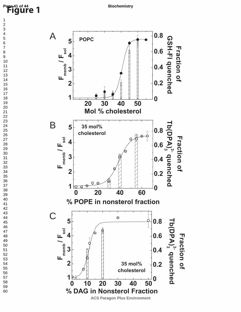
A
B
C
Figure 1Page 41 of 44
ACS Paragon Plus Environment
Biochemistry
123456789101112131415161718192021222324252627282930313233343536373839404142434445464748495051525354555657585960

AA
B
C
Figure 2 Page 42 of 44
ACS Paragon Plus Environment
Biochemistry
123456789101112131415161718192021222324252627282930313233343536373839404142434445464748495051525354555657585960

B
A
C
D E
F G
Figure 3Page 43 of 44
ACS Paragon Plus Environment
Biochemistry
123456789101112131415161718192021222324252627282930313233343536373839404142434445464748495051525354555657585960
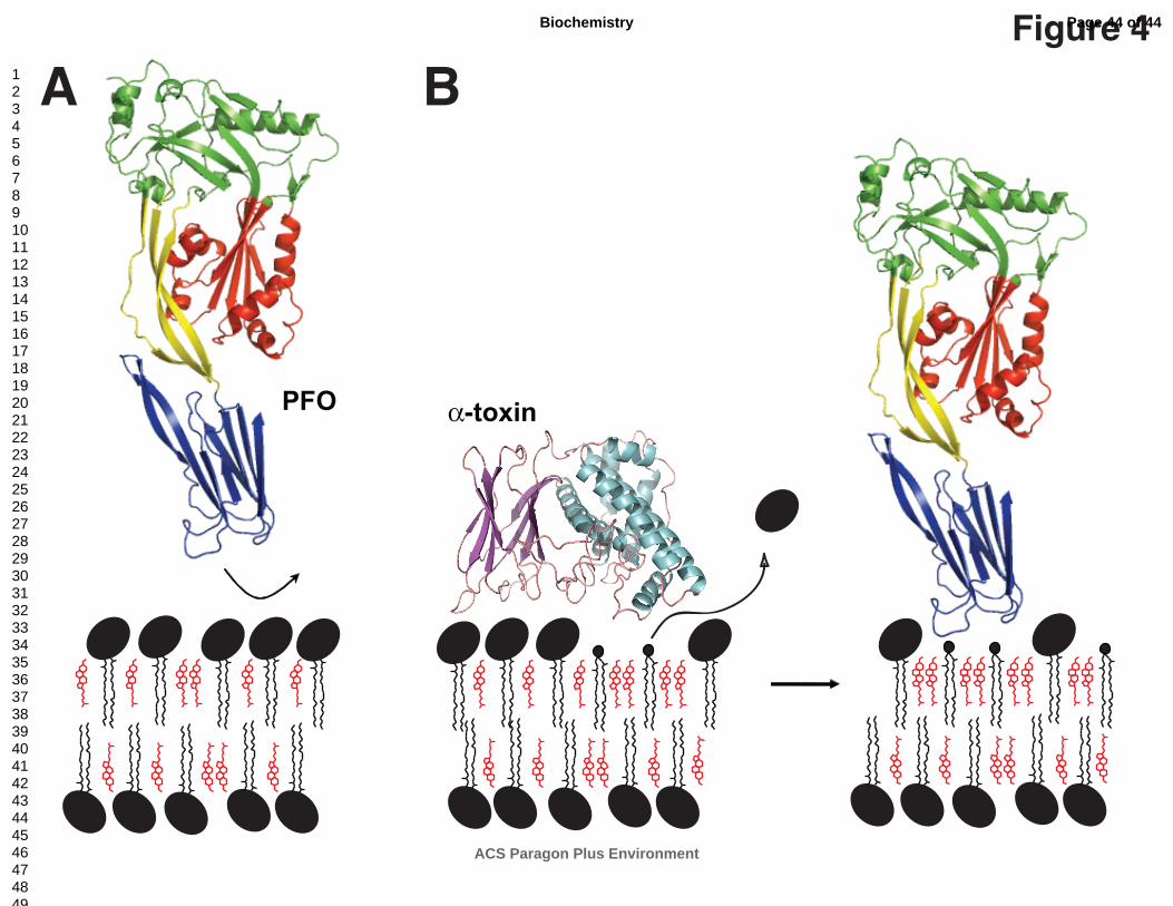
PFO α-toxin
A BFigure 4Page 44 of 44
ACS Paragon Plus Environment
Biochemistry
123456789101112131415161718192021222324252627282930313233343536373839404142434445464748495051525354555657585960


![Manager, Tax Compliance & Reporting Rosalee Lewis€¦ · American Chemical Society 1155 Sixteenth Street, N.W. Washington, D.C. 20036 T [202] 872 6306 F [202] 872 6339 Manager, Tax](https://static.fdocuments.in/doc/165x107/61295d9dac5a6078a120803d/manager-tax-compliance-reporting-rosalee-lewis-american-chemical-society.jpg)