© 2013 Adam C. Mumford All Rights Reserved
142
© 2013 Adam C. Mumford All Rights Reserved
Transcript of © 2013 Adam C. Mumford All Rights Reserved

© 2013
Adam C. Mumford
All Rights Reserved

Microbial Reduction, Precipitation, and Mobilization of Arsenic
by
Adam C. Mumford
A Dissertation submitted to the
Graduate School-New Brunswick
Rutgers, The State University of New Jersey
in partial fulfillment of the requirements
for the degree of
Doctor of Philosophy
Graduate Program in Environmental Sciences
written under the direction of
Professor Lily Y. Young
and approved by
________________________________________________
________________________________________________
________________________________________________
________________________________________________
New Brunswick, New Jersey May, 2013

ii
Abstract of the Dissertation
Microbial Reduction, Precipitation, and Mobilization of Arsenic
by Adam C. Mumford
Dissertation Director: Professor Lily Y. Young
Anaerobic microbes play a critical role in the biogeochemistry of arsenic. In this
Dissertation, I utilize both classical microbiological techniques and cutting edge
genomics to gain a greater understanding of how arsenic reducing bacteria from both
freshwater aquifers and estuarine sediments interact with As(V) as a terminal electron
acceptor. The novel As(V) reducing Strain MPA-C3 was isolated from arsenic
contaminated estuarine sediments in Hong Kong, and when grown in the presence of
sulfide with As(V) as a terminal electron acceptor, removes arsenic from solution by
precipitating it as alacranite (As8S9). Sequencing of both the 16S rRNA gene as well as
the entire genome of Strain MPA-C3 places it among the Deferribacteres. Strain MPA-
C3 is more metabolically versatile than any of the described Deferribacteres, and is able
to utilize NO3-, Se(VI), Se(IV), fumarate, Fe(III) and mixed oxidation state polysulfide as
electron acceptors, and acetate, pyruvate, fructose and benzoate as sources of carbon and
energy. The draft genome of Strain MPA-C3 has allowed for the elucidation of the
diverse pathways this organism uses for the metabolism of carbon sources, as well as
those used for the reduction of both nitrate and sulfur. The role of microbes in the
mobilization of arsenic into groundwater was studied at three sites in New Jersey:

iii
Crosswicks Creek in Upper Freehold, and Six Mile Run and Pike Run along the
Millstone River in Somerset County. At Crosswicks Creek, we determined that microbial
As(V) reduction, driven by inputs of organic carbon, resulted in the mobilization of
arsenic from iron-rich glauconitic sediments into the groundwater. The role of the redox
status of the aquifer was studied by comparing the anoxic subsurface of Six Mile Run
with the oxic subsurface at Pike Run. These two sites were found to have remarkably
similar mineralogy and groundwater chemistry, and we demonstrated that the differing
redox conditions resulted in the development of different microbial communities at each
site. As(V) reducing organisms were found at Six Mile Run by both cultivation based
and molecular methods, while these organisms were absent at Pike Run. These findings
demonstrate that microbial As(V) reduction and mobilization is facilitated by the
reducing conditions present at Six Mile Run, while it is inhibited by the oxidizing
conditions present at Pike Run. Combined, these studies demonstrate how As(V)
reducing microorganisms play a critical role in the biogeochemical cycling of arsenic.

iv
Dedication
This dissertation is dedicated to Florence Mumford and Peggy Marcus
You each inspired me in your own way
And everyone else who has in some way contributed to this Journey
You know who you are, but you cannot know how much you’ve done for me

v
Table Of Contents
Abstract of the Dissertation ................................................................................................ ii
Dedication and Acknowledgment ...................................................................................... iv
Table of Contents .................................................................................................................v
List of Tables ..................................................................................................................... ix
List of Figures ......................................................................................................................x
Chapter 1: Introduction and Literature Review............................................................1
1.1. Introduction .......................................................................................................1
1.2 Microbial Respiratory Arsenate Reduction ......................................................1
1.3. Diversity of Arsenate Reducing Microbes ........................................................3
1.4 arrA as A Biomarker for Microbial Arsenate Reduction ...................................4
1.5 Microbial Precipitation of Arsenic Sulfides ......................................................5
1.6 Microbial Arsenic Mobilization .........................................................................6
1.6.1 Sources of arsenic in the environment ................................................6
1.6.2 Microbial Mobilization of Arsenic into Groundwater ........................8
Chapter 2. Precipitation of Alacranite (As8S9) by a Novel As(V) Respiring
Anaerobe, Strain MPA-C3 ..............................................................................................16
2.1. Introduction .....................................................................................................18
2.2. Materials and Methods ....................................................................................20
2.2.1 Enrichment and Isolation ..................................................................20

vi
2.2.2. Survey of electron donors and terminal electron acceptors .............22
2.2.3. Chemical Analyses...........................................................................23
2.2.4 Molecular Analysis ...........................................................................23
2.2.5 Genomic Analysis .............................................................................24
2.2.6 Mineral Analyses ..............................................................................25
2.3 Results ..............................................................................................................26
2.3.1 Characterization of Strain MPA-C3 ..................................................26
2.3.2 Characterization of Mineral Precipitate ............................................27
2.3.3 Genomic Characterization of Strain MPA-C3 ..................................29
2.4 Discussion ........................................................................................................33
2.5 Conclusions ......................................................................................................36
2.6 Supplemental Material .....................................................................................36
2.6 Acknowledgements ..........................................................................................36
Chapter 3: Microbial Transformations of Arsenic: Mobilization from Glauconitic
Sediments to Water ..........................................................................................................45
3.1 Introduction ......................................................................................................47
3.2 Methods............................................................................................................49
3.2.1 Sample Collection .............................................................................49
3.2.2 Microbiological Analysis ..................................................................50
3.2.3 Chemical and Mineral Analysis of Core Sediments .........................53
3.3 Results and Discussion ....................................................................................55

vii
3.3.1 Sediment Chemistry and Mineralogy ...............................................55
3.3.2 Microbes in Streambed Sediments and the Aquifer .........................56
3.3.3 Molecular Characterization of Groundwater and Streambed
Microbes ...........................................................................................58
3.4 Conclusions ......................................................................................................60
3.5 Supporting Information ....................................................................................62
3.6 Acknowledgement ...........................................................................................62
Chapter 4: Groundwater redox controls microbial release of arsenic from minerals
to groundwater in a fractured rock terrain, New Jersey, USA .......................70
4.1 Introduction ......................................................................................................72
4.2 Methods............................................................................................................74
4.2.1 Groundwater and streamwater sample collection .............................74
4.2.2 Sampling for Microbiological Analysis ............................................76
4.2.3 Streambed-Sediment Sampling .........................................................76
4.2.4 Sample Analysis................................................................................76
4.2.5 DNA Extraction and Amplification ..................................................77
4.2.6 Denaturing Gradient Gel Electrophoresis .........................................78
4.2.7 Sequence Analysis ............................................................................79
4.2.8 Preparation of Groundwater Microcosms .........................................80
4.3 Results ..............................................................................................................81
4.3.1 Stream Sediment Mineralogy and Chemistry ...................................81

viii
4.3.2 Groundwater Geochemistry ..............................................................82
4.3.3 Streamwater Chemistry .....................................................................84
4.3.4 Results of Microcosm Studies ..........................................................85
4.3.5 Molecular Characterization of Microbial Communities at Six Mile
Run and Pike Run ..........................................................................85
4.3.5.1 Characterization of the 16S rRNA gene ............................85
4.3.5.2 Characterization of the Arsenate Respiratory Reductase
(arrA) gene .........................................................................87
4.4 Discussion ........................................................................................................87
4.5 Conclusions ......................................................................................................90
4.6 Supplemental Material .....................................................................................92
4.7 Acknowledgments............................................................................................92
Chapter 5: Conclusions and Future Directions..........................................................101
Appendices ......................................................................................................................107
Appendix to Chapter 2 .........................................................................................107
Appendix to Chapter 3 .........................................................................................109
Appendix to Chapter 4 .........................................................................................113
References .......................................................................................................................118

ix
List of Tables
Chapter 1
Table 1. Summary of Electron Donors and Acceptors used by Characterized Respiratory
As(V) reducing bacteria ...................................................................................12
Chapter 2
Table 1. Summary of Electron Donors and Acceptors used by Characterized Respiratory
As(V) reducing bacteria ...................................................................................38
Chapter 3
Table 1. Contents of selected major and trace elements in a streambed sediment core
and mineral separates from Crosswicks Creek at site C6, August, 2009 ........63
Table 2. Semi-quantitative mineralogy reported in wt% in selected intervals of a
streambed sediment core, Crosswicks Creek, NJ, August 2009 ......................64
Chapter 4
Table 1. Mineralogy of streambed sediments at sampling sites on tributaries Six Mile
Run and Pike Run, Millstone River watershed, 2011 ......................................93
Table 2. Contents of total major and trace elements in streambed sediments from Six
Mile Run and Pike Run, Millstone River watershed .......................................94
Table 3. Concentrations of selected constituents in streamwater and groundwater at
Six Mile Run and Pike Run .............................................................................95

x
List of Figures
Chapter 1
Figure 1. Phylogenetic Tree of arsenic respiring organisms ...........................................13
Figure 2. Cluster analysis of arrA sequences from diverse worldwide sites ..................14
Figure 3. Conceptual model of arsenic mobilization and transport ................................15
Chapter 2
Figure 1. Stoichiometric reduction of As(V) to As(III) by Strain MPA-C3 ...................39
Figure 2. Phylogenetic trees of Strain MPA-C3 based on both 16S rRNA and arrA
genes ................................................................................................................40
Figure 3. Images of Strain MPA-C3 and alacranite precipitate ......................................41
Figure 4. X-Ray Diffractogram of MPA-C3 mineral precipitate ....................................42
Figure 5. Schematic representation of metabolic pathways in MPA-C3 ........................43
Figure 6. Alignments between Nap and Psr genes of MPA-C3 and Deferribacter
Desulfuricans ...................................................................................................44
Chapter 3
Figure 1. Map of NJ Coastal Plain, including Crosswicks Creek Watershed and
Site C6 ..............................................................................................................65
Figure 2. Microcosm results from CRO-C6 demonstrating As(V) reduction .................66
Figure 3. Total arsenic release from amended streambed sediments ..............................67
Figure 4. Microbial communities found in groundwater and sediment at Site C6 .........68
Figure 5. Conceptual diagram of pathways for microbial release of arsenic from
glauconite .........................................................................................................69

xi
Chapter 4
Figure 1. Map of central NJ, with Millstone River, Six Mile Run, and Pike Run sites
Marked .............................................................................................................96
Figure 2. Results from microcosms inoculated with material from Six Mile Run and
Pike Run ...........................................................................................................97
Figure 3. Taxnomic classification of 16S rRNA gene OTUs recovered from Six Mile
Run and Pike Run ............................................................................................98
Figure 4. Image of DGGE comparing 16S rRNA gene amplicons recovered from Six
Mile Run and Pike Run ....................................................................................99
Figure 5. Venn diagram illustrating different microbial communities found at Six Mile
Run and Pike Run ..........................................................................................100

1
Chapter 1
Introduction and Literature Review
1.1 Introduction
Over the course of history, arsenic has gained infamy as a poison. It has only
been in recent years that research has demonstrated that arsenic can also play a vital role
in the microbial world. Current research has demonstrated the ability of microbes to
utilize arsenic ions as both electron donors and acceptors. This research seeks to extend
these discoveries by placing As(V) reducing microorganisms into an environmental and
geochemical context.
1.2 Microbial Respiratory Arsenate Reduction
Microbes generate energy for both heterotrophic and autotrophic growth by
coupling the oxidation of organic carbon to the respiratory reduction of As(V) to As(III)
(3), and by fixing inorganic carbon by coupling the reduction of CO2 to the oxidation of
As(III) to As(V) (105). Microbial respiratory reduction of As(V) to As(III) was first
described in 1994 by Ahmann et al. in the ε-proteobacterium MIT-13 (3, 137). This
organism was found to utilize As(V) as a sole electron acceptor coupled to the oxidation
of lactate, and is reported to release arsenic from ferrous arsenate by the reductive
dissolution of As(V) (3).
Dissimilatory reduction of arsenate to arsenite proceeds through the arsenate
respiratory reductase (ArrAB) system (124). Under standard conditions, the respiratory
reduction of As(V) to As(III) is exergonic, with a ΔG0’ of -175 kJ mol-1 of carbon

2
oxidized (65). When acetate is used as an electron donor, the stoichiometry of this
reaction is CH3COO- + 4HAsO42- + 7H+ 2HCO3
- + 4H3AsO3. The terminal transfer of
electrons to As(V) is catalyzed by the As(V) respiratory reductase (ArrAB) system,
which was first purified from Chrysiogenes arsenatis by Krafft and Macy (66), and has
since also been purified from Bacillus selenitireducens (1) and Shewanella sp. Strain
ANA-3 (82). Arsenate respiratory reductase is reported to be a periplasmic enzyme,
although the ArrAB from B. selenitireducens was reported to be associated with the
external side of the cell membrane (1, 66, 82). In all cases, it appears that the enzyme is
positioned to ensure that arsenate is not brought into the cell (82). The α-subunit of the
enzyme is described to have a molecular mass of 87 kDa (66), 110 kDa (1), or 95 kDa
(82), and the smaller β-subunit is reported to have a molecular mass of 29 kDa (66), 34
kDa (1), or 27 kDa (82). The αβ holoenzyme has a molecular mass reported to be 123
kDa (29), 150 kDa (1), or 131 kDa (35). The ArrA subunit is reported to contain the
molybdopterin catalytic center, as well as at least one [Fe4S4] cluster (1, 66, 82). ArrB
coordinates an iron-sulfur cluster, and is suggested to play a role in electron transfer to
the catalytic α subunit(1). Notably, in 2009, Richey et al. proposed that ArrAB can
function as an arsenite oxidase in Alkalimnicola ehrlichii (119), and this finding has since
been expanded with the description of the arxA clade of arsenite oxidases (152, 153).
This finding, when taken together with the limited number of arsenic reducing
microorganisms which have been isolated, suggests that many new microbial
mechanisms of arsenic metabolism remain to be discovered by both traditional and
molecular techniques.

3
1.3 Diversity of Arsenate Reducing Microbes
Dissimilatory As(V) reducing prokaryotes (DARPs) have been reported from 17
(92, 104) bacteria phyla, as well as 1 archeal phylum (55). A phylogenetic tree of these
organisms demonstrating their diversity based on the 16S rRNA gene is presented in
Figure 1. In Table 1 I summarize 14 characterized As(V) reducing isolates along with
two members of the Deferribacteres as is presented in Mumford et al. (92). A review of
terminal electron acceptors utilized by these isolates allows for the division of these
organisms into two broad groups based on their ability or inability to utilize SO42- and/or
S2O32-. Among the organisms incapable of utilizing SO4
2- or S2O32- we note that three
organisms, G. ferrireducens, D. desulfuricans, and Halarsenitibacter silvermanii SLAS-1
are reported to reduce elemental sulfur and/or polysulfide to S2-, a trait shared by strain
MPA-C3, which I describe in Chapter 2 (25, 34, 92, 143). Of the As(V) respiring
organisms in Table 1 which are unable to utilize SO42- or S2O3
2-, all except Bacillus
selenitireducens MLS-10 (141) are able to respire NO32- and/or Fe(III), suggesting an
adaptation for life at lesser reducing conditions in comparison to the SO42- and/or S2O3
2-
reducing organisms.
At the time of this writing only limited information is available about the range of
organic substrates utilized by the dissimilatory As(V) reducing bacteria. In Chapter 2, I
discuss my isolation of an interesting new organism, Strain MPA-C3, which utilizes a
wider range of organic compounds as a source of carbon and energy than any of the
currently described Deferribacteres, and at least as wide a range as any of the other
described As(V) respiring organisms (Table 1). The ability to utilize pyruvate as a source

4
of carbon and energy appears to be conserved among the described DARPs and, indeed,
only a single isolate among the described DARPs is incapable of utilizing pyruvate (26,
34, 46, 64, 76, 79, 87, 95, 100, 101, 113, 114, 123, 137, 141, 143, 144). Among the
As(V) respiring bacteria, only Bacillus arsenicoselenatis E1H is reported to be incapable
of utilizing pyruvate (141), and only Desulfosporosinus sp. Y5 is reported to utilize
aromatic substrates, including benzoate and toluene (76).
1.4 arrA As a Biomarker for Microbial Arsenate Reduction
The arrA gene has been found in all dissimilatory arsenic reducing bacteria
(DARBs) described to date (1, 79, 82, 83, 100, 104, 107, 124, 136, 138). The use of arrA
as a biomarker for microbial As(V) respiration was first described by Malasarn et al. in
2004, and the gene has been successfully used in a number of studies since (73, 83, 90,
134). While arrA appears to be conserved among the arsenate respiring bacteria, enough
difference appears to exist to determine evolutionary relatedness (73, 83, 134). Figure
two displays a phylogenetic tree based on the arrA genes recovered from freshwater
aquifers, marine, and hypersaline environments, and shows that samples from
groundwater aquifers cluster together (Six Mile Run, Pike Run, and Crosswicks Creek
(90, 91)), distinct from the cluster formed by samples taken from hypersaline lakes
(Mono and Searles Lakes(67)), and marine/estuarine sites (Chesapeake Bay (134) and the
Hackensack River (unpublished data)). This suggests that environmental conditions play
a stronger role in the development of DARP communities than geographical location.

5
1.5 Microbial Precipitation of Arsenic Sulfides
Arsenic sulfide minerals present a major sink of arsenic to the environment.
Arsenic has been found to be closely associated with sulfur, both as arsenic sulfide
minerals (eg orpiment, realgar) as well as iron-sulfide-arsenic minerals, particularly
arsenopyrite (9, 47, 131). Arsenic sulfide minerals have been described as forming
geochemically under the high temperatures and pressures found within the Earth’s crust,
and also under hydrothermal conditions (9, 47, 88, 131). The common arsenic sulfide
mineral, realgar (As4S4), has been described as forming in the condensation zone of
hydrothermal fluids at temperatures of 50-75oC and particularly in veins of silver, lead,
and gold ores (36, 88). Several polymorphs of realgar, including alacranite, uzonite, and
pararealgar have also been described as forming under similar conditions (23, 28, 109,
110). Alacranite, a polymorph of realgar with the formula As8S9 has been identified at
several locations worldwide, as parts of hydrothermal and volcanic deposits (28, 33, 35,
88, 110), and uzonite (As4S5) has been both synthesized (150) and described in nature
(23, 109). To date, microbial activity has never been directly linked to the formation of
realgar or any of its polymorphs under mesophilic conditions, although the microbial
formation of realgar has been described under hyperthermophilic conditions by the
archea Pyrobaculum arsenicum (55), and both O’Day et al.(102) and Demergasso et
al.(39) suggest the potential for microbial formation of realgar under mesophilic
conditions.
The precipitation of arsenic sulfides by microbial activity has been described (39,
63, 74, 100, 120). Rittle et al. (1995) reported the potential for a microbial influence on

6
the precipitation of arsenic sulfides, and the first direct evidence of microbial orpiment
(As2S3) precipitation by the SO42- and As(V) respiring organism Desulfosporosinus
auripigmentum was shown by Newman et al. (1997). Additionally, Demergasso et al.
(2007) suggest that arsenic sulfide deposits comprised of realgar and orpiment in Andean
salt flats are potentially microbial in origin. In Chapter 2, I describe the formation of
alacranite by a novel, As(V) reducing isolate, Strain MPA-C3. Growth of this organism
with As(V) as a terminal electron acceptor and in the presence of sulfide resulted in the
precipitation of alacranite, a mineral previously reported to form only under hydrothermal
conditions. Strain MPA-C3 was isolated from arsenic contaminated sediments from Mai
Po estuary in Hong Kong, where sulfide would be plentiful as a result of the activity of
sulfate reducing microbes. This mineral formed by Strain MPA-C3 provides an
interesting route by which arsenic could be immobilized in the shallow subsurface of
estuarine environments.
1.6 Microbial Arsenic Mobilization
1.6.1 Sources of arsenic in the environment
Arsenic occurs as a major constituent in a wide variety of minerals, however, only
a limited number of these appear to act as major sources of arsenic to groundwater.
Arsenic sulfides comprise a significant source of arsenic in the environment, and of these,
the most common arsenic sulfide mineral is arsenopyrite, FeAsS, which is found
predominantly in mineral veins (131). The sulfide minerals realgar, AsS, and orpiment,
As2O3, have been predominantly found in hydrothermal and volcanic deposits (131). In

7
pyrites, arsenic replaces sulfur within the crystal structure, allowing for very high
concentrations of arsenic to be incorporated (131). For example, pyrites with arsenic
concentrations of up to 77,000 mg/kg have been reported, and concentrations up to
126,000 mg/kg have been observed the iron-sulfur mineral marcasite (131). Iron oxide
minerals also have the potential to contain high levels of arsenic, with concentrations up
to 76,000 mg/kg seen in Fe(III) oxyhydroxides (131). As such, it is not surprising that
sedimentary rocks composed of these minerals have been found to have elevated
concentrations of arsenic. Arsenic concentrations of up to 490 mg/kg have been found in
shales, and iron formations and iron rich sediments are reported to have arsenic
concentrations up to 2900mg/kg (131).
In Southeast Asia arsenic is associated with iron oxide minerals in alluvial
deposits of sediments weathered and transported from the Himalayas, and the
mobilization of this arsenic into drinking water has become a major public health crisis in
Southeast Asia, most notably in Bangladesh, (10), the Bengal Delta of India(56, 133),
Cambodia (73) and Vietnam (19). In Bangladesh elevated levels of arsenic in
groundwater has been implicated in 1 out of every 5 deaths and has been called ”the
world’s greatest mass poisoning”(10). In Southeast Asia, microbial reduction of iron
oxides releases bound arsenic into groundwater, which is then withdrawn from the
aquifer for human consumption and agriculture(6, 20, 44, 51, 52, 81, 111, 140). Harvey
et al. (51) in particular demonstrate that this microbial activity is stimulated by organic
carbon, which has been introduced to the subsurface as a result of intensive abstraction of
groundwater.

8
In New Jersey, black shale underlying the Newark Basin of New Jersey has been
found to have arsenic concentrations up to 240 mg/kg, potentially providing a source of
arsenic to groundwater (130). Arsenopyrite minerals within these shales have been found
to be very highly enriched in arsenic, with arsenic comprising 4% by weight of the
pyrites (130). Wells drawing water from fractured shale bedrock of the Piedmont
Physiographic Province of New Jersey have been found to have the highest
concentrations of arsenic, with nearly 25% of the wells tested having arsenic levels in
excess of 5µg/L (93). The glauconitic sediments of the Coastal Plain of New Jersey have
been found to have levels of arsenic ranging from 7.1-131 mg/kg (41, 90). To date,
however, only a limited number of wells in this region have been tested. While many
thousands of tons of arsenical pesticides have been applied to the agricultural soils of
New Jersey, they are not believed to provide a major source of arsenic to groundwater
(94). It is believed that the arsenic constituent of these pesticides is immobilized on Al
and Fe minerals in the topsoil, and is not leaching into the groundwater (94).
1.6.2 Microbial Mobilization of Arsenic into Groundwater
Microbes have been implicated in the mobilization of arsenic into groundwater
under a variety of conditions. Arsenate in groundwater is bound much more strongly to
iron oxide minerals than arsenite, suggesting that microbial arsenate reduction may play a
role in arsenic mobility (42). The ability of As(V) reducing microbes to release arsenic
into solution was first described by Ahmann et al in 1994 concurrently with the discovery
of the first As(V) reducing microbe, Sulfurospirillum arsenophilum MIT-13 (3). This

9
organism was found to dissolve ferrous arsenate via the reduction of As(V) to As(III),
with the As(III) being released into the mobile phase (3). Further experimentation by
Ahmann et al. demonstrated that S. arsenophilum strain MIT-13 was capable of
mobilizing both As(III) and Fe(II) from arsenic contaminated aquifer sediments,
indicating that iron reduction may also play a role in arsenic mobilization (2). These
findings have since been replicated with the As(V) and Fe(III) reducing organism
Shewanella sp. Strain ANA-3 (56, 140). This organism was demonstrated to reduce
As(V) which had been sorbed to ferrihydrite as well as the ferrihydrite itself, resulting in
the mobilization of arsenic into the mobile phase (56, 140). In addition to the
mobilization of arsenic from iron oxide minerals, Zhu et al. report that sulfide produced
by bacterial sulfate reduction may mobilize arsenic via an arsenide/sulfide exchange
(154). This mechanism has been suggested as a microbial route to arsenic release from
arsenopyrites in black shale aquifers (154). A separate mechanism for mobilization from
arsenopyrites in black shales has been described by Rhine et al., who demonstrated that
under oxidizing conditions, the sulfur oxidizing Strain WAO mobilized arsenic from
arsenopyrite following the oxidation of sulfur (117). In Chapter 3, I present the work
published in Mumford et al. (90). I describe how microbial activity, stimulated by inputs
of organic carbon, drives the mobilization of arsenic from iron-rich glauconitic sediments
into the shallow groundwater beneath Crosswicks Creek in Upper Freehold, NJ. By
spiking sediments from this site with As(V) and utilizing them as inocula for
microcosms, we were able to demonstrate the importance of microbial action in the
mobilization of arsenic. This was demonstrated by spiking microcosms prepared with

10
site sediment, and measuring both the concentration and speciation of arsenic released.
We found that while the sediments had a high capacity to bind As(V), As(III) was
released only by microbial activity, and was present in the mobile phase at a
concentration similar to that observed at the site (~25 µg/L).
In Chapter 4, I describe how, again in collaboration with the USGS, I examined
the potential for microbial As mobilization at two sites along the Millstone River, in
Somerset County, NJ. This research has been submitted to Water Research as
“Groundwater redox controls microbial release of arsenic from minerals to groundwater
in a fractured rock terrain, New Jersey, USA” and is currently under review. The two
sites, Six Mile Run and Pike Run, displayed very similar mineralogy and geochemistry,
with the notable difference being that the groundwater at Six Mile Run was anoxic and
reducing, and the groundwater at Pike Run was oxic and oxidizing. We hypothesized
that these differing redox conditions would lead to the development of differing microbial
communities, and that this would in turn explain why the levels of As present at Six Mile
Run were an order of magnitude higher than those at Pike Run. By following a similar
research methodology as described in Chapter 3, I determined through culture-
independent molecular techniques that the microbial communities at each site were in
fact different, and these findings were supported by the results of microcosm
experiments. In these experiments, I found that microcosms inoculated with groundwater
taken from Six Mile Run were capable of As(V) reduction, while those inoculated with
groundwater from Pike Run were not. This finding was particularly interesting in that the

11
arrA gene was successfully amplified from the Pike Run site, a finding which I suggest
may be due to the very similar As(III) oxidase gene (arxA).
The findings from these two studies, combined with the wealth of information
available from the ongoing studies in Southeast Asia and particularly the Bengal Delta
region, allow for the construction of a conceptual model of arsenic release and transport
in the shallow subsurface. In Figure 3, I present this conceptual model. Both previous
reports and my own work convincingly show that geogenic arsenic is frequently
associated with iron oxide minerals in subsurface aquifers. Arsenic is released into
groundwater by several mechanisms, including direct reduction of As(V) sorbed to
mineral surfaces (155), and release of arsenic following reductive dissolution of the iron
mineral by iron reducing microorganisms (56, 145). As shown in Mumford et al. (90),
released As(V) is reduced to As(III) by As(V) reducing microbes, and this As(III)
remains mobile in the groundwater. On gaining reaches beneath a river, As(III) would
then be brought to the oxic sediment water interface by groundwater flow, at which point
it is oxidized to As(V) either abiotically or by As(III) oxidizing microbes (153), and then
re-sorbs to newly precipitated iron oxides in the streambed (91). During periods of high
flow and disturbance, this streambed material may be carried far downstream and buried,
moving the arsenic far from its original source, and allowing this microbial arsenic cycle
to begin anew.

12
Table 1: N/T: Not Tested. References: 1(64, 95), 2(34), 3(142, 144), 4(79), 5(113, 114),
6(87), 7(25), 8(101), 9(76), 10(99), 11(46), 12(141), 13(123), 14(137)

13
Figure 1:
Phylogenetic tree of arsenic respiring organisms based on the 16S rRNA gene. As(V)
reducing organisms are in blue, As(III) oxidizing organisms are in red.

14
Figure 2:
Cluster analysis of arrA sequences from diverse worldwide sites. Freshwater sites cluster
separately from marine and hypersaline sites.

15
Figure 3:
Conceptual Model of arsenic mobilization and transport. Arsenic can be mobilized by
either (A) direct reduction of As(V) sorbed to mineral surfaces, or (B), reductive
dissolution of iron oxides by iron reducing bacteria. Once released, As(V) is reduced to
As(III) by As(V) reducing microbes (C), and carried to the oxic sediment water interface
by groundwater flow. At the sediment water interface, As(III) is re-oxidized, and binds
to freshly precipitated iron oxides (D).
A
B
C
D

16
Chapter 2
Precipitation of Alacranite (As8S9) by a Novel As(V)-Respiring
Anaerobe Strain MPA-C3
Abstract
Strain MPA-C3 was isolated by incubating arsenic-bearing sediments under
anaerobic, mesophilic conditions in minimal media with acetate as the sole source of
energy and carbon, and As(V) as the sole electron acceptor. Following growth and the
respiratory reduction of As(V) to As(III), a yellow precipitate formed in active cultures,
while no precipitate was observed in autoclaved controls, or in uninoculated media
supplemented with As(III). The precipitate was identified by X-ray diffraction as
alacranite, As8S9, a mineral previously only identified in hydrothermal environments.
Sequencing of the 16S rRNA gene indicated that strain MPA-C3 is a member of the
Deferribacteres family, with relatively low (90%) identity to Denitrovibrio acetiphilus
DSM 12809. The arsenate respiratory reductase gene, arrA, was sequenced, showing
high homology to the arrA gene of Desulfitobacterium halfniense. In addition to As(V),
Strain MPA-C3 utilizes NO3-, Se(VI), Se(IV), fumarate, and Fe(III) as electron acceptors,
and acetate, pyruvate, fructose and benzoate as sources of carbon and energy. Analysis
of a draft genome sequence revealed multiple pathways for respiration and carbon
utilization. The results of this work demonstrate that alacranite, a mineral previously
thought to be formed only chemically under hydrothermal conditions, is precipitated
under mesophilic conditions by the metabolically versatile Strain MPA-C3.

17
This chapter was co-authored with Professor Lily Y. Young and Professor Nathan Yee,
has been submitted to Environmental Microbiology, where it has been accepted for
publication.

18
2.1 Introduction
Arsenic sulfide minerals are widespread in the environment, and are found in
diverse geologic settings, including continental margins, river sediments, and
hydrothermal deposits (9, 47, 131). The common arsenic sulfide minerals realgar (As4S4)
and orpiment (As2S3) are reported to form in the condensation zone at temperatures of
50-75oC in hydrothermal fluids and ore deposits, and particularly in veins of silver, lead,
and gold ores (36, 88). Additionally, the formation of realgar has been reported in
mesophilic, estuary sediments in San Francisco Bay (102). These minerals, along with
iron sulfide minerals such as arsenopyrite and marcasite, are thought to comprise much of
the 2-8 mg/kg of arsenic found in continental margin and river sediments (131). Several
polymorphs of realgar, including alacranite, uzonite, and pararealgar have also been
characterized. Alacranite (As8S9) has been identified at as a component of hydrothermal
and volcanic deposits as several locations (28, 33, 35, 88, 110), and uzonite (As4S5) has
been both synthesized (150) and described in natural samples (23, 109). Although the
microbial formation of realgar has been found to occur under hyperthermophilic
conditions (55), the role of microorganisms in the formation of realgar and its
polymorphs under mesophilic conditions remain poorly understood.
A growing list of anaerobic prokaryotes have been reported to be capable of
generating energy via the respiratory reduction of arsenate [As(V)] to arsenite [As(III)],
(55, 76, 79, 80, 98, 99, 104, 124, 137, 142), and As(V) reduction has been implicated as
an important microbial process in the biologic formation of arsenic sulfide minerals (55,
99). The first direct evidence of microbial orpiment (As2S3) precipitation by the SO42-
19
and As(V) respiring organism Desulfosporosinus auripigmentum was shown by Newman
et al. (1997). Previous studies have also reported the precipitation of arsenic sulfides by
the reaction of biogenic sulfide with dissolved arsenic oxyanions in laboratory
incubations of contaminated aquifer sediments (Rittle et al., 1995; Kirk et al., 2010).
Additionally, Demergasso et al. (2007) suggested that arsenic sulfide deposits comprised
of realgar and orpiment in Andean salt flats have a potential microbial origin. Finally,
recent work has shown that Shewanella species can form arsenic sulfide nanotubes during
As(V) and thiosulfate reduction (58, 74).
While diverse prokaryotes are able to reduce As(V) for growth, this respiratory
process remains poorly understood in the Deferribacteres, a distinct phylum within the
Bacteria consisting of anaerobic organotrophs. Previous studies have shown that
Deferribacteres can utilize terminal electron acceptors such as NO3-, Fe(III), Mn(IV),
Co(III), and S0 (48). To date, a total of 26 isolates have been described, comprising six
genera within the Deferribacteres (96). The majority of these organisms have been
isolated from high-temperature environments, but several strains have been isolated
under more moderate conditions (48). These organisms are reported to utilize acetate and
lactate, however, their ability to utilize more complex carbon sources has not been fully
assessed (34, 48, 95, 143). The type strain of the Deferribacteres, Geovibrio
ferrireducens, is characterized by the ability to respire ferrous iron and elemental sulfur.
It is unable, however, to respire NO3-, and its ability to respire As(V) has not been
determined. Among the described Deferribacteria, only Deferribacter desulfuricans has
been reported to grow at the expense of dissimilatory reduction of As(V) to As(III) (143).
20
In this study, we report the isolation of a novel As(V)-respiring bacterium
designated as strain MPA-C3 from arsenic-bearing sediments collected at Mai Po Marsh,
Hong Kong. Phylogenetic analysis of the 16S rRNA gene indicates that strain MPA-C3 is
a novel member of the Deferribacteres family. We show that during As(V) respiration,
strain MPA-C3 forms a yellow precipitate in close association with the cells. We further
demonstrate that this yellow precipitate formed under circumneutral, mesophilic
conditions by strain MPA-C3 is alacranite, a mineral previously only known to be formed
under hydrothermal conditions. Finally, metabolic and genomic characterization of
strain MPA-C3 reveals that this organism has a wider range of electron acceptors than
any previously described Deferribacteres isolates. The results of this work provide new
insights in the route of microbial arsenic sulfide precipitation under mesophilic
conditions, and elucidate a potential geochemical role for the Deferribacteres in
sedimentary arsenic sulfide biomineralization.
2.2 Materials and Methods
2.2.1 Enrichment and Isolation
Strain MPA-C3 was isolated from sediments sampled from the Mai Po Marsh,
Kowloon, Hong Kong. The marsh is part of the Pearl River Estuary system, and the
sediment at this site contains arsenic concentrations of approximately 20mg/kg (Li Meng,
Personal Communication, 2008). Sediment enrichment cultures from Mai Po site A were
prepared by inoculating 40mL of defined freshwater anaerobic medium (ABF) with
approximately 4 g of composited sediment. The composited sediment was incubated in

21
an anaerobic basal medium composed of 1 g/L NaCl, 400 mg/L MgCl2.2H2O, 100 mg/L
CaCl2.2H2O, 200 mg/L KH2PO4, 500 mg/L KCl, 190 mg/L Na2HPO4, 60 mg/L NaH2PO4,
270 mg/L NH4Cl, 1.05 g/L MOPS buffer (pH 7.2), 31mg/L Na2S, 1.5 mg/L
nitrilotriacetic acid, 0.8 mg/L Fe(NH4)2(SO4)2, 0.2 mg/L Na2SeO3, 0.1 mg/L CoCl2.6H2O,
0.1 mg/L MnSO4.H2O, 0.1 mg/L Na2MoO4
.H2O, 0.1 mg/L NaWO4.H2O, 0.1 mg/L
NiCl2.6H2O, 0.01 mg/L H3BO3, 0.01 mg/L CuSO4
.5H2O, 0.1 µg/L nicotinic acid, 0.1
µg/L calcium pantothenate. 0.1 µg/L pyroxidine HCl, 0.1 µg/L riboflavin, 0.05 µg/L
biotin, 0.05 µg/L folic acid, 0.05 µg/L α-lipoic acid, 0.05 µg/L vitamin B-12, 48.5 mg/L
cysteine. The pH of the media was adjusted to 7.2. Triplicate active enrichment cultures
were inoculated under anaerobic conditions with 4g of Mai Po Marsh sediment, and
amended with 2mM NaH2AsO4 as a terminal electron acceptor and 2mM sodium acetate
as a source of carbon and energy. Duplicate sterile controls were prepared in the same
manner to the active enrichment cultures prior to sterilization by autoclaving (3 times
over 3 consecutive days). A background control was prepared by inoculating sediment
into media which was not supplemented with As(V) or acetate. All cultures were
incubated in the dark at 25oC. The reduction of As(V) to As(III) was monitored by
HPLC as described by Perez-Jimenez et al. (2005). Following the observation of
complete reduction of As(V) to As(III), the enrichment cultures were diluted 1:10 in ABF
media amended with 2mM NaH2AsO4and 2mM sodium acetate. Following six 1:10
dilutions, the enrichment culture was dominated by a single morphology comprised of
bent rods, and a yellow precipitate was noted. The culture was plated under anaerobic
conditions onto ABF media solidified with 1% noble agar and amended with 5mM

22
NaH2AsO4 and 5mM sodium acetate. After 21 days of incubation at 25oC, isolated
yellow colonies were observed, and were transferred to 3mL ABF amended with 2mM
NaH2AsO4 and 2mM sodium acetate.
2.2.2 Survey of electron donors and terminal electron acceptors
The ability of strain MPA-C3 to utilize the following carbon sources was tested:
Acetate (5mM) Benzoate (5mM), fructose (5mM), pyruvate (5mM), palmitate (5mM),
lactate (5mM), toluene (1mM), hexadecane (2mM), and syringic acid (5mM). The
following terminal electron acceptors were tested: As(V), Se(VI), Se(IV), Fe(III) citrate,
amorphous Fe(III)(O)OH, NO3-, NO2
-, SO42-, S2O3
2- and mixed oxidation state
polysulfide. All terminal electron acceptors were supplied at a concentration of 5mM.
Growth on alternative carbon sources was considered positive if reduction of As(V) to
As(III) was observed. Growth on Fe(III) citrate and Fe(O)OH was determined by a
modified ferrozine assay (147). Growth on Se(VI) and Se(IV) was measured by HPLC as
described below. Growth on NO3-, NO2
-, SO42-, S2O3
2-, and polysulfide was determined
by an increase in optical density at 580 nm as measured on a Spectronic Instruments
Spectronic 20D+ spectrophotometer (Thermo Fisher Scientific, Waltham MA) relative to
sterile controls.
Reagents:
All reagents used were of reagent grade and used without further purification.
Stock solutions were prepared by dissolution in degassed MilliQ reagent grade water
(Millipore, Billerica MA) and stored under argon until use.

23
2.2.3 Chemical Analyses
Liquid samples of 600 µL were removed from culture bottles for analysis with an
argon-flushed 1mL syringe with a 22 gauge needle. Particulates were filtered from
samples with Costar SpinX 0.22 µM nylon centrifuge filters (Corning Life Sciences,
Pittston PA), centrifuged at 14,000 x g for 2 minutes.
Arsenic and selenium ions were analyzed by HPLC (Shimadzu, Columbia MD
and Beckman Coulter, Brea CA) using a Hamilton PRP-X100 anion exchange column
(Hamilton, Reno NV). Arsenic ions were eluted with 40mM NaH2PO4 at pH 5.0 flowing
at 1 mL min-1 and detected at 195 nm. Selenium ions were eluted with 12.5mM
(NH4)3PO4 at pH 8.5 flowing at 1 mL min-1 and detected at 201 nm in a modification of
the method reported by Guérin et al. (1997). Acetate was analyzed by HPLC (Beckman
Coulter, Brea CA) using a Resex ROA organic acid column. (Phenomenex, Torrance
CA). Acetate was eluted with 5 µM sulfuric acid flowing at 0.5 mL min-1 and detected at
210 nm.
Soluble ferrous and ferric iron concentrations were analyzed by the modified
ferrozine method described by Viollier et al. (2000).
2.2.4 Molecular Analysis
Genomic DNA was extracted from 1 mL of log phase culture with the MoBio
Ultraclean DNA extraction kit, according to the manufacturer’s instructions (MoBio
Laboratories, Carlsbad, CA). The 16S rRNA gene was amplified using the primers 27F
and 1492R, as described by Lane et al. (70). The As(V) respiratory reductase gene, arrA,

24
was amplified using the primers As1F and As1R as described by Lear et al. (73).
Sequencing of 16S rRNA and arrA amplicons was performed by Genewiz, Inc.
(Genewiz, Inc., South Plainfield NJ). Phylogenetic analysis of the 16S gene was
performed using ARB with the SILVA 106 NR database, and analysis of the arrA gene
was performed using ARB with a ClustalW aligned database of 16 arrA amino acid
sequences from known As(V) reducing organisms downloaded from GenBank (78, 112).
The 16S rRNA gene sequence of strain MPA-C3 has been deposited into
GenBank under accession number JX049127, and the arrA gene sequence has been
deposited into GenBank under accession number JX049128.
2.2.5 Genomic Sequencing
DNA was extracted from strain MPA-C3 using the phenol-chloroform extraction
protocol as described by Wilson (151). A paired end library was constructed using an
Illumina Nextera kit, and sequencing was performed using an Illumina Genome Analyzer
IIX (Illumina Inc., San Diego, CA). Sequence assembly was performed using CLC
Genomics Workbench 5.1 (CLC Bio, Cambridge, MA). Genome annotation and
metabolic pathway reconstruction was performed using RAST (11) and Pathway Tools
(61), and potential rRNA genes were identified with RNAmmr (69). Additional
visualization, analysis and annotation was performed using Geneious (62). Genomic data
has been submitted to NCBI as Bioproject 176465.

25
2.2.6 Mineral Analyses
The mineral precipitate was collected by filtration onto 25 mm Millipore type HA
filters with a nominal pore size of 0.45 µm. (Millipore, Billerica, MA). The filters were
dissolved along with organic debris by washing with acetone followed by centrifugation
at 2,000 x g three times. The mineral precipitate was dried under a stream of argon and
stored under argon prior to analysis.
For X-ray diffraction, samples were dried under an argon stream, then tightly
packed in capillary tubes (outer diameter – 0.5 mm) and sealed under strict anaerobic
conditions. Powder X-ray diffraction patterns were obtained with a Rigaku
MicroMaxTM-007HF with a Cr source operating at 35 KV and 25 mA. The JADE+ V5
(Materials Data Inc., Livermore, CA) software package was used for data analysis.
Inductively Coupled Plasma-Optical Emission Spectroscopy (ICP-OES) was
performed to determine the molar content of arsenic and sulfur in the recovered mineral.
5 mg of alacranite was dissolved in 1 mL of 1 mol L-1 sodium carbonate, and a 50 µL
aliquot of the dissolved mineral solution was then added to 4950 µL of trace metal grade
2% nitric acid and analyzed on a Varian Vista Pro ICP-OES (Agilent Technologies, Santa
Clara, CA) at the Rutgers Inorganic Analytical Laboratory. Data analysis was performed
using Varian ICP-Expert software (Agilent Technologies, Santa Clara, CA).

26
2.3 Results
2.3.1 Characterization of Strain MPA-C3
Sediment enrichment cultures resulted in the isolation of an anaerobic bacterium
with cells 2-4 µm in length and a curved rod morphology. The isolate was designated
strain MPA-C3. Figure 1 demonstrates that strain MPA-C3 is capable of the near-
stoichiometric reduction of 5 mM L-1 As(V) to As(III) in 56 hours. Strain MPA-C3 did
not grow in ABF media supplemented with acetate but without As(V), thus indicating
that As(V) is being used as a terminal electron acceptor for respiration and without which
no growth takes place.
Strain MPA-C3 was found to have a wide range of alternate terminal electron
acceptors (TEAs). Table 1 shows a comparison of the TEAs and electron donors utilized
by strain MPA-C3 with the TEAs and electron donors used by other characterized As(V)
reducing isolates. In addition to As(V), strain MPA-C3 was able to grow by reducing
NO3- to NO2
-, Se(VI) and Se(IV) to elemental selenium, fumarate, a mixed oxidation
state polysulfide, and Fe(III) when supplied as both Fe(III) citrate and amorphous
Fe(III)(OH)3. Strain MPA-C3 was unable to utilize oxygen, SO42-, or S2O3
2- as an
electron acceptor. For electron donors, strain MPA-C3 utilizes acetate, pyruvate, fructose
and benzoate as sources of carbon and energy, but is unable to utilize palmitate,
hexadecane, or toluene
Figure 2A shows the results of the analysis of the 16S rRNA gene from the
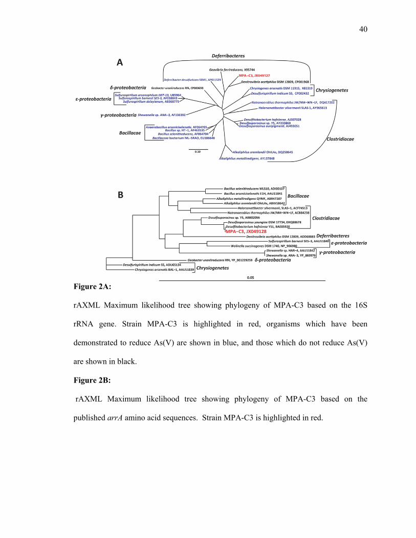
isolated As(V) respiring strain MPA-C3 in relation to its closest relative and other known
respiratory As(V) reducers. Strain MPA-C3 has 90% sequence similarity with

27
Denitrovibrio acetiphilus (AF146526) as calculated by NCBI BLAST (5). Because of its
relatively low sequence similarity to its nearest neighbor, strain MPA-C3 appears to be a
novel member of the Deferribactereres, and may represent a new genus. In addition,
strain MPA-C3 joins Deferribacter desulfuricans as the second member of the
Deferribacteres able to generate energy from the respiratory reduction of As(V) to
As(III) (142). Furthermore, based upon the 2,987,753 bp of sequence obtained from
strain MPA-C3, its genome was found to be most similar to that of Denitrovibrio
acetiphilus, as calculated by RAST (11).
The As(V) respiratory reductase gene, arrA, is recognized as a biomarker for
As(V) respiration (73, 83), and was sequenced to confirm both its presence in strain
MPA-C3 and its homology to other reported As(V) respiring organisms. Figure 2B
illustrates the phylogenetic relationship of the inferred amino acid sequence of the arrA
gene of strain MPA-C3 with the amino acid sequences arrA gene of other known As(V)
reducing organisms. As shown in Figure 2B, the arrA gene of strain MPA-C3 displays
the highest homology (99% BLAST identity) with the arrA gene of a member of the
Clostridia, Desulfitobacterium halfniense Y51 (NC_007907), rather than with the
putative arrA genes identified within the Deferribacteres. This finding suggests that
strain MPA-C3 may have acquired this gene through a horizontal gene transfer event.
2.3.2 Characterization of Mineral Precipitate
A yellow mineral precipitate was observed in cultures of strain MPA-C3 after
approximately one week of growth. Figure 3A shows both this precipitate, and

28
demonstrates that no precipitation occurred in ABF media supplemented with 5 mM
As(III) in the absence of strain MPA-C3, thus demonstrating that an active culture is
necessary for the precipitate to form. In addition, no precipitate occurred in autoclaved
cultures with strain MPA-C3. Inspection of the precipitate under phase contrast
microscopy revealed a yellow mineral closely associated with cells, and similar
observations were made with scanning electron microscopy (Figure 3B). The curved rod
morphology of strain MPA-C3 is shown in Figure 3C.
The mineral precipitate was identified as alacranite, As8S9, by powder X-Ray
diffraction (XRD). The XRD pattern presented in Figure 3 displays 9 well defined
diffraction peaks with corresponding d-spacings of 2.88, 3.03, 3.21, 3.96, 4.86, 5.07,
5.77, and 6.82 Å. The sample X-ray diffractogram exhibited a strong diffraction peak at
23o 2θ (CrKα) that was absent in the orpiment reference pattern. Additionally, well
defined diffraction peak at 26o, 27o, 34o and 47o 2θ were not present in the reference
pattern of realgar. The best match for the sample X-ray diffractogram was the reference
pattern of alacranite reported by Bonazzi et al. (2003). The molar ratio of arsenic to
sulfur in the precipitated alacranite collected from the culture was determined by
inductively coupled plasma-optical emission spectroscopy (ICP-OES) to be
approximately 1:1 (data not shown). This is consistent with the 1:1.1 As:S ratio reported
for alacranite (27).
Assuming all of the 800 µM sulfide present in the media as 400 µM Na2S and 400
µM cysteine reacts with 727 µM of the arsenite which is present in the media in excess, a
maximum of 800 µMoles (710 mg) alacranite can be formed per liter of active strain

29
MPA-C3 culture medium. Given that the molecular weight of alacranite is 887.952 g
mol-1 and that 1.2 L of culture was filtered, this would be a maximum theoretical yield of
852mg. The actual yield of 54 mg of alacranite from 1.2 L of active MPA-C3 culture
medium is 6.3% of the maximum theoretical yield.
2.3.3 Genomic Characterization of Strain MPA-C3
Genomic sequencing of strain MPA-C3 allowed for the assembly of 33
contiguous sequences (contigs) containing a total of 2,985,604 base pairs. The draft
genome has a G+C content of 47%, and 2816 open reading frames (ORFs) have been
identified. Phylogenetic analysis of both the 16S rRNA gene and the entire genome place
strain MPA-C3 among the Deferribacteres. Comparative analysis of the draft genome
with sequenced members of the Deferribacteres and other As(V) reducing bacteria has
allowed for more detailed insight into the metabolic pathways utilized by strain MPA-C3.
Figure 5 provides a schematic overview of some of the pathways identified within
the genome, and connects those pathways with the electron donors and terminal acceptors
identified in culture studies. We identified the complete TCA cycle in strain MPA-C3,
which allows the bacterium to respire simple organic acids such as pyruvate and acetate.
Many of the genes involved in the TCA cycle cluster together on the genome of strain
MPA-C3, including the α, β, γ, and δ subunits of 2-oxoglutarate oxidoreductase
(MPAc_gene_2410-2413), the α and β subunits of succinyl-CoA synthetase
(MPAc_gene_2408-2409), malate dehydrogenase (MPAc_gene_2407), isocitrate
dehydrogenase (MPAc_gene_2406), aconitate hydratase (MPAc_gene_2403), and

30
fumarate hydratase (MPAc_gene_2398-2400) and succinate dehydrogenase
(MPAc_gene_2672-73). Additionally, genes for glycolysis (data not shown) were also
found, allowing for the utilization of more complex carbon sources. This underscores the
physiological data reported in Table 1 that shows fructose can serve as a carbon source
for growth. The aromatic compound benzoate can also be used as a carbon source (Table
1), and genes encoding for a partial benzoyl-CoA reductase (MPAc_gene_0433-4) were
identified in the genome.
Strain MPA-C3 can use a wide range of terminal electron acceptors (TEAs),
similar to other characterized members of the Deferribacteres. Table 1 compares the
TEAs and electron donors utilized by strain MPA-C3 with the TEAs and electron donors
used by the other characterized members of the Deferribacteres as well as the other
characterized non-members of this group that reduce As(V) for respiration. Strain MPA-
C3 generate energy by the reduction of As(V) to As(III), a process catalyzed by the
enzyme ArrAB (66, 124), and this process is included in the schematic view presented in
Figure 5. While the arrAB genes were not identified on the draft genome of strain MPA-
C3, as described above, they were identified by PCR amplification and found to be most
similar to those found in the organism Desulfitobacterium hafniense Y51, which suggests
that they may have been introduced into strain MPA-C3 via horizontal gene transfer. We
suggest that the arrAB genes found by PCR in strain MPA-C3 lie within one of the
unsequenced gaps in the draft genome, and will be found upon completion of the
genome.

31
The reduction of NO3- to NO2
-, but not to N2, was observed in strain MPA-C3
(Table 1). This is similar to the incomplete reduction of NO3- as observed in both D.
acetiphilus and D. desulfuricans (64, 95, 143, 144). In these latter two organisms, the
respiratory reduction of NO3- to NO2
- is catalyzed by the periplasmic NO3- reductase
(Nap) genes (64, 84, 144). A schematic view of this system is provided in Figure 5. In
Figure 6A, we display a cluster of genes from the draft genome of strain MPA-C3
(MPAc_0585-0589) which is homologous to the cluster of Nap genes of D.
desulfuricans, thus supporting the presence of the periplasmic NO3- reductase system in
strain MPA-C3 and the experimental data noted in Table 1. As is consistent with the
experimental data for strain MPA-C3, the Nar genes, which catalyze the reduction of
NO3- to N2 (24), were not found on the genome.
The respiration of fumarate, Se(VI) and Se(IV) are depicted in Figure 5, as we
have demonstrated that strain MPA-C3 generates energy for growth by reducing fumarate
as well as both Se(VI) and Se(IV). Respiratory fumarate reduction is catalyzed by the
FrdABC genes (72), and a homolog of the A subunit of this enzyme was identified near
the end of contig 8 of the draft genome (MPAc_2609). The remaining genes for fumarate
respiration were not found in the preliminary genome sequence, and likely reside in the
gaps within the draft genome. No respiratory selenium reduction genes were found in the
draft genome. Genes with high homology to the selenate reductases SerA (Schröder et
al. (1997), YnfE (49), or SrdA (68) were not identified on the strain MPA-C3 genome.
The selenate reductase genes may also reside in the gaps within the draft genome, or

32
conversely, Se(VI) reduction in this organism is proceeding via a novel, unknown
pathway.
Growth experiments demonstrated that strain MPA-C3 is capable of generating
energy by the dissimilatory reduction of elemental sulfur (Table 1), and this pathway is
represented schematically in Figure 5. Dissimilatory sulfur reduction has also been
reported for two members of the Deferribacteres, Deferribacter desulfuricans and
Geovibrio ferrireducens (34, 143), and is reported to be catalyzed by the polysulfide
reductase (Psr) genes (144). Figure 6B demonstrates the homologs of the polysulfide
reductase (Psr) genes of Deferribacter desulfuricans are found in the genome of strain
MPA-C3. D. desulfuricans utilizes polysulfides produced by the reduction of elemental
sulfur by sulfide (144), and we believe this is also occurring with Strain MPA-C3.
Strain MPA-C3 is capable of growth on both soluble Fe(III) citrate as well as
Fe(III)(OH)3 (Table 1). Fe(III) reduction has been described as a characteristic of the
Deferribacteres (48), and has been reported for Denitrovibrio acetiphilus and Geovibrio
ferrireducens (34, 95). Soluble c-type cytochromes have been suggested as a mechanism
of microbial iron reduction (77), and the genome of strain MPA-C3 contains a cluster of
genes encoding the Ccm c-type cytochrome biogenesis proteins (MPAc_2111-2115).
Figure 5 includes a suite of cytochrome c oxidase genes (MPAc_1085-8), as a similar
mechanism may be involved in the Fe(III) reduction activity observed in strain MPA-C3.

33
2.4 Discussion:
The precipitation of alacranite by strain MPA-C3 is to our knowledge is the first
reported instance of the biogenic formation of this mineral. Alacranite, along with a
similar realgar-like mineral, uzonite (As4S5), was orginally described by Popova et al. in
the Uzon Caldera in Kamchatka, Russia (109, 110). It has since been identified at Conical
Seamount near Lihir Island, Papua New Guinea (33) and as a part of a mineral deposit
formed by the emissions of a burning coal mine in the Czech Republic (27). In each of
these cases, alacranite is described as forming under condition of high temperatures, and
to date, the formation of alacranite has not been described under mesophilic conditions.
While the microbial formation of arsenic sulfide minerals, particularly orpiment, has been
previously reported, (17, 39, 55, 74, 99, 120), this is the first report of the microbial
precipitation of alacranite at any temperature. The formation of realgar in low
temperature arsenic contaminated aquifer sediments was reported by O’Day et al. (2004),
however, the mechanism of realgar formation in this system was not discussed.
Arsenic sulfide mineral precipitation did not occur when As(III) was added to
sterile media with no microbes, clearly demonstrating that strain MPA-C3 is required for
the formation of alacranite. In contrast to the microbial formation of orpiment by D.
auripigmentum (99), the realgar precipitation by Pyrobaculum arsenaticum (55) or the
precipitation of arsenic-sulfide nanotubes by Shewanella HN41 (74), strain MPA-C3 is
unable to reduce SO42- or S2O3
2-, and requires the presence of S2- in the media for the
precipitation of alacranite. In its native estuarine environment, S2- would be supplied to
strain MPA-C3 via the activity of SO42--reducing bacteria of the surrounding microbial

34
community. As shown in figure 2A, the crystals were observed to be larger than the cells
of strain MPA-C3, and appeared to form extracellularly. Mineral precipitation was not
observed in the absence of cells, and we propose that mineral formation is mediated by
cellular metabolism and occurs only in close proximity to the cell, as has been suggested
by Newman et al. (1997) and Lee et al. (2007) for the formation of orpiment and arsenic-
sulfide nanotubes, respectively. The possible role of cell wall nucleation in the formation
of alacranite merits further investigation.
16S rRNA gene and genomic sequencing suggest that strain MPA-C3 is a novel
member of the Deferribacteres. Table 1 summarizes 14 characterized As(V) reducing
isolates along with two members of the Deferribacteres. A review of these isolates
allows for the division of these organisms into two broad groups based on their ability or
inability to utilize SO42- and/or S2O3
2-. Among the organisms incapable of utilizing SO42-
or S2O32-, we note that three organisms, G. ferrireducens, D. desulfuricans, and
Halarsenitibacter silvermanii SLAS-1 are reported to reduce elemental sulfur and/or
mixed oxidation state polysulfide to to S2-, a trait shared by strain MPA-C3 (25, 34, 143).
Of the As(V) respiring organisms in Table 1 which are unable to utilize SO42- or S2O3
2-,
all except Bacillus selenitireducens MLS-10 (141) are able to respire NO32- and/or
Fe(III), suggesting an adaptation for life under lesser reducing conditions in comparison
to the SO42- and/or S2O3
2- reducing organisms. It should be noted that the reduction of
elemental sulfur to S2- provides a source of S2- which can react with As(III) and
precipitate as alacranite.

35
Interestingly, strain MPA-C3 utilizes a wider range of organic compounds as a
source of carbon and energy than any of the currently described Deferribacteres, and at
least as wide a range as any of the other described As(V) respiring organisms (Table 1).
At the time of this writing only limited information is available about the range of
organic substrates utilized by the dissimilatory As(V) reducing bacteria. The ability to
utilize acetate and pyruvate as a source of carbon and energy appears to be conserved
among the Deferribacteres (34, 48, 95, 143). On the other hand, strain MPA-C3 appears
to be unique in this phylum for its ability to utilize the more complex organic compounds
such as fructose and benzoate. Among the other As(V) respiring bacteria, only Bacillus
arsenicoselenatis E1H is reported to be incapable of utilizing pyruvate (141), and only
Desulfosporosinus sp. Y5 has been described as capable of utilizing aromatic substrates,
including benzoate and toluene (76).
The metabolic capabilities of strain MPA-C3 suggest that it lives in the less-
reducing shallow subsurface in its native estuarine environment. Under these conditions
it would be supplied with NO3- from the surface waters, as well as elemental sulfur via
the activity of S2- oxidizing bacteria. Additional sulfide would be present due to the
activity of sulfate reducing bacteria in the deeper, more reducing regions of the
subsurface. This sulfide would allow for the precipitation of alacranite by the As(V)
reducing strain MPA-C3 in the moderate conditions of the shallow subsurface.

36
2.5 Conclusions
We demonstrate that a metabolically flexible anaerobic, As(V) reducing
bacterium, Strain MPA-C3, precipitates alacranite under mesophilic, circumneutral
conditions. This is the first report of this mineral being formed outside of a hydrothermal
system, and supports earlier findings that arsenic sulfide minerals may be formed
biotically. In the environment, therefore, such processes may serve as additional sinks
for arsenic in neutral, reducing ground waters. The evidence presented in this study
indicates that strain MPA-C3 is a novel strain within the phylum Deferribacteres, as
shown by both its metabolic versatility and by the differences between this organism and
the currently described Deferribacteres. We base this conclusion on the physiological
differences, the 16S rRNA gene phylogeny, and the draft genome. We anticipate that
completion of the genome will allow for much greater insights into the metabolism of this
new isolate, as well as a better understanding of its mechanism of alacranite precipitation.
2.6 Supplemental Material
Supplemental material for this chapter is presented in Appendix 1.
2.7 Acknowledgements
We acknowledge the contributions of the following people and organizations: Dr. Li
Meng and Professor Ji-Dong Gu of Hong Kong University for Mai Po sediment samples,
Mark Bowden at the EMSL Facility of the Pacific Northwest National Laboratories for
XRD analysis, Tom Emge of Rutgers University for assistance with XRD interpretation,

37
Dan Sinclair and M. Paul Field of the Rutgers Inorganic Analytical Laboratory for ICP-
OES assistance, Valentin Starovoytov for SEM assistance, Udi Zelzion for genomic
sequencing assistance, and Maria Rivera for laboratory assistance.

38
Table 1: N/T: Not Tested. References: 1(64, 95), 2(34), 3(142, 144), 4(79), 5(113, 114),
6(87), 7(25), 8(101), 9(76), 10(99), 11(46), 12(141), 13(123), 14(137)

39
Figure 1:
Stoichiometric reduction of As(V) to As(III) by strain MPA-C3. No As(V) reduction was
observed in sterile (autoclaved) controls.
40
Figure 2A:
rAXML Maximum likelihood tree showing phylogeny of MPA-C3 based on the 16S
rRNA gene. Strain MPA-C3 is highlighted in red, organisms which have been
demonstrated to reduce As(V) are shown in blue, and those which do not reduce As(V)
are shown in black.
Figure 2B:
rAXML Maximum likelihood tree showing phylogeny of MPA-C3 based on the
published arrA amino acid sequences. Strain MPA-C3 is highlighted in red.

41
Figure 3:
Microscopic analysis of precipitate formed by MPA-C3.
A: Visible yellow precipitate was observed in the bottle containing MPA-C3 (right). No
precipitate was observed in sterile ABF media supplemented with As(III) and sodium
sulfide (left).
B: Electron micrograph (11,800x) of precipitate with associated cells.
C: Light micrograph (1000x) of MPA-C3 cells without precipitate

42
Figure 4:
Powder X-Ray diffractogram (CrKα) of the yellow mineral precipitated by MPA-C3 (top
- black line). Below are reference XRD patterns for, alacranite (red), realgar (green), and
orpiment (purple). ISCD reference numbers are in parentheses.

43
Figure 5:
Schematic representation of metabolic pathways present in MPA-C3. Pathways in italics
represent pathways observed experimentally, and pathways in boxes are those inferred
from genomic analysis with RAST. Nap, periplasmic nitrate reductase; Arr, arsenate
respiratory reductase; CytC, cytochrome C; Nuo, NADH dehydrogenase; Psr, Polysulfide
reductase.

44
Figure 6A-B:
Alignments between the (A) periplasmic nitrate reductase (Nap) and (B) polysulfide
reductase (Psr) genes on the genomes of MPA-C3 and Deferribacter desulfuricans (144).

45
Chapter 3
Microbial Transformations of Arsenic: Mobilization from Glauconitic
Sediments to Water
Abstract
We hypothesize that microbes play an active role in the mobilization of arsenic
from glauconitic subsurface sediments into groundwater in the Inner Coastal Plain of
New Jersey. We have examined the potential impact of microbial activity on the
mobilization of arsenic from subsurface sediments into the groundwater at a site on
Crosswicks Creek in southern New Jersey. The As contents of sediments 33-90 cm
below the streambed were found to range from 15 to 26.4 mg/kg, with siderite forming at
depth. Groundwater beneath the streambed contains arsenic at concentrations up to 89
μg/L. Microcosms developed from site sediments released 23µg/L of total arsenic, and
active microbial reduction of As(V) was observed in microcosms developed from site
groundwater. DNA extracted from site sediments was amplified with primers for the 16S
rRNA gene and the arsenate respiratory reductase gene, arrA, and indicated the presence
of a diverse anaerobic microbial community, as well as the presence of potential arsenic
reducing bacteria. In addition, high iron concentrations in groundwater and the presence
of iron reducing microbial genera suggests that iron reduction in minerals may provide an
additional mechanism for release of associated As, while arsenic-reducing
microorganisms may serve to enhance the mobility of arsenic in groundwater at this site.

46
This Chapter was co-authored with Julia L. Barringer, William M. Benzel, Pamela A.
Reilly, and L. Y. Young. It has been published in Water Research volume 46, pages
2859-2868 (2012).

47
3.1 Introduction
Arsenic (As) is toxic and carcinogenic (37). The effects of chronic exposure to
arsenic contaminated groundwater on populations in Asian and Latin American countries
(21, 32) illustrate the potential for negative human health effects including cancers of the
lung and bladder (132). The arsenic that contaminates groundwater in these countries is
geogenic, and, in the extensively studied Ganges delta area, is thought to be released
from geologic materials by microbial activity (56, 126).
Naturally occurring microorganisms can affect arsenic availability in several
different ways. Microbes can reduce Fe(III) in minerals, promoting mineral
solubilization and subsequent release of sorbed As (60, 116, 122). Anaerobic bacteria
containing the arsenate respiratory reductase gene arrA can use As(V) as a terminal
electron acceptor in respiration (107, 124). These dissimilatory As(V)-reducing
prokaryotes generate energy from the reduction of arsenate (As(V)) to arsenite (As(III))
(76, 117); with the latter being more mobile in groundwater (131). Other
microorganisms may release methyl-arsenic compounds or reduce As(V) to As(III) via
the non-respiratory ars pathway as mechanisms for detoxifying assimilated As(V) (104).
In southern New Jersey’s Inner Coastal Plain Province, geogenic-sourced arsenic
is responsible for high dissolved concentrations in streamwater, shallow groundwater,
and, possibly, deeper groundwater (14, 15). The primary source of arsenic in this
province appears to be glauconite present in the Tertiary and Cretaceous age sediments of
marine and marginal marine origin, which underlie much of the Inner Coastal Plain (14,
15, 40, 41). Other phyllosilicates (illite, micas) in these sediments may contain arsenic,

48
and apatite-rich phosphorite deposits that occur along several formation boundaries are
enriched in As (15).
A cooperative study in 2007-09 by the US Geological Survey (USGS) and the
New Jersey Department of Environmental Protection (NJDEP) and a companion study at
Rutgers University determined that concentrations of arsenic in excess of the NJDEP
Maximum Contaminant Level (MCL) of 5 µg/L were present in shallow groundwater
discharging to two Inner Coastal Plain streams, and that the release appeared to be
microbially mediated (14). This elevated concentration of arsenic required the
calculation of the Total Maximum Daily Load (TMDL) for As in Crosswicks Creek. The
arsenate respiratory reductase gene (arrA), which is considered a marker for arsenic-
respiring bacteria (83), was identified in groundwater beneath a site (C6) on Crosswicks
Creek (CRO), a major Coastal Plain stream (14) (Fig. 1). Only shallow (< 20-m depth)
groundwater discharges to the CRO at C6, which is underlain by clay layers of local
extent.
The sediment underlying CRO at C6 includes material accumulated during two
centuries of human activity (e.g., gristmill, saw mill, farming), and historic records show
the stream was once navigable to C6 from the Delaware River. The accumulated
streambed sediments derive from farm soils and weathered outcrops of clay- and
glauconite-rich sediments and phosphorite deposits upstream. Elevated levels of DOC,
Cl- and NH4+ in the shallow groundwater indicate fertilizers and human and animal
wastes leaching to groundwater during the long span of human activities at the site (14,

49
15); a groundwater NH3 δ15N of 5.96 ‰, which is within the range for soil organics (7)
and for animal wastes (148), supports this interpretation.
Groundwater discharge at CRO-C6 and CRO-C7 (approximately 3km
downstream of CRO-C6) contains high concentrations of Fe and As that precipitate as
As-bearing Fe hydroxides upon reaching the stream (14, 15). The oxidation process
results in a dichotomy between dissolved concentrations in groundwater (16- 89 µg/L)
and those in the stream (generally < 1 µg/L) during low flow (12, 14, 15). Further,
speciation of arsenic in the groundwater in 2007 indicated dimethylarsinate (DMA) at
0.24 and 0.36 µg/L and monomethylarsonate (MMA) at 0.24 and 0.28 µg/L, another
possible indication of microbial activity (18). At CRO site C6, we initiated a pilot
investigation of the microbiological, geochemical, and mineralogical environments
beneath the stream to identify those processes involved in the release of As to
groundwater, streambed sediments, and stream water.
3.2 Methods
3.2.1 Sample collection
In August, 2009, a set of aqueous and streambed samples was collected at C6 on
the CRO. The ultra-clean sampling techniques for stream water and groundwater
(sampled with 1.3-cm inner dia. polyvinyl chloride piezometers) in a gaining reach are
described in detail in (14, 15)). For the microbiological investigations, 300 mL of
groundwater and 200 ml of stream water was filtered (0.45 µm pore-size) aseptically, the

50
filters were then transferred to dry ice for transport and stored at -20o C prior to DNA
extraction, as described in (14).
Two adjacent sediment cores from the streambed at C6 on the CRO were
retrieved by driving 1-m-long stainless steel corers with clean butyl acetate liners into the
sediments about 1 m from the right stream bank. Cleaning and preparation of the corers
is described in detail by (16). The first streambed sediment core was retrieved for
microbiological analysis and frozen on dry ice. Sediments retrieved from the second,
adjacent core were frozen for mineralogical and chemical analysis.
3.2.2 Microbiological analysis
In the laboratory, the organic layer of the top 15 cm of the core was removed and
stored. Sediments from the inner diameter of the core were sectioned, composited by
segment and well mixed under sterile conditions. DNA extractions from sediments
began with the segment from 15 to 30 cm, just above where the screened interval of the
piezometer was positioned in the streambed. DNA also was extracted from the
membranes used to filter groundwater and streamwater using a modified Mo-Bio
Ultraclean protocol (Mo Bio Laboratories, Carlsbad, CA); these extractions,
amplification of the 16S small subunit rRNA gene in sediment and water extracts, and
identification of the arsenic respiratory reductase gene (arrA) are described in detail by
(14). The 16S rRNA gene was amplified with the primers 27F and 1492R (Lane, 1991)
and the arrA gene was amplified with the primers As1F and As1R. 16S and arrA
amplicons were cloned using the Invitrogen TOPO-TA kit (Life Technologies, Grand

51
Island, NY) according to the manufacturer’s instructions, and sequencing was performed
by Genewiz Inc. (South Plainfield, NJ).
All sequence data was initially screened using the NCBI VecScreen tool to
remove vector sequence prior to analysis (97). From the groundwater, 51 16S sequences
were recovered, 44 16S sequences were recovered from sediment, and 16 sequences from
a groundwater microcosm enriched under As(V) reducing conditions were aligned using
the SINA webaligner (112). Results were imported into the ARB software package for
phylogenetic analysis (78, 112). Nearest neighbors were picked from the SILVA 102 Ref
Nonredundant database (112). A ClustalW aligned reference database of 93 arrA
sequences deposited in Genbank was constructed in ARB (71, 112). Thirteen arrA
sequences obtained from groundwater were aligned to the reference arrA sequences using
ClustalW within ARB (71, 112). The phylogenetic tree shown earlier in (14) was
expanded to include all the sequences by using the rAXML maximum likelihood
algorithm within ARB (78). This updated tree can be found in the Supplementary
Material. An ARBNJ distance matrix was constructed within ARB to allow for further
analysis with the mothur software package (128). Sequences were separated into
operational taxonomic units (OTUs) based on a cutoff of 85% similarity using mothur
(128).
We inoculated microcosms using either site sediment or groundwater supplemented
with As(V) (as sodium arsenate) and acetate (as sodium acetate) to enrich for
microorganisms capable of respiration using As(V) as a terminal electron acceptor.
Active microcosms were prepared in triplicate by inoculating 10 g of composited

52
sediment from the 15-33 cm depth or 10 mL of groundwater into 30 mL of defined
anaerobic media (see table S-1 in Supplementary Material for composition) in 120 mL
serum bottles flushed with argon and sealed with butyl rubber stoppers. The microcosms
were supplemented with 2.5 mM As(V) (as sodium arsenate) as an electron acceptor and
2.5 mM acetate (as sodium acetate) as a carbon and energy source to enrich for arsenate
reducing microorganisms. An additional set of sediment microcosms was supplemented
with 2.5 mM acetate as a carbon source but no additional As(V) in order to test for
arsenic mobilization from the sediments in the microcosms. Duplicate sterile controls for
each set of microcosms were prepared by autoclaving inoculated microcosms three times
on three consecutive days. Background controls for each set of microcosms were
prepared by inoculating media which did not contain As(V) or acetate. The reduction of
As(V) to As(III) as well as the utilization of acetate was monitored by high-performance
liquid chromatography (HPLC). After more than 50% of the As(V) was reduced, new
microcosms were established in triplicate, with duplicate autoclaved controls, by
anaerobically transferring 5 mL of the culture to 45 mL of fresh medium.
All microcosms were incubated in the dark under anaerobic conditions at T = 25 oC
(about the same temperature (26.5 oC) as the groundwater). Each week, 600 µL was
sampled using an argon-flushed syringe, filtered through a Costar 0.22 µM nylon
centrifuge tube filter (Corning Inc., Corning, NY) and analyzed for arsenic species until
complete reduction of As(V) to As(III) was observed. Culture supernatant samples from
the sediment microcosm supplemented with acetate only were obtained on days 0 and 23,
and analyzed for As by inductively coupled plasma-optical emission spectrometry (ICP-

53
OES). 50 µL aliquots of the sample were diluted in 950 mL of 1% nitric acid, and ICP-
OES analysis was performed at the Rutgers Inorganic Analytical Laboratory using a
Varian Vista Pro Inductively Coupled Plasma Optical Emissions Spectrometer. ICP-OES
data analysis was performed using Varian ICP-Expert software.
The reduction of arsenic in the microcosms was measured using a Beckman System
Gold HPLC (Beckman Coulter, Brea CA) equipped with a Hamilton PRP X-100 anion
exchange column (Hamilton, Reno NV) as described by Perez-Jimenez et al. (107).
As(V) and As(III) ions were eluted with 40 mM sodium phosphate flowing at 1 mL/min,
and detected at 195 nm. Acetate was measured using a Beckman System Gold HPLC
equipped with a Phenomenex ROA Organic Acids Column (Phenomenex, Torrance CA).
Acetate was eluted with 5 mM sulfuric acid flowing at 0.5 mL/min, and detected at 205
nm.
3.2.3 Chemical and mineralogical analysis of core sediments
The chemical composition and mineralogy of the second sediment core was
determined at the USGS Geologic Discipline Research Laboratory (GDL), Denver, CO.
The frozen core was sectioned into 2 to 4-cm segments; these were retrieved from 33 to
36 cm; 43 to 46 cm; 56 to 58cm; 63 to 66 cm, 71 to 74 cm, and 86 to 90 cm in order to
assess changes in chemistry and mineralogy reflective of changes in redox conditions in
the sediments at and below the interval selected for microbial analysis.
Samples were prepared by removing the outer rim (~0.5 cm) of each sample plug; the
remaining material was freeze-dried, loosely consolidated, and disaggregated. Each bulk
54
sample was riffle split into three 50-g aliquots. One aliquot was ground and chemically
analyzed at the GDL. Analyses for carbon were done by titration and LECO analyzer
(30, 31) at the GDL. Major- and trace-element analysis was done by inductively coupled
plasma/mass spectrometry (ICP/MS), following total acid digestion (29).
Another fraction of the bulk sediment was analyzed for mineralogy by X-ray
diffraction (XRD) to determine mineral abundances. Samples were crushed by hand to
<250 µm, micronized in a McCrone mill (McCrone Microscopes and Accessories,
Westmont IL) with isopropanol, randomly packed into holders, and analyzed on a
Scintag X-1 diffractometer (Scintag Inc, Sunnyvale, CA). The RockJock program (43),
used to quantify the mineralogy, yielded semi-quantitative mineral contents because the
analysis did not consider amorphous and poorly crystalline material. Heavy mineral
concentrates of the five deepest samples were examined optically.
Polished grain mounts were made from aliquots of the five deepest samples.
Unconsolidated sediment was stirred into slow-set epoxy and centrifuged to create graded
grain sizes. The epoxy plugs were cut on an angle, yielding a section through the
graduated grains, and polished. A Scanning Electron Microscope (SEM) scanned several
hundred particles in each mount for minerals with substantial arsenic content. Select
areas and individual grains also were studied by SEM to document texture and growth
habits of the grains. Glauconite and siderite separates were analyzed for total As content
by ICP.

55
3.3 Results and Discussion
3.3.1 Sediment chemistry and mineralogy
The sediment beneath the streambed at CRO-C6 is composed primarily of quartz
and phyllosilicates—mainly glauconite, but also micas (biotite, phlogopite, muscovite,
illite)—with minor amounts of feldspar and siderite. The arsenic content of the sampled
sediments ranged from 15 to 26.4 mg/kg (Table 1), with the highest arsenic and calcium
contents at 33- 36 cm, and at 71-74 cm. Analysis of mineral separates indicates that
arsenic content of the glauconite and siderite was 33.7 mg/kg, and 184 mg/kg,
respectively (Table 1). Fines (< 250 µm illite and Fe hydroxide) contained 46 mg/kg of
arsenic, similar to the precipitate formed at a CRO groundwater seep (15). Regression
analysis indicated the arsenic contents of the sediment were linearly and strongly related
to iron and calcium contents (R2 = 0.91 and 0.95, respectively; p < 0.05), indicating that
iron minerals form the primary source of arsenic at this site. The relation of As with Ca
may be indicative of precipitation of a Ca-As phase; however, speciation calculations
indicated that the groundwater was undersaturated with respect to calcite (Barringer et al.,
2011). The high arsenic and calcium contents of the 33-36 and 71-74-cm intervals also
are distinguished by relatively high contents of micas other than glauconite (Table 1).
SEM scans of selected bulk sediment samples did not detect pyrite or amorphous sulfide,
indicating that arsenical sulfides do not form a significant source of arsenic at this site.
Although siderite was not detected in surficial streambed sediments, speciation
calculations indicated groundwater supersaturation (15). Siderite as grains and cement
was detected in the deeper sediments, along with glauconite in iron hydroxide cement

56
(Figure S3.2). Siderite is a secondary mineral that could be precipitated through activity
of iron-reducing microbes, as described by Anawar et al. (2006) and Islam et al.(2005).
Such biogenic siderite has been proposed as a sink for As(V), but not As(III) (6, 59);
As(III) has been shown (59) to bind weakly to siderite at pH values below 7. Analysis of
the mineral separates from the CRO C6 site shows that siderite is a sink for arsenic
(presumably As (V)) in the New Jersey sediments at slightly acidic pH values
(groundwater pH 6.4-6.9 (15)). Therefore, in addition to primary glauconite in the
accumulated sediments of the streambed, arsenic is present in at least two secondary
phases; these include iron hydroxides that formed in what were once the upper portions
of the streambed sediments, and siderite that precipitates as individual grains and as
cement for other grains.
3.3.2 Microbes in streambed sediments and the aquifer
As illustrated in Figures 2A-2C, As(V) reduction, arising from arsenic respiration,
was observed in both groundwater and sediment microcosms supplemented with acetate
and As(V). The reduction of As(V) to As(III) was observed in active groundwater
microcosms, while no reduction was observed in sterile controls (Fig. 3A). Unlike the
groundwater microcosms, the sediment microcosms (Fig. 2B) sorbed the supplied 2mM
As(V) in the time between preparation of the microcosm, and the initial sampling (<10
minutes). This loss was observed upon analysis of the Day 0 sample, and an additional
spike of 5mM As(V) was added on day 7 (Fig. 2B). As(V) reduction was observed
following the addition of As(V) into the microcosms, Figure 2B also shows that no

57
production of As(III) from As(V) reduction was observed in sterile controls, clearly
demonstrating that reduction was microbially mediated.
In both the groundwater and the sediment microcosms, the product of As(V)
reduction, As(III), persisted in solution, demonstrating that microbial reduction can play a
role in enhancing arsenic mobility in groundwater. Jönsson and Sherman (59) report that
As(III) binds only weakly to siderite, a primary arsenic sink in this system, compared to
the strong binding exhibited by As(V). Although no As(V) reduction occurred in the
sterile controls, As(V) concentrations decreased over time in the sterile sediment
microcosms (Fig. 3B), suggesting continued binding of As(V) to the sediment. Arsenic
mobilization was not detected in background controls (containing sediment inoculum but
no added As(V) or acetate), indicating the requirement for additional organic carbon to
stimulate the native microbial community (data not shown). Following subsequent
transfer of 5 mL of culture to a fresh medium, rapid As(V) reduction was observed, a
clear indicator of an enriched community of arsenic respiring microbes (Fig. 2C).
Furthermore, As(V) loss due to binding to sediment was greatly reduced insofar as very
little sediment was transferred to these subcultures.
Figure 3 shows total arsenic released (mobilized) from native sediment in the
sediment microcosms supplemented with acetate. By day 23, the total soluble arsenic
concentration was 27 µg/L, with no mobilization of soluble arsenic observed in the sterile
control (Fig. 3A). The analyses by ICP-OES do not speciate; nonetheless, it is not
unreasonable to assume that the released arsenic is in the reduced As(III) form since a.)
the microcosms are anoxic and reducing, and b.) our data (Figs. 2A-C) and others (59,
58
131) show very little sorption of As(III). The microbial utilization of acetate was
observed in active microcosms, with acetate concentrations decreasing over the 20 day
period, while no loss of acetate was seen in the sterile microcosm controls (Fig. 3B).
Note that the only available arsenic in these bottles is that which is bound to the
sediments. The fact that this arsenic is mobilized and released into solution when acetate
is available is evidence for microbially mediated release. This indicates that an influx of
organic carbon enhances microbial growth and metabolism, leading to an increase in
microbiologically mediated arsenic release from the CRO-C6 sediments under anaerobic
conditions. It is noteworthy that our observed concentrations of arsenic released in these
microcosms is consistent with the concentration of arsenic in CRO-C6 groundwater
reported by Barringer et al. (2010), and further reinforcing the relevance of microbial
processes to arsenic mobilization at this site.
3.3.3 Molecular characterization of groundwater and sediment microbes
The 16S rRNA gene sequences recovered from the sediment and groundwater of
CRO site C6 suggest the presence of diverse anaerobic bacterial communities in both
groundwater and sediment (Figs. 4A,B). Analysis of 51 cloned sequences from the
groundwater microbial community allowed for the identification of 49 unique OTUs,
based on a 97% cutoff (Fig. 4A). These sequences have been submitted to Genbank
under accession numbers JQ041420 through JQ041470. Sixteen clones were sequenced
from a microcosm inoculated with a 1:1000 dilution of C6 groundwater. Fifteen of these
clones showed 97+% identity to the Fe-reducing bacterium Geobacter sp. Ply1
59
(EF527233), while one showed 99 % identity to Alkaliphilus oremlandii (CP000453)
(formerly Clostridium sp. strain OhlLAs), an organism that has been demonstrated to
respire As(V) (54, 139). The presence of organisms highly similar to Geobacter and
Clostridia suggests that the iron reduction inferred from the groundwater chemistry and
sediment mineralogy is occurring in the CRO sediments, and is one likely mechanism for
release of arsenic (38, 56, 106, 139). The sequences from this enrichment culture have
been submitted to Genbank under accession numbers JQ062862 through JQ062877.
From the sediment core, 44 clones were sequenced, leading to the identification of
11 unique OTUs, suggesting a lower diversity in the sediment than in the groundwater
(Figure 5B). All 16S rRNA gene sequences have been submitted to Genbank, with
accession numbers JQ041471 to JQ041514. Others have also reported on higher
microbial diversity in groundwater than in adjoining sediment (Kilb et al., 1998;
Alfreider et al., 2002), and we believe that is the case here.
The PCR amplification of arrA from DNA extracted from sediment and
groundwater as well as a preliminary analysis of the arrA sequences recovered is
described in detail in Barringer et al. (2010). The 13 partial arrA sequences obtained
were aligned to 93 full length and partial arrA sequences obtained from GenBank using
ClustalW within ARB (71, 78). Following construction of an ARBNJ distance matrix
within ARB, sequences were clustered using mothur (Schloss et al., 2009) into three
OTUs based on a 15% cutoff (85% similarity). As suggested by earlier results (14), two
of the three OTUs showed 75-77% homology to a putative arrA gene found on the
genome of Geobacter uraniireducens Rf4, while the third OTU displayed 71-72%

60
homology to the arxA gene of Alkalilimnicola erlichii MLHE-1, which has been reported
to function as an arsenic oxidase (103). The presence of sequences similar to the arxA
gene of the arsenic oxidizing bacteria A. erlichii MLHE-1, which uses arxA as an As(III)
oxidase under nitrate reducing conditions (103, 119), indicates the potential for microbial
arsenic oxidation at site CRO-C6 as well, particularly at the sediment-water interface
where nitrate may be supplied by surface water. All recovered arrA gene sequences have
been submitted to Genbank under accession numbers JQ041515 through JQ041527.
Successful amplification of the arrA gene, a known biomarker for arsenic
respiration (83) combined with the enrichment of As(V)-reducing microorganisms
provides strong evidence for the presence of arsenic reducing microbes at the CRO C6
site. As suggested by (118), microbial arsenic oxidation can also serve as a mechanism
of mobilization. The presence of an arsenic oxidizing microbe would fit well with the
shallow groundwater chemistry, insofar as As species measured in the water discharging
to the stream in 2007 showed roughly equal concentrations of As(V) and As(III) (15).
3.4 Conclusions
Based upon our findings, we can conclude that the release and transport of arsenic
from geologic materials at the CRO C6 site is mediated and enhanced by microbial
metabolism. Our data indicate that the release of arsenic from stream bed sediment is
strongly enhanced by the addition of organic carbon (Figure 3A,B), indicating that
microbial metabolism plays an important role in arsenic mobilization. Notably, our
finding that reduced As (as As(III)) persists in solution allows us to conclude that the

61
solubility and therefore the transport of arsenic in shallow groundwater at the CRO C6
site is influenced by microbial respiratory reduction of As(V) to As(III) and is consistent
with reports from other studies (59, 131). While siderite was noted as a major sink for
arsenic at this site, it binds As(III) poorly (59), further suggesting that microbially
reduced As(III) will remain in solution at this site.
A complex series of interactions between microbes, primary and secondary
geologic materials, and water is envisioned to result in the levels of arsenic found in
sediments, groundwater and stream water. Figure 5 displays our conceptual model, with
the processes confirmed in this study in black, and major contributing processes
suggested by other reports in gray. Our overall conceptual model of arsenic release at
CRO-C6 includes microbial reduction of Fe(III) enhanced by inputs of organic carbon in
both crystallized and amorphous Fe-bearing materials and subsequent release of As to
groundwater as reported by Islam et al. (2004) and Tufano et al. (2008); direct release of
arsenic from Fe minerals through microbial respiratory arsenic reduction (155) coupled to
the oxidation of organic carbon; microbial respiratory reduction of released As(V) to
As(III) enhanced by inputs of organic carbon as demonstrated in this study; and the
binding of released As(V) to microbially precipitated siderite (59) within the accumulated
streambed sediments. Thus, microbially reduced arsenic will persist in groundwater, and
present a hazard if the groundwater is used for consumption. Insofar as the kinetics of
abiotic oxidation of As(III) in oxic water have been reported to be extremely slow (131),
microbial oxidation of As(III) to As(V) (117, 125) may enhance the co-precipitation of
As and Fe at the oxic/anoxic interface in the upper streambed as described by (15). Based

62
upon the results of our study, we believe that microbial activity plays a significant role in
arsenic cycling at CRO-C6, and that the further elucidation of microbially mediated
geochemical processes is important for understanding water quality in areas with As-
enriched geological materials and agricultural and residential development.
3.5 Supporting Information
Supporting information including XRD data for the sediment core, photograph of
glauconite grains, SEM results showing high P in amorphous material, and the
composition of anaerobic medium can be found in Appendix 2.
3.6 Acknowledgments
Funding for the microbiological work was provided in part by the New Jersey
Water Resources Research Institute and the Aresty Undergraduate Research Center. The
authors are grateful to M. Paul Field of the Rutgers Inorganic Analytical Laboratory for
assistance with ICP-OES analysis. Funding for the geochemical part of the study was
provided by the New Jersey Department of Environmental Protection; thanks go to
Barbara Hirst and Kimberly Cenno for administering the funds and for their assistance in
planning the study. The authors thank LaDonna Choate, Heather Lowers, and Paul
Lamothe (USGS) for ICP/MS and SEM analyses. (Use of trade, product, or firm names is
for descriptive purposes only and does not imply endorsement by the US Government.)

63
Table 1:
Contents of selected major and trace elements in a streambed sediment core and mineral
separates from Crosswicks Creek at site C6, August, 2009.

64
Table 2:
Semi-quantitative mineralogy reported in wt% in selected intervals of a streambed
sediment core, Crosswicks Creek, New Jersey, August 2009.
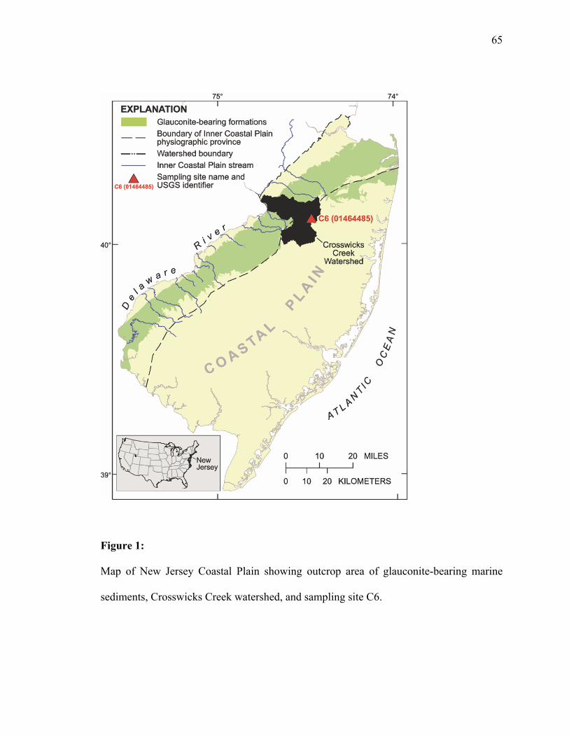
65
Figure 1:
Map of New Jersey Coastal Plain showing outcrop area of glauconite-bearing marine
sediments, Crosswicks Creek watershed, and sampling site C6.

66
Figure 2:
Microcosm results showing As(V) reduction in (a) groundwater microcosms amended
with arsenic and acetate; (b) streambed sediment microcosms amended with arsenic and
acetate (15-30 cm depth); (c) streambed sediment microcosms diluted 1:10 and amended
with arsenic and acetate (15-30 cm depth). As(V) reduction was not noted in sterile
controls. Sterile controls were amended with arsenic and acetate prior to sterilization by
autoclaving, while background controls contain media and sediment, but were not
supplemented with As(V) or acetate.

67
Figure 3:
Total arsenic release from streambed sediments amended with acetate (A), coupled to the
utilization of acetate (B). Arsenic was not detected on day 0 or in control microcosms
sterilized by autoclaving.

68
Figure 4:
Microbial communities in found in groundwater (A) (51 clones) and a streambed
sediment core (B) (44 Clones), Site CRO-C6 August, 2009.

69
Figure 5:
Conceptual diagram of pathways for microbially mediated arsenic release from
glauconite ((K,Na)(Mg,Fe+2)0.33(Fe+3,Al)1.67[(Si,Al)4O10](OH2) ). Processes observed in
this study are in black, those observed in other studies and potentially also occurring at
this site are in gray. Microbial activity may be enhanced by inputs of organic carbon,
leading to an increase in the reduction of iron and mobilization of arsenic. As(V) in
particular may be re-precipitated with, or sorbed to, siderite.
OC: Organic Carbon
IRB: Iron Reducing Bacteria
DARP: Dissimilatory Arsenate Respiring Prokaryotes

70
Chapter 4
Groundwater redox controls microbial release of arsenic from minerals
to groundwater in a fractured rock terrain,
New Jersey, USA
Abstract
The mobilization of arsenic bound to aquifer sediments is a source of
contamination in groundwater in New Jersey. Deep Coastal Plain sediments have
average arsenic concentrations of 15-23 mg/kg, and arsenic concentrations of 26 mg/kg
are found in shallow glauconitic sediments. In this study we investigate microbial As(V)
mobilization in the shallow aquifers beneath the streambeds at two tributaries of the
Millstone River, Six Mile Run and Pike Run, in the black and red shales of the New
Jersey Piedmont Physiographic Province. These two sites offer a distinctive comparison,
namely a similar mineralogy and geochemistry, with the notable difference being that
reducing conditions are present at Six Mile Run (EH=-39.5mV), and oxidizing conditions
are present at Pike Run (EH=+144mv). Oxygen-depleted groundwater at the Six Mile
Run site has an arsenic concentration of 27 µg/L, exceeding the New Jersey Department
of Environmental Protection maximum drinking water contaminant level of 5 µg/L,
whereas the arsenic concentration in more oxidizing groundwater at the Pike Run site is
2.1 µg/L. Groundwater sampled on gaining reaches of these rivers was inoculated into
anaerobic microcosms with acetate as a carbon source and As(V) as an electron acceptor.

71
Microcosms developed from Six Mile Run groundwater reduced 1 mM of As(V) to
As(III) in 30 days, while microcosms from Pike Run did not reduce As(V). The 16S
rRNA gene and the As(V) respiratory reductase gene, arrA, were amplified from DNA
extracted from groundwater. Analysis of the 16S rRNA gene by Denaturing Gradient
Gel Electrophoresis (DGGE) indicated that distinct bacterial communities are present at
each site, a finding supported by cloning and sequencing. Cloning and sequencing of the
As(V) respiratory reductase gene, arrA, revealed distinct arsenic respiring communities at
each site, with 8 unique OTUs found at Six Mile Run and 11 unique OTUs at Pike Run,
which may be representative of the arsenite oxidase gene arxA. Our findings demonstrate
that the microbial As(V) reduction and mobilization is facilitated by the reducing
conditions present at Six Mile Run, while it is inhibited by the oxidizing conditions
present at Pike Run.
This Chapter was co-authored with Julia L. Barringer, Pamela A. Reilly, D.D. Eberl, A.E.
Blum, and L.Y. Young. It has been submitted for publication in Water Research, and is
currently in review.

72
4.1 Introduction
Arsenic is contained in numerous geologic materials that, in parts of the world,
have contributed As to groundwater used as a potable water source. The result has been
exposure of millions of people to hazardous levels of As (up to hundreds of micrograms
per liter) in drinking water, creating illnesses, including cancer (133). The role of
microbes in the release of As into groundwater from the alluvial sediments of the river
deltas of Southeast Asia to groundwater has been studied, particularly in several Asian
countries with widespread As contamination in drinking water(4, 6, 20, 21, 56, 73, 135)
In glauconitic sediments in the Coastal Plain of New Jersey, shallow groundwater
discharging to streams contains As at concentrations that range up to 89 µg/L (14, 90).
These As concentrations have been linked to the microbially mediated release of As from
minerals such as glauconite, other phyllosilicates, apatite, and siderite in the sediments
(Barringer et al., 2010; Mumford et al., 2012). Additionally, similar sediments in the
Atlantic Coastal Plain of the USA have been reported to contain levels of As in
groundwater that exceed the Federal drinking-water maximum contaminant level (MCL)
of 10 µg/L (50, 146).
The Piedmont Physiographic Province of New Jersey is composed of sedimentary
rocks with minor igneous intrusives and extrusives of the Newark Basin and borders the
Coastal Plain to the northwest as shown in Figure 1. The Newark Basin rocks of the
Piedmont Physiographic Province contain As-bearing minerals identified as pyrite,
hematite, and clays (130). Groundwater in various parts of the sedimentary and igneous
fractured-rock aquifers is contaminated with As, with concentrations ranging from 5 µg/L

73
(the State maximum drinking water contaminant level (MCL)) to 215 μg/L (130).
Processes (e.g., oxidation of pyrite in black shales, desorption from red shales) that
release As from bedrock to groundwater have been investigated (108, 118, 130, 154).
Microbially mediated oxidation of S2- in pyrite-containing bedrock by an autotrophic
inorganic-S and As(III) oxidizing bacterium was demonstrated by Rhine et al. (2008).
Although high concentrations of As tend to coincide with low oxygen levels in Piedmont
groundwater (130), the role of microbes in As release under reducing conditions has, to
the authors’ knowledge, not been explored, particularly in shallow groundwater
discharging to the region’s streams.
There are streams and rivers in the New Jersey Piedmont Physiographic Province
in which the stream water contains As at levels exceeding the State Surface Water
Quality Standard (SWQS) that, owing to the carcinogenic nature of As, is a stringent
0.017 μg/L (N.J.A.C. 7:9b). Among these rivers is the Millstone River, which rises in the
glauconitic sediments of the Coastal Plain and continues through the Piedmont’s Newark
Basin fractured rock terrain to its confluence with the Raritan River near Raritan Bay as
shown in Figure 1.
As part of a river-wide study of the Millstone River, two tributaries were targeted
for a combined geochemical/microbiological study to determine if microbially mediated
As release, as seen in the NJ Coastal Plain (14, 90), is occurring in the fractured bedrock
aquifer characteristic to the NJ Piedmont. The tributaries—Six Mile Run and Pike Run—
are underlain by bedrock of the Passaic Formation, which is composed primarily of red
mudstones and siltstones interspersed with black shale and gray mudstone. The dark

74
strata are the richest in As mainly because of the presence of pyrite (Serfes, 2005).
Arsenic contents of the more abundant red strata were found to range from 4.5 to 14.8
mg/kg (Serfes, 2005), and it is over these (presumably) less enriched strata that the
channels of the two tributaries pass at the chosen sampling sites.
Our study at the two sites included determining the mineralogy and chemistry of
streambed sediments, the chemistry of shallow groundwater and streamwater, and the
microbial communities of the shallow aquifer and their abilities to reduce As(V). Results
of the study and discussion of the biogeochemical processes that affect As mobility are
presented herein.
4.2 Methods
4.2.1 Groundwater and streamwater sample collection
The Six Mile Run and Pike Run sites were selected in the Piedmont portion of the
Millstone Basin due to the presence of sufficient weathered material to allow for the
insertion of piezometers into streambed sediments during a period of low flow
conditions. Acid-washed PVC piezometers (1.3-cm inner diam. with a 15-cm-long
slotted screen) were driven into the streambeds in gaining reaches for measurements of
shallow groundwater chemistry (13-15, 90). Production of groundwater from the
piezometers was confirmed by an increase in conductance prior to sample collection, and
samples were obtained using a peristaltic pump through TeflonTM-lined tubing that
extended down through the piezometer to the screen. NJDEP monitoring well MW114,
located approximately 60m south of the Six Mile Run stream sampling site, is

75
hydraulically connected to the aquifer beneath Six Mile Run, and was sampled with the
Six Mile Run site in order to study the processes and conditions deeper beneath Six Mile
Run. MW114 is screened from 3.3 to 6.4 meters below land surface, and samples were
obtained using a Grundfos submersible pump with acid-washed tubing.
Tributary streamwater samples and groundwater samples were collected at the
same time during a period of low-flow conditions. Streamwater was collected as grab
samples using an equal-width-interval technique and composited in an acid-washed
churn, as in previous studies (Barringer et al., 2010a, 2011). Processing and filtering of
each composited sample was done in a new polyethylene bag on a PVC frame (bags were
changed between samples). Ultraclean techniques were followed throughout (Wilde et
al., 2004). All equipment was acid washed (5 percent HCl) and copiously rinsed with de-
ionized water prior to use. De-ionized water (DI) and field-rinsed 0.45-μm-pore-size
capsule filters were used to collect filtered samples for analysis of As and other trace
elements. Disc filters (0.45-μm-pore-size) were used to collect dissolved organic carbon
(DOC) samples. Sample bottles, except the baked glass bottles used for DOC and total
organic carbon (TOC) samples, were DI- rinsed before sampling and field rinsed before
sample collection.
Field parameters (temperature, dissolved oxygen, pH, specific conductance,
turbidity) were measured at all sites using a YSI data sonde (YSI Inc., Yellow Springs,
OH). Filtered and unfiltered samples were collected for analysis of As, metals, DOC and
TOC.

76
4.2.2 Sampling For Microbiological Analysis
Groundwater samples were collected using sterile equipment and filtered with
sterile Pall GN6 (0.45 μm pore-size) filters (Pall Corp. Ann Arbor, Michigan). The filter
membranes were immediately stored on dry ice for transportation and stored at -80oC
until processing. Groundwater samples were transported on ice and stored at 4o C prior
to use as inoculum for microcosms.
4.2.3 Streambed-Sediment Sampling
Streambed-sediment samples were collected under low-flow conditions.
Streambed-sediment grab samples were collected in areas of deposition using an acid-
washed plastic scoop. The sediments were sieved (<2-mm grain size) using an acid-
washed plastic sieve and placed in field-rinsed jars. Attempts to collect cores of
streambed sediments with a hand-driven stainless steel corer were unsuccessful.
4.2.4 Sample Analysis
Streamwater and groundwater samples were analyzed for major ions, nutrients,
acid-neutralizing capacity (ANC), DOC and TOC, As, and other selected trace elements
at the U.S. Geological Survey National Water Quality laboratory (NWQL) in Denver,
CO. The samples included filtered and unfiltered water. Major and trace elements were
determined by inductively coupled plasma optical emission spectroscopy (ICP/OES) and
inductively coupled plasma mass spectrometry (ICP/MS) and collision cell ICP/MS

77
Nutrients were analyzed by colorimetry or photometry, DOC and TOC by
infrared spectrometry, anions by ion chromatography, and ANC by titration at NWQL.
Splits of the sediment samples were analyzed for recoverable (HCl leachable)
metals by ICP/OES and ICP/MS at the NWQL and for total metals (total sediment
digestion by HNO3, HCl, HF, HClO4) by ICP/MS (8, 29), for Hg by cold vapor atomic
fluorescence spectroscopy (CVAFS), and for total carbon and carbonate carbon by
titration and LECO analyzer (30, 31) at the USGS Geologic Discipline (GD) research
laboratory in Denver, CO. Organic carbon (OC) was determined by difference between
total and carbonate carbon. Splits of selected samples were analyzed by X-ray diffraction
for mineralogy, using a Siemens D500 X-ray Diffraction System (Bruker AXS Inc.,
Madison WI) at the USGS Research Laboratory at Boulder, CO. Details of analytical
methods, references, and reporting limits are presented in table S1 in Supplementary
Material.
4.2.5 DNA Extraction and Amplification
DNA was extracted from cells trapped on filter membranes using a Mo Bio
Ultraclean Soil kit (Mo Bio Laboratories, Carlsbad, CA) according to the manufacturer’s
instructions, with modifications as described in Mumford et al. (2012). The 16S small
subunit rRNA gene was amplified from extracted DNA using primers 27F and 1492R
and the As(V) respiratory reductase gene, arrA, was amplified using primers As1F and
As1R, as described by Lane et al. (70), and Lear et al. (73), respectively. The primer
sequences are reported in supplementary table S2. PCR results were visualized by

78
agarose gel electrophoresis and products cloned into a pCR4 vector and transformed into
Escherichia coli TOP10 as described in Barringer et al. (2010a). Positive clones were
sequenced by Genewiz, Inc. (South Plainfield, NJ). A total of 14 16S rRNA gene clones
and 9 arrA gene clones were recovered from Six Mile Run, and a total of 16 16S rRNA
gene clones and 7 arrA gene clones were recovered from Pike Run. Taxonomic
classification was performed based on the 16S rRNA gene using the RDP classifier (149).
Amplification of DNA from filters recovered from MW114 was unsuccessful; DNA was
instead extracted from a microcosm prepared with groundwater from MW114, which
allowed for the recovery of 15 16S rRNA gene clones and 13 arrA gene clones. All
sequence data were screened with the NCBI VecScreen tool to eliminate vector sequence
prior to analysis. 16S rRNA gene sequences have been submitted to Genbank under
accession numbers JX440972 through JX441016. As(V) respiratory reductase (arrA)
gene sequences have been submitted to Genbank under accession numbers JX845248-
JX845276.
4.2.6 Denaturing Gradient Gel Electrophoresis
Denaturing gradient gel electrophoresis (DGGE) was performed to detect
differences between the microbial communities present in the groundwater at Six Mile
Run and Pike Run. Extracted DNA was amplified with the universal 16S primers 27F
and 519R in order to generate a 497bp amplicon for separation by DGGE. The
amplicons were separated using an 8% polyacrylamide gel with a denaturing gradient of
40-80% urea-formamide. Electrophoresis was performed at 55V for 16 hours at 60o using

79
a Bio-Rad DCode Universal Mutation Detection System according to the manufacturer’s
instructions (Bio-Rad Inc., Hercules, CA). The gel was stained for 45 minutes in an
aqueous solution of 10% ethanol, 0.5% acetic acid and 0.1 μL/mL SYBR Green (Life
Technologies, Grand Island, NY). The gel was imaged using a Storm 860 (GE Healthcare
Life Sciences, Piscataway, NJ), scanner with excitation at 450 nm and emission at 525
nm in high sensitivity mode with the photomultiplier tube set at 900V. Major bands were
excised from the gel and eluted in 100 μL of sterile MilliQ water. The eluted DNA was
amplified using the 27F forward primer and 519R reverse primer, and the product of this
amplification was purified using ExoSap-IT (Affymetrix, Santa Clara, CA) according to
the manufacturer’s instructions prior to sequencing by Genewiz, Inc (South Plainfield,
NJ). Sequences recovered from DGGE bands can be found in supplementary table S2.
4.2.7 Sequence Analysis
16S rRNA gene sequences obtained from cloning and DGGE were aligned to the
SILVA 111NR database using the SINA webaligner (112). These aligned 16S rRNA
gene sequences were then imported into the ARB software package for phylogenetic
analysis, and a phylogenetic tree of the sequences and nearest published related
sequences from the SILVA 111NR database was constructed utilizing the RAxML
maximum likelihood algorithm within ARB (78, 112). This tree can be found in the
supplementary material.
The recovered arrA gene sequences were imported into ARB and aligned using
ClustalW within ARB to a database of 392 arrA gene sequences retrieved from Genbank

80
(71, 78). A phylogenetic tree of the recovered arrA gene sequences combined with
selected arrA gene sequences from Genbank was generated using the RAxML maximum
likelihood algorithm within ARB (78) and can be found in the supplementary material.
An ARBNJ distance matrix was constructed to allow for OTU based analysis with
mothur (78, 128). arrA gene similarity between Six Mile Run and Pike Run was
calculated by grouping OTUs by site and generating Venn diagrams within mothur (128).
4.2.8 Preparation of Groundwater Microcosms:
We inoculated microcosms using site groundwater supplemented with As(V) (as
sodium arsenate) and acetate (as sodium acetate) to test for the presence of
microorganisms capable of the respiratory reduction of As(V) to As(III). Active
microcosms were prepared in triplicate by inoculating 10 mL of groundwater into 30 mL
of defined anaerobic freshwater medium as described by Mumford et al. (2012). The
microcosms were supplemented with 5 mM As(V) as an electron acceptor and 5 mM
acetate as a source of carbon and energy. Sterile control microcosms were prepared in
duplicate in the same manner as active microcosms, followed by autoclaving three times
on three successive days. Background microcosms were prepared by inoculating media
which had not been supplemented with As(V) or acetate. The reduction of As(V) to
As(III) and the utilization of acetate was monitored by high-performance liquid
chromatography (HPLC). When reduction of 25% of the As(V) to As(III) was observed,
secondary microcosms were established in triplicate, with duplicate sterile controls, by

81
anaerobically transferring 5 mL of the primary culture to fresh medium. All microcosms
were sampled and analyzed as described in Mumford et al. (2012).
4.3 RESULTS
4.3.1 Stream Sediment Mineralogy and Chemistry
The sampling sites of both Six Mile Run and Pike Run are underlain by shales
and siltstones of the Passaic Formation. As shown in Table 1, streambed sediments at
Six Mile Run contained more organic matter and were more quartz-rich (33.4 wt.
percent) than those at Pike Run; phyllosilicates (mainly illite) constituted 42.8 wt. percent
of the minerals at Pike Run but were only 32.6 wt. percent at Six Mile Run. Feldspars
were major phases at both sites, and Fe oxides and hydroxides were minor phases.
Amorphous Fe hydroxides would not have been detected, however, these probably are
present and may contain As, as in the Coastal Plain site (14, 15, 90). Both total–digestion
and HCl-leachable As contents at Pike Run, Six Mile Run, and 9 other sites in the
Millstone Watershed were strongly and positively related with total-digestion and HCl-
leachable Fe and Mn contents (USGS data not shown).
As shown in Table 2, the As content of streambed sediments at the Six Mile Run
and Pike Run were 16.5 mg/kg and 14.6 mg/kg, respectively—in general agreement with
the maximum As content (14.8) found by Serfes (2005) for the red beds in the Passaic
Formation. The importance of clays, feldspars, and Fe oxides in the sediments is
indicated by the substantial contents of Al, Fe, and K. The P content of sediments at Six

82
Mile Run was substantially higher than that at Pike Run, suggesting that some of the P
content may result from anthropogenic inputs at the Six Mile Run site.
4.3.2 Groundwater Geochemistry
The groundwater chemistry of Six Mile Run, Pike Run, and MW114 is
summarized in table 3, and shows that the Cl- concentration in shallow groundwater
discharging at Pike Run greatly exceeded both the median (18 mg/L) and maximum (100
mg/L) concentrations for Passaic formation groundwater (130). Groundwater from the
Passaic Formation aquifer is mainly Ca-Mg-Na-HCO3- or Ca-SO4
2- (Serfes, 2005); the
enrichment in SO42- with Ca2+ is ascribed to dissolution of gypsum, a secondary mineral
that lines rock fractures. This elevated Cl- concentration, combined with elevated levels
of Na+, NO3-, and B suggest anthropogenic inputs to the aquifer at Pike Run. While the
Na+ concentration at Pike Run does not exceed the Passaic Formation maximum (270
mg/L) reported by Serfes (2005), it greatly exceeds the median concentration of 15 mg/L.
Concentrations of these constituents were substantially lower in the shallow groundwater
discharging at Six Mile Run, however, concentrations of DOC (2.6 mg/L) and P (1.37
mg/L) were higher than at Pike Run (Table 3), suggesting the potential for anthropogenic
inputs to Six Mile Run.
Table 3 summarizes the groundwater chemistry of Six Mile Run, its attendant
monitoring well MW114, and Pike Run. The DO concentration in the Six Mile Run
piezometer was low (2.8 mg/L), and the Fe concentration (20,700 μg/L) was an order of
magnitude higher than that in the well-water sample, indicating the presence of both

83
reducing conditions and the potential for microbial Fe reduction. All of the Fe in the
filtered groundwater is assumed to be Fe(II), as the solubility of the least stable Fe(III)
oxide, ferrihydrite, at a pH of 6.7 as found at Six Mile Run is less than 106 μg/L, less
than 1% of the Fe measured. The EH of the aquifer at Six Mile Run was calculated based
upon the pH and the concentration of Fe(II), and found to be -39mV, a value consistent
with the observed conditions. The water chemistry from well MW114, which is an input
to Six Mile Run, is displayed in Table 3 and clearly indicates that conditions in the
shallow aquifer near Six Mile Run are reducing. The anoxic (0.1 mg/L DO) water at
MW114 contains high Fe (9010 μg/L) and low SO42-(0.015 μg/L) concentrations and As
concentrations (22.1 μg/L) that were nearly as high as the As concentrations in the Six
Mile Run piezometer sample (27.6 μg/L). The EH of the aquifer at MW114 was
calculated in the same manner as Six Mile Run, and found to be a weakly oxidizing
32mV.
The chemistry of the piezometer sample from Pike Run is summarized in Table 3,
and the more oxidizing conditions found there contrasted with the reducing conditions
found at Six Mile Run. The more oxidizing conditions at Pike Run were reflected in the
higher DO concentration (4.9 mg/L), low Fe (197 µg/L) and As (3.4 µg/L)
concentrations, and higher concentrations of SO42- and NO3
- (Table 3). The EH of Pike
Run was calculated based upon the very low concentration of Fe(II) observed, and found
to be an oxidizing 144mV. The oxidizing conditions at Pike Run were further confirmed
by the observation of 6.85 µg/L of U, as U(IV) is insoluble, presumably all U observed
was present as U(VI). The U concentration at Pike Run, presumably all as soluble,

84
oxidized U(VI), was higher than the median concentration reported for Newark Basin
groundwater in Pennsylvania (129), and greatly exceeded that at Six Mile Run (0.03
μg/L) in the samples from the piezometer and MW114 (0.015 μg/L).
4.3.3 Streamwater Chemistry
Streamwater at Pike Run and Six Mile Run was sampled twice, once under high-
flow conditions, and once under low-flow (base-flow) conditions, and the results of this
sampling are summarized in Table 3. The higher concentrations of Na+ and Cl- in
streamwater at both sites during base flow conditions when compared to high flow
conditions points to substantial inputs of dissolved constituents from groundwater
discharge. Groundwater inputs of SO42- and NO3
- at Pike Run, in particular, are reflected
in streamwater concentrations at base flow. As shown in Table 3, the concentrations of
both Fe and As in streamwater at the sites were higher during high-flow conditions than
at base flow. Most of the Fe was particulate (Feunfiltered – Fefiltered = Feparticulate) at high
flow, and about 25% of the As was particulate. Reduced Fe and As in groundwater
discharge apparently oxidizes in the stream and Fe precipitates as a hydroxide, taking
As(V) from solution either by co-precipitation or sorption, similar to the process seen in
the Coastal Plain site described in Mumford et al (2012). Nevertheless, As(III) was
detected in streamwater at both sites, as it was at nearly all Millstone mainstem sites and
tributaries sampled during 2009-10 (USGS unpublished data).

85
4.3.4 Results of Microcosm Studies:
Figure 2A demonstrates that the microcosm inoculated with groundwater from
Six Mile Run stoichiometrically reduced approximately 2.5 mM As(V) to As(III) in 20
days. Further reduction was not observed, possibly due to the toxicity of As(III). Figure
2B demonstrates that microcosms inoculated with groundwater from MW114 began
reducing As(V) after 52 days of incubation, and stoichiometric reduction of 5 mM As(V)
to As(III) was observed over the course of 88 days. The large error bars in figure 2B are
attributed to the fact that one of the three bottles in the set did not reduce As(V). Figure
2C shows that no As(V) reduction was observed in samples from Pike Run, indicating
that As(V) reducing organisms are either not active or present in the groundwater at Pike
Run. As shown in figures 2A-C, no As(V) reduction was observed in the sterile controls,
indicating that microbial reduction, not abiotic processes, are mediating the reduction of
As(V) to As(III).
4.3.5 Molecular Characterization of Microbial Communities at Six Mile Run, and
Pike Run
4.3.5.1 Characterization of the 16S rRNA gene
Figure 3 displays the diversity of the taxonomy of the differing microbial
communities at Six Mile Run (including MW114) and Pike Run as determined by the
RDP Classifier (149). The 11 16S rRNA gene OTUs from Six Mile Run were combined
with the 2 16S rRNA gene OTUs from MW114 (13 OTUs total) and compared with the
14 16S rRNA gene OTUs from Pike Run. These sequences were also used to generate a

86
RAxML maximum likelihood tree with related sequences picked from the SILVA 111NR
database; this tree can be found in the supplemental material as figure S1. The
communities shared several OTUs with high sequence similarity to the Actinobacteria, a
number of important differences were noted. Sequences similar to the Fe-reducing β-
proteobacterium Rhodoferax ferrireducens (45) were identified in both Six Mile Run
groundwater as well as an As(V) reducing microcosm prepared with groundwater from
MW114, indicating the potential for microbial Fe reduction at the Six Mile Run site,
however, no Fe-reducing OTUs were identified from Pike Run. Conversely, sequences
similar to the known Fe-oxidizing β-proteobacterium Gallionella (75) were identified in
groundwater from Pike Run, indicating the potential for microbial Fe oxidation with
concomitant As sequestration (121, 131). No sequences similar to any known Fe-
oxidizing organisms were found at Six Mile Run.
Figure 4 demonstrates the differences in the microbial communities between Six
Mile Run and Pike Run as shown by the unique DGGE banding patterns obtained from
each site. Seven unique major bands were excised and sequenced from Six Mile Run,
and six unique major bands were excised and sequenced from Pike Run. The BLAST
identities of the sequences recovered from the DGGE bands are summarized in table S2.
As each band represents an individual microbial genus, these results indicate that the
differing redox states of the two sites drive the development of different microbial
communities. As such, these different microbial communities, anaerobic at Six Mile Run
and aerobic at Pike Run, may be the determining factor between the elevated
concentration of As at Six Mile Run and the low concentration of As at Pike Run.

87
4.3.5.2 Characterization of the Arsenate Respiratory Reductase (arrA) gene
Figure 5 demonstrates that different As(V) respiring genes, and thus potentially
different As(V) respiring organisms, were present at each of the sites based on arrA gene
sequences. Based on a 97% similarity cutoff, 9 arrA gene OTUs were recovered from
Six Mile Run and MW114, and 6 arrA gene OTUs were recovered from Pike Run. A
phylogenetic tree of the recovered arrA sequences can be found in figure S2 in the
supplementary material.
4.4 Discussion
Six Mile Run and Pike Run are both tributaries of the Millstone River, and lie
approximately 8 km apart within the Piedmont Physiographic Province of New Jersey.
Because the shallow aquifer zones beneath streambeds can be subject to reducing
conditions, the biogeochemical reactions that release As to ground water under oxidizing
conditions found by Rhine et al. (2008) in the Piedmont may not pertain to reactions
beneath some stream channels. Recent studies (14, 15, 90) of shallow groundwater
discharge in the New Jersey Coastal Plain have supported earlier findings where
microbial activity promoted release of As and Fe from aquifer materials under reducing
conditions (56, 57, 85, 135). However, it was not known whether similar microbially
mediated reactions occur in shallow groundwater (<10 m below the water table) in the
fractured rock aquifers of the New Jersey Piedmont, nor was it known what the redox
conditions might be that foster As release. Further, there was little information as to
whether groundwater contributed As to Piedmont streams, although relatively deep As-
88
bearing groundwater was thought to eventually discharge to streams (Serfes, 2005). Both
of these tributaries run over fractured bedrock comprising red shales of the Passaic
Formation, and as shown in Table 1 and Table 2, display similar mineralogy and bed
sediment chemistry. In particular, the As and Fe contents of these sediments are nearly
identical. Despite these similarities in geology, however, the groundwater at Six Mile
Run was found to be reducing, with an (EH=-39.5mV), and the groundwater at Pike Run
was found to be oxidizing, with an EH=+144mv. As shown by our results, this difference
in redox conditions is responsible for the development of differing microbial
communities at each of these sites, and these anaerobic microbial communities are
responsible for the release of both Fe and As into the groundwater at Six Mile Run,
whereas the aerobic microbial communities active at Pike Run release neither As nor Fe
into the groundwater.
The presence of distinct microbial communities at Six Mile Run and Pike Run is
shown in Figure 4, and when the high degree of similarity between physical conditions
found at the sites is taken into account, suggests that microbial community development
is driven by the differences in redox in the groundwater. The hypothesis that the
development of these different microbial communities is driven by difference in
groundwater redox conditions is supported by our microcosm studies, which, as shown in
Figure 2, demonstrate that anaerobic organisms capable of As(V) reduction are present
and active at Six Mile Run, and are either not present or not active at Pike Run. In
addition, sequences similar to the Fe reducing organism Rhodoferax ferrireducens were
found in microcosms prepared with water from a monitoring well (MW114) in the

89
shallow aquifer which feeds Six Mile Run. Microbial Fe reduction has been reported as a
major mechanism for As release (56, 57), and the presence of such organisms suggests
that microbial Fe reduction plays a role in As release at Six Mile Run. Our previous
studies have demonstrated that microbial As(V) reduction plays a major role in the
mobility of As in groundwater (90), and as such, it becomes clear that microbial activity
driven by reducing conditions in the groundwater is responsible for the elevated levels of
As at Six Mile Run. As shown in Figure 5, distinct sequences for the As(V) respiratory
reductase gene (arrA) were found at both Six Mile Run and Pike Run. While arrA has
been reported as to be a biomarker for As(V) respiration (83), this was not the case for
Pike Run. While arrA gene sequences were recovered from Pike Run groundwater,
As(V) reducing activity was not noted in microcosms inoculated with groundwater from
this site, suggesting that the organisms carrying these genes are inactive under the
conditions found at Pike Run. An alternative explanation, however, is that these
sequences represent the arxA gene, which catalyzes the oxidation of As(III) and shares
high homology with arrA (152, 153). The two arrA OTUs shared by Six Mile Run and
Pike Run showed higher similarity to the As(III) oxidase gene, arxA, than to any of the
other described arrA gene sequences. If indeed the arxA gene is present, that would
explain the lack of As(V) reduction in the Pike Run microcosms, and is consistent with
the oxidizing conditions found in the Pike Run groundwater. The As(III) oxidase (arxA)-
like sequences recovered from Six Mile Run suggest the potential for As cycling should
oxidizing conditions become prevalent in the dynamic environment beneath the
streambed. As reported by Rhine et al. (117), shifting redox conditions beneath the

90
streambed could allow for As(V) reduction and mobilization to occur under reducing
conditions, and As(III) oxidation to occur as conditions become more oxidizing.
Alternately, As(III) reduced in the deeper, more reducing portions of the aquifer may be
oxidized in the upper, more oxidizing regions of the aquifer as the groundwater flows
from the aquifer into the stream, as suggested by Mumford et al. (2012).
Our findings along the Millstone River suggest the existence of a pathway by
which As can be mobilized from fractured bedrock aquifers, and then potentially be
transported far from its original source. As shown at a gaining reach of Six Mile Run,
reducing groundwater is enriched in As by the microbial Fe and As reduction, flows from
the aquifer into the stream, and As and Fe then co-precipitate as it crosses the oxic/anoxic
interface in the streambed. The precipitated Fe and As may then be transported during
periods of high flow, and re-deposited far downstream of the original source, as seen in
the accumulation of Himalayan As in the river deltas of Southeast Asia (4, 6, 19-22, 51,
53, 60, 73, 111, 115, 127, 135, 140).
4.5 Conclusions
Our study utilized two sites with highly similar mineralogy and geochemistry to
demonstrate that the redox status of an aquifer plays a role in the microbial reduction and
mobilization of As. Our data demonstrate that suboxic, reducing conditions in shallow
groundwater facilitate microbial respiratory reduction of As(V) to As(III), while
oxidizing conditions inhibit this process. The successful enrichment of As(V) reducing
microbial communities from groundwater sampled at Six Mile Run and MW114

91
indicates the presence and activity of As(V) reducing organisms under the suboxic (Six
Mile Run) and anoxic (MW114) conditions present at these sites. Conversely, no
reduction of As(V) was observed in microcosms prepared with oxic groundwater from
Pike Run, suggesting that As(V) reducing organisms are not present or not active. While
As(V) reductase gene (arrA) sequences were recovered at each of the sites, they were not
predictive of As(V) reducing activity as has been previously reported (83). However, the
presence of sequences similar to the recently discovered As(III) oxidase gene, arxA,
suggests the potential for As(III) oxidation under the oxidizing conditions found at Pike
Run (119, 152, 153). In addition, the high NO3- concentration found at Pike Run would
promote NO3- reduction coupled to As(III) oxidation via arxA, as proposed by Zargar et
al. (2010, 2012). This finding underlines the importance of cultivation-based studies to
confirm the presence of active microorganisms when assessing the potential for As(V)
reduction. The differing redox conditions below the streambeds at Six Mile Run and
Pike Run result in the development of differing microbial communities at the two sites,
which in turn determines how microbial activity affects the mobility of As and Fe. Based
on our findings at the Millstone River tributaries of Six Mile Run and Pike Run, we
conclude that under similar geochemical and mineralogical conditions, the potential for
microbial reduction and mobilization of As is highly dependent on the redox status of the
aquifer. These findings suggest a mechanism by which microbial activity can mobilize
As from fractured rock aquifers, and allow it to be transported far from its original
source.
92
4.6 Supplemental Material
Supplemental information for this chapter may be found in Appendix 3.
4.7 Acknowledgements
Funding for the microbiological work was provided in part by the New Jersey
Water Resources Research Institute and the Aresty Undergraduate Research Center. The
authors are grateful to M. Paul Field of the Rutgers Inorganic Analytical Laboratory for
assistance with ICP-OES analysis. We thank Ashley Jennings, Maria Rivera, Abigail
Porter, and Alexandra Walczak for field and laboratory support. Funding for the
geochemical part of the study was provided by the New Jersey Department of
Environmental Protection; thanks go to Barbara Hirst and Kimberly Cenno for
administering the funds and for their assistance in planning the study. The authors thank
LaDonna Choate, Heather Lowers, and Paul Lamothe (USGS) for ICP/MS and SEM
analyses. (Use of trade, product, or firm names is for descriptive purposes only and does
not imply endorsement by the US Government.)

93
Table 1:
Mineralogy of streambed sediments at sampling sites on tributaries Six-Mile Run and
Pike Run, Millstone River watershed, 2011.
[%, percent; organic-matter content is entered as peat; weight percents are normalized to
total 100 percent; mineral formulas are from Eberl and Smith (2009); weight percents of
varieties of oligoclase and illite are combined]
Site name and USGS number Six-Mile Run (01401900)
Pike Run (01401700)
Mineral and approximate formula Weight % Weight % Framework silicates Quartz (SiO2) 33.4 22.3 Potassium feldspar (microcline) (KAlSi3O8) 4.5 3.3 Potassium feldspar (orthoclase) (KAlSi3O8) 0.9 0.0 Plagioclase (albite) (NaAlSi3O8) 9.1 11.4 Plagioclase (oligoclase) ((Na,Ca)(AlSi)4O8) 2.8 5.1 Metal oxides Hematite (Fe2O3) 2.5 2.1 Goethite (FeO(OH) 0.2 0.2 Maghemite (Fe2O3) 0.8 0.7 Ilmenite (FeTiO3) 0.2 2.8 Organic matter Peat (C4H6O3) 13.0 9.3 Phyllosilicates Halloysite (AlSi2O5(OH)4.nH2O) 8.1 1.5 Kaolinite (disordered) (AlSi2O5(OH)4) 0.0 0.0 Chlorite ((Mg,Fe,Al6)[(Si,Al)4O10](OH8)) 4.1 0.0 Muscovite (KAl2(Si3Al)O10(OH)2) 20.4 11.1 Illite (K0.8(Al,Fe,Mg)2(Si,Al)4O10(OH)2.nH2O) 0.0 24.4 Vermiculite 0.0 5.8 Total 100 100

94
Table 2:
Contents of total major and trace elements in streambed sediments from Pike Run and Six
Mile Run, Millstone River watershed, New Jersey, USA.

95
Table 3:
Concentrations of selected constituents in streamwater and groundwater at Pike Run and
Six Mile Run, Millstone River watershed, New Jersey, 2010.
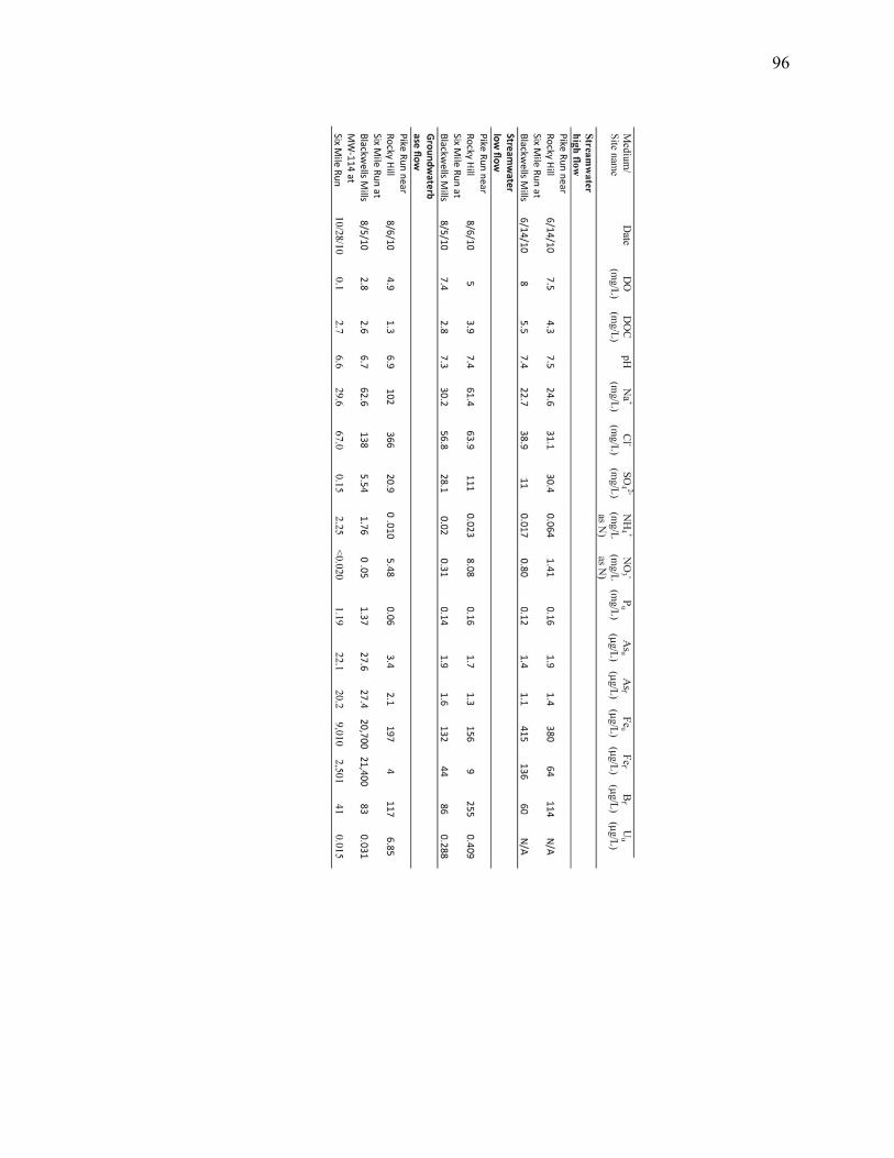
96

97
Figure 1:
Map of Central New Jersey, with sampling sites along the Millstone River marked. The
Highlands, Piedmont, and Coastal Plain Physiographic provinces are delineated by red
lines.

98
Figure 2:
Stoichiometric As(V) reduction was observed in microcosms inoculated with
groundwater from Six Mile Run (A) and MW114 (B). No As(V) reduction was observed
in microcosms inoculated with groundwater from Pike Run (C).

99
Figure 3:
Taxonomic classification of 16S rRNA gene OTUs recovered from Six Mile Run, Pike
Run, and NJDEP MW114. Sequences recovered from Six Mile Run are combined with
those from MW114.
0%
10%
20%
30%
40%
50%
60%
70%
80%
90%
100%
Six Mile Run Pike Run
Unclassified bacteriaActinobacteriaCandidate Division OP11Unclassified BacteriodetesVerrucomicrobia Subdivision 1ClostridiaNitrospiraUnclassified PlanctomycetesVerrucomicrobia Subdivision 3Unclassified ChloroflexiAcidobacteria Group 3SphingobacteriaAcidobacteria Group 6Acidobacteria Group 22Alpha ProteobacteriaBeta ProteobacteriaGamma ProteobacteriaDeferribacteres

100
Figure 4:
Image of the 492bp 16S rRNA gene amplicons from Six Mile Run and Pike Run as
separated by DGGE. Unique banding patterns were obtained from each site. Circled
bands were excised from the gel and sequenced.

101
Figure 5:
Venn diagram illustrating the different microbial communities found at Six Mile Run and
Pike Run based on As(V) respiratory reductase (arrA).

102
Chapter 5
Conclusions and Future Directions
Chapter 1 describes how an As(V) reducing organism I isolated, Strain MPA-C3,
is required for the precipitation of an arsenic sulfide mineral, alacranite (As8S9). This
research has been submitted to Environmental Microbiology, and is currently in review.
In this chapter, I describe how I isolated a novel, As(V) reducing strain, MPA-C3, from
estuarine sediments sampled in the Mai Po Marsh in Hong Kong, China. I found that
Strain MPA-C3 is metabolically diverse, and capable of using a wide range of electron
donors and acceptors. Sequencing of first the 16S rRNA gene and then the entire genome
demonstrated that Strain MPA-C3 is a member of the Deferribacteres, a genus of
anaerobic heterotrophs. The genome sequence allowed for elucidation of the metabolic
pathways utilized by Strain MPA-C3, including those for nitrate reduction, sulfur
reduction, and the utilization of simple carbon sources. While Strain MPA-C3 has a
wider metabolic range than any of the described Deferribacteres, its most notable trait is
that, when grown with As(V) as a terminal electron acceptor, it precipitates a yellow
mineral from the culture medium. Analysis of this precipitate identified it as alacranite
(As8S9), a mineral which has previously only been described as forming under
hydrothermal conditions. This finding suggests that the precipitation of a wider range of
arsenic sulfides may be mediated by microbial activity than previously thought, and such
activity may play an important role in the biogeochemical cycling of arsenic.
At this point in time, several aspects of this project present useful topics for
further study. Foremost among these is the mechanism by which Strain MPA-C3
catalyzes the precipitation of alacranite. This process appears to occur extracellurlarly,

103
and we have hypothesized that it is a result of an as-yet unidentified product released
from the cell. Newman et al. suggest that the precipitation of orpiment occurs both in the
periplasm of Desulfosporosinus auripigmentum and and extracellularly, however, no
mechanism for this precipitation is discussed (99). As I demonstrated in Chapter 2,
neither alacranite nor any other arsenic sulfide mineral formed when As(III) and sulfide
were added to the media used to grow Strain MPA-C3, thus demonstrating the conditions
necessary for low-temperature formation as suggested by O’Day et al. (102) are not
found in the absence of cells. We did note, however, that the mineral appeared to form
either in direct contact or in the immediate vicinity of the cells, which suggests that the
cells may be providing a nucleation site for crystal formation. This could in turn by
studied by TEM, which would allow for a much closer look at the environment in the
immediate vicinity of the cell, and could potentially show if crystal nucleation is
occurring at the cell surface, in the periplasm, or simply very near to the cells.
The As(V) respiratory reductase (arrA) gene of Strain MPA-C3 was successfully
amplified by PCR, sequenced, and found to be most similar to that of Desulfitobacterium
hafniense Y51, an organism in a distant phylum than the Deferribacteres. In Chapter 2, I
suggest that this is the result of the arrA gene of Strain MPA-C3 having been
incorporated via horizontal gene transfer. However, this gene was not found on the
genome of Strain MPA-C3, making determination of the surrounding genes, and perhaps
confirmation of this hypothesis, much more challenging. I anticipate that completion and
closure of the genome would allow for the identification of the arrA gene identified by
PCR, as well as the elucidation of more of the metabolic pathways utilized by this
fascinating isolate.

104
Strain MPA-C3 provides an interesting example of a microbe biologically
catalyzing a process under moderate conditions which has been thought to only occur
chemically at very high temperatures and pressures. In its native estuarine environment,
Strain MPA-C3, and organisms like it, may in fact be responsible for the formation of
minerals which would have otherwise been assumed to have formed geologically and
transported to a moderate environment. This finding provides an example of the
importance of biological processes in the geochemical cycling of both trace and major
elements. A greater understanding of the process by which Strain MPA-C3 precipitates
alacranite could potentially lead to the development of biological treatment methods for
the removal of arsenic contaminated drinking water.
In Chapter 3, I present the paper “Microbial transformation of Arsenic:
Mobilization from glauconitic sediments to water” (90) in which I collaborated with the
United States Geological Survey (USGS) to assess the potential for microbial activity in
the release of As to groundwater at Crosswicks Creek, in Upper Freehold, NJ. In this
chapter, I describe how we utilized cultivation-based techniques to determine that
microbial activity releases As from aquifer sediments, and combined these findings with
cultivation-independent molecular characterization of the microbial community present at
our study site. Through sequencing of the 16S rRNA gene, I found that the microbial
communities present in the groundwater of the site were more diverse than those present
on recovered sediments. In addition, I describe how sequencing of the As(V) respiratory
reductase gene (arrA) led to the finding that As(V) reducing organisms were present at
the site. In this chapter, I describe how we determined that As release from the sediments
is most likely driven by Fe reducing microorganisms, and once released, As mobility in

105
groundwater is enhanced by the reduction of the less mobile As(V) to the more mobile
As(III).
In Chapter 4, I describe how, again in collaboration with the USGS, I examined
the potential for microbial As mobilization at two sites along the Millstone River, in
Somerset County, NJ. This research has been submitted to Water Research as (TITLE)
and is currently under review. The two sites, Six Mile Run and Pike Run, displayed very
similar mineralogy and geochemistry, with the notable difference being that the
groundwater at Six Mile Run was anoxic and reducing, and the groundwater at Pike Run
was oxic and oxidizing. We hypothesized that these differing redox conditions would
lead to the development of differing microbial communities, and that this would in turn
explain why the levels of As present at Six Mile Run were an order of magnitude higher
than those at Pike Run. By following a similar research methodology as described in
Chapter 3, I determined through culture-independent molecular techniques that the
microbial communities at each site were in fact different, and these findings were
supported by the results of microcosm experiments. In these experiments, I found that
microcosms inoculated with groundwater taken from Six Mile Run were capable of
As(V) reduction, while those inoculated with groundwater from Pike Run were not. This
finding was particularly interesting in that the arrA gene was successfully amplified from
the Pike Run site, a finding which I suggest may be due to the very similar As(III)
oxidase gene (arxA).
Both Chapter 3 and Chapter 4 would be enhanced and better completed by further
work in several directions. First and foremost, it is likely that the incubation of sediment
and groundwater from Crosswicks Creek, Six Mile Run, and Pike Run would allow for

106
the identification of microbial As(III) oxidation by both cultivation-based and molecular
techniques. This would allow for better understanding of how arsenic is biologically
cycled at this site, and would provide field confirmation for the arsenic cycling
demonstrated in vitro by Rhine et al. (117). Additionally, the recent advances in high-
throughput sequencing would allow for a more complete picture of the microbial
communities present, which would in turn allow for more detailed comparisons between
the sites. Furthermore, such information would allow for a more detailed model of the
geomicrobiological processes occurring in the subsurface aquifer, the groundwater, and
the streambed to be constructed.
A deep understanding of the microbial processes which drive arsenic release into
drinking water is of critical importance, not only when one considers the highly
publicized issue of arsenic contaminated drinking water in Bangladesh, but also when
one considers the potential impacts of adding organic carbon to any aquifer which resides
in arsenic-bearing sediments or bedrock. As I have shown in this Dissertation, microbial
arsenic reduction can result in both the mobilization and immobilization of arsenic,
depending both on the geochemistry of the environment, and the organisms present. In
all cases, the organisms present are heterotrophs, requiring the introduction of organic
carbon to survive. As I demonstrated in Chapter 3, inputs of organic carbon drove the
release of arsenic into the shallow subsurface aquifer at Crosswicks Creek, NJ.
Following the example, it is not impossible to imagine a scenario in the addition of
organic contaminants to an otherwise carbon-limited aquifer by industrial processes
might result in the release of arsenic into drinking water where arsenic contamination had
never before been observed. Conversely, the ability of microbes such as Strain MPA-C3

107
to precipitate insoluble arsesnic-sulfide minerals such as alacranite presents great promise
as a biological treatment method for drinking and irrigation waters contaminated with
arsenic. Current techniques for arsenic removal are based upon sorption to iron
hydroxides, which while effective, also retain the risk of re-mobilization of arsenic
should redox conditions change upon disposal (86). A treatment based upon microbial
arsenic sulfide precipitation would instead result in the removal of arsenic as an insoluble
phase, without the volumes of arsenic-laden waste and risk of re-mobilization as present
with current iron oxide based techniques.
As the evidence for the importance of microbial processes in geochemical cycles
mounts, it is becoming more and more apparent just how little is known about these
processes. My research fits into a growing body of evidence for the importance of
microbial processes in geochemical cycles, demonstrating both how microbes can form
‘hydrothermal’ minerals, and drive the dissolution and mobilization of sequestered
elements. Much remains to be studied on the mechanisms behind these biogeochemical
cycles, and their elucidation will help to enable the rational, harmonious utilization of our
limited natural resources.

108
Appendix 1: Supplemental Material For Chapter 2
Assembly Statistics for MPA-C3:

109
Subsystems Found in the Genome of Strain MPA-C3 as annotated by RAST

110
Appendix 2: Supplemental Material For Chapter 3
Table S-1. Composition of anaerobic medium used in microcosm experiments using Crosswicks Creek groundwater and streambed sediments. Compound Concentration NaCl 1g/L MgCl2·2H2O 400 mg/L CaCl2·2H2O 100 mg/L KH2PO4 200 mg/L KCl 500 mg/L Na2HPO4 0.19 g/L NaH2PO4 0.06 g/L NH4Cl 0.27 g/L MOPS 1.05 g/L Nitrilotriacetic Acid 1.5 mg/L Fe(NH4)2(SO4)2 0.8 mg/L Na2SeO3 0.2 mg/L CoCl2·6H2O 0.1 mg/L MnSO4·H2O 0.1 mg/L Na2MoO4·H2O 0.1 mg/L NaWO4·H2O 0.1 mg/L NiCl2·6H2O 0.1 mg/L H3BO3 0.01 mg/L CuSO4·5H2O 0.01 mg/L nicotinic acid 0.1µg/L calcium pantothenate 0.1µg/L pyroxidine HCl 0.1 µg/L riboflavin 0.1 µg/L thiamine HCl 0.1 µg/L biotin 0.05 µg/L folic acid 0.05 µg/L α-lipoic acid 0.05 µg/L vitamin B-12 0.05 µg/L Na2S 31 mg/L Cysteine 48.5 mg/L
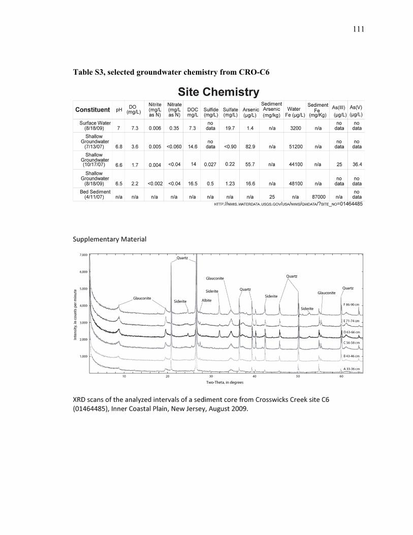
111
Table S3, selected groundwater chemistry from CRO-C6

112
Figure S3.2. Photograph of glauconite grains from CRO-C6
Figure S3.3 SEM scans of sediment particles from streambed sediment core from
Crosswicks Creek, site C6 (01464485), Inner Coastal Plain, New Jersey, August 2009.

113
Note P content of cement surrounding grains. The SEM scans indicated that amorphous
phases likely are composed of Fe hydroxide; Fe-phosphate cement also may be present.
Such cement is reported elsewhere, formed though the activity of nitrate-reducing, iron-
oxidizing microbes(89).

114
Appendix 3: Supplemental Material for Chapter 4
Table S4.1 Primers used in Chapter 4
Primer Name Primer Sequence Reference 27F 5’-AGAGTTTGATCCTGGCTCAG-3’ (70)
519R 5’-GTATTACCGCGGCAGCTGGCAC-3’ (70) 1492R 5’-GGTTACCTTGTTACGACTT-3’ (70) As1F 5’-CGAAGTTCGTCCCGATHACNTGG-3’ (73) As1R 5’-GGGGTGCGGTCYTTNARYTC-3’ (73)
Table S4.2 Sequences amplified from DGGE Gel Bands
Six Mile Run Band 1 gctTAcCaTGCAGTCGACGGNAGCACGGACTTCGGTCTGGTGGCGAGTGG
CGAACGGGTGAGTAATATATCGGAACGTGCCCAGTCGTGGGGGATAACGT AgTGAAAATTACGCTAATACCGCATACGATCTACGGATGAAAGCGGGGGA TCGCAAGACCTCGCGCGATTGGAGCGGCCGATATCAGATTAGGTAGTTGG TGGGGTAAAGGCTCACCAAGCCAACGATCTGTAGCTGGTCTGAGAGGACA ACCAGCCACACTGGAACTGAGACACGGTCCAGACTCCTACGGGAGGCAGC AGTGGGGAATTTTGGACAATGGACGCAAGTCTGATCCAGCCATTCCGCGT GCAGGACGAAGGCCTTCGGGTTGTAAACTGCTTTTGTACAGAACGAAAAG GTTTCTATTAATACTAGGAGCTCATGACGGTACTGTAAGAATAAGCACCG GCTAACTACGTGCCAGCCGCCGCGGNAATTCAAAAAA
Six Mile Run Band 2 CTaCCaTGCAGTCGACGGCAgCACGGACTTCGGTCTGGTGGCGAGTGGCG
AACGGGTGAGTAATATATCGGAACGTGCCCACTCGTGGGGGATAACGTAT TGAAAATTACGCTAATACCGCATACGATCTACGGATGAAAGCGGGGGATC GCAAGACCTCGCGCGATTGGAGCGGCCGATATCAGATTAGGTAGTTGGGG AGGTGAAgGCTCACCAAgGCAACGATATGTttcTGGTCTCAAAAGACaAA CAGgCaCCCTGGGNATGcgAAACNgGGAagAAATCCCCTGGGGCGGtT
Six Mile Run Band 3 CTAcaaTGCAGTCGACGGTGTCTTCGGAGATAGCGGGGGACGGGGGCGGA
ACACGTATGGACCTACCTTACATTGGGGGACAGCCTTGCGAAAGGGAGAT TAATACCGCATAATACCGTAGCTGGGCATCCAGCAGCTGTTAAAGATTTA TCGATGTACTCTGGGGATGGGTCCAATTAatTAcTTGGTGAGGAAACGGC TCACCAAGGCTATGATTGGTAGGGGAACTGAgAGGGCAATCCCCCACACT GGCACTGAgATACGGGCCAGACTCCTACGGGAGGCAGCAGTAAGGAATAT TGGGCAATGGACGCAAGTCTGACCCAGCCATGCCGCGTGCAGGATGAAGG CGTTATGCGTTGTAAACTGCTTTTATACAGGAATAAACGACTCTTGCGAG AGGCATTGACGGTACTGTATGAATAAGCACCGGCTAACTCCGTGCCAGCC GCCGCGGAtAAATtCAAAAA
Six Mile Run Band 4 gCtACctTGCAGTCGAACGGgattGGtGTTTCCGGTGGCGGACGGGTGAG
TAACGCGTAAGAACCTGCCCTTGGGAGGGGAACAACCGGTGGAAACGGCT GCTAATACCCCATAAGCTGAGGAACAAAAGGAGGAATCCGCCCAAGGAGG GGCTCGCGTCTGATTAGTTAGTTGGTGAGGCAATGGCTTACCAAGGCGAC GATCAGTANCTGGTCCGAGAGGATGATCAGCCACACTGGGACTGAGACAC GGCCCANACTCCTACGGGAGGCAGCAGTGGGGAATTTTCCGCAATGGGCG AAAGCCTGACGGAGCAATGCCGCGTGAAGGAAGAAGGCCCACGGGTCGTG AACTTCTTTTCTCGGAGAAGAAACAATGACGGTATCTGAGGAATAANCAT CGGCTAACTCTGTGCCAGCCGCCGCGGTAATTCaAAAAa
Six Mile Run Band 5 CTAccaTGCAGTCGAGCGATGAgCGCCTTCTGGCGTGAtTAGCGGCGAAC
GGGTGAGGAACACGTGAGAAATCTGCCTTCTCTCTGGGATAACTCCGGGA AAACGGGGGTAATACCGGATATGAAATCTACAGGCATCTGTGGATCTGGA AAGTTTTTCGGTTGAANATGATCTCGCGGCCTATCAACTTGTTGGTGAGG TAATGGCTCACCAAGGCGACGACGGGatTCCCGGCTGAaAAGgctTcCgG

115
CCACACTGGGACTGAGACACGGcCCAGACTCCTATTTGGGagcTCTTCGG GGTCCATTgNACTGTGGCgCTGCACTCGAGCCTTCGaCTGCCCGCGNGGG AATCAACTCACTCaCAGGCGTAAATACggTTcTCCGCGGAATCaNNCGAT GGGCGGGATNTAACATGTGAgCAAAAATCCANTTATgTGCCAgCCGCCCT
Six Mile Run Band 6 gcgCTAcTGCAGTCACGtgacTGGTCTTGTgGGGGGataGGGGANGGGGG
AGATaTACCTGACGGTCTGCCCTTgGCTCTGGGACCCTGCGGgAGCTGTA ATACCTGCCtTCAACctNGgCTCaAGCGGGCGAGGGCTGGACTTCTTTTG TGAAGAGTTCGCTCTCGATTTATTTGCTGGTTGGGGAAGGAATGGCCCAC CCgACGAACtGTAGGTGGGCTGACAGGACGACCGACCACCCTGGAACTGA GACACAGcCCAgACTCCTACCGGAGGGAGGAGTGGGGAATTTTGTTTAAT GGGNGAAACTCTGACCCACCCATGCCgCGTGCGGGATGAAAGcCTTCCTT TTGTTTACTACTTTTTCTTCAGGAAAAAAAGaGAATAAAgTTTNAGTAAc CACgcAANCAAAAcaCCaCTATATGTCCCGCGCGCCGCCAAN
Six Mile Run Band 7 tAccaTGCAGTCGAGCGATGAAGCACCTTCGGGTGTGAATTAgCGGCGAA CGGGTGAGGAACACGTGAGAAATCTGCCTTCAACTCTGGGATAACTCCGG GAAACCGGGGCTAATACCGGATATGAGCCTTGATCGCATGATCGGGGCTG GAAAGTTTTTCGGTTGAAGATGATTTCGCGGCCTATCAGCTTGTTGGTGA GGTAATGGCTCACCAAGGCGACGACGGGTAGCCGGCCTGAGAGGGCGACC GGCCACACTGGGACTGAGACACGGCCCAGACTCCTACGGGAGGCAGCAGT GGGGAATATTGGGCAATGGAGGAAACTCTGACCCAGCGACGCCGCGTGAG GGATGAAGGCCTTCGGGTTGTAAACCTCTTTCAGTAGGGAAGAAGCGAAA GTGACGGTACCTACAGAAGAAGCACCGGCTAACTATGTGCCAGCCGCCGC GGGTAATTCAaaaa
Pike Run Band 1 Sequencing unsuccessful Pike Run Band 2 TGCaccgAGGaCNaAGgatGcCCCCAGcGCtgTCCNggNGcTacCcNTaN
aTTCCCGTTTGNTGNCgGGggaGGGGGGGAATACCCCGCTCGGGCcNANa ACAAAAAACTCGTTGAttTGTTTGgTTCGgcCCGAANTGTGaTTcGgANg CTaaATaCtTtNATTcTTTGGgTCGaGACCgcTTaCcAAGCcgATATCAC TNNGGNNCTNAANAATGAAcCGccAACTGGAGGtGAAAaCGGcTAtNAtt ccAgrGcTCgAAcaATTGAAcAtatttcTGgACAATCTgcATTCgCCaTC CaAaaac
Pike Run Band 3 agaTacTGcagTCgACGgAtTActTTcgTAAgAAACGGGGNaGANGNTGN
aCTCTTTCTCaTcTGTATTATTCGGGNGAAAAGGAcAaTAATACCTAATA CTTCCTAATGAAATCCTGGGGTTAtCTTCTGAGCTCTCATGATCATGCTC TGCCAGcATCGCTATATCGAGTTGGTGGGGCAGCGTNCCATAAAATCAAA GAGACGGAACTGTCTCTGACAGGGTGATGATGNACNATCCCCCCGGATAG CgCTGAgNACCCCcNCNGGAGcCTAACAAGGTgAGTACCCCTACgTTGGc GAAAAATTGCGCGATCTTTgCANTACCNGACGGGAAATGAACTCNTNNNA CNAAAAAAAAGNNTN
Pike Run Band 4 TaccatGCAGTCgAACGGtattGtGTNCGggcGCTGACGGGaGGCGAACG
GGTGAgAAATTGTTATTGcgTaCCTTTAGtGGGGGATAACGcTGCAataC CCAATAAATACCGaATTAAAACAAAGGATCTGCCTGGCGAGGGGCTCGCG NGTGATTTGTTAGATGGCGATATGTTGGATTACCTAGGTGGCGATCGAAA AGCGGaCCGAGGGGAAGATCAGTCTCCCTGGGAATGAGACGATGCCCcaA CTCGGACGGGAGGCACGGCTGGGACATTTTCCGCAgTGGGCAAaGGCCTG ATTtActGATGGCGCGTGAGCCTGGAaCCCCaTGGGTCGCGACCTTCTTT TCTCTGACANTTATAATGACCTTTTgTGAGaANTaAACATCCTGTAACTC TGTACCACCGCCGCGgTAATTCAaCAACAATAGtgCCTTCTcaCaAAgTG gCtCCCGCCCAGAACtTTCAAAcagNCAcCgNtAATacAAcAAN
Pike Run Band 5 gctAcACATGCAGTCGAACGAgAagTTCGGGTGTAGCAATACATTCGGGC
GAGTAAAGTGGCGTCCGGGTGAGTAACACGTANCTAACCTACCCTCGAGC GGGGGATAACCTGGGGAAACTCGGGCTAATACCGCATAACGTCCAGGCTA CATATGTAGCTTGGATCAAAGGCCCGCAAGGGCCACTGGAGGAGGGGGCT GCGGCCGATTAGCTAGTTGGTGGGGTAATGGCCTACCAAGGCGATGATCG GTACCCGGCCTGAGAGGGCGCACGGCCACACTGGCACTGAAATACGGGCC AGACTCCTACGGGAGGCAGCAGTGGGGAATTTTGCACAATGCCCGAAAGG GTGATGCAGCAACGCCGCGTGAGGGATGAANGtCTTCGGGTCGTAAACCT CTTTCGACCGGGAAGAATGGCTCCGTGAACAAAGGACCCTGACAGTCCCG GNGAAAAAAAACCCGGCTAaCTACgTGCCtgCCNCCN

116
Pike Run Band 6 GCtAccaTGCAGTCGACGAgAagtGcGGGTGggcatGCATTCGGGCGAGA
ACGGGGGAGTCCGGGTGAGAAACACGTACTTAAgGTACCCTCGAGCGGGG GATAAcccGGGCACACTTCCACACAACCCGcGAGGCGGAAAGGTAAAGTG TGAAGCCTGGATCGCCTAATGAGTGANCTAACTCACATTAATTGCGTTGC GCTCACTGCCCGCTTTCCAGTCAGGAAACCTGTCGTGCCAGCTGCATTAA TGAaTCGGCCAACGCGCGGGGAGAGGCGGTTTGCgTATTGGGCGCTCTTC CGCTTCCTCGCTCANTGNNTCGCTGCGCTCGGTCGTTCGGCTGCGGCGAG CGGtATCAaCTCACTCAaAGGCNgTAATACGGTTATCCACAaAATCAaGg GAtAaCgcaGGAAaga

117
Figure S4.1 Annotated rAXML tree of 16S rRNA sequences from Six Mile Run and
Pike Run

118
Figure S4.2 Annotated rAXML tree of arrA sequences obtained from Six Mile Run and
Pike Run.

119
References:
1. Afkar, E., J. Lisak, C. Saltikov, P. Basu, R. S. Oremland, and J. F. Stolz. 2003. The respiratory arsenate reductase from Bacillus selenitireducens strain MLS10. FEMS Microbiology Letters 226:107-112.
2. Ahmann, D., L. R. Krumholz, H. F. Hemond, D. R. Lovley, and F. M. M. Morel. 1997. Microbial Mobilization of Arsenic from Sediments of the Aberjona Watershed. Environmental Science & Technology 31:2923-2930.
3. Ahmann, D., A. L. Roberts, L. R. Krumholz, and F. M. M. Morel. 1994. Microbe grows by reducing arsenic. Nature 371:750-750.
4. Akai, J., K. Izumi, H. Fukuhara, H. Masuda, S. Nakano, T. Yoshimura, H. Ohfuji, H. Md Anawar, and K. Akai. 2004. Mineralogical and geomicrobiological investigations on groundwater arsenic enrichment in Bangladesh. Applied Geochemistry 19:215-230.
5. Altschul, S. F., W. Gish, W. Miller, E. W. Myers, and D. J. Lipman. 1990. Basic local alignment search tool. Journal of Molecular Biology 215:403-410.
6. Anawar, H., J. Akai, T. Yoshioka, E. Konohira, J. Lee, H. Fukuhara, M. Tari Kul Alam, and A. Garcia-Sanchez. 2006. Mobilization of arsenic in groundwater of Bangladesh: evidence from an incubation study. Environmental Geochemistry and Health 28:553-565.
7. Annable, W. K., S. K. Frape, O. Shouakar-Stash, T. Shanoff, R. J. Drimmie, and F. E. Harvey. 2007. 37Cl, 15N, 13C isotopic analysis of common agro-chemicals for identifying non-point source agricultural contaminants. Applied Geochemistry 22:1530-1536.
8. Arbogast, B. F. 1996. Analytical methods manual for the Mineral Resource Surveys Program. In U. S. G. Survey (ed.).
9. Arehart, G. B., S. L. Chryssoulis, and S. E. Kesler. 1993. Gold and arsenic in iron sulfides from sediment-hosted disseminated gold deposits; implications for depositional processes. Economic Geology 88:171-185.
10. Argos, M., T. Kalra, P. J. Rathouz, Y. Chen, B. Pierce, F. Parvez, T. Islam, A. Ahmed, M. Rakibuz-Zaman, R. Hasan, G. Sarwar, V. Slavkovich, A. van Geen, J. Graziano, and H. Ahsan. 2010. Arsenic exposure from drinking water, and all-cause and chronic-disease mortalities in Bangladesh (HEALS): a prospective cohort study. The Lancet 376:252-258.
11. Aziz, R. K., D. Bartels, A. A. Best, M. DeJongh, T. Disz, R. A. Edwards, K. Formsma, S. Gerdes, E. M. Glass, M. Kubal, F. Meyer, G. J. Olsen, R. Olson, A. L. Osterman, R. A. Overbeek, L. K. McNeil, D. Paarmann, T. Paczian, B. Parrello, G. D. Pusch, C. Reich, R. Stevens, O. Vassieva, V. Vonstein, A. Wilke, and O. Zagnitko. 2008. The RAST Server: rapid annotations using subsystems technology. BMC genomics 9:75.
12. Babechuk, M. G., C. G. Weisener, B. J. Fryer, D. Paktunc, and C. Maunders. 2009. Microbial reduction of ferrous arsenate: Biogeochemical implications for arsenic mobilization. Applied Geochemistry 24:2332-2341.
13. Barringer, J., M. Riskin, Z. Szabo, P. Reilly, R. Rosman, J. Bonin, J. Fischer, and H. Heckathorn. 2010. Mercury and Methylmercury Dynamics in a Coastal Plain Watershed, New Jersey, USA. Water, Air, & Soil Pollution 212:251-273.

120
14. Barringer, J. L., A. Mumford, L. Y. Young, P. A. Reilly, J. L. Bonin, and R. Rosman. 2010. Pathways for arsenic from sediments to groundwater to streams: Biogeochemical processes in the Inner Coastal Plain, New Jersey, USA. Water Research 44:5532-5544.
15. Barringer, J. L., P. A. Reilly, D. D. Eberl, A. E. Blum, J. L. Bonin, R. Rosman, B. Hirst, M. Alebus, K. Cenno, and M. Gorska. 2011. Arsenic in sediments, groundwater, and streamwater of a glauconitic Coastal Plain terrain, New Jersey, USA--Chemical "fingerprints" for geogenic and anthropogenic sources. Applied Geochemistry 26:763-776.
16. Barringer, J. L., Szabo, Z., Barringer, T.H. 1998. Arsenic and metals in soils in the vincintiy of the Imperial Oil Company Superfund site, Marlboro Township, Monmouth County, New Jersey. . In U.S.G.S. (ed.).
17. Battaglia-Brunet, F., C. Crouzet, A. Burnol, S. Coulon, D. Morin, and C. Joulian. 2012. Precipitation of arsenic sulphide from acidic water in a fixed film bioreactor. Water Research.
18. Bentley, R., and T. G. Chasteen. 2002. Microbial Methylation of Metalloids: Arsenic, Antimony, and Bismuth. Microbiology and Molecular Biology Reviews 66:250-271.
19. Berg, M., H. C. Tran, T. C. Nguyen, H. V. Pham, R. Schertenleib, and W. Giger. 2001. Arsenic Contamination of Groundwater and Drinking Water in Vietnam: A Human Health Threat. Environmental Science & Technology 35:2621-2626.
20. Berg, M., P. T. K. Trang, C. Stengel, J. Buschmann, P. H. Viet, N. Van Dan, W. Giger, and D. Stüben. 2008. Hydrological and sedimentary controls leading to arsenic contamination of groundwater in the Hanoi area, Vietnam: The impact of iron-arsenic ratios, peat, river bank deposits, and excessive groundwater abstraction. Chemical Geology 249:91-112.
21. Bhattacharya, P., G. Jacks, K. M. Ahmed, J. Routh, and A. A. Khan. 2002. Arsenic in Groundwater of the Bengal Delta Plain Aquifers in Bangladesh. Bulletin of Environmental Contamination and Toxicology 69:538-545-545.
22. Bhowmick, S., B. Nath, D. Halder, A. Biswas, S. Majumder, P. Mondal, S. Chakraborty, J. Nriagu, P. Bhattacharya, M. Iglesias, G. Roman-Ross, D. G. Mazumder, J. Bundschuh, and D. Chatterjee. Arsenic mobilization in the aquifers of three physiographic settings of West Bengal, India: understanding geogenic and anthropogenic influences. Journal of Hazardous Materials.
23. Bindi, L., V. Popova, and P. Bonazzi. 2003. Uzonite, As4S5, from the type locality: single crystal x-ray study and effects of exposure to light The Canadian Mineralogist 41:1463-1468.
24. Blasco, F., C. Iobbi, G. Giordano, M. Chippaux, and V. Bonnefoy. 1989. Nitrate reductase of Escherichia coli: Completion of the nucleotide sequence of the nar operon and reassessment of the role of the α and β subunits in iron binding and electron transfer. Molecular and General Genetics MGG 218:249-256.
25. Blum, J. S., S. Han, B. Lanoil, C. Saltikov, B. Witte, F. R. Tabita, S. Langley, T. J. Beveridge, L. Jahnke, and R. S. Oremland. 2009. Ecophysiology of “Halarsenatibacter silvermanii” Strain SLAS-1T, gen. nov., sp. nov., a Facultative

121
Chemoautotrophic Arsenate Respirer from Salt-Saturated Searles Lake, California. Applied and Environmental Microbiology 75:1950-1960.
26. Blum, J. S., S. Han, B. Lanoil, C. Saltikov, B. Witte, F. R. Tabita, S. Langley, T. J. Beveridge, L. Jahnke, and R. S. Oremland. 2009. Ecophysiology of “Halarsenatibacter silvermanii” Strain SLAS-1T, gen. nov., sp. nov., a Facultative Chemoautotrophic Arsenate Respirer from Salt-Saturated Searles Lake, California. Applied and Environmental Microbiology 75:5437.
27. Bonazzi, P., L. Bindi, F. Olmi, and S. Menchetti. 2003. How many alacranites do exist? A structural study of non-stoichiometric As8S9-x crystals. European Journal of Mineralogy 15:283-288.
28. Bonazzi, P., L. Bindi, V. Popova, G. Pratesi, and S. Menchetti. 2003. Alacranite, As8S9: structural study of the holotype and re-assignment of the original chemical formula. American Mineralogist 88:1796-1800.
29. Briggs, P. H., Meier, A.L. 2002. The determination of forty-two elements in geological materials by inductively coupled plasma-mass spectrometry for NAWQA. In U.S.G.S. (ed.).
30. Brown, Z. A., Curry, K.J. 2002. Total carbon by combustion. In U.S.G.S. (ed.). 31. Brown, Z. a., Papp, C., Brandt, E., Aruscavage, P. 2002. Carobonate carbon by
coulometric titration. In U.S.G.S. (ed.). 32. Bundschuh, J., M. Litter, V. S. T. Ciminelli, M. E. Morgada, L. Cornejo, S.
G. Hoyos, J. Hoinkis, M. T. Alarcón-Herrera, M. A. Armienta, and P. Bhattacharya. 2010. Emerging mitigation needs and sustainable options for solving the arsenic problems of rural and isolated urban areas in Latin America - A critical analysis. Water Research 44:5828-5845.
33. Burns, P. C., and J. B. Percival. 2001. Alacranite, As4S4: A new occurance, new formula, and determination of the crystal structure. Canadian Mineralogist 39:809-818.
34. Caccavo Jr, F., J. D. Coates, R. A. Rossello-Mora, W. Ludwig, K. H. Schleifer, D. R. Lovley, and M. J. McInerney. 1996. Geovibrio ferrireducens, a phylogenetically distinct dissimilatory Fe(III)-reducing bacterium. Archives of Microbiology 165:370-376.
35. Clark, A. H. 1970. Alpha-arsenic sulfide, from Mina Alacràn, Pampa Larga, Chile. American Mineralogist 55:1338-1344.
36. Cline, J. S. 2001. Timing of Gold and Arsenic Sulfide Mineral Deposition at the Getchell Carlin-Type Gold Deposit, North-Central Nevada. Economic Geology 96:75-89.
37. Council, N. R. 1999. Arsenic in drinking water. National Academy Press, Washington D.C.
38. Cummings, D. E., F. Caccavo, S. Fendorf, and R. F. Rosenzweig. 1999. Arsenic Mobilization by the Dissimilatory Fe(III)-Reducing Bacterium Shewanella alga BrY. Environmental Science & Technology 33:723-729.
39. Demergasso, C. S., C. D. Guillermo, E. G. Lorena, J. J. P. Mur, and C. Pedrós-Alió. 2007. Microbial Precipitation of Arsenic Sulfides in Andean Salt Flats. Geomicrobiology Journal 24:111-123.
40. Dooley, J. H. 2001. N.J. Geological Survey Investigation Report: Baseline concentrations of arsenic, beryllium, and associated elements in glauconite and

122
glauconitic soils in the New Jersey Coastal Plain. N.J. Department of Environmental Protection, Trenton.
41. Dooley, J. H. 1998. NJGS Technical Memorandum 98-1: Comprehensive Chemistry of Select Greensand from the New Jersey Coastal Plain. N.J Department of Environmental Protection, Trenton.
42. Driehaus, W., R. Seith, and M. Jekel. 1995. Oxidation of arsenate(III) with manganese oxides in water treatment. Water Research 29:297-305.
43. Eberl, D. D. 2003. User's Guide to RockJock - A program for determining quantitatative mineralogy from powder x-ray diffraction data. In U.S.G.S. (ed.).
44. Farooq, S. H., D. Chandrasekharam, Z. Berner, S. Norra, and D. Stüben. 2010. Influence of traditional agricultural practices on mobilization of arsenic from sediments to groundwater in Bengal delta. Water Research 44:5575-5588.
45. Finneran, K. T., C. V. Johnsen, and D. R. Lovley. 2003. Rhodoferax ferrireducens sp. nov., a psychrotolerant, facultatively anaerobic bacterium that oxidizes acetate with the reduction of Fe(III). International Journal of Systematic and Evolutionary Microbiology 53:669-673.
46. Fisher, E., A. M. Dawson, G. Polshyna, J. Lisak, B. Crable, E. Perera, M. Ranganathan, M. Thangavelu, P. Basu, and J. F. Stolz. 2008. Transformation of Inorganic and Organic Arsenic by Alkaliphilus oremlandii sp. nov. Strain OhILAs. Annals of the New York Academy of Sciences 1125:230-241.
47. Fleet, M. E., Mumin, A. H. . 1997. Gold-Bearing arsenian pyrite and marcasite and arsenopyrite from Carlin Trend gold deposits and laboratory synthesis. American Mineralogist 82:182-193.
48. Garrity, G. M., and J. M. Holt. 2001. Bergey’s Manual® of Systematic Bacteriology Phylum BIX. Deferribacteres phy. nov. , p. 465-471. In D. R. Boone and R. W. Castenholz (ed.). Springer, New York.
49. Guymer, D., J. Maillard, and F. Sargent. 2009. A genetic analysis of in vivo selenate reduction by <i>Salmonella enterica serovar Typhimurium LT2 and <i>Escherichia coli K12. Archives of Microbiology 191:519-528.
50. Haque, S., J. Ji, and K. H. Johannesson. 2008. Evaluating mobilization and transport of arsenic in sediments and groundwaters of Aquia aquifer, Maryland, USA. Journal of Contaminant Hydrology 99:68-84.
51. Harvey, C. F., C. H. Swartz, A. B. M. Badruzzaman, N. Keon-Blute, W. Yu, M. A. Ali, J. Jay, R. Beckie, V. Niedan, D. Brabander, P. M. Oates, K. N. Ashfaque, S. Islam, H. F. Hemond, and M. F. Ahmed. 2002. Arsenic Mobility and Groundwater Extraction in Bangladesh. Science 298:1602-1606.
52. Héry, M., A. G. Gault, H. A. L. Rowland, G. Lear, D. A. Polya, and J. R. Lloyd. 2008. Molecular and cultivation-dependent analysis of metal-reducing bacteria implicated in arsenic mobilisation in south-east asian aquifers. Applied Geochemistry 23:3215-3223.
53. Hery, M., B. E. Van Dongen, F. Gill, D. Mondal, D. J. Vaughan, R. D. Pancost, D. A. Polya, and J. R. Lloyd. Arsenic release and attenuation in low organic carbon aquifer sediments from West Bengal. Geobiology 8:155-168.
54. Holmes, D. E., R. A. O'Neil, H. A. Vrionis, L. A. N'Guessan, I. Ortiz-Bernad, M. J. Larrahondo, L. A. Adams, J. A. Ward, J. S. Nicoll, K. P. Nevin, M. A. Chavan, J. P. Johnson, P. E. Long, and D. R. Lovley. 2007. Subsurface clade

123
of Geobacteraceae that predominates in a diversity of Fe(III)-reducing subsurface environments. ISME J 1:663-677.
55. Huber, R., M. Sacher, A. Vollmann, H. Huber, and D. Rose. 2000. Respiration of Arsenate and Selenate by Hyperthermophilic Archaea. Systematic and Applied Microbiology 23:305-314.
56. Islam, F. S., A. G. Gault, C. Boothman, D. A. Polya, J. M. Charnock, D. Chatterjee, and J. R. Lloyd. 2004. Role of metal-reducing bacteria in arsenic release from Bengal delta sediments. Nature 430:68-71.
57. Islam, F. S., R. L. Pederick, A. G. Gault, L. K. Adams, D. A. Polya, J. M. Charnock, and J. R. Lloyd. 2005. Interactions between the Fe(III)-Reducing Bacterium Geobacter sulfurreducens and Arsenate, and Capture of the Metalloid by Biogenic Fe(II). Applied and Environmental Microbiology 71:8642-8648.
58. Jiang, S., J.-H. Lee, M.-G. Kim, N. V. Myung, J. K. Fredrickson, M. J. Sadowsky, and H.-G. Hur. 2009. Biogenic Formation of As-S Nanotubes by Diverse Shewanella Strains. Applied and Environmental Microbiology 75:6896-6899.
59. Jönsson, J., and D. M. Sherman. 2008. Sorption of As(III) and As(V) to siderite, green rust (fougerite) and magnetite: Implications for arsenic release in anoxic groundwaters. Chemical Geology 255:173-181.
60. Kar, S., J. P. Maity, J.-S. Jean, C.-C. Liu, B. Nath, H.-J. Yang, and J. Bundschuh. 2010. Arsenic-enriched aquifers: Occurrences and mobilization of arsenic in groundwater of Ganges Delta Plain, Barasat, West Bengal, India. Applied Geochemistry 25:1805-1814.
61. Karp, P. D., S. M. Paley, M. Krummenacker, M. Latendresse, J. M. Dale, T. J. Lee, P. Kaipa, F. Gilham, A. Spaulding, L. Popescu, T. Altman, I. Paulsen, I. M. Keseler, and R. Caspi. 2010. Pathway Tools version 13.0: integrated software for pathway/genome informatics and systems biology. Briefings in Bioinformatics 11:40-79.
62. Kearse, M., R. Moir, A. Wilson, S. Stones-Havas, M. Cheung, S. Sturrock, S. Buxton, A. Cooper, S. Markowitz, C. Duran, T. Thierer, B. Ashton, P. Meintjes, and A. Drummond. 2012. Geneious Basic: An integrated and extendable desktop software platform for the organization and analysis of sequence data. Bioinformatics 28:1647-1649.
63. Kirk, M. F., E. E. Roden, L. J. Crossey, A. J. Brealey, and M. N. Spilde. 2010. Experimental analysis of arsenic precipitation during microbial sulfate and iron reduction in model aquifer sediment reactors. Geochimica et Cosmochimica Acta 74:2538-2555.
64. Kiss, H., E. Lang, A. Lapidus, A. Copeland, M. Nolan, T. Glavina Del Rio, F. Chen, S. Lucas, H. Tice, J. F. Cheng, C. Han, L. Goodwin, S. Pitluck, K. Liolios, A. Pati, N. Ivanova, K. Mavromatis, A. Chen, K. Palaniappan, M. Land, L. Hauser, Y. J. Chang, C. D. Jeffries, J. C. Detter, T. Brettin, S. Spring, M. Rohde, M. Goker, T. Woyke, J. Bristow, J. A. Eisen, V. Markowitz, P. Hugenholtz, N. C. Kyrpides, and H. P. Klenk. 2010. Complete genome sequence of Denitrovibrio acetiphilus type strain (N2460). Standards in genomic sciences 2:270-279.

124
65. Konhauser, K. 2007. Introduction to Geomicrobiology. Blackwell Science Ltd, Malden, MA.
66. Krafft, T., and J. M. Macy. 1998. Purification and characterization of the respiratory arsenate reductase of Chrysiogenes arsenatis. European Journal of Biochemistry 255:647-653.
67. Kulp, T. R., S. E. Hoeft, L. G. Miller, C. Saltikov, J. N. Murphy, S. Han, B. Lanoil, and R. S. Oremland. 2006. Dissimilatory Arsenate and Sulfate Reduction in Sediments of Two Hypersaline, Arsenic-Rich Soda Lakes: Mono and Searles Lakes, California. Applied and Environmental Microbiology 72:6514-6526.
68. Kuroda, M., M. Yamashita, E. Miwa, K. Imao, N. Fujimoto, H. Ono, K. Nagano, K. Sei, and M. Ike. 2011. Molecular cloning and characterization of the srdBCA operon, encoding the respiratory selenate reductase complex, from the selenate-reducing bacterium Bacillus selenatarsenatis SF-1. J Bacteriol 193:2141-2148.
69. Lagesen, K., P. Hallin, E. A. Rødland, H.-H. Stærfeldt, T. Rognes, and D. W. Ussery. 2007. RNAmmer: consistent and rapid annotation of ribosomal RNA genes. Nucleic Acids Research 35:3100-3108.
70. Lane, D. J. 1991. In E. Stackebrandt, Goodfellow, M. (ed.), Nucleic Acid Techniques in Bacterial Systematics. John Wiley and Sons New York.
71. Larkin, M. A., G. Blackshields, N. P. Brown, R. Chenna, P. A. McGettigan, H. McWilliam, F. Valentin, I. M. Wallace, A. Wilm, R. Lopez, J. D. Thompson, T. J. Gibson, and D. G. Higgins. 2007. Clustal W and Clustal X version 2.0. Bioinformatics 23:2947-2948.
72. Lauterbach, F., C. Kortner, S. P. Albracht, G. Unden, and A. Kroger. 1990. The fumarate reductase operon of Wolinella succinogenes. Sequence and expression of the frdA and frdB genes. Arch Microbiol 154:386-393.
73. Lear, G., B. Song, A. G. Gault, D. A. Polya, and J. R. Lloyd. 2007. Molecular Analysis of Arsenate-Reducing Bacteria within Cambodian Sediments following Amendment with Acetate. Applied and Environmental Microbiology 73:1041-1048.
74. Lee, J.-H., M.-G. Kim, B. Yoo, N. V. Myung, J. Maeng, T. Lee, A. C. Dohnalkova, J. K. Fredrickson, M. J. Sadowsky, and H.-G. Hur. 2007. Biogenic formation of photoactive arsenic-sulfide nanotubes by Shewanella sp. strain HN-41. Proceedings of the National Academy of Sciences 104:20410-20415.
75. Li, D., Z. Li, J. Yu, N. Cao, R. Liu, and M. Yang. 2010. Characterization of Bacterial Community Structure in a Drinking Water Distribution System during an Occurrence of Red Water. Applied and Environmental Microbiology 76:7171-7180.
76. Liu, A., E. Garcia-Dominguez, E. D. Rhine, and L. Y. Young. 2004. A novel arsenate respiring isolate that can utilize aromatic substrates. FEMS Microbiology Ecology 48:323-332.
77. Lovley, D. R., S. J. Giovannoni, D. C. White, J. E. Champine, E. J. Phillips, Y. A. Gorby, and S. Goodwin. 1993. Geobacter metallireducens gen. nov. sp. nov., a microorganism capable of coupling the complete oxidation of organic

125
compounds to the reduction of iron and other metals. Arch Microbiol 159:336-344.
78. Ludwig, W., O. Strunk, R. Westram, L. Richter, H. Meier, Yadhukumar, A. Buchner, T. Lai, S. Steppi, G. Jobb, W. Forster, I. Brettske, S. Gerber, A. W. Ginhart, O. Gross, S. Grumann, S. Hermann, R. Jost, A. Konig, T. Liss, R. Lussmann, M. May, B. Nonhoff, B. Reichel, R. Strehlow, A. Stamatakis, N. Stuckmann, A. Vilbig, M. Lenke, T. Ludwig, A. Bode, and K.-H. Schleifer. 2004. ARB: a software environment for sequence data. Nucleic Acids Research 32:1363-1371.
79. Macy, J. M., K. Nunan, K. D. Hagen, D. R. Dixon, P. J. Harbour, M. Cahill, and L. I. Sly. 1996. Chrysiogenes arsenatis gen. nov., sp. nov., a new arsenate-respiring bacterium isolated from gold mine wastewater. International Journal of Systematic and Evolutionary Microbiology 46:1153-1157.
80. Macy, J. M., J. M. Santini, B. V. Pauling, A. H. O’Neill, and L. I. Sly. 2000. Two new arsenate/sulfate-reducing bacteria: mechanisms of arsenate reduction. Archives of Microbiology 173:49-57.
81. Mailloux, B. J., E. Alexandrova, A. R. Keimowitz, K. Wovkulich, G. A. Freyer, M. Herron, J. F. Stolz, T. C. Kenna, T. Pichler, M. L. Polizzotto, H. Dong, M. Bishop, and P. S. K. Knappett. 2009. Microbial Mineral Weathering for Nutrient Acquisition Releases Arsenic. Applied and Environmental Microbiology 75:2558-2565.
82. Malasarn, D., J. R. Keeffe, and D. K. Newman. 2008. Characterization of the Arsenate Respiratory Reductase from Shewanella sp. Strain ANA-3. The Journal of Bacteriology 190:135-142.
83. Malasarn, D., C. W. Saltikov, K. M. Campbell, J. M. Santini, J. G. Hering, and D. K. Newman. 2004. arrA Is a Reliable Marker for As(V) Respiration. Science 306:455.
84. Marietou, A., D. Richardson, J. Cole, and S. Mohan. 2005. Nitrate reduction by Desulfovibrio desulfuricans: A periplasmic nitrate reductase system that lacks NapB, but includes a unique tetraheme c-type cytochrome, NapM. FEMS Microbiology Letters 248:217-225.
85. McLean, J. E., R. R. Dupont, and D. L. Sorensen. 2006. Iron And Arsenic Release From Aquifer Solids In Response To Biostimulation. J. Environ. Qual. 35:1193-1203.
86. Meng, X., G. P. Korfiatis, C. Christodoulatos, and S. Bang. 2001. Treatment of arsenic in Bangladesh well water using a household co-precipitation and filtration system. Water Research 35:2805-2810.
87. Mesbah, N. M., D. B. Hedrick, A. D. Peacock, M. Rohde, and J. Wiegel. 2007. Natranaerobius thermophilus gen. nov., sp. nov., a halophilic, alkalithermophilic bacterium from soda lakes of the Wadi An Natrun, Egypt, and proposal of Natranaerobiaceae fam. nov. and Natranaerobiales ord. nov. International Journal of Systematic and Evolutionary Microbiology 57:2507-2512.
88. Migdisov, A. A., and A. Y. Bychkov. 1998. The behaviour of metals and sulphur during the formation of hydrothermal mercury–antimony–arsenic mineralization, Uzon caldera, Kamchatka, Russia. Journal of Volcanology and Geothermal Research 84:153-171.

126
89. Miot, J., K. Benzerara, G. Morin, S. Bernard, O. Beyssac, E. Larquet, A. Kappler, and F. Guyot. 2009. Transformation of vivianite by anaerobic nitrate-reducing iron-oxidizing bacteria. Geobiology 7:373-384.
90. Mumford, A. C., J. L. Barringer, W. M. Benzel, P. A. Reilly, and L. Y. Young. 2012. Microbial transformations of arsenic: Mobilization from glauconitic sediments to water. Water Research 46:2859-2868.
91. Mumford, A. C., Barringer, J.L., Reilly, P.A., Eberl, D.D., and Blum, A.E., Young, L.Y. 2012. Groundwater redox controls microbial release of arsenic from minerals to groundwater in a fractured rock terrain, New Jersey, USA. Water Res In Review.
92. Mumford, A. C., Yee, Nathan, and Young, L.Y. . 2012. Precipitation of Alacranite (As8S9) by a Novel As(V)-Respiring Anaerobe Strain MPA-C3. Environ Microbiol In Review.
93. Murphy, E. A. 2002. Arsenic and Mercury in Residential Well Water from Readington and Raritan Townships, Hunterdon County, New Jersey. New Jersey Department of Environmental Protection.
94. Murphy, E. A., and M. Aucott. 1998. An assessment of the amounts of arsenical pesticides used historically in a geographical area. The Science of The Total Environment 218:89-101.
95. Myhr, S., and T. Torsvik. 2000. Denitrovibrio acetiphilus, a novel genus and species of dissimilatory nitrate-reducing bacterium isolated from an oil reservoir model column. International Journal of Systematic and Evolutionary Microbiology 50:1611-1619.
96. NCBI 2012, posting date. NCBI Taxonomy Browser. [Online.] 97. NCBI 2/11/2009 2009, posting date. NCBI VecScreen. [Online.] 98. Newman, D. K., D. Ahmann, and F. M. M. Morel. 1998. A brief review of
microbial arsenate respiration. Geomicrobiol 15:255-268. 99. Newman, D. K., T. J. Beveridge, and F. M. M. Morel. 1997. Precipitation of
Arsenic Trisulfide by Desulfotomaculum auripigmentum. Applied and Environmental Microbiology 63:2022-2028.
100. Newman, D. K., E. K. Kennedy, J. D. Coates, D. Ahmann, D. J. Ellis, D. R. Lovley, and F. M. M. Morel. 1997. Dissimilatory arsenate and sulfate reduction in Desulfotomaculum auripigmentum sp. nov. Archives of Microbiology 168:380-388.
101. Niggemyer, A., S. Spring, E. Stackebrandt, and R. F. Rosenzweig. 2001. Isolation and Characterization of a Novel As(V)-Reducing Bacterium: Implications for Arsenic Mobilization and the Genus Desulfitobacterium. Applied and Environmental Microbiology 67:5568-5580.
102. O'Day, P. A., D. Vlassopoulos, R. Root, and N. Rivera. 2004. The influence of sulfur and iron on dissolved arsenic concentrations in the shallow subsurface under changing redox conditions. Proceedings of the National Academy of Sciences of the United States of America 101:13703-13708.
103. Oremland, R. S., S. E. Hoeft, J. M. Santini, N. Bano, R. A. Hollibaugh, and J. T. Hollibaugh. 2002. Anaerobic Oxidation of Arsenite in Mono Lake Water and by a Facultative, Arsenite-Oxidizing Chemoautotroph, Strain MLHE-1. Applied and Environmental Microbiology 68:4795-4802.

127
104. Oremland, R. S., and J. F. Stolz. 2003. The Ecology of Arsenic. Science 300:939-944.
105. Osborne, F. H., and H. L. Ehrlich. 1976. Oxidation of Arsenite by a Soil Isolate of Alcaligenes. Journal of Applied Microbiology 41:295-305.
106. Pearcy, C. A., D. A. Chevis, T. J. Haug, H. A. Jeffries, N. Yang, J. Tang, D. A. Grimm, and K. H. Johannesson. 2011. Evidence of microbially mediated arsenic mobilization from sediments of the Aquia aquifer, Maryland, USA. Applied Geochemistry 26:575-586.
107. Perez-Jimenez, J. R., C. DeFraia, and L. Y. Young. 2005. Arsenate respiratory reductase gene (arrA) for Desulfosporosinus sp. strain Y5. Biochemical and Biophysical Research Communications 338:825-829.
108. Peters, S. C., and J. D. Blum. 2003. The source and transport of arsenic in a bedrock aquifer, New Hampshire, USA. Applied Geochemistry 18:1773-1787.
109. Popova, V. I., Polyakov, V.O. 1985. Uzonite As4S5 - A new arsenic sulfide from Kamchatka. Zap. Vses. Mineral. Obshchest 114:369-373.
110. Popova, V. I., Popov, V.A., Clark, A., Polyakov, V.O., Borisovski, S.E. . 1986. Alacranite As8S9; a new mineral. Proceedings of the Russian Mineralogical Society (ZVMO) 115:360-368.
111. Postma, D., F. Larsen, N. T. Minh Hue, M. T. Duc, P. H. Viet, P. Q. Nhan, and S. Jessen. 2007. Arsenic in groundwater of the Red River floodplain, Vietnam: Controlling geochemical processes and reactive transport modeling. Geochimica et Cosmochimica Acta 71:5054-5071.
112. Pruesse, E., C. Quast, K. Knittel, B. M. Fuchs, W. Ludwig, J. Peplies, and F. O. Glockner. 2007. SILVA: a comprehensive online resource for quality checked and aligned ribosomal RNA sequence data compatible with ARB. Nucleic Acids Research 35:7188-7196.
113. Rauschenbach, I., P. Narasingarao, and M. M. Häggblom. 2011. Desulfurispirillum indicum sp. nov., a selenate- and selenite-respiring bacterium isolated from an estuarine canal. International Journal of Systematic and Evolutionary Microbiology 61:654-658.
114. Rauschenbach, I., N. Yee, M. M. Häggblom, and E. Bini. 2011. Energy metabolism and multiple respiratory pathways revealed by genome sequencing of Desulfurispirillum indicum strain S5. Environmental Microbiology 13:1611-1621.
115. Reza, A. H. M. S., J.-S. Jean, M.-K. Lee, C.-C. Liu, J. Bundschuh, H.-J. Yang, J.-F. Lee, and Y.-C. Lee. 2010. Implications of organic matter on arsenic mobilization into groundwater: Evidence from northwestern (Chapai-Nawabganj), central (Manikganj) and southeastern (Chandpur) Bangladesh. Water Research 44:5556-5574.
116. Reza, A. H. M. S., J.-S. Jean, M.-K. Lee, H.-J. Yang, and C.-C. Liu. 2010. Arsenic enrichment and mobilization in the Holocene alluvial aquifers of the Chapai-Nawabganj district, Bangladesh: A geochemical and statistical study. Applied Geochemistry 25:1280-1289.
117. Rhine, E. D., E. Garcia-Dominguez, C. D. Phelps, and L. Y. Young. 2005. Environmental Microbes Can Speciate and Cycle Arsenic. Environmental Science and Technology 39:9569-9573.

128
118. Rhine, E. D., K. M. Onesios, M. E. Serfes, J. R. Reinfelder, and L. Y. Young. 2008. Arsenic Transformation and Mobilization from Minerals by the Arsenite Oxidizing Strain WAO. Environmental Science & Technology 42:1423-1429.
119. Richey, C., P. Chovanec, S. E. Hoeft, R. S. Oremland, P. Basu, and J. F. Stolz. 2009. Respiratory arsenate reductase as a bidirectional enzyme. Biochemical and Biophysical Research Communications 382:298-302.
120. Rittle, K. A., J. I. Drever, and P. J. S. Colberg. 1995. Precipitation of arsenic during bacterial sulfate reduction. Geomicrobiology Journal 13:1-11.
121. Root, R. A., S. Dixit, K. M. Campbell, A. D. Jew, J. G. Hering, and P. A. O’Day. 2007. Arsenic sequestration by sorption processes in high-iron sediments. Geochimica et Cosmochimica Acta 71:5782-5803.
122. Rowland, H. A. L., C. Boothman, R. Pancost, A. G. Gault, D. A. Polya, and J. R. Lloyd. 2009. The Role of Indigenous Microorganisms in the Biodegradation of Naturally Occurring Petroleum, the Reduction of Iron, and the Mobilization of Arsenite from West Bengal Aquifer Sediments, p. 1598-1607, vol. 38.
123. Saltikov, C. W., A. Cifuentes, K. Venkateswaran, and D. K. Newman. 2003. The ars Detoxification System Is Advantageous but Not Required for As(V) Respiration by the Genetically Tractable Shewanella Species Strain ANA-3. Applied and Environmental Microbiology 69:2800-2809.
124. Saltikov, C. W., and D. K. Newman. 2003. Genetic identification of a respiratory arsenate reductase. Proceedings of the National Academy of Sciences 100:10983-10988.
125. Santini, J. M., vanden Hoven, R. N., Macy, J. M. . 2002. Characteristics of Newly Discovered Arsenic-Oxidizing Bacteria. In W. T. Frankenberger (ed.), Environmental Chemistry of Arsenic. Marcel Dekker, New York, NY.
126. Saunders, J. A., M. K. Lee, M. Shamsudduha, P. Dhakal, A. Uddin, M. T. Chowdury, and K. M. Ahmed. 2008. Geochemistry and mineralogy of arsenic in (natural) anaerobic groundwaters. Applied Geochemistry 23:3205-3214.
127. Saunders, J. A., M. K. Lee, A. Uddin, S. Mohammad, R. T. Wilkin, M. Fayek, and N. E. Korte. 2005. Natural arsenic contamination of Holocene alluvial aquifers by linked tectonic, weathering, and microbial processes. Geochem. Geophys. Geosyst. 6:Q04006.
128. Schloss, P. D., S. L. Westcott, T. Ryabin, J. R. Hall, M. Hartmann, E. B. Hollister, R. A. Lesniewski, B. B. Oakley, D. H. Parks, C. J. Robinson, J. W. Sahl, B. Stres, G. G. Thallinger, D. J. Van Horn, and C. F. Weber. 2009. Introducing mothur: Open-Source, Platform-Independent, Community-Supported Software for Describing and Comparing Microbial Communities. Applied and Environmental Microbiology 75:7537-7541.
129. Senior, L. A., Sloto, R. A. . 2006. Arsenic, Boron, and Fluoride Concentrations in Ground Water in and Near Diabase Intrusions, Newark Basin, Southeastern Pennsylvania. In USGS (ed.). USGS, Reston, VA.
130. Serfes, M. E. 2005. Arsenic Occurance, Sources, Mobilization, Transport and Prediction in the Major Bedrock Aquifers of the Newark Basin. Rutgers University, New Brunswick.
131. Smedley, P. L., and D. G. Kinniburgh. 2002. A review of the source, behaviour and distribution of arsenic in natural waters. Applied Geochemistry 17:517-568.

129
132. Smith, A. H., P. A. Lopipero, M. N. Bates, and C. M. Steinmaus. 2002. Arsenic Epidemiology and Drinking Water Standards. Science 296:2145-2146.
133. Smith, A. H., P. A. Lopipero, M. N. Bates, and C. M. Steinmaus. 2002. PUBLIC HEALTH: Enhanced: Arsenic Epidemiology and Drinking Water Standards. Science 296:2145-2146.
134. Song, B., Chyun, E., Jaffé, P.R., Ward, B.B. 2009. Molecular methods to detect and monitor dissimilatory arsenate-respiring bacteria (DARB) in sediments. FEMS Microbiol Ecol 68:108-117.
135. Sthiannopkao, S., K. W. Kim, S. Sotham, and S. Choup. 2008. Arsenic and manganese in tube well waters of Prey Veng and Kandal Provinces, Cambodia. Applied Geochemistry 23:1086-1093.
136. Stolz, J. F., P. Basu, J. M. Santini, and R. S. Oremland. 2006. Arsenic and Selenium in Microbial Metabolism*. Annual Review of Microbiology 60:107-130.
137. Stolz, J. F., D. J. Ellis, J. S. Blum, D. Ahmann, D. R. Lovley, and R. S. Oremland. 1999. Note: Sulfurospirillum barnesii sp. nov. and Sulfurospirillum arsenophilum sp. nov., new members of the Sulfurospirillum clade of the ε-Proteobacteria. International Journal of Systematic and Evolutionary Microbiology 49:1177-1180.
138. Stolz, J. F., and R. S. Oremland. 1999. Bacterial respiration of arsenic and selenium. FEMS Microbiology Reviews 23:615-627.
139. Stolz, J. F., E. Perera, B. Kilonzo, B. Kail, B. Crable, E. Fisher, M. Ranganathan, L. Wormer, and P. Basu. 2007. Biotransformation of 3-Nitro-4-hydroxybenzene Arsonic Acid (Roxarsone) and Release of Inorganic Arsenic by Clostridium Species. Environmental Science & Technology 41:818-823.
140. Stüben, D., Z. Berner, D. Chandrasekharam, and J. Karmakar. 2003. Arsenic enrichment in groundwater of West Bengal, India: geochemical evidence for mobilization of As under reducing conditions. Applied Geochemistry 18:1417-1434.
141. Switzer Blum, J., A. Burns Bindi, J. Buzzelli, J. F. Stolz, and R. S. Oremland. 1998. Bacillus arsenicoselenatis, sp. nov., and Bacillus selenitireducens, sp. nov.: two haloalkaliphiles from Mono Lake, California that respire oxyanions of selenium and arsenic. Archives of Microbiology 171:19-30.
142. Takai, K., H. Kobayashi, K. H. Nealson, and K. Horikoshi. 2003. Deferribacter desulfuricans sp. nov., a novel sulfur-, nitrate- and arsenate-reducing thermophile isolated from a deep-sea hydrothermal vent. International Journal of Systematic and Evolutionary Microbiology 53:839-846.
143. Takai, K., H. Kobayashi, K. H. Nealson, and K. Horikoshi. 2003. Deferribacter desulfuricans sp. nov., a novel sulfur-, nitrate- and arsenate-reducing thermophile isolated from a deep-sea hydrothermal vent. Int J Syst Evol Microbiol 53:839-846.
144. Takaki, Y., S. Shimamura, S. Nakagawa, Y. Fukuhara, H. Horikawa, A. Ankai, T. Harada, A. Hosoyama, A. Oguchi, S. Fukui, N. Fujita, H. Takami, and K. Takai. 2010. Bacterial Lifestyle in a Deep-sea Hydrothermal Vent Chimney Revealed by the Genome Sequence of the Thermophilic Bacterium Deferribacter desulfuricans SSM1. DNA Research 17:123-137.

130
145. Tufano, K. J., C. Reyes, C. W. Saltikov, and S. Fendorf. 2008. Reductive Processes Controlling Arsenic Retention: Revealing the Relative Importance of Iron and Arsenic Reduction. Environmental Science & Technology 42:8283-8289.
146. USEPA. 2002. Implementation Guidance for the Arsenic Rule. In USEPA (ed.), EPA-816-K-02-018.
147. Viollier, E., P. W. Inglett, K. Hunter, A. N. Roychoudhury, and P. Van Cappellen. 2000. The ferrozine method revisited: Fe(II)/Fe(III) determination in natural waters. Applied Geochemistry 15:785-790.
148. Vitòria, L., A. Soler, À. Canals, and N. Otero. 2008. Environmental isotopes (N, S, C, O, D) to determine natural attenuation processes in nitrate contaminated waters: Example of Osona (NE Spain). Applied Geochemistry 23:3597-3611.
149. Wang, Q., G. M. Garrity, J. M. Tiedje, and J. R. Cole. 2007. Naïve Bayesian Classifier for Rapid Assignment of rRNA Sequences into the New Bacterial Taxonomy. Applied and Environmental Microbiology 73:5261-5267.
150. Whitfield, H. J. 1973. Crystal and molecular structure of tetra-arsenic pentasulfide. J. Chem. Soc. Dalton Trans:1800-1803.
151. Wilson, K. 1987. Preparation of genomic DNA from bacteria. Wiley and Sons, NY.
152. Zargar, K., A. Conrad, D. L. Bernick, T. M. Lowe, V. Stolc, S. Hoeft, R. S. Oremland, J. Stolz, and C. W. Saltikov. 2012. ArxA, a new clade of arsenite oxidase within the DMSO reductase family of molybdenum oxidoreductases. Environmental Microbiology 14:1635-1645.
153. Zargar, K., S. Hoeft, R. Oremland, and C. W. Saltikov. 2010. Identification of a Novel Arsenite Oxidase Gene, arxA, in the Haloalkaliphilic, Arsenite-Oxidizing Bacterium Alkalilimnicola ehrlichii Strain MLHE-1. Journal of Bacteriology 192:3755-3762.
154. Zhu, W., L. Y. Young, N. Yee, M. Serfes, E. D. Rhine, and J. R. Reinfelder. 2008. Sulfide-driven arsenic mobilization from arsenopyrite and black shale pyrite. Geochimica et Cosmochimica Acta 72:5243-5250.
155. Zobrist, J., P. R. Dowdle, J. A. Davis, and R. S. Oremland. 2000. Mobilization of Arsenite by Dissimilatory Reduction of Adsorbed Arsenate. Environmental Science & Technology 34:4747-4753.