Antigen specificity can be irrelevant to immunocytokine ... · Antigen specificity can be...
6
Antigen specificity can be irrelevant to immunocytokine efficacy and biodistribution Alice Tzeng a,b , Byron H. Kwan a,b , Cary F. Opel b,c , Tejas Navaratna c , and K. Dane Wittrup a,b,c,1 Departments of a Biological Engineering and c Chemical Engineering and b Koch Institute for Integrative Cancer Research, Massachusetts Institute of Technology, Cambridge, MA 02139 Edited by Ira Pastan, National Cancer Institute, National Institutes of Health, Bethesda, MD, and approved February 9, 2015 (received for review August 20, 2014) Cytokine therapy can activate potent, sustained antitumor re- sponses, but collateral toxicity often limits dosages. Although antibody–cytokine fusions (immunocytokines) have been designed with the intent to localize cytokine activity, systemic dose-limiting side effects are not fully ameliorated by attempted tumor targeting. Using the s.c. B16F10 melanoma model, we found that a nontoxic dose of IL-2 immunocytokine synergized with tumor-specific anti- body to significantly enhance therapeutic outcomes compared with immunocytokine monotherapy, concomitant with increased tumor saturation and intratumoral cytokine responses. Examination of cell subset biodistribution showed that the immunocytokine associ- ated mainly with IL-2R–expressing innate immune cells, with more bound immunocytokine present in systemic organs than the tumor microenvironment. More surprisingly, immunocytokine antigen specificity and Fcγ receptor interactions did not seem necessary for therapeutic efficacy or biodistribution patterns because immu- nocytokines with irrelevant specificity and/or inactive mutant Fc domains behaved similarly to tumor-specific immunocytokine. IL-2–IL-2R interactions, rather than antibody– antigen targeting, dic- tated immunocytokine localization; however, the lack of tumor target- ing did not preclude successful antibody combination therapy. Mathematical modeling revealed immunocytokine size as another driver of antigen targeting efficiency. This work presents a safe, straightforward strategy for augmenting immunocytokine efficacy by supplementary antibody dosing and explores underappreciated factors that can subvert efforts to purposefully alter cytokine biodistribution. immunocytokine | biodistribution | immunotherapy | antibody | IL-2 C ytokines constitute a class of small immunomodulatory pro- teins, many of which possess tumoricidal properties useful for cancer immunotherapy (1, 2). One of the earliest successful cytokine therapies, IL-2, which has antitumor functions that include the activation of natural killer (NK) and cytotoxic T cells, can induce durable remissions in 5–10% of patients with meta- static melanoma and renal cell carcinoma, malignancies with poor prognoses (3). However, IL-2’s potent immunostimulatory abili- ties often cause serious side effects, including life-threatening vascular leak syndrome, during systemic IL-2 administration. In theory, cytokine side effects might be mitigated by localizing cyto- kine activity to tumor tissues. One approach for tumor targeting has been to link cytokines to antibodies specific for tumor-associated antigens, generating immunocytokines (1, 2). Treatment with IL-2 immunocytokines has proven to be superior to treatment with equivalent antibody and cytokine given as separate agents (4–7). Nonetheless, systemic dose-limiting side effects have been ob- served after IL-2 immunocytokine administration, despite its ex- pected tumor localization (8, 9). Based on previous findings that antibody-dependent cell- mediated cytotoxicity (ADCC) plays an important role in immu- nocytokine efficacy (2), we hypothesized that IL-2 immunocytokine efficacy could be enhanced without introducing toxicity by the administration of additional tumor-specific antibody. We show in a syngeneic solid tumor model that IL-2 immunocytokine in- deed synergized with antitumor antibody to significantly prolong survival. Although the antibody component has generally been expected to mediate immunocytokine localization, we show instead that the IL-2 moiety entirely governed biodistribution, explaining our unexpected observation that immunocytokines recognizing ir- relevant antigen performed equivalently to tumor-specific immu- nocytokines when combined with antibody. Mathematical modeling complemented our experimental results and highlights molecular size as another determinant of tumor-targeting outcome. Results Generation and Characterization of IL-2 Immunocytokines. Cytokine fusion to the IgG light-chain rather than the heavy-chain C ter- minus yields constructs that are more stable and possess greater effector functions (10, 11). To generate immunocytokines of this format, we fused murine IL-2 to full-length mouse IgG2a mol- ecules with variable regions specific for either TRP1 antigen ex- pressed by murine melanomas or CEA, a human tumor marker, to produce TA99-IL2 and sm3E-IL2, respectively (Fig. 1A and Dataset S1). The purified proteins were predominantly homog- enous monomers (Fig. S1A) and showed the expected masses on SDS/PAGE (Fig. S1B). As confirmed by flow cytometry, both immunocytokines retained the ability to bind simultaneously to cell surface antigens and anti–IL-2 antibody (Fig. S1C). Assess- ment of simultaneous binding function also revealed excellent thermal stability and reasonable proteolytic stability for the two constructs (Fig. S1D). Moreover, the immunocytokines exhibited similar in vitro specific activity to WT IL-2 as measured by IL-2– dependent CTLL-2 cell proliferation (Fig. 1B). When injected into mice, TA99-IL2 and sm3E-IL2 displayed plasma β half-lives of 11–14 h (Fig. 1C and Table S1), considerably longer than that of WT IL-2, which is on the order of minutes (12). Both the Significance Cytokines (potent immunostimulatory proteins) exert powerful antitumor effects but often cause severe whole-body in- flammation when used as cancer therapies. Contrary to the cur- rent paradigm that fusion to antitumor antibodies can constrain cytokine activity to tumors, we have found that, for some im- munocytokines incorporating the cytokine IL-2, the cytokine moi- ety overrides antibody-mediated targeting, localizing the fusion protein to IL-2 receptor-expressing cells rather than tumor cells. Although the IL-2 immunocytokines did not selectively home to tumors, they persisted longer in circulation than free IL-2, such that a nontoxic immunocytokine dose could synergize with tumor- specific antibody to cure mice with aggressive solid tumors. Author contributions: A.T. and K.D.W. designed research; A.T., B.H.K., C.F.O., and T.N. performed research; A.T., B.H.K., C.F.O., T.N., and K.D.W. analyzed data; and A.T. and K.D.W. wrote the paper. The authors declare no conflict of interest. This article is a PNAS Direct Submission. 1 To whom correspondence should be addressed. Email: [email protected]. This article contains supporting information online at www.pnas.org/lookup/suppl/doi:10. 1073/pnas.1416159112/-/DCSupplemental. 3320–3325 | PNAS | March 17, 2015 | vol. 112 | no. 11 www.pnas.org/cgi/doi/10.1073/pnas.1416159112
Transcript of Antigen specificity can be irrelevant to immunocytokine ... · Antigen specificity can be...

Antigen specificity can be irrelevant toimmunocytokine efficacy and biodistributionAlice Tzenga,b, Byron H. Kwana,b, Cary F. Opelb,c, Tejas Navaratnac, and K. Dane Wittrupa,b,c,1
Departments of aBiological Engineering and cChemical Engineering and bKoch Institute for Integrative Cancer Research, Massachusetts Institute ofTechnology, Cambridge, MA 02139
Edited by Ira Pastan, National Cancer Institute, National Institutes of Health, Bethesda, MD, and approved February 9, 2015 (received for reviewAugust 20, 2014)
Cytokine therapy can activate potent, sustained antitumor re-sponses, but collateral toxicity often limits dosages. Althoughantibody–cytokine fusions (immunocytokines) have been designedwith the intent to localize cytokine activity, systemic dose-limitingside effects are not fully ameliorated by attempted tumor targeting.Using the s.c. B16F10 melanoma model, we found that a nontoxicdose of IL-2 immunocytokine synergized with tumor-specific anti-body to significantly enhance therapeutic outcomes compared withimmunocytokine monotherapy, concomitant with increased tumorsaturation and intratumoral cytokine responses. Examination ofcell subset biodistribution showed that the immunocytokine associ-ated mainly with IL-2R–expressing innate immune cells, with morebound immunocytokine present in systemic organs than the tumormicroenvironment. More surprisingly, immunocytokine antigenspecificity and Fcγ receptor interactions did not seem necessaryfor therapeutic efficacy or biodistribution patterns because immu-nocytokines with irrelevant specificity and/or inactive mutant Fcdomains behaved similarly to tumor-specific immunocytokine.IL-2–IL-2R interactions, rather than antibody–antigen targeting, dic-tated immunocytokine localization; however, the lack of tumor target-ing did not preclude successful antibody combination therapy.Mathematical modeling revealed immunocytokine size as anotherdriver of antigen targeting efficiency. This work presents a safe,straightforward strategy for augmenting immunocytokine efficacy bysupplementary antibody dosing and explores underappreciated factorsthat can subvert efforts to purposefully alter cytokine biodistribution.
immunocytokine | biodistribution | immunotherapy | antibody | IL-2
Cytokines constitute a class of small immunomodulatory pro-teins, many of which possess tumoricidal properties useful
for cancer immunotherapy (1, 2). One of the earliest successfulcytokine therapies, IL-2, which has antitumor functions thatinclude the activation of natural killer (NK) and cytotoxic T cells,can induce durable remissions in 5–10% of patients with meta-static melanoma and renal cell carcinoma, malignancies with poorprognoses (3). However, IL-2’s potent immunostimulatory abili-ties often cause serious side effects, including life-threateningvascular leak syndrome, during systemic IL-2 administration. Intheory, cytokine side effects might be mitigated by localizing cyto-kine activity to tumor tissues. One approach for tumor targeting hasbeen to link cytokines to antibodies specific for tumor-associatedantigens, generating immunocytokines (1, 2). Treatment with IL-2immunocytokines has proven to be superior to treatment withequivalent antibody and cytokine given as separate agents (4–7).Nonetheless, systemic dose-limiting side effects have been ob-served after IL-2 immunocytokine administration, despite its ex-pected tumor localization (8, 9).Based on previous findings that antibody-dependent cell-
mediated cytotoxicity (ADCC) plays an important role in immu-nocytokine efficacy (2), we hypothesized that IL-2 immunocytokineefficacy could be enhanced without introducing toxicity by theadministration of additional tumor-specific antibody. We showin a syngeneic solid tumor model that IL-2 immunocytokine in-deed synergized with antitumor antibody to significantly prolong
survival. Although the antibody component has generally beenexpected to mediate immunocytokine localization, we show insteadthat the IL-2 moiety entirely governed biodistribution, explainingour unexpected observation that immunocytokines recognizing ir-relevant antigen performed equivalently to tumor-specific immu-nocytokines when combined with antibody. Mathematical modelingcomplemented our experimental results and highlights molecularsize as another determinant of tumor-targeting outcome.
ResultsGeneration and Characterization of IL-2 Immunocytokines. Cytokinefusion to the IgG light-chain rather than the heavy-chain C ter-minus yields constructs that are more stable and possess greatereffector functions (10, 11). To generate immunocytokines of thisformat, we fused murine IL-2 to full-length mouse IgG2a mol-ecules with variable regions specific for either TRP1 antigen ex-pressed by murine melanomas or CEA, a human tumor marker,to produce TA99-IL2 and sm3E-IL2, respectively (Fig. 1A andDataset S1). The purified proteins were predominantly homog-enous monomers (Fig. S1A) and showed the expected masses onSDS/PAGE (Fig. S1B). As confirmed by flow cytometry, bothimmunocytokines retained the ability to bind simultaneously tocell surface antigens and anti–IL-2 antibody (Fig. S1C). Assess-ment of simultaneous binding function also revealed excellentthermal stability and reasonable proteolytic stability for the twoconstructs (Fig. S1D). Moreover, the immunocytokines exhibitedsimilar in vitro specific activity to WT IL-2 as measured by IL-2–dependent CTLL-2 cell proliferation (Fig. 1B). When injectedinto mice, TA99-IL2 and sm3E-IL2 displayed plasma β half-livesof 11–14 h (Fig. 1C and Table S1), considerably longer than thatof WT IL-2, which is on the order of minutes (12). Both the
Significance
Cytokines (potent immunostimulatory proteins) exert powerfulantitumor effects but often cause severe whole-body in-flammation when used as cancer therapies. Contrary to the cur-rent paradigm that fusion to antitumor antibodies can constraincytokine activity to tumors, we have found that, for some im-munocytokines incorporating the cytokine IL-2, the cytokine moi-ety overrides antibody-mediated targeting, localizing the fusionprotein to IL-2 receptor-expressing cells rather than tumor cells.Although the IL-2 immunocytokines did not selectively home totumors, they persisted longer in circulation than free IL-2, suchthat a nontoxic immunocytokine dose could synergize with tumor-specific antibody to cure mice with aggressive solid tumors.
Author contributions: A.T. and K.D.W. designed research; A.T., B.H.K., C.F.O., and T.N.performed research; A.T., B.H.K., C.F.O., T.N., and K.D.W. analyzed data; and A.T. and K.D.W.wrote the paper.
The authors declare no conflict of interest.
This article is a PNAS Direct Submission.1To whom correspondence should be addressed. Email: [email protected].
This article contains supporting information online at www.pnas.org/lookup/suppl/doi:10.1073/pnas.1416159112/-/DCSupplemental.
3320–3325 | PNAS | March 17, 2015 | vol. 112 | no. 11 www.pnas.org/cgi/doi/10.1073/pnas.1416159112
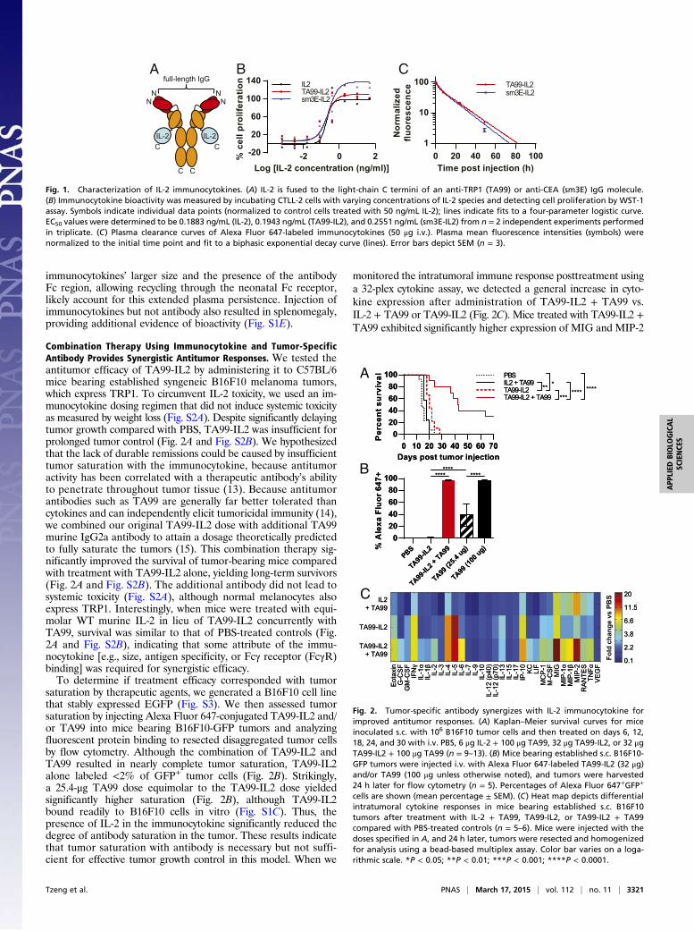
immunocytokines’ larger size and the presence of the antibodyFc region, allowing recycling through the neonatal Fc receptor,likely account for this extended plasma persistence. Injection ofimmunocytokines but not antibody also resulted in splenomegaly,providing additional evidence of bioactivity (Fig. S1E).
Combination Therapy Using Immunocytokine and Tumor-SpecificAntibody Provides Synergistic Antitumor Responses. We tested theantitumor efficacy of TA99-IL2 by administering it to C57BL/6mice bearing established syngeneic B16F10 melanoma tumors,which express TRP1. To circumvent IL-2 toxicity, we used an im-munocytokine dosing regimen that did not induce systemic toxicityas measured by weight loss (Fig. S2A). Despite significantly delayingtumor growth compared with PBS, TA99-IL2 was insufficient forprolonged tumor control (Fig. 2A and Fig. S2B). We hypothesizedthat the lack of durable remissions could be caused by insufficienttumor saturation with the immunocytokine, because antitumoractivity has been correlated with a therapeutic antibody’s abilityto penetrate throughout tumor tissue (13). Because antitumorantibodies such as TA99 are generally far better tolerated thancytokines and can independently elicit tumoricidal immunity (14),we combined our original TA99-IL2 dose with additional TA99murine IgG2a antibody to attain a dosage theoretically predictedto fully saturate the tumors (15). This combination therapy sig-nificantly improved the survival of tumor-bearing mice comparedwith treatment with TA99-IL2 alone, yielding long-term survivors(Fig. 2A and Fig. S2B). The additional antibody did not lead tosystemic toxicity (Fig. S2A), although normal melanocytes alsoexpress TRP1. Interestingly, when mice were treated with equi-molar WT murine IL-2 in lieu of TA99-IL2 concurrently withTA99, survival was similar to that of PBS-treated controls (Fig.2A and Fig. S2B), indicating that some attribute of the immu-nocytokine [e.g., size, antigen specificity, or Fcγ receptor (FcγR)binding] was required for synergistic efficacy.To determine if treatment efficacy corresponded with tumor
saturation by therapeutic agents, we generated a B16F10 cell linethat stably expressed EGFP (Fig. S3). We then assessed tumorsaturation by injecting Alexa Fluor 647-conjugated TA99-IL2 and/or TA99 into mice bearing B16F10-GFP tumors and analyzingfluorescent protein binding to resected disaggregated tumor cellsby flow cytometry. Although the combination of TA99-IL2 andTA99 resulted in nearly complete tumor saturation, TA99-IL2alone labeled <2% of GFP+ tumor cells (Fig. 2B). Strikingly,a 25.4-μg TA99 dose equimolar to the TA99-IL2 dose yieldedsignificantly higher saturation (Fig. 2B), although TA99-IL2bound readily to B16F10 cells in vitro (Fig. S1C). Thus, thepresence of IL-2 in the immunocytokine significantly reduced thedegree of antibody saturation in the tumor. These results indicatethat tumor saturation with antibody is necessary but not suffi-cient for effective tumor growth control in this model. When we
monitored the intratumoral immune response posttreatment usinga 32-plex cytokine assay, we detected a general increase in cyto-kine expression after administration of TA99-IL2 + TA99 vs.IL-2 + TA99 or TA99-IL2 (Fig. 2C). Mice treated with TA99-IL2 +TA99 exhibited significantly higher expression of MIG and MIP-2
-2 0 2-20
20
60
100
140
Log [IL-2 concentration (ng/ml)]
% c
ell p
rolif
erat
ion IL2
TA99-IL2sm3E-IL2
A
N
IL-2IL-2
NN N
C C
C C
full-length IgGCB
Time post injection (h)
Nor
mal
ized
fluor
esce
nce
0 20 40 60 80 1001
10
100 TA99-IL2sm3E-IL2
Fig. 1. Characterization of IL-2 immunocytokines. (A) IL-2 is fused to the light-chain C termini of an anti-TRP1 (TA99) or anti-CEA (sm3E) IgG molecule.(B) Immunocytokine bioactivity was measured by incubating CTLL-2 cells with varying concentrations of IL-2 species and detecting cell proliferation by WST-1assay. Symbols indicate individual data points (normalized to control cells treated with 50 ng/mL IL-2); lines indicate fits to a four-parameter logistic curve.EC50 values were determined to be 0.1883 ng/mL (IL-2), 0.1943 ng/mL (TA99-IL2), and 0.2551 ng/mL (sm3E-IL2) from n = 2 independent experiments performedin triplicate. (C) Plasma clearance curves of Alexa Fluor 647-labeled immunocytokines (50 μg i.v.). Plasma mean fluorescence intensities (symbols) werenormalized to the initial time point and fit to a biphasic exponential decay curve (lines). Error bars depict SEM (n = 3).
A
B
C
Fig. 2. Tumor-specific antibody synergizes with IL-2 immunocytokine forimproved antitumor responses. (A) Kaplan–Meier survival curves for miceinoculated s.c. with 106 B16F10 tumor cells and then treated on days 6, 12,18, 24, and 30 with i.v. PBS, 6 μg IL-2 + 100 μg TA99, 32 μg TA99-IL2, or 32 μgTA99-IL2 + 100 μg TA99 (n = 9–13). (B) Mice bearing established s.c. B16F10-GFP tumors were injected i.v. with Alexa Fluor 647-labeled TA99-IL2 (32 μg)and/or TA99 (100 μg unless otherwise noted), and tumors were harvested24 h later for flow cytometry (n = 5). Percentages of Alexa Fluor 647+GFP+
cells are shown (mean percentage ± SEM). (C) Heat map depicts differentialintratumoral cytokine responses in mice bearing established s.c. B16F10tumors after treatment with IL-2 + TA99, TA99-IL2, or TA99-IL2 + TA99compared with PBS-treated controls (n = 5–6). Mice were injected with thedoses specified in A, and 24 h later, tumors were resected and homogenizedfor analysis using a bead-based multiplex assay. Color bar varies on a loga-rithmic scale. *P < 0.05; **P < 0.01; ***P < 0.001; ****P < 0.0001.
Tzeng et al. PNAS | March 17, 2015 | vol. 112 | no. 11 | 3321
APP
LIED
BIOLO
GICAL
SCIENCE
S

(P < 0.0001 vs. IL-2 + TA99 and TA99-IL2), chemokines knownto recruit antitumor effector cells, including NK cells, T cells, andneutrophils (16, 17). The chemokine IP-10, a close relative ofMIG, was also significantly up-regulated in mice treated withTA99-IL2 + TA99 compared with those treated with IL-2 + TA99(P < 0.05). Overall, elevated intratumoral cytokine responsescorresponded with better prognosis, linking immune activationwith antitumor efficacy.
Synergy Between Tumor-Specific Antibody and ImmunocytokinesRequires Neither Immunocytokine Antigen Specificity nor FcγRInteractions. Fusion of a full-length IgG to a cytokine allows notonly targeting through the antibody variable region, but also ADCCand complement-dependent cytotoxicity effector functions andextended half-life through the Fc region. To examine which ofthese properties contributes to immunocytokine synergy with TA99,we compared the effectiveness of various immunocytokines. Sur-prisingly, treatment with sm3E-IL2, which recognizes an antigenabsent in mice, had an almost identical effect on B16F10 tumorgrowth as TA99-IL2 therapy, acting synergistically with TA99 topromote long-term survival (Fig. 3A and Fig. S2). This unexpectedobservation shows that tumor targeting by the antibody variableregion is dispensable for synergy with TA99. A single point mu-tation (D265A) in the antibody Fc region ablates interactionswith FcγRs and complement, abolishing IgG effector functions(18). We introduced the D265A mutation to TA99-IL2 andsm3E-IL2 and found that the D265A immunocytokines did notperform significantly differently from their parent immunocyto-kines (Fig. 3A and Fig. S2), suggesting that immunocytokineFc-mediated effector functions are also nonessential for effectivecombination therapy with TA99. Neither antigen specificity noreffector function competence affected immunocytokine satura-tion of established B16F10-GFP tumors, because there were nosignificant labeling differences among groups treated with fluo-rescently labeled immunocytokines (Fig. 3B). This observationcontradicts predictions that tumor-targeted agents should displaygreater tumor accumulation than their nontargeted counter-parts. After coinjection of fluorescently labeled TA99, ∼100%of GFP+ cells were bound to fluorescent therapeutic agents(Fig. 3B). Because immunocytokine antigen specificity and Fceffector functions both were unnecessary for therapeutic syn-ergy with TA99, IL-2 plasma persistence is likely the key factorin the combination therapy. Moreover, IL-2 with a short half-life did not produce durable remissions when coinjected withTA99 (Fig. 2A).
The IL-2 Moiety Governs Immunocytokine Localization. Our un-anticipated findings that immunocytokine antigen specificity didnot affect therapeutic efficacy or immunocytokine distribution totumor cells (Figs. 2 and 3) prompted us to develop an assay fortracking immunocytokine and antibody localization at a cellularlevel. A similar approach has been successfully used in moni-toring nanoparticle trafficking (19). We first examined the dis-tribution of TA99 antibody and found that, as expected, TA99binding was enriched intratumorally for immune populations[e.g., dendritic cells (DCs), macrophages, and NK cells] knownto express FcγR (14, 20), which can interact with the antibodyFc region (Fig. 4). This enrichment presumably stems from tu-mor localization mediated by antibody recognition of the TRP1antigen on the surface of tumor cells. In the blood and spleen,monocytes/macrophages exhibited the greatest percentage ofbinding to TA99 (Fig. 4), consistent with the high expression ofFcγR by these cells (20). However, IL-2 fusion drastically alteredthe binding distribution pattern of its partner, and for TA99-IL2,NK and natural killer T (NKT) cells dominated binding. Nearlyall NK cells and a high percentage of NKT cells in the analyzedorgans were bound to TA99-IL2 (Fig. 4). In contrast, a lowerpercentage of monocytes/macrophages, particularly in the tumor,
showed binding to TA99-IL2 vs. TA99 (Fig. 4 and Fig. S4A), al-though no differences in absolute cell numbers were observed(Fig. S4B). Notably, ∼80% of circulating DCs were TA99-IL2–bound (Fig. 4). Greater percentages of intratumoral CD8+ T cellsand Tregs bound to the immunocytokine compared with corre-sponding populations from other organs, which is attributable tothe IL-2 moiety given that both cell types do not express FcγRand did not bind to naked TA99 (Fig. 4). Remarkably, thebinding distribution of sm3E-IL2 closely mirrored that of TA99-IL2 (Fig. 4), showing that the antigen specificity of the parentantibody had negligible effects on immunocytokine localization.To determine the contribution of Fc–FcγR interactions to im-
munocytokine localization, we monitored the binding distributionof D265A mutant immunocytokines. In most cases, ablation ofFc–FcγR interactions did not change the distribution patterns.However, for all analyzed compartments, a higher percentage ofTregs bound to the D265A than to the corresponding parentimmunocytokine (Fig. S4A). Intratumoral CD8+ T cells alsoshowed greater binding to sm3E-IL2 D265A than to sm3E-IL2(Fig. S4A). Although the parent immunocytokines could con-ceivably mediate immune cell depletion through their intact
A
B
****sm3E-IL2 D265A + TA99 ***
***sm3E-IL2 D265A
0 10 20 30 40 50 60 700
20406080
100
Days post tumor injection
PBS
*******TA99-IL2 D265A + TA99
020406080
100
Per
cent
sur
viva
l PBSTA99-IL2 D265A ***
****sm3E-IL2 + TA99
020406080
100 PBSsm3E-IL2 ****
******** ****
PBS
sm3E
-IL2
sm3E
-IL2 +
TA99
TA99-IL
2 D26
5A
TA99-IL
2 D26
5A +
TA99
sm3E
-IL2 D
265A
sm3E
-IL2 D
265A
+ TA99
020406080
100
% A
lexa
Flu
or 6
47+
Fig. 3. IL-2 immunocytokine antigen specificity and Fc receptor interactionsare dispensable for therapeutic synergy with tumor-specific antibody.(A) Kaplan–Meier survival curves for mice inoculated s.c. with 106 B16F10tumor cells and then treated on days 6, 12, 18, 24, and 30 with i.v. PBS, 32 μgsm3E-IL2, 32 μg sm3E-IL2 + 100 μg TA99, 32 μg TA99-IL2 D265A, 32 μg TA99-IL2 D265A + 100 μg TA99, 32 μg sm3E-IL2 D265A, or 32 μg sm3E-IL2 D265A +100 μg TA99 (n = 10–13). (B) Mice bearing established s.c. B16F10-GFP tumorswere injected i.v. with therapeutic dosages of Alexa Fluor 647-labeled ver-sions of the indicated agents, and tumors were harvested 24 h later for flowcytometry (n = 5). Percentages of Alexa Fluor 647+GFP+ cells are shown(mean percentage ± SEM). ***P < 0.001; ****P < 0.0001.
3322 | www.pnas.org/cgi/doi/10.1073/pnas.1416159112 Tzeng et al.

effector activity, there were no significant differences in Treg orCD8+ T-cell numbers between mice treated with D265A andparent immunocytokines (Fig. S4B). Overall, the four examinedimmunocytokines had only the IL-2 component in common butdisplayed distribution patterns strikingly similar to one anotherand distinct from TA99 (Fig. 4 and Fig. S4A). In addition, theimmune cell lineages exhibiting the highest percentage bindingto immunocytokines have all been documented to expressIL-2R but not necessarily FcγR at appreciable levels in previousstudies (14, 21) and our own experience (Fig. S5). Ex vivo bloodbinding analysis further corroborated the existence of distinctFcγR- and IL-2R-expressing cell populations that act as sinks fortherapeutic proteins containing intact Fc or IL-2 domains, re-spectively, in a dose- and affinity-dependent manner (Fig. S4C).These data strongly suggest that the IL-2–IL-2R interaction wasthe main determinant of these immunocytokine distributions, withFc–FcγR interactions playing, if anything, a minor role.
Immunocytokines Predominantly Remain Systemic and Associatewith Innate Immune Cells Regardless of Antigen Specificity or FcγRInteractions. Quantifying the percentages of immunocytokine-bound (IC+) cells highlights the immune cell lineages showing thegreatest relative binding to immunocytokines but does not allowcomparisons of immunocytokine abundances between differentorgans or cell types. For each immune lineage of interest, weapproximated the amounts of immunocytokine associated witheach organ by multiplying the total number of IC+ cells from thatorgan by the median fluorescence intensity, which is proportionalto the number of fluorescent molecules binding to the cells. Forall four immunocytokines, the partitioning patterns among organswere remarkably similar (Fig. 5A). Immunocytokines bound tothe DC, NK cell, and NKT cell populations were mostly foundin the spleen, with a smaller percentage located in the blood.Meanwhile, more than 80% of immunocytokines bound to CD8+
T cells and Tregs were found in the spleen, with very little in theblood. In contrast, only a small proportion of immunocytokineslocalized in the tumor, and even less was found in the draininglymph node. Because these immunocytokines nonetheless showedtherapeutic efficacy when combined with TA99 antibody (Figs.2A and 3A), high tumor uptake is not necessary for effectiveantitumor therapy. Ablation of Fc–FcγR interactions did notappreciably alter organ distribution (Fig. 5A). Although the tu-mor-specific TA99-IL2 had been expected to accumulate to
a greater extent in tumors than the nontumor-specific sm3E-IL2,the two immunocytokines actually displayed similar tumor uptakeproportions (Fig. 5A). These results indicate that neither antigenspecificity nor Fc mutations greatly impact immunocytokine dis-tribution at the organ level and that the majority of immunocy-tokine associates with systemic rather than tumor-proximalimmune cells. Next, we estimated the fraction of immunocytokineor antibody bound to each immune cell lineage by summing theproducts of IC+ lineage cells and median fluorescence intensityfor the analyzed organs. The largest proportion of all fourimmunocytokines was associated with NK cells, with DCs andNKT cells harboring the second and third largest proportions,respectively (Fig. 5B). In contrast, NK cells bound a smallerpercentage of TA99 antibody, and monocytes/macrophages andNKT cells played far greater roles in interacting with TA99 thanwith the immunocytokines (Fig. 5B). Once again, the distributionpatterns of TA99-IL2 were distinct from those of TA99 andclosely resembled those of sm3E-IL2, TA99-IL2 D265A, andsm3E-IL2 D265A, suggesting that for these immunocytokines,antibody variable and Fc region-mediated interactions were largelysuperseded by IL-2–mediated interactions.
Mathematical Modeling Reveals Size Dependence of ImmunocytokineTargeting. To further explore additional parameters for immu-nocytokine targeting effectiveness and the generalizability of ourobservations, we developed a simple ordinary differential equa-tion (ODE) model (Fig. 6A and Fig. S6). We modeled the twomost common formats for immunocytokines currently in clinicaltrials (1, 2): large (full-length IgG) and small (diabody consist-ing of dimeric single-chain variable fragment). Moreover, we
PBS TA99-IL2 TA99-IL2 D265A
tumor
spleen
blood
dLN
80%60%40%20%0%
TA99 sm3E-IL2 sm3E-IL2 D265A
tumor
spleen
blood
dLN
%AF647+100%
Fig. 4. The IL-2 moiety targets immunocytokines to IL-2R–expressing im-mune cells both systemically and tumor proximally, regardless of antigenspecificity or Fc receptor interactions. Mice bearing established s.c. B16F10tumors were injected i.v. with equimolar doses of Alexa Fluor 647-labeledproteins (n = 5); 24 h later, organs were collected, rendered into single-cellsuspensions, and stained with immune cell lineage markers before flowcytometric analysis. Heat maps depict Alexa Fluor 647+ percentages of indi-cated cell lineages in the blood, spleen, tumor, or draining lymph node (dLN).
% o
f tot
al fl
uore
scen
ce
A
B
dLNtumorspleenblood
TA99-IL
2
TA99-IL
2 D26
5A
sm3E
-IL2
sm3E
-IL2 D
265A
NKT cells
TA99-IL
2
TA99-IL
2 D26
5A
sm3E
-IL2
sm3E
-IL2 D
265A
0
50
100DCs
TA99-IL
2
TA99-IL
2 D26
5A
sm3E
-IL2
sm3E
-IL2 D
265A
NK cells
Tregs
0
50
100CD8+ T cells
TA99-IL
2
TA99-IL
2 D26
5A
sm3E
-IL2
sm3E
-IL2 D
265A
TA990
50
100
% o
f tot
alflu
ores
cenc
e CD8+ T cellsCD4+ cellsTregs
DCsMo/MΦ
NK cellsNKT cells
Fig. 5. Most immunocytokine localizes outside the tumor and associatesmainly with innate immune cells, regardless of antigen specificity or Fcreceptor interactions. Mice bearing established s.c. B16F10 tumors wereinjected i.v. with equimolar doses of Alexa Fluor 647-labeled proteins, andorgans were collected and processed as in Fig. 4 (n = 5). (A) For each indicatedimmune cell lineage, the total fluorescence resulting from cell-boundimmunocytokines was calculated by summing the following quantity for theexamined organs: total IC+ cell number × median fluorescence intensity.(B) The total fluorescence resulting from immunocytokines bound to all ex-amined immune cell lineages was calculated by summing the total fluores-cence values for each cell lineage calculated in A. dLN, draining lymph node.
Tzeng et al. PNAS | March 17, 2015 | vol. 112 | no. 11 | 3323
APP
LIED
BIOLO
GICAL
SCIENCE
S

considered different rates of tumor antigen endocytosis toaccount for antigen targets with varying turnover rates. Asvalidation, we input pharmacokinetic parameters from severalprior studies of therapeutic protein distribution into our modeland found that the predicted biodistributions closely matchedexperimental data (Fig. S7). Unlike experiments using radio-or fluorescently labeled immunocytokines, this model dis-tinguishes when immunocytokines are active (extracellular andcapable of signaling through cytokine receptors). To assess theeffects of tumor targeting, we computed expected tumor-to-blood exposure ratios for large and small immunocytokines.Consistent with our experimental results, the predicted tumor-to-blood ratios for active large immunocytokines are poor,even with a very slow rate of antigen endocytosis (Fig. 6B). No-tably, large immunocytokines recognizing rapidly endocytosedantigens have tumor-to-blood ratios <1, localizing to tumors to aneven lesser extent than immunocytokines with irrelevant antigenspecificity, because fast turnover translates to a depletion effect.In contrast, small immunocytokines have much better predictedtumor-to-blood exposure ratios at varying endocytosis rates (Fig.6B). These results indicate that another major determinant ofimmunocytokine targeting efficiency is the balance between sys-temic and tumor clearance. Small immunocytokines have muchfaster blood clearance than large ones, minimizing systemic ex-posure, whereas tumor clearance mostly depends on antigenturnover rates, which are unaffected by immunocytokine size.Hence, for the same antigen target, small immunocytokines canachieve higher tumor-to-blood ratios than large ones. Anotherway to view the benefits of immunocytokine tumor targeting is todetermine relative cytokine activities in different compartmentsbased on the EC50 of IL-2. Although large immunocytokinesmaintain constant activities ∼100% in both the blood and tumor,small immunocytokines can achieve a window in which the cy-tokine is active in the tumor but not in the blood (Fig. S8). Im-portantly, in the model, we did not account for immunocytokineuptake into extratumoral cellular sinks to parse out the effectsof size independent of this process. However, as we have shown(Figs. 2B and 5), immunocytokine consumption outside the tumorcan dominate overall distribution.
DiscussionImmunocytokines are designed to localize cytokine therapy totumors but still suffer from systemic dose-limiting toxicities(5, 9). We have shown that tumor-specific antibody synergizeswith IL-2 immunocytokines to produce long-term remissions in thepoorly immunogenic B16F10 tumor model. Similarly, cotherapywith a tumor vasculature-directed IL-2 immunocytokine and ananti-CD20 antibody significantly enhanced survival in a lymphomaxenograft model in immunocompromised mice lacking T cells (6);we report here for the first time, to our knowledge, an immu-nocytokine/antibody combination strategy for the treatment ofestablished syngeneic solid tumors in mice with an intact im-mune system. Surprisingly, it is the IL-2 moiety rather than
immunocytokine antigen specificity that almost completelydetermines cellular and organ biodistribution. Neither immuno-cytokine tumor targeting nor effector functions are required for oreven seem to contribute to effective combination therapy. Usinga mathematical model, we show that, in general, slow systemicclearance may further impair antigen targeting efficiency by IgG-based immunocytokines, such as the ones tested here.This study shows that the only essential contribution of anti-
body fusion to IL-2 efficacy in this model is prolonging IL-2plasma half-life from minutes to roughly one-half of a day. Thisobservation is consistent with previous hypotheses that IL-2’srapid clearance hampers its therapeutic use (21, 22). In the clinic,IL-2 therapy requires frequent infusions (3), and in a preclinicaltrial using the hu14.18-IL2 immunocytokine, which has a half-lifeof 3–4 h, complete eradication of murine neuroblastoma requiredcombination with continuous IL-2 administration (23). Saturationof tumor cells by ADCC-activating molecules and a robust intra-tumoral cytokine response likely account for the impressive anti-tumor activity exhibited by the immunocytokine/TA99 antibodycombination therapies (Figs. 2 and 3). Additional work is nec-essary to definitively identify key immune cell types and mecha-nisms underlying the synergistic tumor control observed for IL-2immunocytokines and TA99.Numerous reports have characterized immunocytokine bio-
distribution at the organ level (1), but to our knowledge, this isthe first to examine the cellular-level distribution of multispecificproteins, allowing us to identify unexpected trends in localizationpatterns. Although naked TA99 was driven by antigen recogni-tion to accumulate intratumorally, fusion to IL-2 introduceda competing IL-2–IL-2R interaction that overrode both antigenspecificity and FcγR interactions as the key mechanism for lo-calization (Figs. 4 and 5). In retrospect, IL-2R binding probablyexplains the significantly reduced association of TA99-IL2 withtumor cells compared with that of equimolar TA99 (Fig. 2B).Similar instances of cytokine–cytokine receptor-dominated bio-distribution for tumor vasculature-targeted IFN-γ immunocyto-kines have been observed (24, 25). Moreover, the markedly fasterplasma clearance of immunocytokines relative to their parentantibodies as reported by several groups (26, 27) may reflectextensive binding of immunocytokines to IL-2R–expressing cells.Circulating immune cells expressing CD122, which may correspondto the immunocytokine-bound NK cells that we detected, havebeen linked to IL-2 toxicity (28) but may also contribute to ther-apeutic efficacy. Overall, the relationship between immunocytokinebinding and efficacy or toxicity requires additional investigation.It may seem paradoxical that NK and NKT cells, which express
the intermediate-affinity IL-2R, bind a greater proportion ofimmunocytokine than Tregs and activated CD8+ T cells, whichexpress the high-affinity IL-2R. However, NK and NKT cells aregenerally more abundant than Tregs and activated CD8+ T cells,especially in the blood (29), the first point of contact for i.v.-administered immunocytokines. On entering the blood, immu-nocytokines first encounter and bind to NK and NKT cells,
0 15 30 45 60 750.01
0.1
1
10
100 Small IC Format
Tumor Binding IC (t1/2-endo 2h)Tumor Binding IC (t1/2-endo 10h)Tumor Binding IC (t1/2-endo 50h)Non-Binding IC
Plasma Clearance
Internalization of Antigen-Bound IC
Bolus Dose
Extravasation/Intravasation
A
Tumor
Blood
B
0 15 30 45 60 750.01
0.1
1
10
100
Time (h)
Act
ive
IL-2
tum
or:b
lood
ratio
Large IC Format
Fig. 6. A simple ODE-based mathematical model of immunocytokine distribution shows that immunocytokine targeting depends highly on construct size.(A) A simplified model schematic. In this two-compartment model, the immunocytokine can be cleared from the blood or the tumor or undergo inter-compartmental transport by extravasation or intravasation. (B) Tumor-to-blood ratios of active immunocytokine exposure for (Left) large, full-length IgG and(Right) small diabody-based formats. The dotted black line indicates equal exposure in tumor and blood. IC, immunocytokine.
3324 | www.pnas.org/cgi/doi/10.1073/pnas.1416159112 Tzeng et al.
which may preclude efficient distribution to other sites thatcontain Tregs and activated CD8+ T cells. Analogously, althoughthe affinities of TA99 for TRP1 and IL-2 for intermediate-affinity IL-2R are both ∼10−9 M (21, 30), i.v.-injected immuno-cytokines encounter and associate with circulating IL-2R–
expressing immune cells well before they can reach TRP1+ tumorcells, resulting in predominant immunocytokine accumulation insystemic rather than tumor-proximal compartments.Our results suggest that in targeted therapy development, the
biodistribution of any construct with multiple potential targetinginteractions should be directly assessed rather than assumed. Ifthe desired antigen targeting is not achieved, several generalstrategies may be used to promote selective localization, with thecaveat that such targeting may not be required for therapeuticefficacy, which was the case for this study. For accessible solidtumors, intratumoral administration may exhibit superior anti-tumor activity and reduced systemic toxicity compared with thei.v. route (31–33), consistent with our findings that i.v.-injectedimmunocytokine resided predominantly in the blood and spleenrather than the tumor (Fig. 5A). Antigen choice also impactstargeting efficacy: in contrast to our results, several studies haveshown that immunocytokines specific for antigens expressed bytumor vasculature or liquid/disseminated tumors perform betterthan irrelevant immunocytokines (4, 7, 34), likely because theseantigens are in closer contact with the blood and thus, moreaccessible than solid tumor antigens. Alternatively, the affinity ofthe cytokine for its receptor can be weakened, so that cytokinefunction is potentially restored only on high-avidity binding totumor cells (35). Finally, our mathematical model suggests thatsmall-format immunocytokines may intrinsically exhibit betterantigen-targeting specificity than large-format immunocytokinesbecause of size-dependent transport properties (rapid systemicclearance and diffusion by smaller molecules).
In conclusion, we have explored a facile strategy for improvingimmunocytokine antitumor efficacy and uncovered limitations forselective targeting by multicomponent constructs. Addition oftumor-specific antibody to existing immunocytokine therapies maybe a promising option for enhancing clinical outcomes withoutintroducing incremental toxicity. Because extended plasma per-sistence seems to be far more important than tumor localizationfor effective IL-2 synergy with naked antibody, simply prolongingIL-2 circulation by fusion to serum albumin or antibody Fc regionmay allow equivalent efficacy when combined with tumor-specificantibody. This nontargeted, persistent cytokine could be readilycoadministered with clinically approved antibodies, circumventingthe need to develop a new immunocytokine for each tumor anti-gen. Future studies will be needed to validate these experimentaland theoretical concepts in additional tumor models using a varietyof cytokine moieties, antigen targets, and immunocytokine formats.
Materials and MethodsDetailed methods are provided in SI Materials and Methods. In summary,IL-2 immunocytokines were generated by PCR-based cloning, produced bytransient HEK293-F cell transfection, purified using protein A beads, andcharacterized in vitro and in vivo. TA99 antibody was produced by a stableHEK293-F cell line and purified using protein A beads. Fluorescently labeledproteins were generated by reaction with Alexa Fluor 647 NHS ester. C57BL/6mice were inoculated s.c. with 106 B16F10 or B16F10-GFP tumor cells, andtumors were allowed to establish before treatment with i.v. IL-2, immunocy-tokine, and/or TA99 for tumor control assessment, intratumoral cytokinemeasurement, or flow cytometry analysis. Mathematical modeling was per-formed using MATLAB.
ACKNOWLEDGMENTS. We thank W. Overwijk for experimental suggestionsand the staff of the Swanson Biotechnology Center at the Koch Institute forIntegrative Cancer Research for technical assistance. This work was fundedby National Cancer Institute Grant CA174795. A.T., B.H.K., and C.F.O. weresupported by National Science Foundation Graduate Research Fellowships.
1. Pasche N, Neri D (2012) Immunocytokines: A novel class of potent armed antibodies.Drug Discov Today 17(11-12):583–590.
2. Sondel PM, Gillies SD (2012) Current and potential uses of immunocytokines as cancerimmunotherapy. Antibodies (Basel) 1(2):149–171.
3. Rosenberg SA (2014) IL-2: The first effective immunotherapy for human cancer.J Immunol 192(12):5451–5458.
4. Becker JC, Pancook JD, Gillies SD, Furukawa K, Reisfeld RA (1996) T cell-mediatederadication of murine metastatic melanoma induced by targeted interleukin 2 ther-apy. J Exp Med 183(5):2361–2366.
5. Gillies SD, et al. (2011) A low-toxicity IL-2-based immunocytokine retains antitumor ac-tivity despite its high degree of IL-2 receptor selectivity. Clin Cancer Res 17(11):3673–3685.
6. Schliemann C, et al. (2009) Complete eradication of human B-cell lymphoma xenograftsusing rituximab in combination with the immunocytokine L19-IL2. Blood 113(10):2275–2283.
7. Gillies SD, et al. (2005) An anti-CD20-IL-2 immunocytokine is highly efficacious ina SCID mouse model of established human B lymphoma. Blood 105(10):3972–3978.
8. Shusterman S, et al. (2010) Antitumor activity of hu14.18-IL2 in patients withrelapsed/refractory neuroblastoma: A Children’s Oncology Group (COG) phase II study.J Clin Oncol 28(33):4969–4975.
9. Yu AL, et al.; Children’s Oncology Group (2010) Anti-GD2 antibody with GM-CSF,interleukin-2, and isotretinoin for neuroblastoma. N Engl J Med 363(14):1324–1334.
10. Rossi EA, Chang CH, Cardillo TM, Goldenberg DM (2013) Optimization of multivalentbispecific antibodies and immunocytokines with improved in vivo properties. Bio-conjug Chem 24(1):63–71.
11. Gillies SD (2013) A new platform for constructing antibody-cytokine fusion proteins(immunocytokines) with improved biological properties and adaptable cytokine ac-tivity. Protein Eng Des Sel 26(10):561–569.
12. Mühlradt PF, Opitz HG (1982) Clearance of interleukin 2 from the blood of normaland T cell-depleted mice. Eur J Immunol 12(11):983–985.
13. Freeman DJ, et al. (2012) Tumor penetration and epidermal growth factor receptorsaturation by panitumumab correlate with antitumor activity in a preclinical modelof human cancer. Mol Cancer 11:47.
14. Weiner LM, Surana R, Wang S (2010) Monoclonal antibodies: Versatile platforms forcancer immunotherapy. Nat Rev Immunol 10(5):317–327.
15. Wittrup KD, Thurber GM, Schmidt MM, Rhoden JJ (2012) Practical theoretic guidancefor the design of tumor-targeting agents. Methods Enzymol 503:255–268.
16. Franciszkiewicz K, Boissonnas A, Boutet M, Combadière C, Mami-Chouaib F (2012)Role of chemokines and chemokine receptors in shaping the effector phase of theantitumor immune response. Cancer Res 72(24):6325–6332.
17. Wolpe SD, et al. (1989) Identification and characterization of macrophage inflammatoryprotein 2. Proc Natl Acad Sci USA 86(2):612–616.
18. Baudino L, et al. (2008) Crucial role of aspartic acid at position 265 in the CH2 domain formurine IgG2a and IgG2b Fc-associated effector functions. J Immunol 181(9):6664–6669.
19. Kourtis IC, et al. (2013) Peripherally administered nanoparticles target monocyticmyeloid cells, secondary lymphoid organs and tumors in mice. PLoS ONE 8(4):e61646.
20. Guilliams M, Bruhns P, Saeys Y, Hammad H, Lambrecht BN (2014) The function of Fcγreceptors in dendritic cells and macrophages. Nat Rev Immunol 14(2):94–108.
21. Boyman O, Sprent J (2012) The role of interleukin-2 during homeostasis and activa-tion of the immune system. Nat Rev Immunol 12(3):180–190.
22. Rao BM, Driver I, Lauffenburger DA, Wittrup KD (2004) Interleukin 2 (IL-2) variantsengineered for increased IL-2 receptor alpha-subunit affinity exhibit increased po-tency arising from a cell surface ligand reservoir effect.Mol Pharmacol 66(4):864–869.
23. Neal ZC, et al. (2004) Enhanced activity of hu14.18-IL2 immunocytokine againstmurine NXS2 neuroblastoma when combined with interleukin 2 therapy. Clin CancerRes 10(14):4839–4847.
24. Ebbinghaus C, et al. (2005) Engineered vascular-targeting antibody-interferon-gammafusion protein for cancer therapy. Int J Cancer 116(2):304–313.
25. Hemmerle T, Neri D (2014) The dose-dependent tumor targeting of antibody-IFNγfusion proteins reveals an unexpected receptor-trapping mechanism in vivo. CancerImmunol Res 2(6):559–567.
26. Kendra K, Malkovska V, Allen M, Guzman J, Albertini M (1999) In vivo binding andantitumor activity of Ch14.18. J Immunother 22(5):423–430.
27. Gillies SD, Young D, Lo KM, Roberts S (1993) Biological activity and in vivo clearanceof antitumor antibody/cytokine fusion proteins. Bioconjug Chem 4(3):230–235.
28. Shanafelt AB, et al. (2000) A T-cell-selective interleukin 2 mutein exhibits potentantitumor activity and is well tolerated in vivo. Nat Biotechnol 18(11):1197–1202.
29. Maddatu TP, Grubb SC, Bult CJ, Bogue MA (2012) Mouse phenome database (MPD).Nucleic Acids Res 40(database issue):D887–D894.
30. Patel D, et al. (2007) Generation and characterization of a therapeutic human anti-body to melanoma antigen TYRP1. Hum Antibodies 16(3-4):127–136.
31. Johnson EE, et al. (2008) Intratumoral immunocytokine treatment results in enhancedantitumor effects. Cancer Immunol Immunother 57(12):1891–1902.
32. Yang RK, et al. (2012) Intratumoral hu14.18-IL-2 (IC) induces local and systemic anti-tumor effects that involve both activated T and NK cells as well as enhanced IC re-tention. J Immunol 189(5):2656–2664.
33. Marabelle A, Kohrt H, Caux C, Levy R (2014) Intratumoral immunization: A newparadigm for cancer therapy. Clin Cancer Res 20(7):1747–1756.
34. Halin C, et al. (2002) Enhancement of the antitumor activity of interleukin-12 bytargeted delivery to neovasculature. Nat Biotechnol 20(3):264–269.
35. Garcin G, et al. (2014) High efficiency cell-specific targeting of cytokine activity.Nat Commun 5:3016.
Tzeng et al. PNAS | March 17, 2015 | vol. 112 | no. 11 | 3325
APP
LIED
BIOLO
GICAL
SCIENCE
S