


Languages
Pages
Legal


(X-Ray Crystallography)
X-RAY DIFFRACTION

I. X-Ray Diffraction Uses X-Rays to identify the arrangement of atoms, molecules, or ions
within a crystalline solid
Quantitative and qualitative
Ooi, L. Principles of X-ray Crystallography (2010)
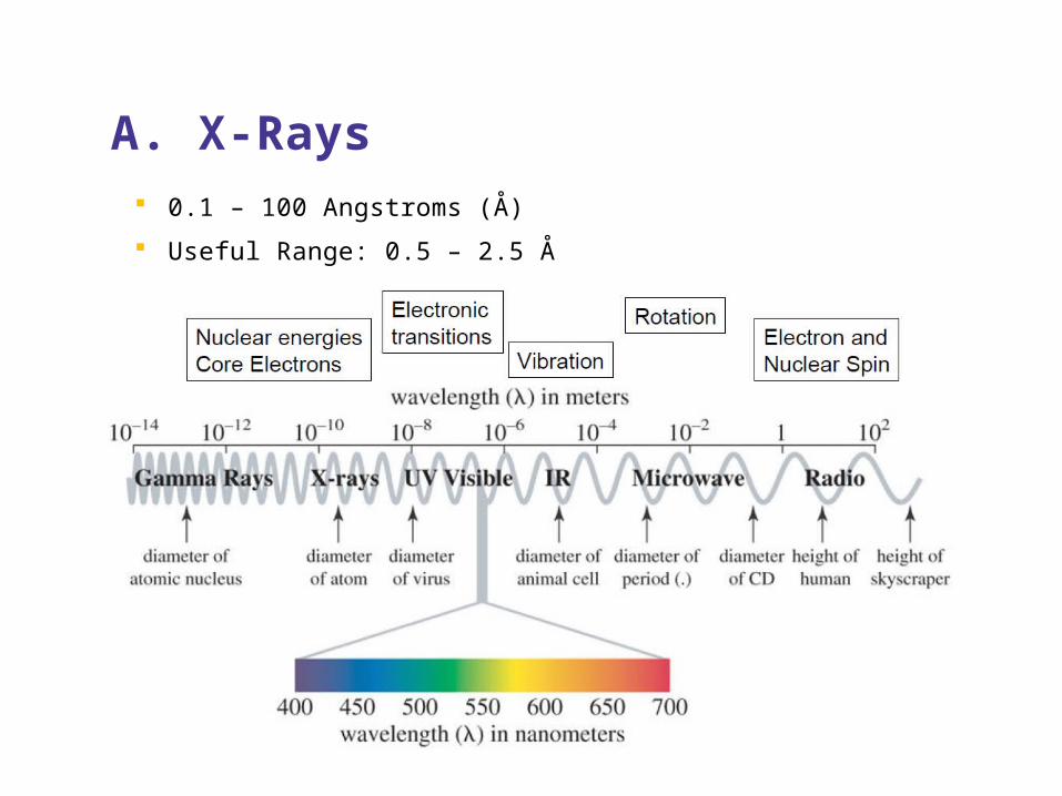
A. X-Rays 0.1 – 100 Angstroms (Å)
Useful Range: 0.5 – 2.5 Å

B. Amorphous Substances1. Gases and Liquids
Extremely difficult
2. Non-crystalline Solids
Atoms are not regularly arranged or regularly shaped
Interference
Fiber Diffraction

Atoms are regularly arranged
“The Unit Cell” – a cookie cutter
C. Crystalline Solids
Rhodes, G. Crystallography Made Crystal Clear, 3rd ed. (2006)

II. X-Ray CrystallographyA. Small-molecule crystallography
Up to ~100 atoms
Organic molecules, catalysts, newly synthesized drugs, etc.
Identify each atom
B. Macromolecular (protein) crystallography
Large biological molecules – nucleic acids and proteins
Identify 2° structure
Note: must show that the crystal structure (asymmetric unit) is comparable to structure in solution (biological unit)

III. X-Ray Diffractometer
Ooi, L. Principles of X-ray Crystallography (2010)
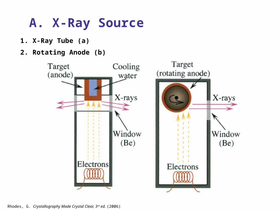
A. X-Ray Source
Rhodes, G. Crystallography Made Crystal Clear, 3rd ed. (2006)
1. X-Ray Tube (a)
2. Rotating Anode (b)

3. Particle Storage Ring (Synchrotron Radiation)
Particle Accelerator
Ooi, L. Principles of X-ray Crystallography (2010)

Rhodes, G. Crystallography Made Crystal Clear, 3rd ed. (2006)
National Synchrotron Light Source at Brookhaven National Lab (Long Island)

B. Collimator Narrow metal tube that selects and reflects the X-Rays into parallel paths
Ooi, L. Principles of X-ray Crystallography (2010)

C. Crystal (Sample)
Rhodes, G. Crystallography Made Crystal Clear, 3rd ed. (2006)
1. Growth – Screens
Crystal vs. useless blob
2. Optimization
Quantity
3. Crystal Quality
Purity
4. Mount for Data Collection
Cryocrystallography
Note: Diffracted in “mother liquor”
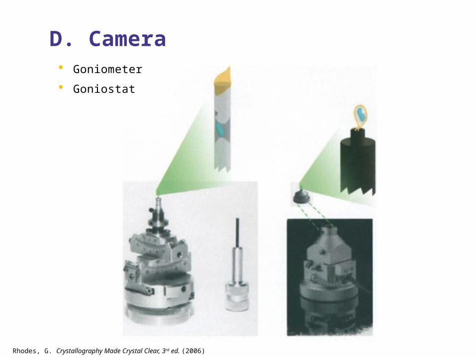
D. Camera
Rhodes, G. Crystallography Made Crystal Clear, 3rd ed. (2006)
Goniometer
Goniostat

Rhodes, G. Crystallography Made Crystal Clear, 3rd ed. (2006)

E. Detector
Rhodes, G. Crystallography Made Crystal Clear, 3rd ed. (2006)
1. Charged Couple Device (CCD)
2. Image Plate (IP)
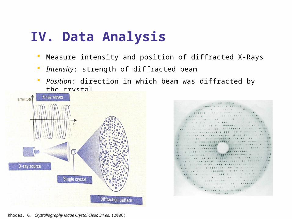
IV. Data Analysis Measure intensity and position of diffracted X-Rays
Intensity: strength of diffracted beam
Position: direction in which beam was diffracted by the crystal
Rhodes, G. Crystallography Made Crystal Clear, 3rd ed. (2006)

Ooi, L. Principles of X-ray Crystallography (2010)
Computer calculates this data from the diffraction pattern

A. Fournier Sum Based on simple waves
f(x) = F cos 2π (hx + α)
F = f0 + f1 + f2 + …
Rhodes, G. Crystallography Made Crystal Clear, 3rd ed. (2006)

B. Bragg’s Law States: diffraction spots occur when 2 d sin θ = n λ
Rhodes, G. Crystallography Made Crystal Clear, 3rd ed. (2006)


Molecular structure in solid crystalline state with extreme certainty
Direct inference of data
Provides limitless info.
Crystals
Slow
Hydrogen
Still just a model
Benefits Downfalls
Top Related