


Languages
Pages
Legal

Virus infections – emerging threats
and management strategies
Prof. Per Ljungman
Department of Hematology
Karolinska University Hospital
Stockholm, Sweden

Pneumonia Encephalitis Hepatitis GI disease
CMV CMV CMV CMV
Influenza Adenovirus EBV HSV
Adenovirus HSV Adenovirus Adenovirus
RSV VZV HBV EBV
Parainfluenza HHV-6 HCV VZV
Metapneumovirus Measles VZV Rotaviruses
Measles JCV HAV Noroviruses
VZV EBV HSV
EBV Rabies HHV-6 (?)
New respiratory viruses
West Nile virus
Some viruses important for differential
diagnoses in SCT patients

Protection against viral infection is different from
protection from viral disease!
Antibodies protect against primary infection
The innate and adaptive cell-mediated immunity protect
against disease (T-cells, NK-cells)
Some principles
How can we manage viral infections?
Prevent a patient being infected
Prevent an infected patient to develop disease
Treat an established viral disease

Prevention of infection
Select the right donor
Safe blood products
Infection control
Antiviral prophylaxis
Vaccination

Select the right donor
Viral infection possibly transmitted through the donor:
CMV HIV
EBV HBV
HHV-6 HCV
West Nile virus HTLV-1/2
Other viruses that give viremia
Influenza (?), adeno (?)
Prevention of disease
Early diagnosis
Monitoring of patients
Antiviral prophylaxis
Vaccination

How to diagnose a viral infection!
answer
Microbiology
question
General points
What is the question?
Do you suspect a specific virus?
Where should you look for the virus?

Diagnostic techniques
Detection of an immune response
Serology / antibody detection
Detection of virus or virus components
Isolation
Antigen
Nucleic acid(s) – DNA, RNA
Quantification of viral load

Timing of management options V
iral
rep
lica
tio
n viral
disease
Gra
ftin
g Time
Diagnosis
of viral
infection
Treatment of
established
disease
Pre-emptive
therapy
Prophylaxis
Alain, ILTS 2008

A virological ”smorgasbord”
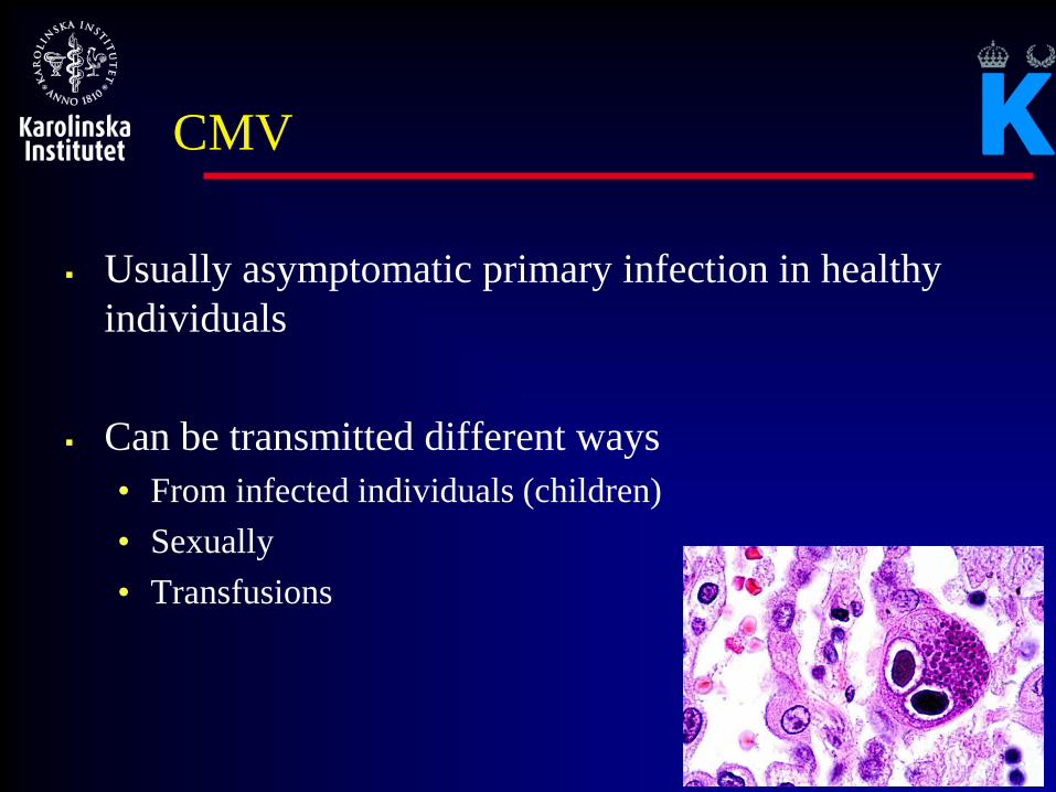
CMV
Usually asymptomatic primary infection in healthy
individuals
Can be transmitted different ways
• From infected individuals (children)
• Sexually
• Transfusions

CMV pneumonia

Other forms of disease
Gastrointestinal disease (frequently together with GVHD)
Encephalitis
Hepatitis
Bone marrow suppression.
Retinitis


Risk factors for CMV disease.
The patient’s serological status
The donor’s serological status
The type of stem cell donor (sibling, unrelated, haplo)
The type of transplant (allogeneic, autologous, reduced conditioning)
What is the influence of CMV on outcome
of a HSCT?
Being CMV seropositive is associated with decreased
survival
Having a CMV seropositive donor for a CMV
seronegative patient is associated with decreased survival
Having a CMV sernegative unrelated donor for a CMV
seropositive patient is associated with decreased survival

19
Effect of donor status – CMV seropositive patients
The protective effect of a CMV matched unrelated donor is seen only in patients receiving myeloablative conditioning
MAC RIC
OS 0.91 (0.86-0.97; p=.01) 1.06 (0.96-1.17; p = ns)
RFS 0.94 (0.88-1.00; p=.05) 1.08 (0.99-1.19; p = .08)
NRM 0.87 (0.81-0.95; p=.01) 1.09 (0.96-1.23; p = ns)
RI 1.05 (p = ns) 1.05 (p = ns)

Treatment of CMV
Ganciclovir = foscarnet
Valganciclovir = ganciclovir
Effects are the similar
Side effects are different

CMV DNA levels and antiviral therapy
There might therefore be at least a week before the DNA levels
decrease
Sample taken
Result to you
Antiviral therapy
Effective ic konc
d.0 d.3-5 d.7
Sample taken
d.1-2

Repeated CMV reactivations
Common in high risk patients
Associated with poor T-cell control of CMV
Increased risk for antiviral resistance
Increased risk for toxicity from antiviral drugs

New drugs/options
Maribavir
Letermovir
CMX001
CMV specific T-cell infusions
CMV vaccines

Adoptive T-cell therapy
In development for > 20 years
Major advances in technology have been achieved over
the last few years
However, still far away from routine therapy in most
centers

HSV

HSV virus
Common
Can give “uncharacteristic” signs and symptoms in SCT
recipients
Prophylaxis usually effective
Visceral and CNS manifestations are rare

Aciclovir resistance
Usually mediated through mutations in the HSV TK
Usually less pathogenic than wild type
Reported in up to 12% of HSCT recipients
Foscarnet or cidofovir is possible alternatives

VZV
Both primary and reactivated infections can cause severe disease
Primary infection (usually children) is a serious complication
VZV disease without skin lesions can occur (GI, liver, CNS)
Severe abdominal pains
Increasing liver function tests
Neurological symptoms
VZV visceral disease has high mortality

Herpes zoster

All varicella-zoster infections should be treated in HSCT
patients!
IV acyclovir for varicella and disseminated zoster
PO acyclovir, valacyclovir, or famciclovir can be used for local HZ

EBV
Might cause symptoms of various types after SCT
Encephalitis
Pneumonia
Hepatitis
However, these symptoms are rare!

Where does the EBV come from?
From the patient (reactivation/increased replication)
From the outside
a) The stem cell donor (both in pretransplant
seropositive and seronegative patients)
b) Blood transfusions
c) ”True” primary infection – oral transmission

EBV PTLD
Important complication in SCT patients
EBV-driven B-cell proliferation
Increasing frequency over the last decade
High mortality

What can we do to prevent PTLD?
Anti-CD20 monoclonal antibody (rituximab)
Reduced immunosuppression
Cell therapy (CTL or donor lymphocytes)
Antiviral therapy –most likely not
35
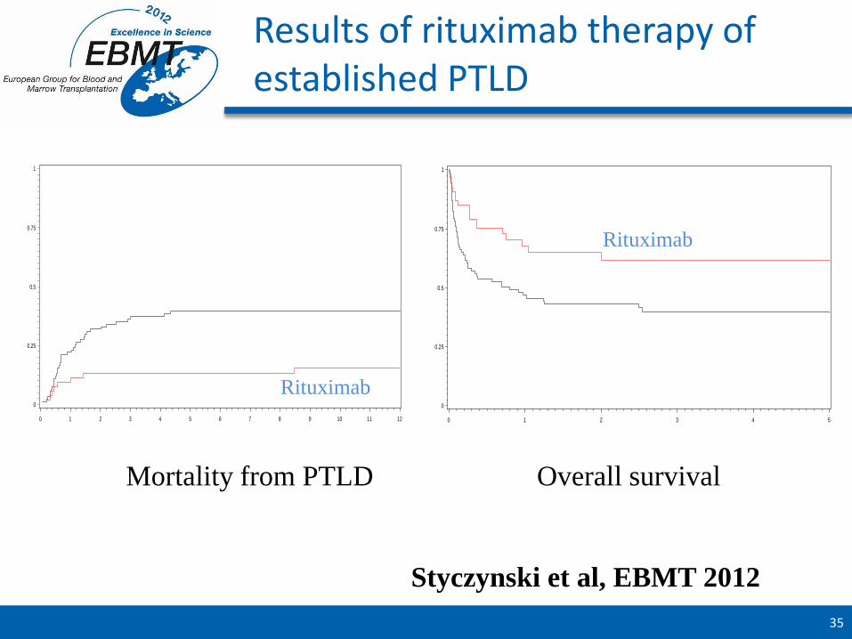
Results of rituximab therapy of established PTLD
ci1
0
0.25
0.5
0.75
1
s_time_ptld_mm
0 1 2 3 4 5 6 7 8 9 10 11 12
red_ist No Ye
survival
0
0.25
0.5
0.75
1
s_time_ptld
0 1 2 3 4 5
red_ist No Ye
Mortality from PTLD Overall survival
Styczynski et al, EBMT 2012
Rituximab
Rituximab

Adenovirus infections
DNA virus.
Many subtypes (currently 51)
Divided inte 6 subgenuses (A-F)
Upper and lower respiratory infections
Renal infections / hemorrhagic cystitis
Gastrointestinal infections
Hepatitis
CNS disease

Possible sources of adenovirus
in SCT patients
Infection from an outside source
• Respiratory route
• GI-route
There are described outbreaks within units
Activation/reactivation of persistent/latent virus
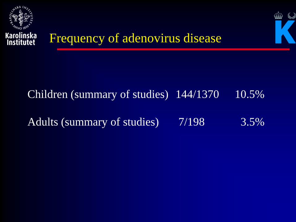
Frequency of adenovirus disease
Children (summary of studies) 144/1370 10.5%
Adults (summary of studies) 7/198 3.5%

Diagnostic procedures
qPCR for monitoring
Important to detect in tissue for diagnosis of
visceral infections (not PCR)
Presumed infections (GI disease, HC) with
adenovirus detected in stools or urine in
symptomatic patients

Outcome of antiviral therapy (mortality)
Children (summary of studies) 84/234 37%
Adults (summary of studies) 13/56 23%

Possible antiviral agents
Cidofovir
(Ribavirin)
(Ganciclovir)
CMX001
Specific T-cells

Cidofovir and viral load
Neofytos et al BBMT 2007

Papovaviruses
BK-, JC-virus and two new respiratory víruses
DNA viruses
Ubiquitous viruses in the population
Symptoms from the primary infections are mild and uncharacteristic
Reactivates in severely immunocompromised patients

Symptoms in transplant patients
JC-virus PML (rare)
BK-virus Nephropathy in renal tx patients
Hemorrhagic cystitis in SCT patients
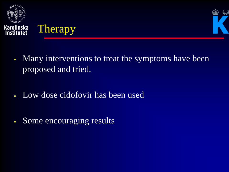
Therapy
Many interventions to treat the symptoms have been
proposed and tried.
Low dose cidofovir has been used
Some encouraging results

Hepatitis viruses and HSCT patients
Before HSCT
- latent infection with no liver disease
- chronic asymptomatic hepatitis
- acute ,clinically overt hepatitis (rare)
Following HSCT
- reactivation of latent infection ± LD
- de novo infection ± LD
- unmodified ongoing chronic hepatitis
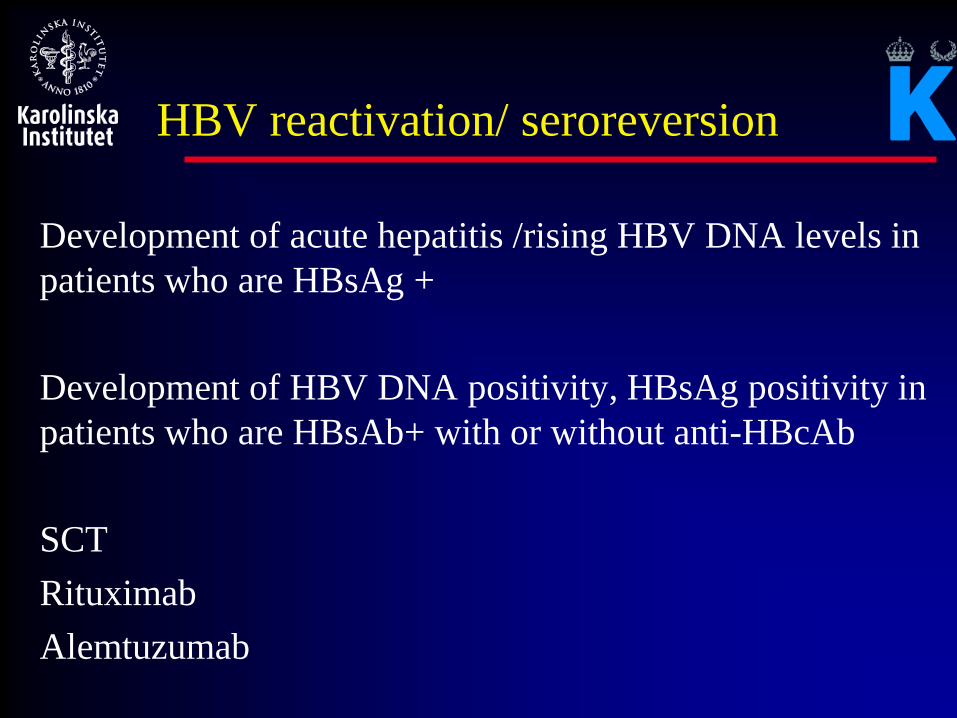
HBV reactivation/ seroreversion
Development of acute hepatitis /rising HBV DNA levels in
patients who are HBsAg +
Development of HBV DNA positivity, HBsAg positivity in
patients who are HBsAb+ with or without anti-HBcAb
SCT
Rituximab
Alemtuzumab

Norovirus –a nuisance or an
important pathogen?
Family: Caliciviridae
Genus: Norovirus
Different subtypes
Liknande virus: sapovirus
Single stranded RNA viruses
Diagnosis: PCR or elektronemicroskopy
Incubation time: 12-48 hs

12 patients
All had prolonged diarrhoea (0.5 – 14 mths)
6 required nutritional support

Karolinska experience
67 patients (42 hematology, 25 SCT)
PCR positivity – median 2 d (1-216) • 42% positive > 1 week
• 32% positive > 2 weeks
• 18% positive > 4 weeks
Ljungman et al poster ASH 2009

Respiratory viruses
“Old “ “New”
RSV Metapneumo
Parainfluenza Boca
Influenza Papova
Rhino Avian influenza
Corona SARS

Respiratory virus infections
Frequency of infections associated to the epidemiological situation in the community
Major risk for nosocomial transmission within units (RSV, parainfluenza, influenza)
No controlled studies
Varying treatment schedules and combinations

Community Acquired Respiratory Viruses
Recommendations
Prevention ■ Good personal hygiene should be observed including frequent hand
washing, cover the mouth when coughing & sneezing, and safe disposal of oral & nasal secretions. (II-A)
■ Leukaemia patients and HCT patients should avoid contact with
individuals with RTI in the hospital and in the community. (II-A)
■ Young children should be restricted from visiting to patients and wards because of the higher risk of CARV exposure and transmission. (II-B)
■ All visitors and health care workers (HCW) with RTI should be restricted from access to patients and wards. (II-A)

RSV infection
Severe immunodef. Moderate immunodef
UTI 12 10
Treatment 9 5
Progression 2 0
Death 1 0
LTI 10 2
Treatment 10 2
Death 5 0
Khanna et al CID 2008

Treatment options
Ribavirin iv, po or inhaled
Palivizumab
Immune globulin
New drugs?

RSV Treatment
Review of outcome of any ribavirin combinations
URTI treated (n=161) % P-value OR (95% CI)
Progression to LRTID (n=26) 16
URTI untreated (n=342)
Progression to LRTID (n=150) 44 <.001 4.1 (2.5 – 6.5)
LRTI treated (n=240)
Mortality (n=87) 36
LRTI untreated (n=35)
Mortality (n=28) 80 <.001 7.0 (2.9 – 16.8)
(Shah & Chemaly 2011, Blood 117: 2755; see Table 4)

Data on influenza in
transplant recipients
GETH
Study Neuramidase
inhibitors
LRT Death
Whimbey 1994 HSCT No 75% 17%
Ljungman 2001 HSCT No 15%
Nicholls 2004 HSCT Yes
No
0%
28%
0%
9%
Machado 2004 HSCT Yes 5.1% 0%
Kumar 2010 SOT Yes 31.7% 4%
Tramontana 2010 HSCT+HM Yes 22%

H1N1 characteristics, prevention, and
therapy
GETH
Median time to H1N1 from HSCT was 19.4 months (0-204.9)
92 patients were hospitalized due to H1N1 infection
33 patients (11.%) became infected while in hospital
(nosocomial infection)
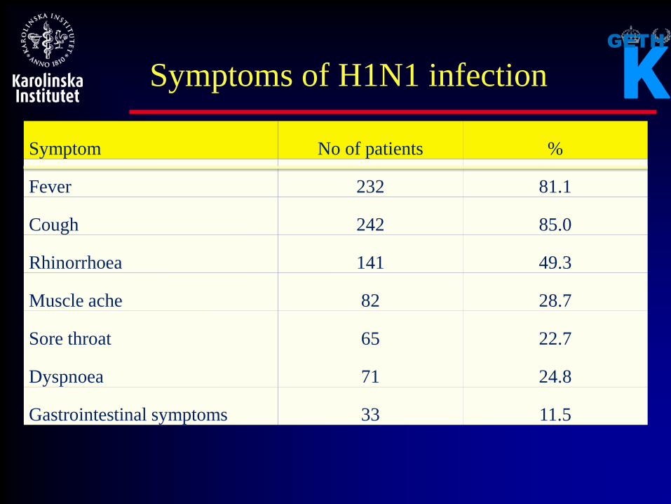
Symptom No of patients %
Fever 232 81.1
Cough 242 85.0
Rhinorrhoea 141 49.3
Muscle ache 82 28.7
Sore throat 65 22.7
Dyspnoea 71 24.8
Gastrointestinal symptoms 33 11.5
GETH
Symptoms of H1N1 infection

Symptom No of patients %
LRT disease 93 32.5
Mechanical ventilation 33 11.5
Neurological symptoms 10 3.5
Death from H1N1 18 6.3
Death from other causes 8 2.8
Outcome of H1N1 infection
EBMT/GETH survey (n=286)
Time to H1N1 from HSCT in fatal cases: Median 1.1 years (0 – 15.3)

Influenza vaccination
Recommended to HSCT patients
Clinical support for a protective effect (Machado)
When after HSCT is it meaningful to vaccinate?
Better immune responses later after HSCT
Current recommendations are to start when the season arrives but not earlier than 3 months after HSCT
Are there any risks?
No evident risks with the seasonal vaccine

Staff vaccination is strongly
recommended!
Data in nursing home
residents show that staff
vaccination works!
No negative effects of
repeated vaccinations
with the seasonal vaccine

Metapneumovirus
Paramyxovirus
5-10 or respiratory virus infections in children
Similar outcome as RSV if pneumonia develops
Ribavirin????

Human Rhinovirus (HRhV)
HRhV are throughout the year the most common cause of
URTID (rhinorhoea, postnasal drip, cough) and occasionally
bronchitis
In allogeneic HCT recipient, HRhVs are the most frequent
CARV reaching a cumulative incidence of 22.3% by day 100.
LRTID in allogeneic HCT is rare (<10%)
The role of treatment is limited by the lack of agents and RCTs.

Rhinovirus pneumonia; overall survival
Seo et al Tandem 2013

New threats
New viruses are emerging usually from animals and
crossing over to humans:
What will be the impact on transplant patients?
Might appear in the donor and transferred to the patient
Might appear in the patient
Might appear in contacts and transmitted to the patient

Some examples
New respiratory viruses - Boca, papova
New coronaviruses - last year in the middle east
New influenza viruses - just now in China (H7N9)
West Nile virus - increasing in some areas
Dengue virus - Outbreak in Madeira
Travel to endemic areas

A common practical question
What do I do with a HSCT patient travelling to
…………….. ?

What vaccines might come up?
HBV No risk / data exist HAV No risk / limited data Polio (inactivated) No risk / data exist Measles Some risks? / some data exist BCG Poor risk / benefit ratio? Typhoid No data / should be no risk Japanese encephalitis No data / should be no risk Yellow fever Limited data / risk?

Thank you for your attention!
Top Related