


Languages
Pages
Legal


Usefulness and Limitation of
Transesophageal Echocardiography
January 2, 2006
Joe M. Moody, Jr, MD
UTHSCSA and STVHCS
The availability of TEE should in NO WAY
adversely influence TTE technical quality

ACC/AHA Practice Guideline, Echo 2003; p. 6.
Where TTE is inadequate, TEE can
usually obtain the desired information.
Usefulness and Limitations
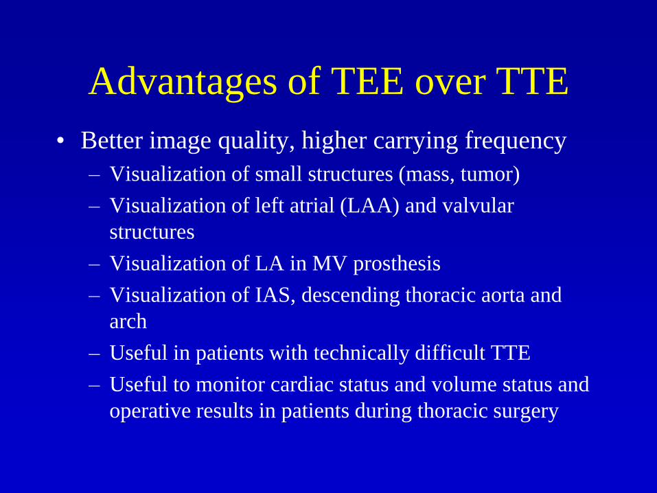
Advantages of TEE over TTE
• Better image quality, higher carrying frequency
– Visualization of small structures (mass, tumor)
– Visualization of left atrial (LAA) and valvular
structures
– Visualization of LA in MV prosthesis
– Visualization of IAS, descending thoracic aorta and
arch
– Useful in patients with technically difficult TTE
– Useful to monitor cardiac status and volume status and
operative results in patients during thoracic surgery

Limitations of TEE Compared to TTE
• Windows limited to different esophageal levels and transgastric window (less uniformly helpful)– Doppler gradients may be falsely low
• Higher risk of complications
• Greater discomfort
• More expensive– Procedure
– Equipment
– Personnel (RN and physician in attendance)
– Time

Indications for TEE
• Endocarditis and valvular disease
• Dyspnea, edema, and cardiomegaly
• Cardioembolic source
• Pre cardioversion
• Critically ill patients
• Imaging coronary ostia in congenital heart
disease
ACC/AHA Practice Guideline, Echo 2003.
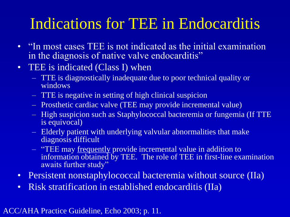
Indications for TEE in Endocarditis
• “In most cases TEE is not indicated as the initial examination in the diagnosis of native valve endocarditis”
• TEE is indicated (Class I) when – TTE is diagnostically inadequate due to poor technical quality or
windows
– TTE is negative in setting of high clinical suspicion
– Prosthetic cardiac valve (TEE may provide incremental value)
– High suspicion such as Staphylococcal bacteremia or fungemia (If TTE is equivocal)
– Elderly patient with underlying valvular abnormalities that make diagnosis difficult
– “TEE may frequently provide incremental value in addition to information obtained by TEE. The role of TEE in first-line examination awaits further study”
• Persistent nonstaphylococcal bacteremia without source (IIa)
• Risk stratification in established endocarditis (IIa)
ACC/AHA Practice Guideline, Echo 2003; p. 11.

Indications for TEE in Valvular
Heart Disease
• Native valve disease or mitral valve prolapse – no
indication for TEE as initial test
• For intervention (Echo indication is Class I):
– For selection of alternative therapies for MS/MR
(valvuloplasty, repair, replacement)*
– Guiding performance of valvuloplasty, repair or
replacement*
– Suspected prosthetic valve dysfunction (changing signs
and symptoms)*
*TEE may provide incremental value in addition to TTE
ACC/AHA Practice Guideline, Echo 2003; p. 13-4.
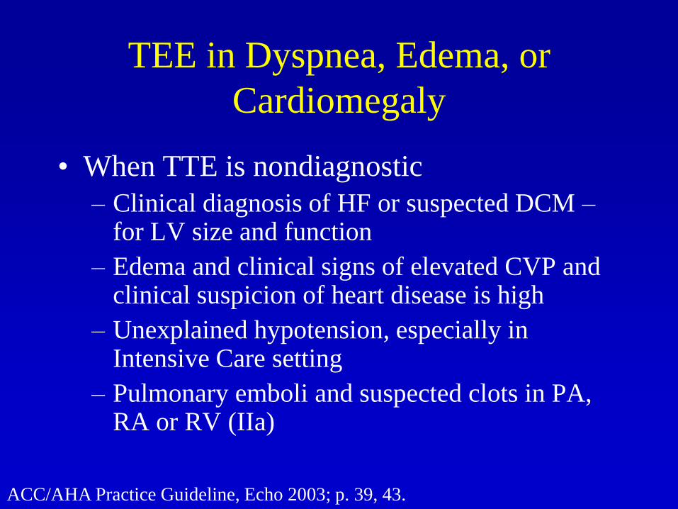
TEE in Dyspnea, Edema, or
Cardiomegaly
• When TTE is nondiagnostic
– Clinical diagnosis of HF or suspected DCM –for LV size and function
– Edema and clinical signs of elevated CVP and clinical suspicion of heart disease is high
– Unexplained hypotension, especially in Intensive Care setting
– Pulmonary emboli and suspected clots in PA, RA or RV (IIa)
ACC/AHA Practice Guideline, Echo 2003; p. 39, 43.

ACC/AHA Practice Guideline, Echo 2003; p. 46.
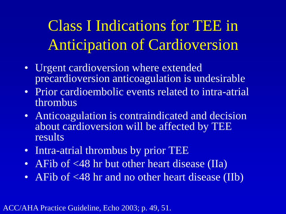
Class I Indications for TEE in
Anticipation of Cardioversion
• Urgent cardioversion where extended precardioversion anticoagulation is undesirable
• Prior cardioembolic events related to intra-atrial thrombus
• Anticoagulation is contraindicated and decision about cardioversion will be affected by TEE results
• Intra-atrial thrombus by prior TEE
• AFib of <48 hr but other heart disease (IIa)
• AFib of <48 hr and no other heart disease (IIb)
ACC/AHA Practice Guideline, Echo 2003; p. 49, 51.
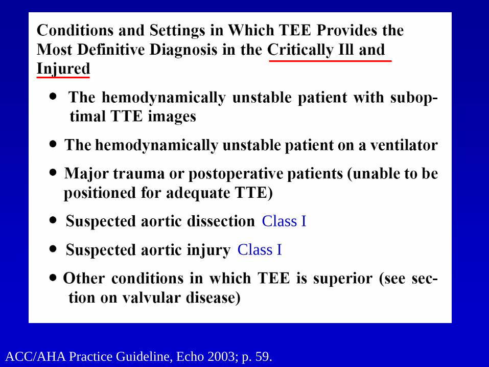
ACC/AHA Practice Guideline, Echo 2003; p. 59.
Class I
Class I

Preparation for TEE Examination
• Patient preparation
– Contraindications: Esophageal pathology (stricture, varices,
tumors, diverticula, scleroderma), severe atlantoaxial joint
disease, prior XRT to chest, perforated viscus; hemodynamic
instability, respiratory compromise
– Fasting 4-6 h (urgent: only clear fluids for 2 hr*); hx of prior
endoscopy or gastroesophageal sx; IV access; suction; crash
cart; monitor; O2, pulse oximeter
• Local anesthesia peak effect 2-5 minutes
– Gargle 2% viscous xylocaine
– 10% Cetacaine spray
– NPO for at least 30 minutes after procedure
Weyman AE. Principles and Practice of Echocardiography, 2nd ed. 1994; p. 332

Preparation for TEE Examination - 2
• Anticholinergic is optional
– Atropine 0.5 mg SQ or Glycopyrrolate (Robinul) 0.1-0.2 mg
– Blurred vision so no driving, increase HR
• Sedation and Analgesia
– Midazolam 0.5-5.0 mg (initial dose ≤2 mg IV over 1-2 min, 0.05 mg/kg)) or Diazepam 5-10 mg
– Meperidine with acetaminophen or morphine can be used for associated discomfort
• Antibiotics for endocarditis prophylaxis are debatable, many laboratories use with prosthetic valves or intracardiac prostheses, poor dentition or prior endocarditis
Weyman AE. Principles and Practice of Echocardiography, 2nd ed. 1994; p. 332

Technique of TEE Examination
• Patient left lateral decubitus position
• Neck gently flexed; a bite guard should always be used except in edentulous patients*
• Index or index and middle fingers of nondominant hand advanced to base of tongue
• Probe advanced with dominant hand and passed beneath the index finger and guided using gentle downward pressure toward the mouth of the esophagus (manually depressing the back of the tongue provides more room, allowing the TEE probe to assume a less acute angle*)
• Anticipate transient gagging
Weyman AE Text 1994; p. 333 *Otto CM. The Practice of Echocardiography, 2nd ed. P. 3.
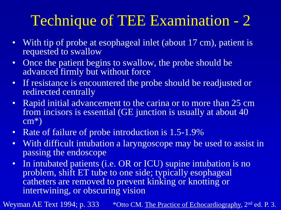
Technique of TEE Examination - 2
• With tip of probe at esophageal inlet (about 17 cm), patient is requested to swallow
• Once the patient begins to swallow, the probe should be advanced firmly but without force
• If resistance is encountered the probe should be readjusted or redirected centrally
• Rapid initial advancement to the carina or to more than 25 cm from incisors is essential (GE junction is usually at about 40 cm*)
• Rate of failure of probe introduction is 1.5-1.9%
• With difficult intubation a laryngoscope may be used to assist in passing the endoscope
• In intubated patients (i.e. OR or ICU) supine intubation is no problem, shift ET tube to one side; typically esophageal catheters are removed to prevent kinking or knotting or intertwining, or obscuring vision
Weyman AE Text 1994; p. 333 *Otto CM. The Practice of Echocardiography, 2nd ed. P. 3.

Clemente CD. Anatomy
3rd Ed, 1987, fig 219.
Veins

Clemente CD.
Anatomy
3rd Ed, 1987, fig
184.

Ref?

Clemente CD. Anatomy
3rd Ed, 1987, fig 216, 7.

Malouf JF et al. Ch. 3, “Functional Anatomy of the Heart” in Hurst’s The Heart 11th ed. 2004

Mechanical Complications of TEE
• Esophagogastric trauma
• Dental trauma
• Minor oral trauma
• Laryngospasm
• Transient vocal-cord paralysis
• Buckling of probe in esophagus
St. John Sutton MG et al. Atlas of Multiplane Transesophageal Echocardiography,
2003, p. 6-8.
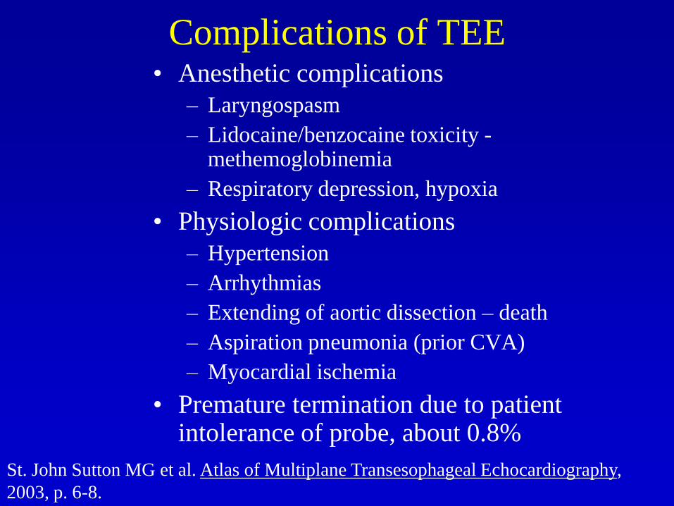
Complications of TEE• Anesthetic complications
– Laryngospasm
– Lidocaine/benzocaine toxicity -methemoglobinemia
– Respiratory depression, hypoxia
• Physiologic complications
– Hypertension
– Arrhythmias
– Extending of aortic dissection – death
– Aspiration pneumonia (prior CVA)
– Myocardial ischemia
• Premature termination due to patient intolerance of probe, about 0.8%
St. John Sutton MG et al. Atlas of Multiplane Transesophageal Echocardiography,
2003, p. 6-8.

TEE Complications, Mayo Clinic
Series
• Transient hypertension 0.3% 1/255
• SVT 0.3% 1/294
• Blood-tinged sputum 0.2% 1/425
• Hypoxia 0.3% 1/294
• Transient hypotension 0.3% 1/294
• NSVT 0.2% 1/478
• Laryngospasm 0.14% 1/765
• Heart failure 0.05% 1/1913
• VT 0.02% 1/3827
• Death 0.01% 1/3827
• Miscellaneous 0.8% 1/123
Seward JB et al. J Am Soc Echocardiogr 1992;5:228.

Esophageal Trauma from TEE• Review of 10,000 TEE’s in one institution
• 3 cases of perforation, all after difficult intubation and multiple attempts and resistance to passage– Age of patients: 73, 79, 84 … 2 anticoagulated
– Symptoms occurred 12, 4, and 22 hours after the procedure
– Symptoms were hemoptysis and mild dyspnea, severe sore throat and subsequent dyspnea and hypoxia, and dyspnea and cough and pain swallowing
– 2 had subcutaneous emphysema, all had elevated WBC, 2 had drop in Hct, 2 had hypoxia
– All had surgery: 2 patients failed conservative therapy and required surgery, and the third patient had primary surgery
• Mucosal tear in the left piriform sinus with abscess
• Mucosal laceration of cervicoesophageal mucosa
• Esophageal perforation of cervical esophagus
– All survived, with prolonged hospitalization of 19-20 days
Min JK et al. J Am Soc Echocardiogr 2005;18:925-29.

Methemoglobinemia
• Topical anesthesia with benzocaine or lidocaine
may result in acute toxic methemoglobinemia
• Also can be caused by nitrates (Na nitrate is
saltpetre), nitrites, and acetaminophen, and
hundreds of other chemicals
• Risk may be increased in patients with G-6-PD
deficiency, may be increased in patients on
acetaminophen and receiving benzocaine
Otto CM. The Practice of Echocardiography, 2nd ed. 2002; P. 18.
Dougherty AH. Circulation 1995;92:268. Wong DH. Mayo Clin Proc. 1995;70:197.

Methemoglobinemia: Fe++ Fe+++
• Hemoglobin is oxidized by the topical agent and
is unable to carry oxygen to the tissues
• Usually normal metabolism but it is possible that
cytochrome B5 reductase, methemoglobin
reductase, or diaphorase, is overwhelmed, or the
hemoglobin is an abnormal M hemoglobin
Otto CM. The Practice of Echocardiography, 2nd ed. 2002; P. 18.
Dougherty AH. Circulation 1995;92:268. Wong DH. Mayo Clin Proc. 1995;70:197.
Ferrous to ferric

Methemoglobinemia: Fe++ Fe+++
• pO2 is normal but oxyhemoglobin level is low
• Cyanosis (total methemoglobin ~1.5 g/dL = 8-12%) and dyspnea
• Pulse oximeter saturation appears only mildly reduced because this technique cannot distinguish well between methemoglobin and reduced hemoglobin
– It measures red and infrared transmission of light (660 nm and 940 nm respectively
– methemoglobin (equal absorption at 940 and 660)
– oxyhemoglobin (more at 940 whereas reduced Hb is more at 660)
– in high methemoglobin concentration the display will read about 85%
• Diagnosis by ABG with measurement of methemoglobin level at >10% (nl is 0.4-1.5%), symptoms if 30-40% and CNS depression if >50%, usually fatal at >70%
Otto CM. The Practice of Echocardiography, 2nd ed. 2002; P. 18.
Dougherty AH. Circulation 1995;92:268. Wong DH. Mayo Clin Proc. 1995;70:197.
Ferrous to ferric

Methemoglobinemia: Fe++ Fe+++
• Management: Methylene blue (1-2 mg/kg) as 1% solution (1,000 mg/100 ml) over 5 minutes will result in prompt resolution of cyanosis;
• If patient has G6PD deficiency, methylene blue will not help, and ascorbic acid should be used.
– Could prescreen African Americans, subjects of Mediterranean discent, southeast Asians
– Also hyperbaric O2 and/or exchange transfusion have been used
Otto CM. The Practice of Echocardiography, 2nd ed. 2002; P. 18.
Dougherty AH. Circulation 1995;92:268. Wong DH. Mayo Clin Proc. 1995;70:197.

Methemoglobinemia
UpToDate; 2005.
Top Related