


Languages
Pages
Legal


DOI: 10.1002/chem.200900484
Unique Twisted Ribbons Generated by Self-Assembly of Oligo(p-phenyleneethylene) Bearing Dimeric Bile Acid Pendant Groups
Yan Li,[a, b] Guangtao Li,*[a] Xinyan Wang,[a, c] Weina Li,[a, b] Zhixiang Su,[a]
Yihe Zhang,[a, c] and Yong Ju*[a, b]
Introduction
The controlled organization of p-conjugated molecules intohighly ordered arrays at the supramolecular level is of greatimportance, owing to the potential applications of thesearrays in optical devices and supramolecular electronics.[1]
Nature elegantly utilizes the self-assembly of biomoleculesto construct functional superstructures. In a manner thatmimics natural processes, the introduction of biogenetic
compounds (e.g., amino acids and nucleic acids) to p-conju-gated systems provides one convenient way to create a vari-ety of fascinating morphologies for the development of ad-vanced materials.[2]
Steroids make up a class of naturally occurring com-pounds containing four fused rings, generally aliphatic rings,examples of which include the biologically important bileacids and cholesterol.[3] In addition to their physiologicalrole, steroidal compounds have been presented as an inex-pensive source of chirality. Due to the rigid steroidal skele-ton and its hydrophobic nature, such molecules tend to formsupramolecular aggregates in which the position and orien-tation of the molecule is well organized.[4] In this respect,cholesterol is widely used as a pendant group, which can beattached to p-conjugated systems to induce the chiral pack-ing of chromophores.[5] However, p-conjugated compoundsbearing dimeric bile acid moieties have been far less system-atically investigated. In contrast to cholesterol, bile acids arestrikingly diverse in nature; therefore, it is highly interestingto study and compare the self-assembly behavior of thesestructurally closely related compounds.
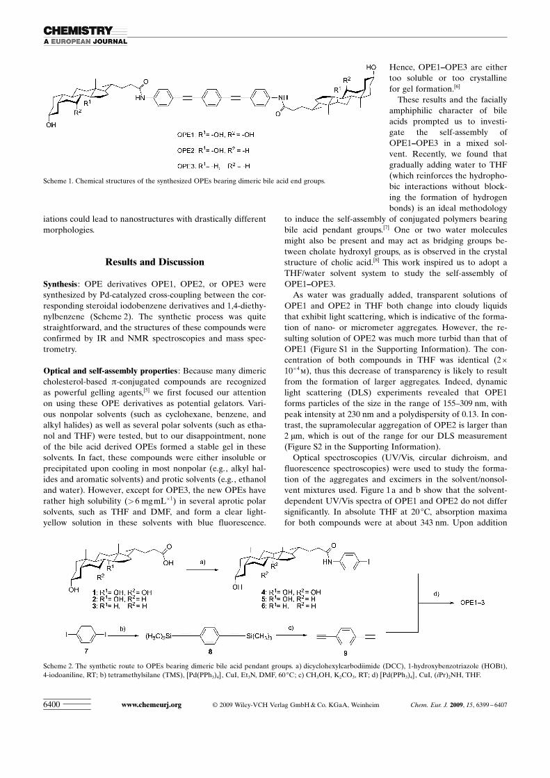
Herein, a new series of compounds consisting of anoligo(p-phenylene ethylene) (OPE) spacer and a bile acidend group was designed. Scheme 1 shows the synthesizedOPE derivatives bearing dimeric cholic acid (OPE1), deoxy-cholic acid (OPE2), and lithocholic acid units (OPE3),which are differentiated only by the number of hydroxylgroups. Very interestingly, it was found that such subtle var-
Abstract: A series of novel oligo(p-phenylene ethynylene) (OPE) deriva-tives bearing dimeric cholic acid(OPE1), deoxycholic acid (OPE2) andlithocholic acid (OPE3) was synthe-sized. The self-assembly behavior ofthese derivatives were systematicallystudied and compared in a THF/watersolvent mixture. The addition of waterto THF can induce the helical stacking
of oligo(p-phenylene ethylene) bearingdimeric deoxycholic acid end groups,which leads to highly interesting twist-ed assemblies. Variation of the bileacid group led to nanostructures with
drastically different morphologies. Theapplication of spectroscopy in combi-nation with X-ray diffraction tech-niques provided a reasonable view ofthe final self-assembled structures. Theobserved distinctive aggregate shapesand the preference in the type of mo-lecular packing of these steroid–OPEconjugates are attributed to the subtledifferences in their molecular features.
Keywords: bile acids · chirality ·conjugation · self-assembly · supra-molecular chemistry
[a] Y. Li, Prof. Dr. G. Li, X. Wang, W. Li, Z. Su, Prof. Dr. Y. Zhang,Prof. Dr. Y. JuDepartment of ChemistryKey Lab of Organic Optoelectronics & Molecular EngineeringMinistry of Education, Tsinghua UniversityBeijing, 100084 (China)Fax: (+86) 10-6279-2905E-mail : [email protected]
[b] Y. Li, W. Li, Prof. Dr. Y. JuDepartment of ChemistryKey Lab of Bioorganic Phosphorus Chemistry & Chemical BiologyMinistry of Education, Tsinghua UniversityBeijing, 100084 (China)Fax: (+86) 10-6278-1695E-mail : [email protected]
[c] X. Wang, Prof. Dr. Y. ZhangSchool of Material Science and TechnologyChina University of GeosciencesBeijing 100083 (China)
Supporting information for this article is available on the WWWunder http://dx.doi.org/10.1002/chem.200900484.
Chem. Eur. J. 2009, 15, 6399 – 6407 � 2009 Wiley-VCH Verlag GmbH & Co. KGaA, Weinheim 6399
FULL PAPER
iations could lead to nanostructures with drastically differentmorphologies.
Results and Discussion
Synthesis : OPE derivatives OPE1, OPE2, or OPE3 weresynthesized by Pd-catalyzed cross-coupling between the cor-responding steroidal iodobenzene derivatives and 1,4-diethy-nylbenzene (Scheme 2). The synthetic process was quitestraightforward, and the structures of these compounds wereconfirmed by IR and NMR spectroscopies and mass spec-trometry.
Optical and self-assembly properties : Because many dimericcholesterol-based p-conjugated compounds are recognizedas powerful gelling agents,[5] we first focused our attentionon using these OPE derivatives as potential gelators. Vari-ous nonpolar solvents (such as cyclohexane, benzene, andalkyl halides) as well as several polar solvents (such as etha-nol and THF) were tested, but to our disappointment, noneof the bile acid derived OPEs formed a stable gel in thesesolvents. In fact, these compounds were either insoluble orprecipitated upon cooling in most nonpolar (e.g., alkyl hal-ides and aromatic solvents) and protic solvents (e.g., ethanoland water). However, except for OPE3, the new OPEs haverather high solubility (>6 mgmL�1) in several aprotic polarsolvents, such as THF and DMF, and form a clear light-yellow solution in these solvents with blue fluorescence.
Hence, OPE1–OPE3 are eithertoo soluble or too crystallinefor gel formation.[6]
These results and the faciallyamphiphilic character of bileacids prompted us to investi-gate the self-assembly ofOPE1–OPE3 in a mixed sol-vent. Recently, we found thatgradually adding water to THF(which reinforces the hydropho-bic interactions without block-ing the formation of hydrogenbonds) is an ideal methodology
to induce the self-assembly of conjugated polymers bearingbile acid pendant groups.[7] One or two water moleculesmight also be present and may act as bridging groups be-tween cholate hydroxyl groups, as is observed in the crystalstructure of cholic acid.[8] This work inspired us to adopt aTHF/water solvent system to study the self-assembly ofOPE1–OPE3.
As water was gradually added, transparent solutions ofOPE1 and OPE2 in THF both change into cloudy liquidsthat exhibit light scattering, which is indicative of the forma-tion of nano- or micrometer aggregates. However, the re-sulting solution of OPE2 was much more turbid than that ofOPE1 (Figure S1 in the Supporting Information). The con-centration of both compounds in THF was identical (2�10�4
m), thus this decrease of transparency is likely to resultfrom the formation of larger aggregates. Indeed, dynamiclight scattering (DLS) experiments revealed that OPE1forms particles of the size in the range of 155–309 nm, withpeak intensity at 230 nm and a polydispersity of 0.13. In con-trast, the supramolecular aggregation of OPE2 is larger than2 mm, which is out of the range for our DLS measurement(Figure S2 in the Supporting Information).
Optical spectroscopies (UV/Vis, circular dichroism, andfluorescence spectroscopies) were used to study the forma-tion of the aggregates and excimers in the solvent/nonsol-vent mixtures used. Figure 1 a and b show that the solvent-dependent UV/Vis spectra of OPE1 and OPE2 do not differsignificantly. In absolute THF at 20 8C, absorption maximafor both compounds were at about 343 nm. Upon addition
Scheme 1. Chemical structures of the synthesized OPEs bearing dimeric bile acid end groups.
Scheme 2. The synthetic route to OPEs bearing dimeric bile acid pendant groups. a) dicyclohexylcarbodiimide (DCC), 1-hydroxybenzotriazole (HOBt),4-iodoaniline, RT; b) tetramethylsilane (TMS), [Pd ACHTUNGTRENNUNG(PPh3)4], CuI, Et3N, DMF, 60 8C; c) CH3OH, K2CO3, RT; d) [Pd ACHTUNGTRENNUNG(PPh3)4], CuI, (iPr)2NH, THF.
www.chemeurj.org � 2009 Wiley-VCH Verlag GmbH & Co. KGaA, Weinheim Chem. Eur. J. 2009, 15, 6399 – 64076400
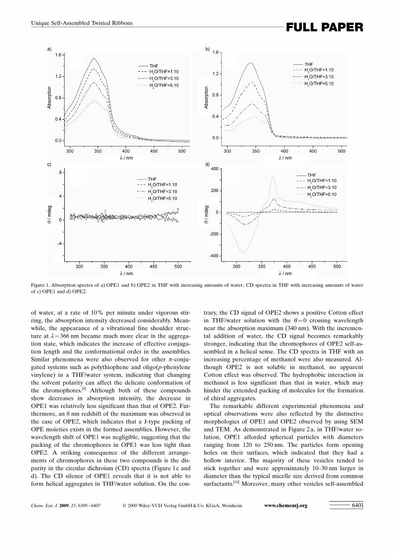
of water, at a rate of 10 % per minute under vigorous stir-ring, the absorption intensity decreased considerably. Mean-while, the appearance of a vibrational fine shoulder struc-ture at l= 366 nm became much more clear in the aggrega-tion state, which indicates the increase of effective conjuga-tion length and the conformational order in the assemblies.Similar phenomena were also observed for other p-conju-gated systems such as polythiophene and oligo(p-phenylenevinylene) in a THF/water system, indicating that changingthe solvent polarity can affect the delicate conformation ofthe chromophores.[9] Although both of these compoundsshow decreases in absorption intensity, the decrease inOPE1 was relatively less significant than that of OPE2. Fur-thermore, an 8 nm redshift of the maximum was observed inthe case of OPE2, which indicates that a J-type packing ofOPE moieties exists in the formed assemblies. However, thewavelength shift of OPE1 was negligible, suggesting that thepacking of the chromophores in OPE1 was less tight thanOPE2. A striking consequence of the different arrange-ments of chromophores in these two compounds is the dis-parity in the circular dichroism (CD) spectra (Figure 1 c andd). The CD silence of OPE1 reveals that it is not able toform helical aggregates in THF/water solution. On the con-
trary, the CD signal of OPE2 shows a positive Cotton effectin THF/water solution with the q=0 crossing wavelengthnear the absorption maximum (340 nm). With the incremen-tal addition of water, the CD signal becomes remarkablystronger, indicating that the chromophores of OPE2 self-as-sembled in a helical sense. The CD spectra in THF with anincreasing percentage of methanol were also measured. Al-though OPE2 is not soluble in methanol, no apparentCotton effect was observed. The hydrophobic interaction inmethanol is less significant than that in water, which mayhinder the extended packing of molecules for the formationof chiral aggregates.
The remarkable different experimental phenomena andoptical observations were also reflected by the distinctivemorphologies of OPE1 and OPE2 observed by using SEMand TEM. As demonstrated in Figure 2 a, in THF/water so-lution, OPE1 afforded spherical particles with diametersranging from 120 to 250 nm. The particles form openingholes on their surfaces, which indicated that they had ahollow interior. The majority of these vesicles tended tostick together and were approximately 10–30 nm larger indiameter than the typical micelle size derived from commonsurfactants.[10] Moreover, many other vesicles self-assembled
Figure 1. Absorption spectra of a) OPE1 and b) OPE2 in THF with increasing amounts of water; CD spectra in THF with increasing amounts of waterof c) OPE1 and d) OPE2.
Chem. Eur. J. 2009, 15, 6399 – 6407 � 2009 Wiley-VCH Verlag GmbH & Co. KGaA, Weinheim www.chemeurj.org 6401
FULL PAPERUnique Self-Assembled Twisted Ribbons

from small amphiphilic molecules immediately collapsed onsolid surfaces,[11] but these OPE vesicles showed remarkablestability and retained their shape upon drying, presumablydue to the rigid p-conjugated spacer and the steroid nuclei.The TEM image in Figure 2 b further confirmed theserobust capsule-like aggregates with diameters of about200 nm, which is in agreement with the DLS experimentalresults.[12] It is interesting to note that this linear structureleads to a spherical morphology.
On the other hand, OPE2, with dimeric deoxycholatemoieties, shows a very interesting assembly behavior inwater/THF (1:2 v/v): remarkably broad ribbons twisted in aright-handed helical structure were observed (Figure 2 c andd). The ribbons are approximately 1.5 mm wide and a fewhundred micrometers in length, with an average helicalpitch length of 2 mm. Moreover, the dimensions of thesesupramolecular aggregations were large enough to be ob-served by using normal optical microscopy (Figure 3), whichprovides a convenient approach to capture the image ofthese twisted ribbons in situ. Polarizing microscopy showsthat periodic color strips appear on the self-assembled twist-ed ribbons, which suggests the existence of a periodic or-dered packing of molecular structures. The twisted ribbonsderived from OPE2 exhibited strong blue fluorescence thatcan be clearly detected by fluorescent optical microscopy(Figure 4 a). The fluorescent spectra of the aggregate statewere slightly broader and exhibited a redshift (~6 nm) com-pared with those observed in THF, which is indicative of aJ-type packing of p-conjugated chains in the molecular as-semblies (Figure 4 b).
We wondered how the pres-ence of nonsolvent could affectthe morphological transition ofOPE2 that finally leads to sucha unique microstructure. To ad-dress this question, the self-as-sembly of OPE2 in THF (2�10�4
m) with increasing additionof water was monitored bySEM (Figure 5). At the 5 %volume fraction of water,bundle-like structures with a di-ameter of less than 1 mm werefound to be the dominant mor-phology. These bundles have alow aspect ratio and possessseveral branches at their ends,which is in sharp contrast to thenanofibers formed by similarcholesterol derivatives.[5,13]
Upon increasing the water con-tent to 20 %, the twisted senseseems to start to form from thetips of the branches. In themeantime, the diameter of thebundles was increased consider-
ably and leads to a ribbon-like structure. Further increasingthe volume of water to 30 % finally yields helical ribbonswith an almost regular pitch length of 2 mm, in line with thestrong positive cotton effect observed in the CD spectrum.
Figure 2. SEM and TEM images of supramolecular assemblies of OPE1 (a, b) and OPE2 (c, d) in water/THF(1:2 v/v).
Figure 3. Photographs (1000 � magnification) of the twisted ribbons inwater/THF 1:2 observed by optical microscopy under a) white light andb) polarized light. The scale bar is 10 mm.
www.chemeurj.org � 2009 Wiley-VCH Verlag GmbH & Co. KGaA, Weinheim Chem. Eur. J. 2009, 15, 6399 – 64076402
G. Li, Y. Ju et al.

The progressive changes of the aggregate morphologies withthe addition of water indicate that a relaxation process maybe involved throughout the entire assembly.[14] In fact, therelaxation process is evidenced by the fact that when OPE2was directly dissolved in a fixed ratio of THF/water mixtureat elevated temperature, it did not yield any helical structureupon cooling (Figure S3 in the Supporting Information). Inour experiment, it seems that the incremental addition ofwater to THF under ultrasound irradiation or vigorous stir-ring is necessary for the formation of these twisted ribbons.Even though bile acids possess more than ten chiral carbonatoms, the transfer of chirality from the molecular level tohelical structures on a supramolecular scale that can beclearly visualized using techniques such as SEM, TEM, andeven optical microscopy makes this system really unusual.
In contrast to OPE1 and OPE2, OPE3 (which possessesonly two hydroxyl groups) yields large microcrystalline rodsunder the same conditions (Figure S4 in the Supporting In-formation). This compound is so insoluble in aqueous mediathat the addition of water leads to rapid precipitation.
Mechanism of self-assembly : The mechanism for the forma-tion of the described superstructures is at present not veryclear. The molecular structure of bile acid is considerably
different from conventional surfactants and the mechanismof their formation is quite complex, often involving compli-cated “secondary assemblies”.[15] As mentioned above, thespectroscopy studies (UV/Vis, CD, and fluorescence) dem-onstrate that OPE-derived steroidal compounds undergo ex-tensive conformational changes in solvent/nonsolvent (THF/water) mixtures. It is believed that the cooperation of aro-matic stacking, hydrogen bonding, and van der Waals inter-actions contributes significantly to the observed uniquemesoscopic structures, whereas the orientation of the chro-mophore plays a crucial role in the optical and chiropticalproperties. With reference to the existing literature, possiblemolecular packing models for the superstructures formedfrom OPE1 and OPE2 are suggested (Scheme 3).
As demonstrated in Scheme 3 a, OPE1 possesses a p-con-jugated spacer (OPE) and two amphiphilic pendant groups
Figure 4. a) Photographs (1000 � magnification) of the twisted ribbonsobserved under illumination with 330–380 nm UV light. The arrow indi-cates the helical sense. The scale bar is 10 mm. b) Normalized emissionspectra of OPE2 in THF solution (solid line) and in aggregation state(dashed line).
Figure 5. SEM images of hierarchical superstructures of OPE2 in THF/water solution with a volume fraction of water of a) 5 %, b) 15%, andc) 30%. The scale bar is 2 mm.
Chem. Eur. J. 2009, 15, 6399 – 6407 � 2009 Wiley-VCH Verlag GmbH & Co. KGaA, Weinheim www.chemeurj.org 6403
FULL PAPERUnique Self-Assembled Twisted Ribbons
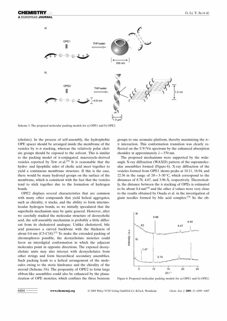
(cholate). In the process of self-assembly, the hydrophobicOPE spacer should be arranged inside the membrane of thevesicles by p–p stacking, whereas the relatively polar chol-ate groups should be exposed to the solvent. This is similarto the packing model of p-conjugated, macrocycle-derivedvesicles reported by Tew et al.[16] It is reasonable that thehydro- and lipophilic sides of cholic acid meet together toyield a continuous membrane structure. If this is the case,there would be many hydroxyl groups on the surface of themembrane, which is consistent with the fact that the vesiclestend to stick together due to the formation of hydrogenbonds.
OPE2 displays several characteristics that are commonwith many other compounds that yield helical aggregates,such as chirality, p stacks, and the ability to form intermo-lecular hydrogen bonds, so we initially speculated that thesuperhelix mechanism may be quite general. However, afterwe carefully studied the molecular structure of deoxycholicacid, the self-assembly mechanism is probably a little differ-ent from its cholesterol analogue. Unlike cholesterol, bileacid possesses a curved backbone with the thickness ofabout 0.6 nm (C3-C18).[17] To make the extended packing ofchromophores possible, the deoxycholate moieties couldfavor an interdigital conformation in which the adjacentmolecules point in opposite directions. The exposed deoxy-cholate units may also interact with deoxycholates fromother strings and form hierarchical secondary assemblies.Such packing leads to a helical arrangement of the mole-cules owing to the steric hindrance and the chirality of thesteroid (Scheme 3 b). The propensity of OPE2 to form largeribbon-like assemblies could also be enhanced by the plana-rization of OPE moieties, which confines the three benzene
groups to one aromatic platform, thereby maximizing the p–p interaction. This conformation transition was clearly re-flected on the UV/Vis spectrum by the enhanced absorptionshoulder at approximately l=370 nm.
The proposed mechanisms were supported by the wide-angle X-ray diffraction (WAXD) pattern of the supramolec-ular assemblies formed (Figure 6). X-ray diffraction of thevesicles formed from OPE1 shows peaks at 10.11, 18.94, and22.56 in the range of 2q= 3–30 8C, which correspond to thedistances of 8.78, 4.67, and 3.96 �, respectively. Theoretical-ly, the distance between the p stacking of OPEs is estimatedto be about 0.4 nm[18] and the other d values were very closeto the results obtained by Osada et al. in the investigation ofgiant needles formed by bile acid complex.[19] So the ob-
Scheme 3. The proposed molecular packing models for a) OPE1 and b) OPE2.
Figure 6. Proposed molecular packing models for a) OPE1 and b) OPE2.
www.chemeurj.org � 2009 Wiley-VCH Verlag GmbH & Co. KGaA, Weinheim Chem. Eur. J. 2009, 15, 6399 – 64076404
G. Li, Y. Ju et al.

served d spacings of 8.78 and 4.67 � are possibly caused bythe ordered packing of the adjacent cholate units, whereasthe d spacing of 0.40 nm is a result of the center-to-centerdistance between the OPE spacers. In other words, this self-assembly aggregation preserved some of the basic packingpatterns of bile acid, combined with the p–p stacking ofOPE moieties. The X-ray pattern of OPE2 was similar tothat of OPE1, and several peaks appeared in the samerange. It is interesting to note that the peaks correspondingto 1.47 and 0.74 nm (1.47/2) were not observed for OPE1.This periodicity indicates a lamellar packing structure. Con-sidering that the space between the amide group (C24atom) and the end of the steroid nucleus (C3 atom) is about1.5 nm, based on the assumption that the alkyl spacer takesa fully extended conformation, the distance of 1.47 nm verylikely corresponds to the distance between the planesformed by adjacent OPE strips, which gives clear evidencefor the proposed self-assembly pattern of OPE2. The helicalribbons of OPE2 were solely right-handed, presumably re-flecting the characteristic asymmetric nature of the deoxy-cholic acid. In our experiment, we also noticed that thesignal-to-noise ratio of OPE1 in the XRD result was consis-tently lower than that of OPE2, which suggests that the mol-ecules packing in the membrane of the vesicle were less or-ganized than the twisted ribbons.
The distinctive self-assembly behavior of bile acid derivedOPEs also raised the question of why the helical twist deter-iorated when the number of hydroxyl groups either in-creased or decreased. To approach this question, it is essen-tial to analyze the molecular arrangements in the supra-molecular assemblies and the structures of these OPE deriv-atives. According to the molecular packing model proposed,one significant difference between these two supramolecularassemblies is that more steroid moieties in the vesicles areexposed to the solvent media than in the twisted ribbons inthe every unit volume. In other words, many deoxycholicacid units were buried inside the large ribbon rather than indirect contact with the solvent. On the other hand, OPE1has more hydroxyl groups on the cholate unit than OPE2,which makes the pendant groups of OPE1 more hydrophilicthan their OPE2 counterparts. Considering the fact that thesolvent used (THF/water mixture) is a relatively polarmedium, it is reasonable that the pendant groups in OPE2tend to be less exposed to the solvents than those in OPE1,which facilitates the reduction of the interfacial energy.
In the case of OPE3, the lack of hydrophilic functionalgroups means that the hydrophobic interactions of the largehydrophobic steroid nuclei become the dominant force,leading to either crystallization or precipitation. There isalso a stronger tendency for growth in a one-dimensionalfashion and loss of the curvature of the aggregates.
The number, location, and configuration of the hydroxylgroups play a crucial role in the formation of aggregatesfrom bile acid derivatives.[20] In particular, two researchgroups found that among seven kinds of bile acid salts, onlysodium deoxycholate aggregated in aqueous solution. In thiscase, an elongated right-handed helical structure with mac-
romolecular dimensions (10–100 �) was obtained.[21] Com-pared with our result, we do not know whether the specialbehavior of deoxycholic acid is a coincidence. The backboneof deoxycholic acid presumably possesses an “appropriate”hydrophilic–hydrophobic balance, which may endow deli-cate influences on hydrogen-bond formation and hydropho-bic interactions in aqueous solution. However, understand-ing and predicting the intermolecular forces in these com-plex systems and how it determines their self-assembly mor-phology remains a significant challenge.
Conclusions
The current work employs naturally occurring bile acids aschirality-inducing moieties and offers a new means to con-trol the packing and orientation of the chromophore. Veryinteresting is that subtle structure variations could drastical-ly change the morphologies of the nanostructures formed.In this respect, it is possible to prepare many other interest-ing bile acid based molecular assemblies for this purpose byvarying the type of bile acids and p-conjugated bridginggroups of the scaffold. Based on the study of the self-assem-bly behavior of these structurally closely related compounds,this work provides new insights into the formation of chiralsupramolecular assemblies starting from the molecular chir-ality of steroids. On the other hand, the microstructuresformed from them may find use in hybrid-material synthesisor as a useful chiral medium for manipulating chemical sep-arations. Some of these possibilities are being explored inour laboratory.
Experimental Section
Instruments : 1H NMR spectra were recorded at 300 MHz on JOELJNM-ECA 300 spectrometers. Chemical shifts (d) are given in ppm rela-tive to TMS (d =0.0). High-resolution electrospray ionization mass spec-trometry (HRMS) measurements were recorded on a Bruker APEXspectrometer in positive mode. UV/Vis characterization was carried outon a Perkin–Elmer Lambda35 spectrometer. The fluorescence emissionmeasurements were carried out using a Hitachi F-4500 fluorescence spec-trometer. CD results were obtained on a Jasco-720 spectrometer. IRspectra were recorded on an AVATAR 360 ESP FTS spectrophotometerwith KBr pellets. Elemental analyses were performed on a Carlo–Erba-1106 instrument. DLS measurements were carried out by using a Mal-vern Zetasizer 3000HS instrument, which supplies vertically polarizedlight with a wavelength of 633 nm. SEM experiments were performedwith a LEO-1530 scanning electron microscope. TEM was performed ona MODEL H-800 electron microscope. All of the samples were stainedwith 1.5% phosphotungstic acid hydrate and filter paper was used todrain the excess solution. The polarizing behavior of the self-assembledaggregations and the fluorescence images were observed by using anOlympus 146 Tokyo incident-light optical microscope (� 1000 magnifica-tion).
X-ray diffraction : The vesicles and helical assemblies formed by OPE1and OPE2 in solution were placed directly on the glass plate and dried inair. Both diffraction patterns were recorded on a Rigaku D/max diffrac-tometer by using a CuKa X-ray irradiation source (40 kV, 30 mA). Datawere measured at room temperature between 3 and 308 in 2q/q scanmode and the scanning rate was kept at 18min�1.
Chem. Eur. J. 2009, 15, 6399 – 6407 � 2009 Wiley-VCH Verlag GmbH & Co. KGaA, Weinheim www.chemeurj.org 6405
FULL PAPERUnique Self-Assembled Twisted Ribbons

Chemicals : Cholic acid, deoxycholic acid, lithocholic acid, and 4-iodoani-line were purchased from Sigma and used as received. Trimethylsilylace-tylene was purchased from Shanghai Reagent Coporation. [Pd ACHTUNGTRENNUNG(PPh3)4]was obtained from Pingyang Chemical Company and washed with coldethanol prior to use to remove the oxidized impurity. Other reagents andsolvents were received from Beijing Chemical Company without furtherpurification unless otherwise stated. 1,4-Diethynylbenzene was preparedand characterized according to a previously published procedure.[22]
N-Cholyl-4-iodoaniline 4 : Cholic acid (818 mg, 2.0 mmol), HOBt(297 mg, 2.2 mmol), and 4-iodoaniline (438 mg, 2.0 mmol) were dissolvedin CH2Cl2/pyridine (12 mL, 5:1 v/v). After stirring at 0 8C for 10 min,DCC (453 mg, 2.2 mmol) was added and the resulting reaction mixturewas kept at ambient temperatures for a further 20 h. A white precipitatewas filtered from the purple solution and washed with acetone. Thecrude product was purified by column chromatography on neutral Al2O3
(5 % MeOH/CH2Cl2) to obtain 4 (926 mg, 76%). 1H NMR (300 MHz,[D6]DMSO): d =9.58 (s, 1 H; NH), 7.52 (d, J =7.2 Hz, 2H; Ar-H), 7.42(d, J =7.2 Hz, 2 H; Ar-H), 3.90 (s, 1H; 12-CH), 3.74 (s, 1H; 7-CH), 3.39–3.55 (m, 1H; 3-CH, overlap with the water signal in [D6]DMSO), 1.01–2.39 (m, 27H; alkyl-H), 1.01 (d, J =6.0 Hz, 3H; 21-CH3), 0.87 (s, 3H; 19-CH3), 0.61 ppm (s, 3H; 18-CH3); 13C NMR (300 MHz, [D6]DMSO): d=
172.5, 139.0, 137.1, 121.4, 85.7, 72.1, 71.1, 67.3, 46.4, 46.1, 41.4, 35.2, 35.1,34.7, 34.5, 33.7, 31.3, 30.3, 28.3, 27.3, 26.1, 24.6, 23.0, 22.4, 17.2, 12.4 ppm;HRMS (ESI): m/z calcd for [C30H44INO4+Na]+ : 632.2213; found:632.2200.
N-Deoxycholyl-4-iodoaniline 5 : Compound 5 was synthesized as de-scribed for compound 4 by using deoxycholic acid instead of cholic acid.Yield: 77 %; 1H NMR (300 MHz, [D6]DMSO): d=9.58 (s, 1 H; NH), 7.52(d, J=7.2 Hz, 2 H; Ar-H), 7.42 (d, J=7.2 Hz, 2H; Ar-H), 3.90 (s, 1H; 12-CH), 3.74 (s, 1H; 7-CH), 3.39–3.55 (m, 1 H; 3-CH, overlap with the watersignal in [D6]DMSO), 1.01–2.39 (m, 27H; alkyl-H), 1.01 (d, J =6.0 Hz,3H; 21-CH3), 0.87 (s, 3 H; 19-CH3), 0.61 ppm (s, 3 H; 18-CH3); 13C NMR(300 MHz, [D6]DMSO): d=172.4, 139.8, 137.4, 121.7, 86.7, 71.5, 70.5,55.4, 48.0, 46.7, 46.5, 42.1, 36.8, 36.2, 35.7, 35.6, 34.3, 34.0, 33.5, 31.8, 30.8,30.0, 27.7, 27.5, 26.7, 24.1, 23.6, 22.4, 17.6, 13.0 ppm; HRMS (ESI): m/zcalcd for [C30H44INO3+H]+ : 594.2444; found: 594.2439.
N-Lithocholyl-4-iodoaniline 6 : Compound 6 was synthesized as describedfor compound 4 by using lithocholic acid instead of cholic acid. Yield:80%; 1H NMR (300 MHz, [D6]DMSO): d=9.95 (s, 1 H; NH), 7.60 (d, J=
7.2 Hz, 2H; Ar-H), 7.41 (d, J =7.2 Hz, 2 H; Ar-H), 4.44 (br s, 1H; OH),3.39–3.55 (m, 1H; 3-CH, overlap with the signal of water in [D6]DMSO),1.05–2.35 (m, 28H; alkyl-H), 1.01 (d, J= 6.0 Hz, 3 H; 21-CH3), 0.87 (s,3H; 19-CH3), 0.61 ppm (s, 3H; 18-CH3); 13C NMR (300 MHz,[D6]DMSO): d =172.5, 140.0, 137.9, 121.7, 85.7, 71.9, 71.1, 55.00, 54.96,41.8, 41.3, 39.5, 39.3, 35.0, 34.6, 33.5, 30.4, 29.5, 27.3, 26.2, 25.4, 23.3, 22.4,19.7, 17.5, 12.0 ppm; HRMS (ESI): m/z calcd for [C30H44INO2+H]+ :578.2495; found: 578.2489.
OPE1: [PdACHTUNGTRENNUNG(Ph3P)4] (46.0 mg, 0.04 mmol) and CuI (7.6 mg, 0.04 mmol)were added to a stirred solution of 4 (632.2 mg, 1 mmol) and 1,4-diethy-nylbenzene (64.3 mg, 0.51 mmol) in DMF (10 mL) under nitrogen. Thereaction mixture was stirred under nitrogen at 40 8C for 20 h, then fil-tered. The filtrate was evaporated under vacuum and further purified bychromatography on neutral Al2O3 (CHCl3/CH3OH 15:1). The crudeproduct was dissolved in a small amount of THF and added dropwiseinto acetone. OPE1 was obtained as a light yellow solid by filtration andwas dried under vacuum for 24 h (337.6 mg, 62 %). 1H NMR (300 MHz,[D6]DMSO): d=10.08 (s, 2H; NH), 7.65 (d, J =7.2 Hz, 4H; Ar-H), 7.55(s, 4H; Ar-H), 7.49 (d, J =7.2 Hz, 4H; Ar-H), 4.32 (d, J =4.2 Hz, 2H; 12-OH), 4.12 (d, J= 3.9 Hz, 2H; 7-OH), 4.05 (d, J =3.8 Hz, 2H; 3-OH), 3.80(s, 2 H; 12-CH), 3.62 (s, 2 H; 7-CH), 3.19–3.26 (m, 2 H; 3-CH), 1.00–2.38(m, 48 H; alkyl-H), 0.98 (d, J =5.6 Hz, 6 H; 18-CH3), 0.81 (s, 6H; 19-CH3), 0.60 ppm (s, 6H; 21-CH3); 13C NMR (300 MHz, [D6]DMSO): d=
172.6, 140.6, 132.7, 132.0, 120.0, 119.0, 118.7, 92.2, 88.8, 71.5, 71.0, 66.8,46.6, 46.3, 41.9, 36.3, 35.8, 35.7, 35.5, 34.1, 32.1, 31.9, 29.1, 27.8, 26.8, 23.4,23.2, 17.0, 12.9 ppm; IR (KBr): n =3428, 2923, 2862, 2215, 1669, 1593,1520, 1046 cm�1; MS (ESI): m/z : 1112 [M+Na]+ ; elemental analysis calcd(%) for C70H92N2O8: C 77.17, H 8.51, N 2.57; found: C 76.93, H 8.90, N2.41.
OPE2 : OPE2 was synthesized as described for OPE1 by using 5 insteadof 4. Yield: 68%; 1H NMR (300 MHz, [D6]DMSO): d= 10.08 (s, 2 H;NH), 7.65 (d, J =7.2 Hz, 4 H; Ar-H), 7.55 (s, 4 H; Ar), 7.49 (d, J =7.2 Hz,4H; Ar-H), 4.46 (d, J=4.2 Hz, 2H; 12-OH), 4.20 (d, J =3.9 Hz, 2 H; 3-OH), 3.80 (s, 2 H; 12-CH), 3.33–3.48 (m, 2H; 3-CH, overlap with thewater signal in [D6]DMSO), 0.85–2.37 (m, 52H; alkyl-H), 0.91 (d, J=
6.0 Hz, 6H; 21-CH3), 0.85 (s, 6 H; 19-CH3), 0.61 ppm (s, 6 H; 18-CH3);13C NMR (300 MHz, [D6]DMSO): d =172.6, 140.6, 132.7, 132.0, 123.0,119.4, 116.5, 92.2, 88.7, 71.6, 70.5, 48.0, 46.7, 46.5, 36.8, 36.2, 35.6, 34.4,34.1, 33.5, 31.9, 30.8, 29.2, 27.7, 27.5, 26.7, 24.1, 23.6, 17.7, 13.0 ppm; IR(KBr): n=3412, 2934, 2862, 2214, 1670, 1591, 1520, 1041, 838 cm�1; MS(ESI): m/z : 1080 [M+Na]+ ; elemental analysis calcd (%) for C70H92N2O6:C 79.50, H 8.77, N 2.65; found: C 79.00, H 8.52, N 2.94.
OPE3 : OPE3 was synthesized as described for OPE1 by using 6 insteadof 4. Yield: 55%; 1H NMR (300 MHz, [D6]DMSO): d= 10.07 (s, 2 H;NH), 7.65 (d, J =7.2 Hz, 4H; Ar-H), 7.54 (s, 4H; Ar-H), 7.48 (d, J =
7.2 Hz, 4H; Ar-H), 4.41 (s, 2 H; 3-OH), 3.20–3.50 (m, 2 H; 3-CH), 1.02–1.93 (m, 60H; alkyl-H), 0.94 (d, J =5.6 Hz, 6H; 18-CH3), 0.87 (s, 6H; 19-CH3), 0.61 ppm (s, 6H; 21-CH3); 13C NMR (300 MHz, [D6]DMSO): d=
172.4, 141.1, 136.0, 132.7, 119.9, 118.0, 92.2, 71.0, 57.1, 56.2, 43.0, 42.3,41.3, 40.5, 36.3, 35.9, 34.5, 32.8, 31.1, 29.1, 27.7, 26.5, 23.7, 23.1, 21.4, 17.0,11.9 ppm; IR (KBr): n=3481, 3426, 2930, 2850, 2214, 1672, 1585,1527 cm�1; MS (ESI): m/z : 1048 [M+Na]+ ; elemental analysis calcd (%)for C70H92N2O4: C 81.99, H 9.04, N 2.73; found: C 81.56, H 9.49, N 2.52.
Acknowledgements
We gratefully acknowledge the financial support from the National Sci-ence Foundation of China (20533050, 20772071, and 50673048), 973 Pro-gram (2006CB806200) and the transregional project (TRR6). We wouldalso like to thank Prof. Chun Li for help during the circular dichroism ex-periment.
[1] a) M. Grell, D. D. C. Bradley, Adv. Mater. 1999, 11, 895 – 905;b) J. A. A. W. Elemans, R. van Hameren, R. J. M. Nolte, A. E.Rowan, Adv. Mater. 2006, 18, 1251 –1266; c) U. H. F. Bunz, Chem.Rev. 2000, 100, 1605 –1644.
[2] a) M. J. Plater, J. P. Sinclair, S. Aiken, T. Gelbrich, M. B. Hursthouse,Tetrahedron 2004, 60, 6385 –6394; b) B. Francesco, C. Donato, D. B.Lorenzo, M. F. Gianluca, H. O. Omar, N. Francesco, P. Gennaro,Macromolecules 2006, 39, 5206 – 5212.
[3] Y. Li, J. R. Dias, Chem. Rev. 1997, 97, 283 – 304.[4] a) J. Wu, T. Yi, T. Shu, M. Yu, Z. Zhou, M. Xu, Y. Zhou, H. Zhang,
J. Han, F. Li, C. Huang, Angew. Chem. 2008, 120, 1079 –1083;Angew. Chem. Int. Ed. 2008, 47, 1063 –1067; b) X. Wang, Y. Lu, Y.Duan, L. Meng, C. Li, Adv. Mater. 2008, 20, 462 – 465.
[5] a) S. Kawano, N. Fujita, S. Shinkai, Chem. Eur. J. 2005, 11, 4735 –4742; b) P. Xue, R. Lu, D. Li, M. Jin, C. Bao, Y. Zhao, Z. Wang,Chem. Mater. 2004, 16, 3702 –3707.
[6] M. S. Neralagatta, U. Maitra, Chem. Soc. Rev. 2005, 34, 821 – 836.[7] Y. Li, G. Li, X. Wang, C. Lin, Y. Zhang, Y. Ju, Chem. Eur. J. 2008,
14, 10331 –10339.[8] M. Shibakami, M. Tamura, A. Sekiya, J. Am. Chem. Soc. 1995, 117,
4499 – 4505.[9] a) F. Brustolin, F. Goldoni, E. W. Meijer, N. A. J. M. Sommerdijk,
Macromolecules 2002, 35, 1054 –1059; b) R. Iwaura, F. J. M.Hoeben, M. Masuda, A. P. H. J. Schenning, E. W. Meijer, T. Shimizu,J. Am. Chem. Soc. 2006, 128, 13298 –13304.
[10] S. Verma, T. Hauck, M. E. El-Khouly, P. A. Padmawar, T. Canteen-wala, K. Pritzker, O. Ito, L. Y. Chiang, Langmuir 2005, 21, 3267 –3272.
[11] J. H. Fuhrhop, H. H. David, J. Mathieu, U. Liman, H. J. Winter, E.Boekema, J. Am. Chem. Soc. 1986, 108, 1785 –1791.
[12] DLS measurements may slightly overestimate the size of the parti-cles due to agglomeration in aqueous solution.
www.chemeurj.org � 2009 Wiley-VCH Verlag GmbH & Co. KGaA, Weinheim Chem. Eur. J. 2009, 15, 6399 – 64076406
G. Li, Y. Ju et al.

[13] A. Ajayaghosh, C. Vijayakumar, R. Varghese, S. J. George, Angew.Chem. 2006, 118, 470 –474; Angew. Chem. Int. Ed. 2006, 45, 456 –460.
[14] L. Li, H. Jiang, B. W. Messmore, S. R. Bull, S. I. Stupp, Angew.Chem. 2007, 119, 5977 – 5980; Angew. Chem. Int. Ed. 2007, 46, 5873 –5876.
[15] a) Y. Zhao, Curr. Top. Mol. Simul. Curr. Opin. Colloid. Interface Sci.2007, 12, 92– 97; b) V. H. S. Tellini, A. Jover, F. Meijide, J. V. Tato,L. Galantini, N. V. Pavel, Adv. Mater. 2007, 19, 1752 – 1756.
[16] S. H. Seo, J. Y. Chang, G. N. Tew, Angew. Chem. 2006, 118, 7688 –7692; Angew. Chem. Int. Ed. 2006, 45, 7526 – 7530.
[17] M. Alvarez Alcalde, A. Jover, F. Meijide, L. Galantini, N. V. Pavel,A. Antelo, J. Tato, Langmuir 2008, 24, 6060 – 6066.
[18] Y. Wang, B. Erdogan, J. N. Wilson, U. H. F. Bunz, Chem. Commun.2003, 1624 – 1625.
[19] T. Kaneko, A. Ichikawa, J. P. Gong, Y. Osada, Macromol. RapidCommun. 2003, 24, 789 –792.
[20] a) Nonappaa, U. Maitra, Org. Biomol. Chem. 2008, 6, 657 –669;b) L. B. Partay, M. Sega, P. Jedlovszky, Langmuir 2008, 24, 10729 –10736.
[21] a) A. Rich, D. M. Blow, Nature 1958, 182, 423 –426; b) N. Ramana-than, A. L. Currie, J. R. Colvin, Nature 1961, 190, 779 – 781.
[22] M. J. Plater, J. P. Sinclair, S. Aiken, T. Gelbrich, M. B. Hursthouse,Tetrahedron 2004, 60, 6385 –6394.
Received: February 22, 2009Published online: May 26, 2009
Chem. Eur. J. 2009, 15, 6399 – 6407 � 2009 Wiley-VCH Verlag GmbH & Co. KGaA, Weinheim www.chemeurj.org 6407
FULL PAPERUnique Self-Assembled Twisted Ribbons
Top Related